1. Introduction
In recent years, research on amphibian tumors, though relatively limited, has garnered increasing attention. By 1986, there had been 491 documented cases of spontaneous neoplasms in anurans and approximately 253 in caudates[
1]. Pathological reports describe amphibian tumors such as dermal papillomas and lymphomas, primarily affecting the skin and hematopoietic systems[
2,
3]. In addition to skin and hematopoietic tumors, soft tissue tumors, such as sarcomas, originating from connective tissues, including muscle and fat, have also been reported [
4]. While the incidence of these tumors in amphibians is relatively low, their presence highlights the diversity of neoplastic conditions in these species. Although the pathology of amphibian cancers remains understudied, the documentation of these tumor cases, along with related data, offers valuable insights into amphibian disease patterns and can contribute significantly to biological conservation efforts.
Soft tissue sarcoma is an extremely rare malignant tumor that originates from the mesoderm, commonly affecting connective tissue, muscle, and adipose tissue[
5]. The concept of dedifferentiated liposarcoma (DDLPS) was first proposed in 1979[
6]. Initially, it was believed that the dedifferentiated component developed from atypical lipomatous tumor/well-differentiated liposarcoma (ALT-WDLPS) over time. However, further research has shown that the dedifferentiated component exists independently and does not transform from ALT-WDLPS. This finding suggests that DDLPS can arise as a distinct tumor type rather than as a progression from a less malignant form[
7,
8].
Histologically, DDLPS presents as areas of well-differentiated fat tissue mixed with poorly differentiated, non-fatty tissue. Immunohistochemical markers, including MDM2 and CDK4, play a key role in diagnosing this subtype of liposarcoma[
9]. Previous research demonstrates that MDM2 and CDK4 exhibit sensitivities of 95% and 92%, and specificities of 81% and 95%, respectively, for DDLPS diagnosis. Therefore, immunohistochemical analysis is critical for ensuring accurate diagnosis[
10].
Research on liposarcoma in animals is less common than in humans, with even fewer reports on subtypes, leading to an imbalance in the understanding of liposarcoma across species[
11]. Currently, there are no reports of DDLPS in animals, highlighting a gap in veterinary medicine.
In this study, we report a case of DDLPS in Maculopaa medogensis, diagnosed using micro-CT scanning, gross anatomy, histological examination, and immunohistochemistry. Due to the absence of previous reports on DDLPS in non-human species, this case provides critical insights that can enhance our understanding of cancer in non-human species. Considering the unique evolutionary position of amphibians as a transitional group between aquatic and terrestrial environments, this discovery of DDLPS in Maculopaa medogensis not only advances our understanding of cancer development in wildlife but also provides critical insights for conservation efforts.
2. Materials and Methods
The animal described in this case report was found on October 18, 2023, in a drainage culvert on a winding mountain road in Renqingbeng Township, Medog County, Xizang Autonomous Region. The culvert, an artificial cave with abundant water and water storage capacity, provided a suitable living environment for the animal. The animal was then transported to the Chengdu Institute of Biology, Chinese Academy of Sciences, for feeding.
Under artificial conservation conditions, the animal was housed individually in an enclosure designed to simulate its natural habitat, including a water purification system with a pump to mimic running water, and rocks and moss. The temperature was maintained at 15–20°C, and the animal was fed mealworms every other day.
On November 10, 2023, we observed a subcutaneous mass on the right dorsal-ventral side of one animal. Initially, the mass was soft to the touch, but it increased in volume over time, eventually becoming partially stiff. Additionally, the animals exhibited signs of mental weakness and reduced activity.
December 14, 2023, following ether anesthesia, the frog was send for CT scanning at the Chengdu Institute of Biology, Chinese Academy of Sciences. A high-resolution X-ray scanner (Quantum GX micro-CT Imaging System, PerkinElmer) and RadiAnt DICOM Viewer software (Medixant, Poland) were used for CT imaging and analysis. Images were scanned along the coronal axis at resolution of 2 000 × 2 000 pixels. Each scan was performed at a voltage of 90 kV, a current of 88μA, and 512 projections in 4 min[
12].
After the animal’s death on December 15, 2023, a tissue autopsy was conducted, and samples were collected for further analysis. The extracted pathological tissue was fixed in 4% paraformaldehyde solution for two days, then sent to Wuhan Servicebio Co. Ltd. for immunohistochemistry (IHC) and Hematoxylin and Eosin (H&E) staining. The primary antibodies used in IHC were S100A4 (GB11397, dilution 1:500), CDK4 (GB11238-2, dilution 1:100), MDM2 (GB111161, dilution 1:500), CD34 (GB13584, dilution 1:100), Vimentin (GB111308, dilution 1:1000), and Leptin (GB11309, dilution 1:600). All antibodies were sourced from Servicebio, Wuhan, China. The stained sections were examined using an optical microscope (Optec B302, Chongqing Optec Instrument Co., Ltd.) equipped with a CCD camera (ICX285A, Sony, Tokyo, Japan).
3. Results
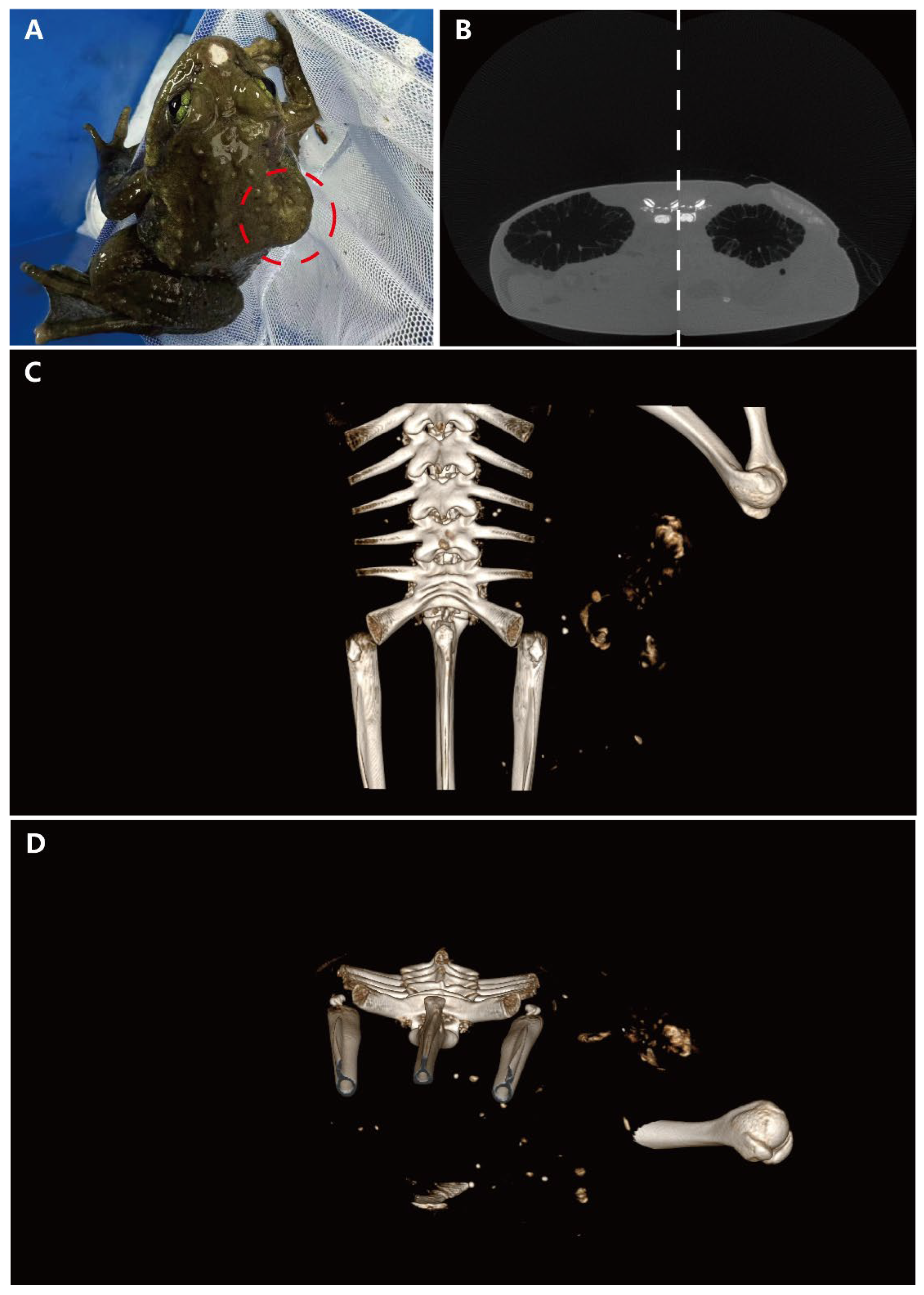
On gross examination, a subcutaneous bulge was observed on the right dorsoventral surface of the frog, along with a mechanical injury at the rostral end (
Figure 1A). Micro-CT scans were performed, revealing a region with relatively clear edges but a complex internal structure on the dorsal side of the body cavity, compared to the healthy side. This finding indicated uneven tissue composition in the affected region. Notably, a circular, uneven mass of similar density was also found on the liver (
Figure 1B). The spatial relationships of these masses were further illustrated using three-dimensional imaging techniques. The general shape of the largest dorsoventral mass was viewed from the dorsal angle, demonstrating its irregular structure (
Figure 1C). Additionally, a cross-sectional image of the body cavity revealed that the nodular mass was dispersed in multiple locations within the cavity, suggesting significant heterogeneity and potential aggressiveness (
Figure 1D).
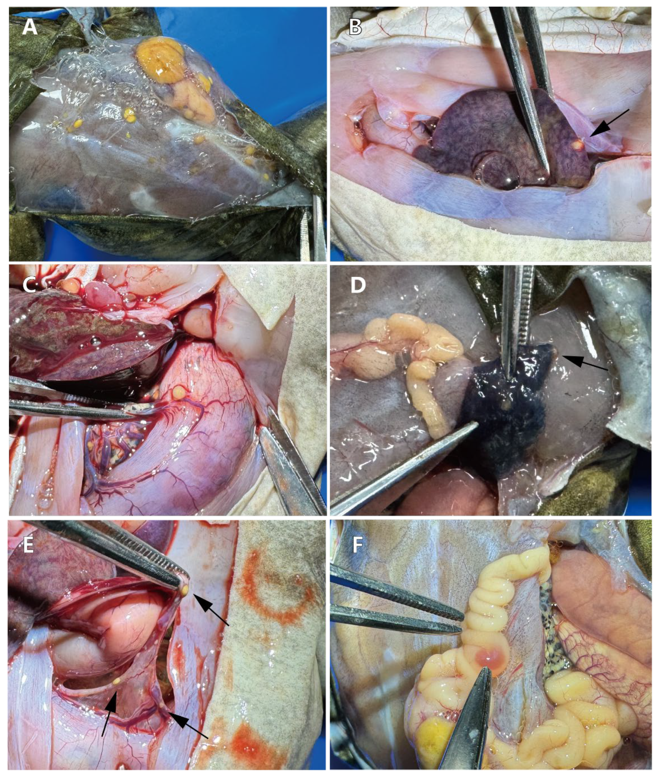
Dissection from the dorsal side revealed a large mass accompanied by multiple scattered yellow masses, the largest measuring 1.0 × 0.7 × 0.5 cm (
Figure 2A). Within the body cavity, a yellow embedded mass was observed in the liver (
Figure 2B). Additionally, a yellow mass with a rich blood supply was found on the surface of the stomach (
Figure 2C). Similar small tumors were present in the lung adjacent to the larger tumor, though the boundaries of the lung tumors were less distinct compared to those in other locations (
Figure 2D). Further examination of the body cavity revealed significant lesions in the mesentery (
Figure 2E) and intestines (
Figure 2F), all characterized by a predominantly yellow coloration.
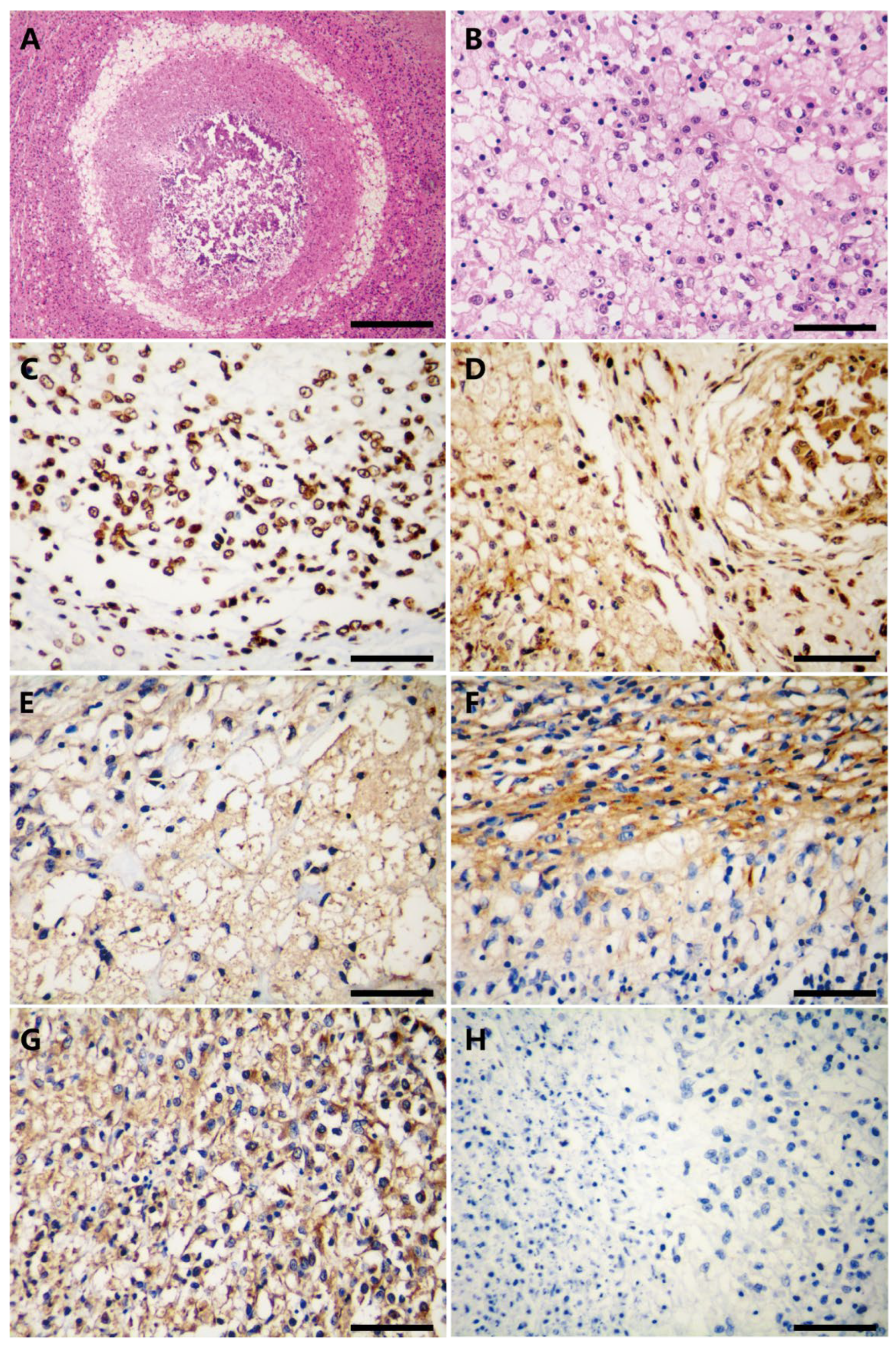
The following results were observed in the hematoxylin and eosin (H&E) stained pathological sections. At low magnification (
Figure 3A), a central pink area lacking structural organization and showing necrosis is visible, indicating a highly malignant feature characterized by rapid progression. This rapid growth deprives cells in the center of the tumor of adequate blood supply and oxygen, leading to ischemia and necrosis. The high metabolic demands and fast proliferation of the tumor cells are indicative of their aggressiveness and destructive potential [
13], suggesting that the tumor cells are highly invasive. The cells surrounding the necrotic area are densely packed, with high cell density and irregular morphology, pointing to dedifferentiated malignant tumor cells. A ring of adipocyte components is observed at the periphery of the high-density cell area, suggesting that the tumor may originate in adipose tissue, which is consistent with the features of dedifferentiated liposarcoma (DDLPS). DDLPS typically presents as a dense mixture of fat cells and undifferentiated tumor cells.
At higher magnification (
Figure 3B), cells in the well-differentiated region exhibit pleomorphism, with variable nuclear size and shape, partial chromatin thickening, and prominent nucleoli. The cytoplasm is mostly vacuolated or transparent, suggesting that these cells may be adipocytes or lipoid cells. Some nuclei are large and irregular, with pronounced nuclear polymorphism. Although the cell distribution is relatively uniform, there are localized dense areas.
Immunohistochemistry was used for differential diagnosis. Tumor cells exhibited strong nucleolar positive expression of S100A4, with no apparent cytoplasmic staining. The nuclei were diverse, with some being large and irregular (
Figure 3C). CDK4 staining showed strong positive expression in both the nuclei and cytoplasm, resulting in a brown coloration of these cellular components. The cells were densely packed, with prominent interstitial spaces in some areas, and the nuclei varied in size and shape, with some deeply stained and irregular (
Figure 3D). MDM2 staining revealed numerous adipocytes, with dark brown staining of the nuclei and partial cytoplasmic staining (
Figure 3E). CD34 expression was significant in both the membrane and cytoplasm of highly differentiated adipocytes and dedifferentiated spindle cells (
Figure 3F). Vimentin staining revealed a large number of positive cells, uniformly distributed throughout the tissue, indicating a substantial presence of mesenchymal cells or epithelial-mesenchymal transition (EMT) phenomena. The positive cells exhibited varied morphologies, with some appearing elongated or fibrous, consistent with stromal cells or cells undergoing EMT (
Figure 3G). Leptin expression was negative (
Figure 3H).
4. Discussion
According to the 2020 update of the WHO’s Classification of Soft Tissue Tumors, fat cell tumors are categorized into benign, intermediate, and malignant types based on their invasive characteristics. Malignant liposarcomas include well-differentiated liposarcoma, dedifferentiated liposarcoma, pleomorphic liposarcoma, and myxoid liposarcoma[
13]. Dedifferentiated liposarcomas (DDLPS), which account for 18% of all liposarcomas, are characterized by the transformation of well-differentiated liposarcomas (ALT/WDLPS) into non-lipogenic sarcomas. Occasionally, these dedifferentiated components present as pleomorphic liposarcomatoid forms, which are highly aggressive [
14].
Diagnosing DDLPS is particularly challenging due to its heterogeneous histological features. DDLPS often exhibit extensive morphological changes, making it difficult to distinguish them from other liposarcoma types and soft tissue sarcomas[
15]. To improve diagnostic accuracy, pathologists commonly use immunohistochemical staining, with MDM2 and CDK4 being key markers for differential diagnosis[
10].
In this case, CT imaging and pathological examination revealed that the tumor had clear boundaries but exhibited invasion into surrounding tissues and distal organs, indicating high aggressiveness. Pathological examination further showed metastases and central necrotic areas, which suggest rapid tumor growth and high metabolic activity. The presence of both highly differentiated and dedifferentiated components supports the diagnosis of DDLPS. Additionally, inflammatory cells were observed, indicating a possible immune response to the tumor.
Immunohistochemical analysis demonstrated positive expression of S100A4, CDK4, MDM2, CD34, and vimentin, along with negative expression of leptin, confirming the tumor’s high aggressiveness and malignancy. These markers are critical for confirming the diagnosis of DDLPS and distinguishing it from other types of fat cell tumors. In particular, the negative expression of leptin helps differentiate tumor cells from normal adipose tissue, indicating a deviation from normal adipose differentiation pathways[
16]. S100A4, a calc-binding protein associated with cell migration, invasion, and tumor metastasis, indicates high invasive and metastatic potential when expressed[
17]. Vimentin, a mesenchymal cell marker, highlights the mesenchymal properties of tumor cells, which are common in tumors of mesenchymal origin[
18]. Positive MDM2 expression is often seen in well-differentiated liposarcomas and DDLPS, making it a key diagnostic marker. CDK4, a cell cycle-dependent kinase involved in cell cycle regulation, is frequently used to identify DDLPS. Moreover, CD34, a marker of vascular endothelial cells, indicates the presence of new blood vessels, reflecting the tumor’s angiogenic capacity[
19].
Currently, DDLPS is primarily observed in humans, with no known cases in animals. However, the causes of cancer are multifactorial, including genetics, diet, and environmental factors, all of which may contribute to disease development[
20]. Amphibians, in particular, are highly sensitive to environmental changes, and the occurrence of liposarcoma in amphibians may signal ecological stress. Studying tumors across species helps researchers understand general tumorigenesis mechanisms and species-specific differences. This comparative approach sheds light on fundamental principles of tumor biology and provides new perspectives and methodologies for human cancer research[
21]. By investigating the occurrence and characteristics of liposarcomas in different species, valuable insights can be gained into the underlying mechanisms of the disease. This study not only advances our understanding of veterinary oncology but also provides important insights into amphibian conservation, emphasizing the potential role of environmental stressors in tumor development.
5. Conclusions
This study reports the first documented case of dedifferentiated liposarcoma (DDLPS) in veterinary medicine and presents the first pathological evidence of DDLPS in a wild amphibian, Maculopaa medogensis. These findings are significant for enhancing the understanding of cancer in wildlife species. DDLPS is known for its pronounced histological heterogeneity and invasive potential, which was evident in this case through multiple metastases observed on subcutaneous muscle surfaces and internal organs. The diagnostic process relied primarily on immunohistochemistry, with micro-CT scans, pathological sections, and gross autopsy providing crucial support for a definitive diagnosis.
Author Contributions
Conceptualization, R.-L.Z. and J.-P.J.; methodology, R.-L.Z., T.-Y.Q. and J.-P.J.; software, R.-L.Z.; Validation, R.-L.Z.; investigation, R.-L.Z., T.-Y.Q. and Y.-H.W.; resources, J.-P.J.; data curation, R.-L.Z. and T.-Y.Q.; writing—original draft preparation, R.-L.Z.; writing—review and editing, R.-L.Z., T.-Y.Q., W.Z and J.-P.J.; visualization, R.-L.Z.; supervision, W.Z., and J.-P.J.; project administration, B.W., F.X., C.L., and J.-P.J.; funding acquisition, B.W. and J.-P.J. All authors have read and agreed to the published version of the manuscript.
Funding
This research was funded by the Strategic Priority Research Program of the Chinese Academy of Sciences (XDA042020304), Survey of Wildlife Resources in Kev Areas of Xizang (Phase II, ZL202303601), and China Biodiversity Observation Networks (Sino BON -Amphibian & Reptile).
Acknowledgments
The authors thank for the Meihua Zhang, Yan Wang, Zhonghao Luo, and Wei Li for help in equipment operation, animal collection and care.
Conflicts of Interest
The authors declare no conflict of interest. The funders had no role in the design of the study; in the collection, analyses, or interpretation of data; in the writing of the manuscript, or in the decision to publish the results.
References
- Torres-Dimas E, Cruz-Ramírez A, Bermúdez-Cruz RM: Cancer in Amphibia, a rare phenomenon? Cell Biology International 2022, 46(12):1992-1998. [CrossRef]
- Hopewell E, Harrison SH, Posey R, Duke EG, Troan B, Harrison T: Analysis of Published Amphibian Neoplasia Case Reports. Journal of Herpetological Medicine and Surgery 2020, 30(3):148-155. [CrossRef]
- O’Brien MF, Justice WSM, Beckmann KM, Denk D, Pocknell AM, Stidworthy MF: Four cases of neoplasia in amphibians at two zoological institutions: Alpine newt Ichthyosaura alpestris, Red-eyed tree frog Agalychnis callidryas, Common frog Rana temporaria and Puerto Rican crested toad Peltophryne lemur. International Zoo Yearbook 2017, 51(1):269-277. [CrossRef]
- Balls M: Spontaneous Neoplasms in Amphibia: A Review and Descriptions of Six New Cases*. Cancer Research 1962, 22(10):1142-1154.
- Sabhavath M, Annamaraju SS, Amanchi NR, Bhavanam KR, Kancha RK: Soft Tissue Sarcoma. In: Biomedical Aspects of Solid Cancers. Edited by Kancha RK. Singapore: Springer Nature Singapore; 2024: 279-288. [CrossRef]
- Evans HL: Liposarcoma: a study of 55 cases with a reassessment of its classification. Am J Surg Pathol 1979, 3(6):507-523. [CrossRef]
- Coindre J-M, Pédeutour F, Aurias A: Well-differentiated and dedifferentiated liposarcomas. Virchows Archiv 2010, 456(2):167-179. [CrossRef]
- Henricks WH, Chu YC, Goldblum JR, Weiss SW: Dedifferentiated liposarcoma: a clinicopathological analysis of 155 cases with a proposal for an expanded definition of dedifferentiation. Am J Surg Pathol 1997, 21(3):271-281.
- Coindre J-M, Mariani O, Chibon F, Mairal A, de Saint Aubain Somerhausen N, Favre-Guillevin E, Bui NB, Stoeckle E, Hostein I, Aurias A: Most Malignant Fibrous Histiocytomas Developed in the Retroperitoneum Are Dedifferentiated Liposarcomas: A Review of 25 Cases Initially Diagnosed as Malignant Fibrous Histiocytoma. Modern Pathology 2003, 16(3):256-262. [CrossRef]
- Binh MBN, Sastre-Garau X, Guillou L, de Pinieux G, Terrier P, Lagacé R, Aurias A, Hostein I, Coindre JM: MDM2 and CDK4 immunostainings are useful adjuncts in diagnosing well-differentiated and dedifferentiated liposarcoma subtypes: a comparative analysis of 559 soft tissue neoplasms with genetic data. Am J Surg Pathol 2005, 29(10):1340-1347. [CrossRef]
- Lee E-J, Jeong K-S: Histopathological Features of Myxoid Pleomorphic Liposarcoma in an African Pygmy Hedgehog (Atelerix Albiventris). Veterinary Sciences 2022, 9(11):642. [CrossRef]
- Zhang M, Chen X, Ye C-y, Fei L, Li P-p, Jiang J, Wang B: Osteology of the Asian narrow-mouth toad Kaloula borealis (Amphibia, Anura, Microhylidae) with comments on its osteological adaptation to fossorial life. Acta Zoologica 2020. [CrossRef]
- Sbaraglia M, Bellan E, Dei Tos AP: The 2020 WHO Classification of Soft Tissue Tumours: news and perspectives. Pathologica 2020, 113:70 - 84. [CrossRef]
- Zhao M, Xu M, Wang Y, He X: Histological diagnosis and differential diagnosis of dedifferentiated liposarcoma(in Chinese). Chinese Journal of Pathology 2019, 48(7):573-579.
- Mocellin S: Dedifferentiated Liposarcoma. In: Soft Tissue Tumors : A Practical and Comprehensive Guide to Sarcomas and Benign Neoplasms. Edited by Mocellin S. Cham: Springer International Publishing; 2021: 209-212. [CrossRef]
- Picó C, Palou M, Pomar CA, Rodríguez AM, Palou A: Leptin as a key regulator of the adipose organ. Reviews in Endocrine & Metabolic Disorders 2021, 23:13 - 30. [CrossRef]
- Fei F, Qu J, Zhang M, Li Y, Zhang S: S100A4 in cancer progression and metastasis: A systematic review. Oncotarget 2017, 8:73219 - 73239. [CrossRef]
- Satelli A, Li S: Vimentin in cancer and its potential as a molecular target for cancer therapy. Cellular and Molecular Life Sciences 2011, 68:3033-3046. [CrossRef]
- de la Puente P, Muz B, Azab F, Azab AK: Cell Trafficking of Endothelial Progenitor Cells in Tumor Progression. Clinical Cancer Research 2013, 19(13):3360-3368. [CrossRef]
- Sankpal UT, Pius H, Khan M, Shukoor MI, Maliakal P, Lee CM, Abdelrahim M, Connelly SF, Basha R: Environmental factors in causing human cancers: emphasis on tumorigenesis. Tumor Biology 2012, 33(5):1265-1274. [CrossRef]
- LeBlanc AK, Mazcko CN, Khanna C: Defining the Value of a Comparative Approach to Cancer Drug Development. Clin Cancer Res 2016, 22(9):2133-2138. [CrossRef]
|
Disclaimer/Publisher’s Note: The statements, opinions and data contained in all publications are solely those of the individual author(s) and contributor(s) and not of MDPI and/or the editor(s). MDPI and/or the editor(s) disclaim responsibility for any injury to people or property resulting from any ideas, methods, instructions or products referred to in the content. |
© 2024 by the authors. Licensee MDPI, Basel, Switzerland. This article is an open access article distributed under the terms and conditions of the Creative Commons Attribution (CC BY) license (http://creativecommons.org/licenses/by/4.0/).


