Preprint
Article

Development of a Herb-Based Dietary Ingredient with Potential Nootropic Properties: From Bench to Bedside

This version is not peer-reviewed.

Submitted:

25 September 2024

Posted:

26 September 2024

You are already at the latest version

A peer-reviewed article of this preprint also exists.

Abstract
Brain health is a matter of great importance as we age, where memory loss and other cognitive declines become slowly apparent. There are numerous natural solutions, called nootropics, that can help mitigate this natural process. While some, such as ginkgo biloba, have been traditionally used for centuries, in many cases there is a lack of scientific evidence on their potential mechanisms and efficacy. To this end, the objective of the current study is to evaluate and compare several standardized herb extracts with potential nootropic effects, and from this analysis develop a formula and confirm its effect in healthy volunteers with mild cognitive impairment. A total of 9 standardized herb extracts were chosen based on previous scientific evidence and tested in human neural stem cell cultures exposed to oxidative stress. The expression of nerve growth factor (NGF), NRF2 and vascular endothelial growth factor (VEGF) were also analyzed. As a result, it was observed that an extract from Salvia officinalis revealed to have the most noticeable effect among the herbs analyzed. In further tests it was confirmed that its combination with rutin, a phenolic compound with potential nootropic effects, increased brain derived neurotrophic factor (BDNF) expression, as well as inhibiting acetylcholine esterase (AChE) activity. Finally, a randomized, double-blind placebo-controlled clinical study was conducted, testing the effects of the combination at two different doses, for 12 weeks, in healthy volunteers aged 50+ with mild cognitive impairment. Two questionnaires were applied, the MoCA and ADAS-Cog. As a result, both doses significantly improved several characteristics related to memory, particularly memory recall, with a more noticeable effect in the higher dose. Therefore, the results suggest that the long-term intake, at least 6-12 weeks, of a natural herb blend comprised of Salvia officinalis and rutin can help improve certain brain health-related parameters in healthy subjects with mild cognitive impairment.
Keywords: 
Subject: 
Medicine and Pharmacology  -   Dietetics and Nutrition

1. Introduction

Nootropics, also known as “smart drugs”, are substances found in the market that can potentially contribute to improve cognitive health, such as memory, focus or concentration [1]. These substances are commonly found in food supplements and are usually of natural origin [2]. Overall, nootropics do not have an immediate effect and generally require continuous consumption for at least several weeks [3].
There are numerous described mechanisms by which nootropics may exert their effect (for a review, see reference 4). For example, some act by increasing blood flow to the brain, thereby providing more access to glucose and oxygen [3,5]. Others reduce oxidative stress and contribute to eliminating free radicals [6,7,8,9]. Still others increase the production of trophic factors that contribute to promote neuronal survival, axon growth or synaptic transmission [10,11,12]. Overall, due to the variety of active compounds found in these herbs, nootropics usually have a multifaceted effect upon the brain.
While there are numerous herbs that traditionally hav been used for their potential nootropic effect [13], there are few that are backed by solid scientific evidence and supported with clinical studies. To this end, the objective of the current study is to assess and compare the nootropic potential of several botanical extracts, standardized in certain active compounds. Some of these extracts, such as ginkgo biloba or bacopa, are well known for their traditional use, while others are less known. The strategy applied in the study was to first analyze in vitro various aspects related to nootropics (antioxidant effect, neuronal survival, stimulation of growth factors, etc), and compare them to find the best candidate. Then, it was tested to assess various aspects related to improving neuronal functioning (BDNF expression and acetylcholine esterase inhibition), in isolation or combined with rutin, a purified polyphenol with scientific evidence as a nootropic [14]. Then, the formula was tested in a clinical setting in healthy older adults with mild cognitive impairment. The results of the current study may provide scientific and clinical evidence of a natural, botanical-based formula with nootropic effects.

2. Materials and Methods

2.1. Analytical equipment

Inverted microscope with integrated camera, laminar flow hood, cell culture incubator (37°C, 5% CO2, 90% relative humidity (RH), Bürker chamber, pipettes, rack, plate reader spectrophotometer, Nano-Drop spectrophotometer, Spectrophotometer Synergy, vortex, Quant studio 5 (Applied Biosystem) Quantitative real-time PCR, heating block, thermocycler and consumables, statistical analysis software.

2.2. Reagents

Distilled Water (Braun), Human Neural Stem Cells (NSC) specific culture medium and supplements (Phenocell), PBS (Gibco), Trypan Blue Solution (Bio-Rad), Trypsin/EDTA (SigmaAldrich), Dimethylsulfoxide (DMSO, Sigma-Aldrich), MTT reagent [3-(4,5-Dimethylthiazol-2-yl)-2,5-Diphenyltetrazolium Bromide] (Invitrogen), Ethanol (Sigma-Aldrich), DNAse-I (Qiagen), RNeasy extraction kit (Qiagen), PrimeScript RT reagent kit (Perfect Real Time- Takara Clontech), ROS/RNS detection kit (Abcam), BDNF ELISA kit (Proteintech KE00096-96T), AChE activity ELISA kit (Abcam).

2.3. Botanical Extracts

The following plant extracts were used, standardized in the active compounds described: Vaccinium myrtillus fruits (bilberry, 25% anthocyanidines by UV), Vaccinium angustifolium fruits (blueberry, 25% anthocyanins by UV), Bacopa monnieri whole plants (water hyssop, 45% bacosides by HPLC), Salvia officinalis leaves (sage, 4% rosmarinic acid by HPLC), Crocus sativa stigmas (saffron, 2% safranal & 11% crocines by UV), Rhodiola rosea roots (arctic root, 5% rosavins by HPLC), Ginkgo biloba leaves (24% flavonoids by HPLC), Panax ginseng roots (ginseng, 5% ginsenosides by HPLC), Centella asiatica whole plant (gotu kola, 20% total asiaticosides by HPLC). Sophora japonica standardized in 95% rutin was used as source of rutin.
These extracts were acquired by various manufacturers, obtained generally through water-ethanol extractions of the raw material. Internal analysis by UV or HPLC, depending on the extract, was performed in the laboratory to ensure the quality of the extract.

2.4. Cell viability – MTT assay

For seeding cells, cell numbers and viability were determined using Trypan-Blue staining and counting in a Bürker chamber under the microscope. For the MTT assay, NSC were cultured overnight in supplemented growth medium. 24 hours later, the culture medium was replaced with fresh medium and the different products at 8 different concentrations. After 24 hours of incubation, the medium was removed, and MTT solution was added to each well. Plates were incubated in the refrigerated incubator at 37ºC for 3 hours. MTT reactive was removed, and DMSO 100% was added to each well to solubilize formazan crystals prior to absorbance measurements at 550 nm and 620 nm as reference on a scanning multi-well spectrophotometer.
In the case of hippocampal cell cultures, mouse hippocampal neuronal cells were cultured overnight at a 40.000 cells/well density in a 96 well plate, in supplemented growth medium. 24 hours later, the culture medium was replaced with fresh medium at 8 different concentrations (0.01, 0.003, 0.001, 0.0003, 0.0001, 0.00003, 0.00001 and 0.000003%). After 48 hours of incubation, the medium was removed, and MTT solution was added to each well. Plates were incubated at 37ºC for 3 hours. MTT reagent was removed and DMSO at 100 % was added to each well to solubilize formazan crystals, then the absorbance was measured at 550 nm and 620 nm as a reference on a scanning multi-well spectrophotometer.

2.5. ROS/RNS Assay

NSC were seeded and after 24 h, medium was replaced with fresh medium containing the tested products at two or three non-cytotoxic concentrations for 24 hours (h). After that, oxidative stress was induced with H2O2 and ROS and RNS were quantified through a specific Cellular ROS/RNS assay kit. ROS and RNS concentrations were measured by spectrophotometry at Ex/Em: 490/525nm and Ex/Em: 550/620nm, respectively.

2.6. Gene expression analysis by qRT-PCR

For gene expression quantification of selected genes, NSC were supplemented with medium containing the tested products at two or three non-cytotoxic concentrations for 24 hours (h). After that, neural toxicity was induced with H2O2 for 24 h. After the incubation period, cells were collected in lysis buffer to proceed with the RNA extraction. Total RNA was extracted using RNeasy kit (Qiagen) and treated with DNAse-I to remove any contamination from genomic DNA. RNA quality and quantity were checked in a Nano-Drop spectrophotometer, and cDNA was synthesized. Finally, quantitative PCR (qPCR) was performed in a real time PCR machine (QuantStudio 5, Applied BioSystem). To perform raw data analysis, we used the 2–∆∆Ct method (Livak & Schmittgen, 2001) to calculate the gene relative expression ratio of NRF2, NGF, BDNF, VEGF, CASP9, BAX and BCL2 to H2O2-treated control (C). Actin (ACT) was used as a reference housekeeping gene.

2.7. Quantification of BDNF protein levels by ELISA

To assess the BDNF protein levels, NSC were seeded at 250,000 cells/well in 12-well plates and kept for 24 h at 37ºC. Then, cell culture media was replaced by fresh medium containing the tested products and incubated for 24 h. Thereafter, supernatants were collected and processed for the quantification of BDNF protein levels by an ELISA kit, following the manufacturer’s instructions.

2.8. Quantification of AChE protein levels by ELISA

For the assessment of the AChE activity, cells were seeded in 12-well plates at a density of 70,000 cells/well and kept for 24 hours at 37ºC. Then, cells were cultured in fresh medium at the selected concentrations (0.001% and 0.0002%) and incubated for 48 h. Thereafter, cell lysates were harvested and used for the measurement of the AChE activity by ELISA, following the manufacturer’s instructions.

2.9. Clinical trial information

A total of 121 healthy subjects, ages 50-65 years old, with mild cognitive impairment were enrolled in the study. The established exclusion criteria were: (i) allergy or reactivity to some of the components of the products or a product with a similar category than tested one, (ii) in-use relevant pharmacological or hormonal treatment, (iii) forecast of change of routine or relevant way of life during the period of study, and (iv) nursing, pregnant, or planning to become pregnant during the study according to subject self-report. Subjects were asked to (i) respect the conditions of use of the treatment, (ii) do not take antidepressants, anxiolytics, hypnotics or sleeping pills, analgesics that affect the central nervous system (morphines and derivatives), (iii) do not take memory supplements during the study, (iv) do not take Disgren, Somacin or Aspirin, and (v) do not use products similar to the one in the trial during the time of participation. The standard protocol and test conditions were submitted to and approved by the Ethical Committee of Bionos Biotech (Date 18/07/2023 and Code number 0058-2023). Subjects were randomly assigned to 3 groups: 42 subjects for placebo group, 40 subjects for sage/rutin at 250mg group and 39 subjects for sage/rutin at 400mg group. Subjects ingested by oral intake one capsule per day of the corresponding product, after breakfast. The cognitive tests were performed before the ingestion of the product (day 0), 6 weeks and 12 weeks after the first ingestion of the product.

2.10. Statistical analysis

The MTT assay was set with 8 replicates per condition. ROS/RNS quantification assay and qRT-PCR assay was set with 5 replicates per condition. Both Data outliers were identified with ROUT method (Q = 5%) and excluded from the analysis if found. Data were statistically analyzed by Ordinary one-way ANOVA and Dunnett’s post hoc multiple test or Brown-Forsythe ANOVA test and Dunnet’s T3 post hoc multiple comparisons test. Statistical significance was declared at p < 0.05, 95% of confidence. Bars in the charts represent the mean value for each condition and error bars indicate the standard error of the mean (SEM) for each group of values.

3. Results

3.1. MTT analysis of individual extracts

MTT analysis was first performed in the neural stem cells cultures to test cell viability in presence of the various plant extracts (Figure 1). A total of 8 concentrations were used in each case. The results indicated that each extract presented different effects on the cell viability, indicated in the graphs as values under 1. The following extracts did not affect cell viability at any concentration tested: sage, saffron, arctic root, ginkgo, ginseng, and gotu kola. However, some did present a tendency to lower cell viability despite not being statistically significant. For this reason, 3 concentrations were considered for future experiments in the case of sage, ginseng, and gotu kola. Also, an intermediate concentration of 0.0005% was considered for ginkgo and saffron for this same reason. Bilberry only presented toxicity at the highest concentration, 0.01%, while blueberry and bacopa had various levels of toxicity, especially at the higher concentrations.
Based on these results, the following working concentrations were used in the subsequent studies: bilberry (0.0001%, 0.001%), blueberry (0.0001%, 0.001%), bacopa (0.0001%, 0.001%), sage (0.0001%, 0.001%, 0.01%), saffron (0.0001%, 0.00005%), arctic root (0.0001%, 0.001%), ginkgo biloba (0.0001%, 0.00005%), ginseng (0.0001%, 0.001%, 0.01%), gotu kola (0.0001%, 0.001%, 0.01%).

3.2. ROS/RNS and Cell Death Markers in Individual Extracts

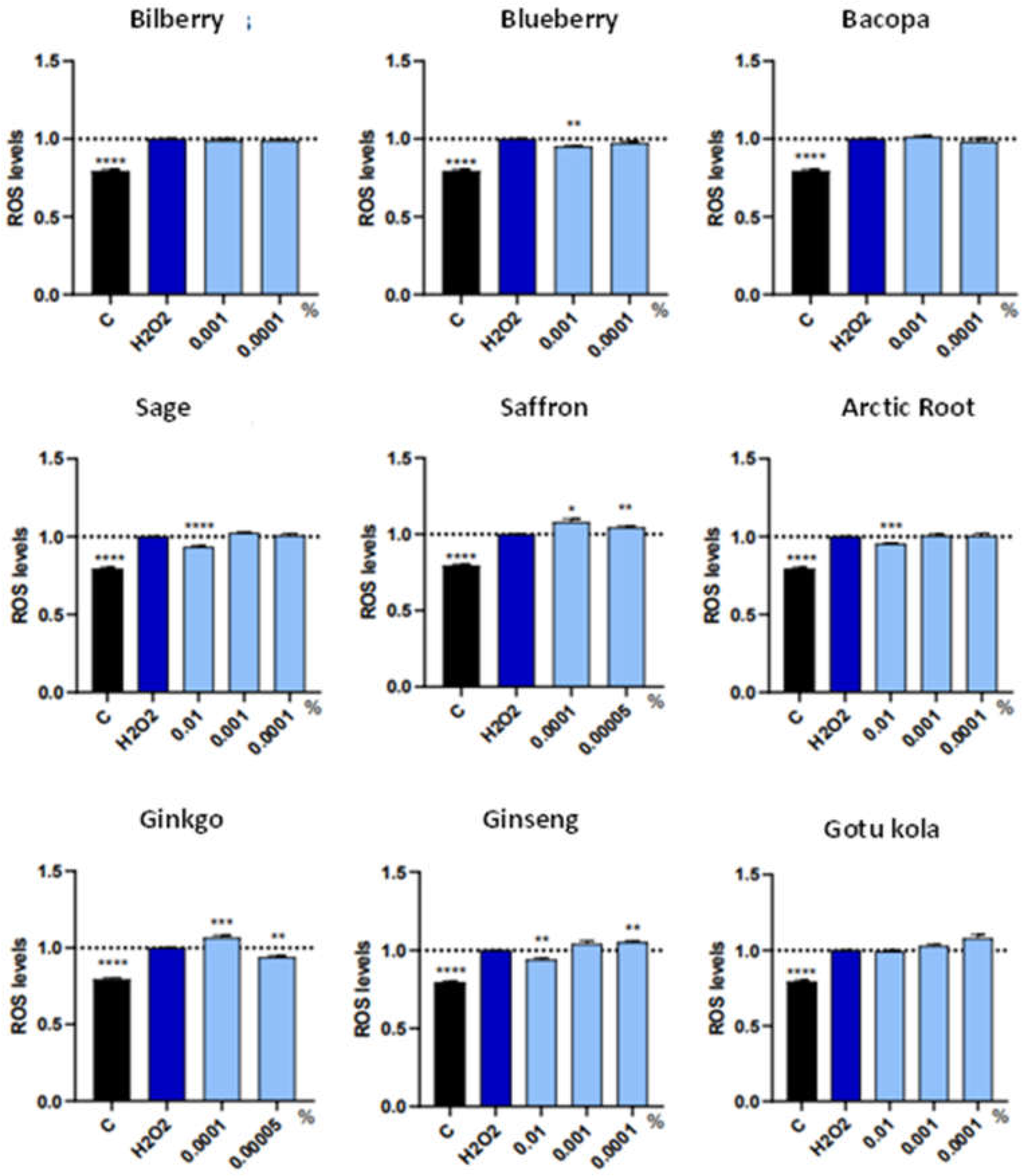
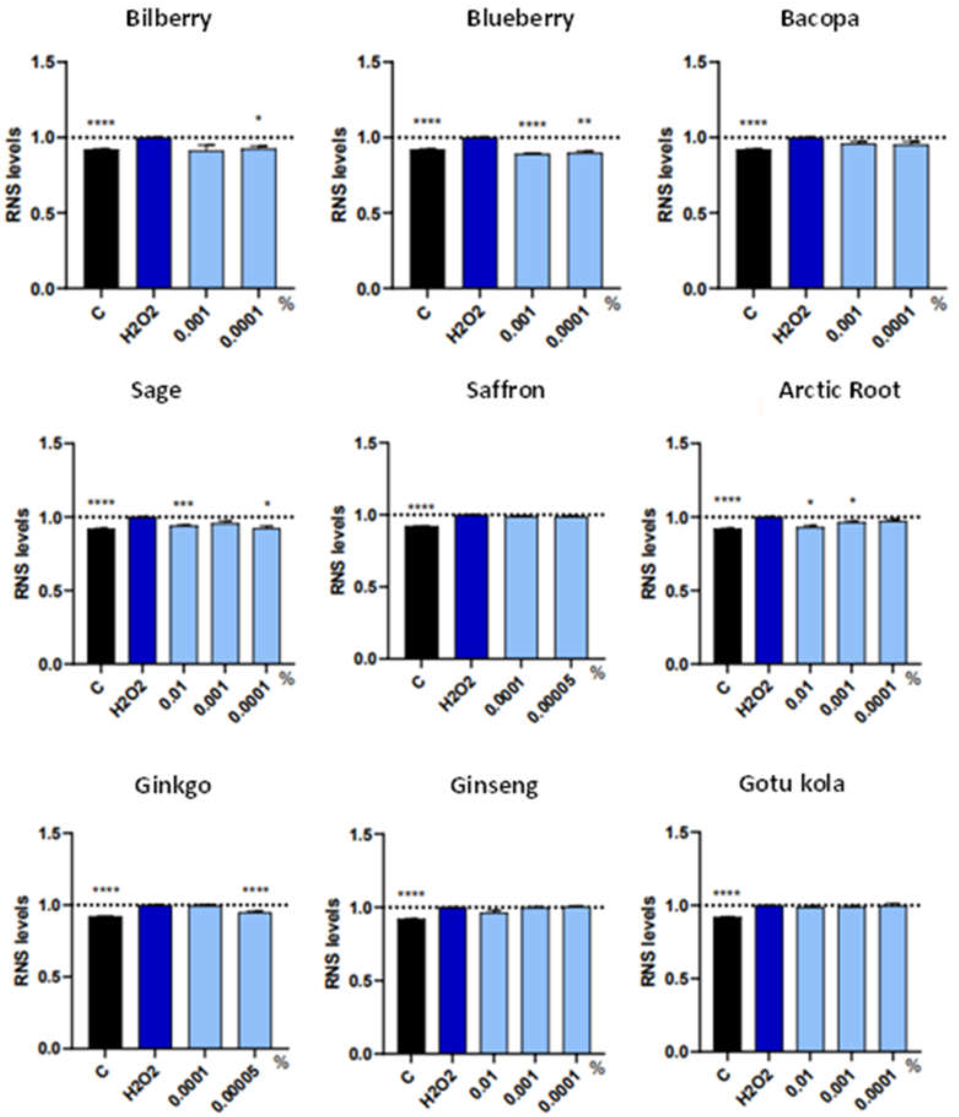
Reactive oxygen species (ROS) and reactive nitrogen species (RNS) was performed in neural stem cells submitted to oxidative stress by H2O2. The results can be observed in Figure 2 and 3, respectively.
NSC exposure to oxidative stress increased ROS on average by 20.5%. The cultures exposed to oxidative stress and the botanical extracts revealed different levels of effectiveness in reducing ROS.
Specifically, bilberry, bacopa, saffron, and gotu kola did not have any effect on reducing oxidative stress. Surprisingly, a pro-oxidant effect was observed in the case of saffron, in both concentrations used.
Both ginkgo and ginseng had antioxidant and pro-oxidant effects, depending on the concentration used in the cell cultures. The extracts where only an antioxidant effect observed was blueberry (at 0.001%), sage (at 0.01%) and arctic root (at 0.01%).
Regarding RNS, H2O2 treatment increased this parameter by 7.9%. In this case, the results, as observed in Figure 3, indicated that the following extracts did not have an effect on RNS: bacopa, saffron, ginseng, and gotu kola. In turn, the extracts that significantly reduced RNS were: bilberry (at 0.0001%), blueberry (at both concentrations), sage (at 0.01% and 0.0001%), arctic root (at 0.01% and 0.001%) and ginkgo (at 0.00005%).
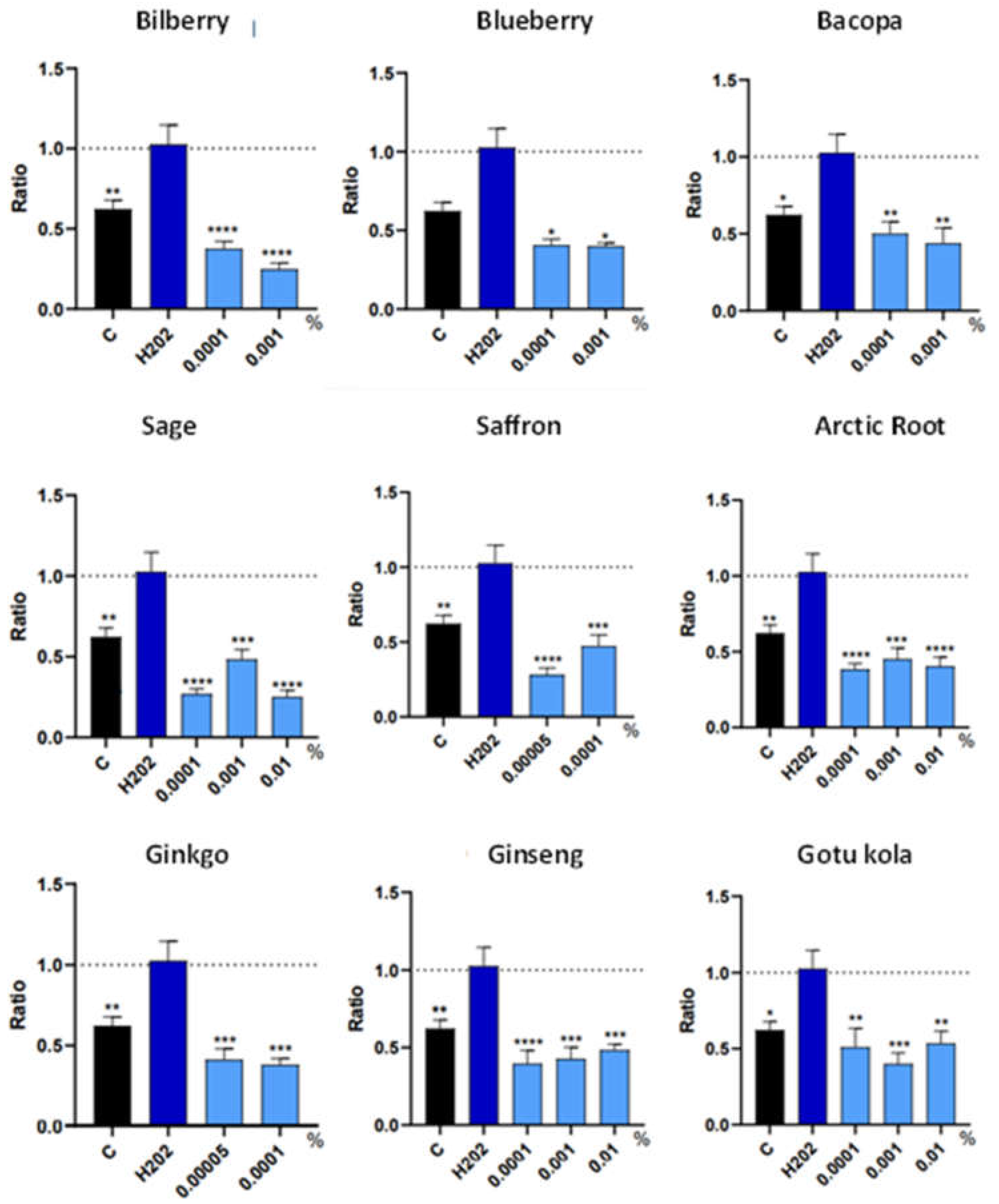
Finally, several genes related to cell death were analyzed, specifically caspase-9, Bcl2 and Bax. There was no effect observed in any of the extracts for Bcl2 and Bax, under the culture conditions used (data not shown). On the other hand, all extracts significantly reduced caspase-9 in NSC exposed to oxidative stress, in all the concentrations used (Figure 4). As caspase-9 is implicated in the initiation process of the apoptotic pathway, this result suggests that all the extracts contain active compounds that contribute to inhibit this gene.

3.3. Trophic Factor Release in Individual Extracts

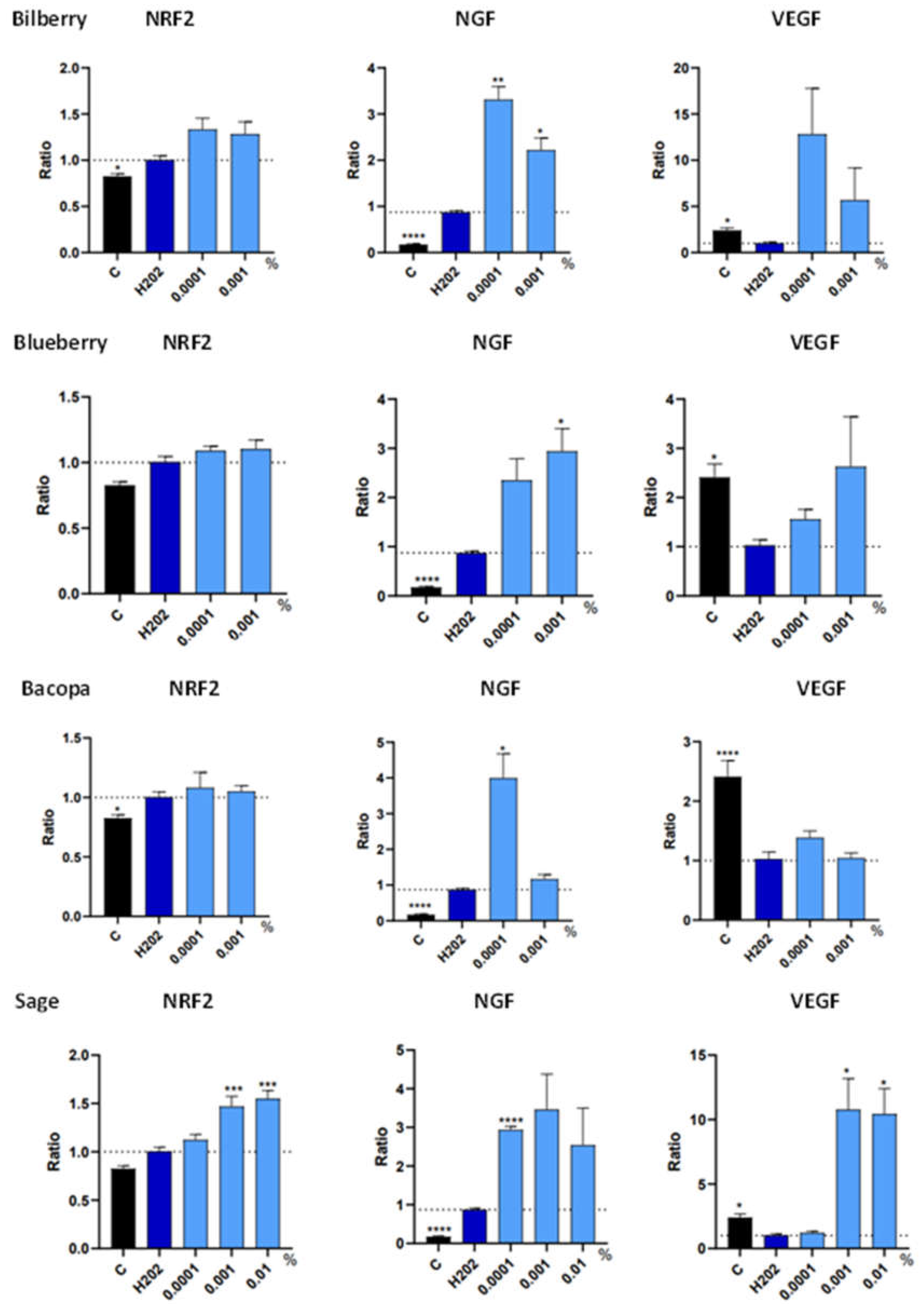
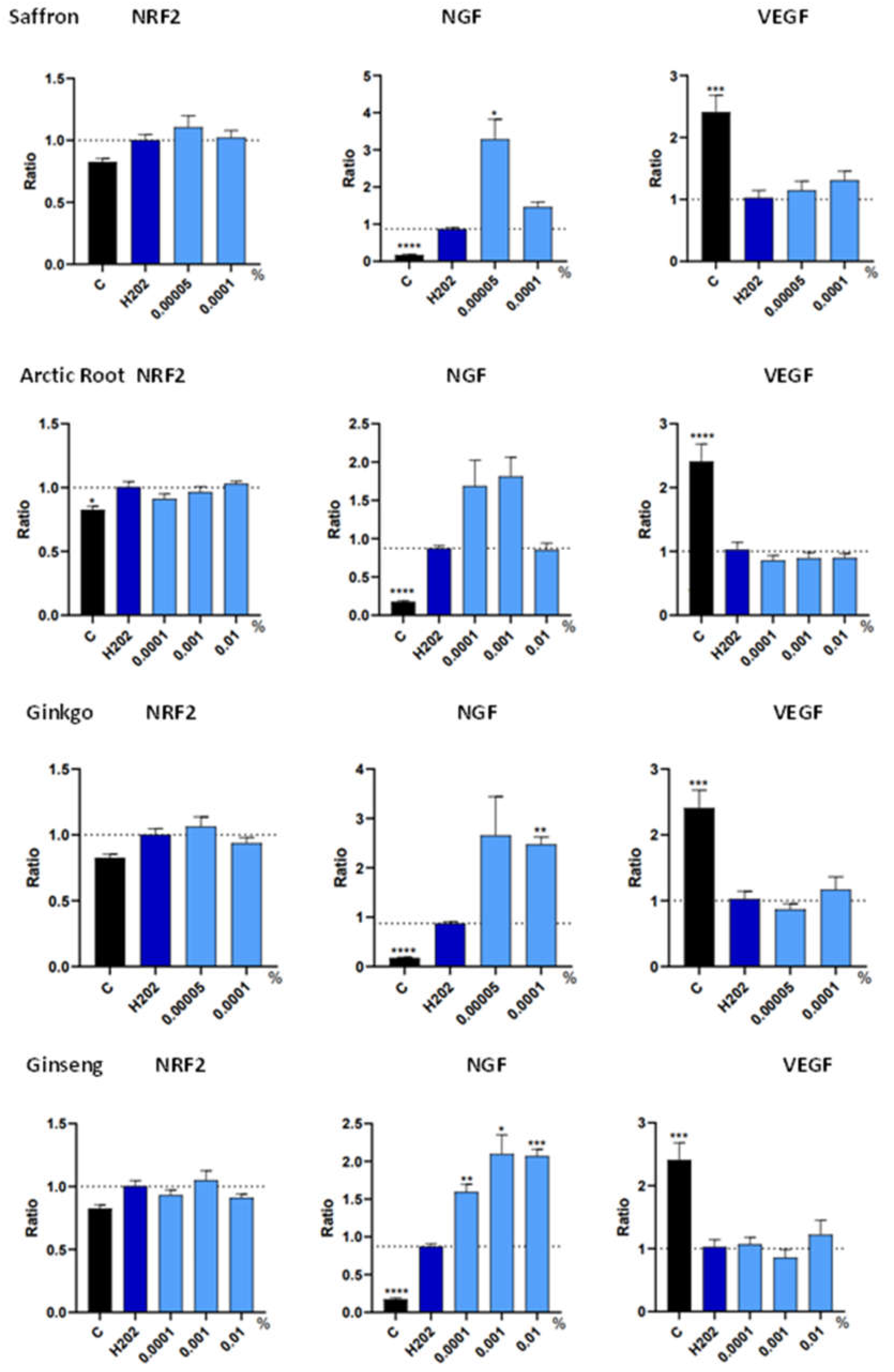
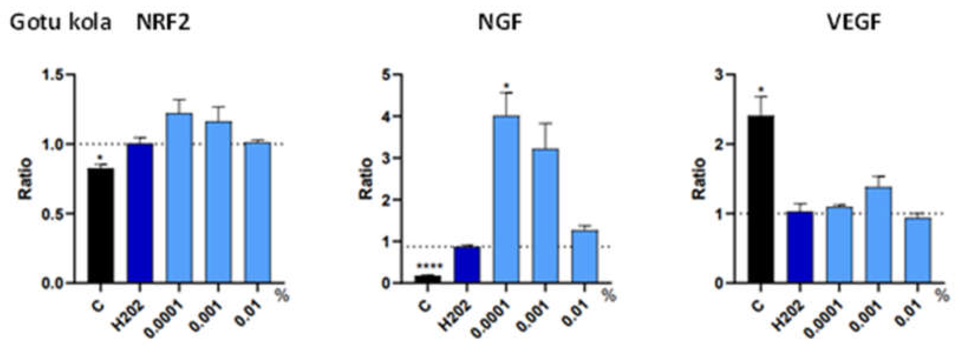
The gene expression levels of three trophic factors were analyzed in the human neural stem cell cultures exposed to oxidative stress; NRF2, NGF and VEGF (Figure 5).
Regarding NRF2, this trophic factor was significantly increased by approximately 17.8% when the NSCs were submitted to oxidative stress. Among the botanical extracts, only sage significantly increased the expression of NRF2 by approximately 1.5 fold, at 0.001% and 0.01%.
As for NGF, H2O2 exposure caused an average 70.1% increase of this growth factor in NSC. With the exception of arctic root, all botanical extracts further increased its expression. Bilberry (at 0.0001%), blueberry (at 0.001%), sage (at 0.0001%), saffron (at 0.00005%), increased NGF by around 3-fold. Bacopa (at 0.0001%) and gotu kola (at 0.0001%) seemed to be the extracts with the highest increase in NGF, around 4-fold. On the other hand, ginkgo (at 0.0001%) and ginseng (at 0.01%) increased only around 2-fold.
Finally, VEGF expression was significantly decreased in NSC submitted to oxidative stress, by an average 40.3%. As in the case of NRF2, only sage revealed a significant increase, at both 0.001% and 0.01% concentrations, with an over 10-fold increase in expression.
A summary of the results observed, with the exception of cell death markers, among the botanical extracts can be found in Table 1.
Based on the results obtained with the various individual extracts, sage revealed to possess the most significant effects overall, with positive results observed in both ROS and RNS, reduction in the pro-apoptotic marker caspase-9, and was the only extract to significantly increase the expression of all three trophic factors analyzed; NRF2, NGF and VEGF.

3.4. In vitro Results of Sage in combination with Rutin

Based on the results observed in the individual botanical extracts, sage (Salvia officinalis) standardized in 4% rosmarinic acid revealed to present the most noticeable effects on reducing oxidative stress and cell death, while increasing the gene expression of trophic factors implicated in neuronal survival.
Next, in vitro studies were performed related to the potential effect of sage in promoting neuronal function. Specifically, brain-derived neurotrophic factor (BDNF) protein expression was analyzed in human neural stem cells, and acetylcholine esterase (AChE) activity was analyzed in mouse hippocampal cells.
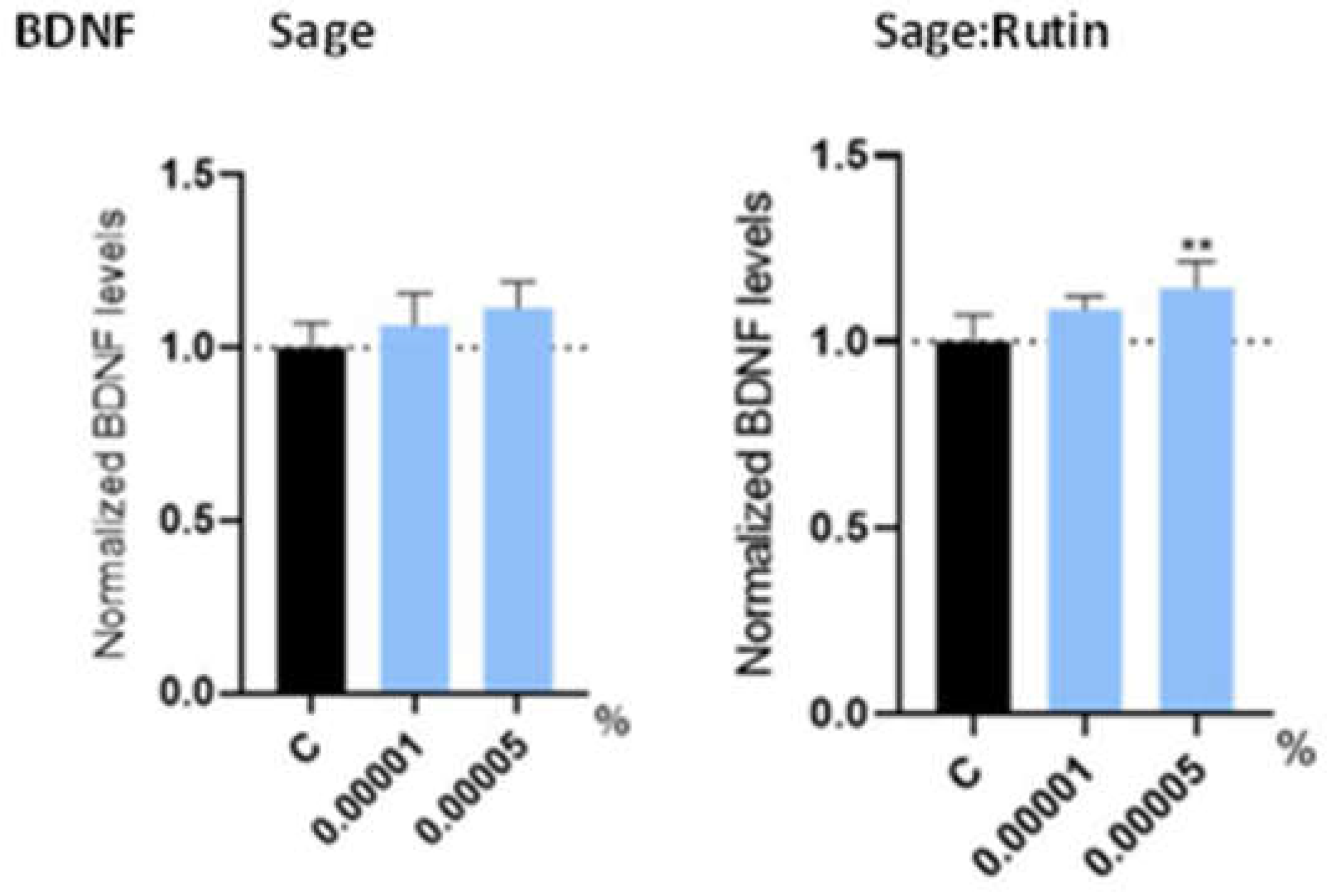
BDNF presents numerous effects in the nervous system, and is especially relevant for learning and memory, particularly long-term memory [15]. Based on this premise, the expression of this trophic factor in neural stem cells exposed to sage extract was analyzed (Figure 6). Furthermore, the combination of sage with rutin, at a 65:35 ratio, was also assessed and compared. Rutin is a phenolic compound with multiple health benefits, including potential neuroprotective effects [14,16]. The reason for this was to evaluate if the combination of these two extracts could have possible complementary effects. The concentrations of the extracts in culture were chosen based on MTT analysis (data not shown).
As a result, it was observed that sage extract presented a tendency, but not quite significant (p=0.11), effect in increasing BDNF protein levels. However, when combined with rutin, BDNF expression was significantly increased, confirming a potential combinatory effect.
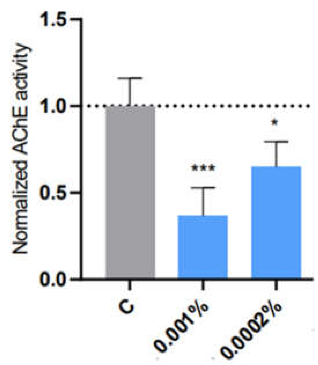
Finally, AChE activity was assessed in mouse hippocampus cells with the combination of sage and rutin (Figure 7). The results here showed that AChE activity was significantly reduced by approximately 63.4% at the highest dose and 35.2% at the lowest, confirming the inhibitory effect of the combination in hippocampal cell cultures.

3.5. Clinical Trial Results

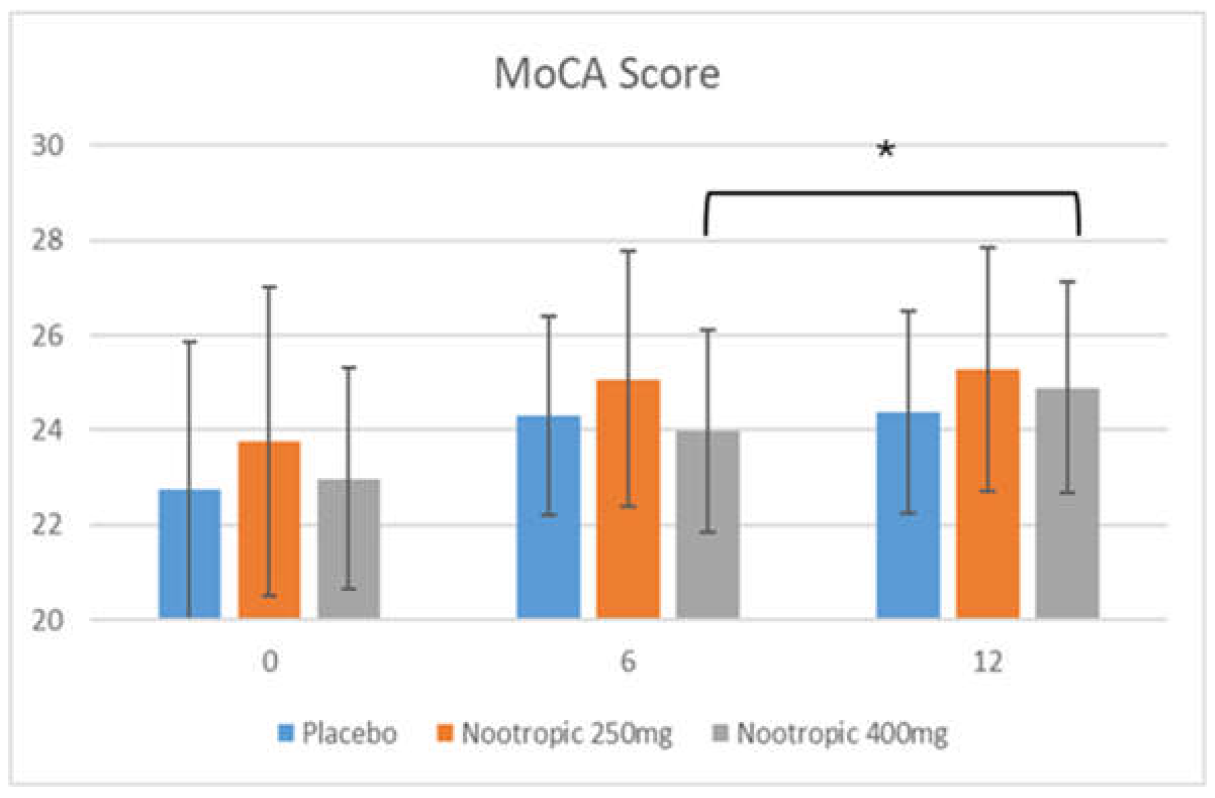
The combinatorial formula of sage and rutin was tested in healthy human volunteers, aged 50+, with mild cognitive impairment as assessed by the MoCA test. The study subjects were randomly assigned to take one of the three capsules: placebo, sage/rutin at 250mg, and sage/rutin at 400mg (branded as Mindrevive®). They were instructed to take the capsules daily, during the morning, for 84 days. MoCA and ADAS-Cog tests, which are standardized questionnaires to assess mild cognitive impairment [17,18], were conducted at baseline, week 6 and end of the study (week 12).

3.5.1. MoCA Test

The overall score obtained through the MoCA test can be found in Figure 8. Baseline values revealed that the subjects presented mild cognitive impairment, with values below 26 (from a total of 30) [17]. Briefly, all study groups, including placebo, significantly improved compared to their baseline values, at weeks 6 and 12. However, the higher dose of sage and rutin revealed a significant improvement comparing week 6 and 12, which was not observed in the placebo group nor in the lower dose. This suggests that from baseline to week 6 there was an acquired learning process, but from week 6 to 12 there is no significant improvement, and that the improvement observed in the higher dose is due to the product’s effect.
The MoCA test assesses 7 aspects of memory: short-term memory recall, visuospatial memory, executive function, attention/concentration, language, abstract reasoning and orientation in time and place. These aspects were analyzed independently to evaluate the product´s main area of effect (Table 2).
Regarding intragroup analysis, all three groups revealed significant improvements throughout time in the following aspects: visuospatial memory, executive function, and orientation in time and space. However, there were certain aspects where only the experimental groups revealed significant improvements compared to baseline values: short-term memory recall (250mg at week 6 and 12, 400mg at week 12 only) and abstract reasoning (400mg at week 12 only). No significant differences were observed in attention nor in language (except for a slight but significant improvement in the placebo and 250mg groups at week 6).
As for intergroup analysis, a significant effect was observed only in the 250mg group regarding short term memory recall, at week 12.
Therefore, the results seem to indicate that the overall MoCA score seems to improve significantly after 12 weeks of intake when consuming the 400mg dose. Regarding the subcategories, the 250mg dose contributes to improve short-term memory recall only (starting at week 6), while the 400mg contributes to improve both short-term memory recall and abstract reasoning after 12 weeks of intake.

3.5.2. ADAS-Cog Test

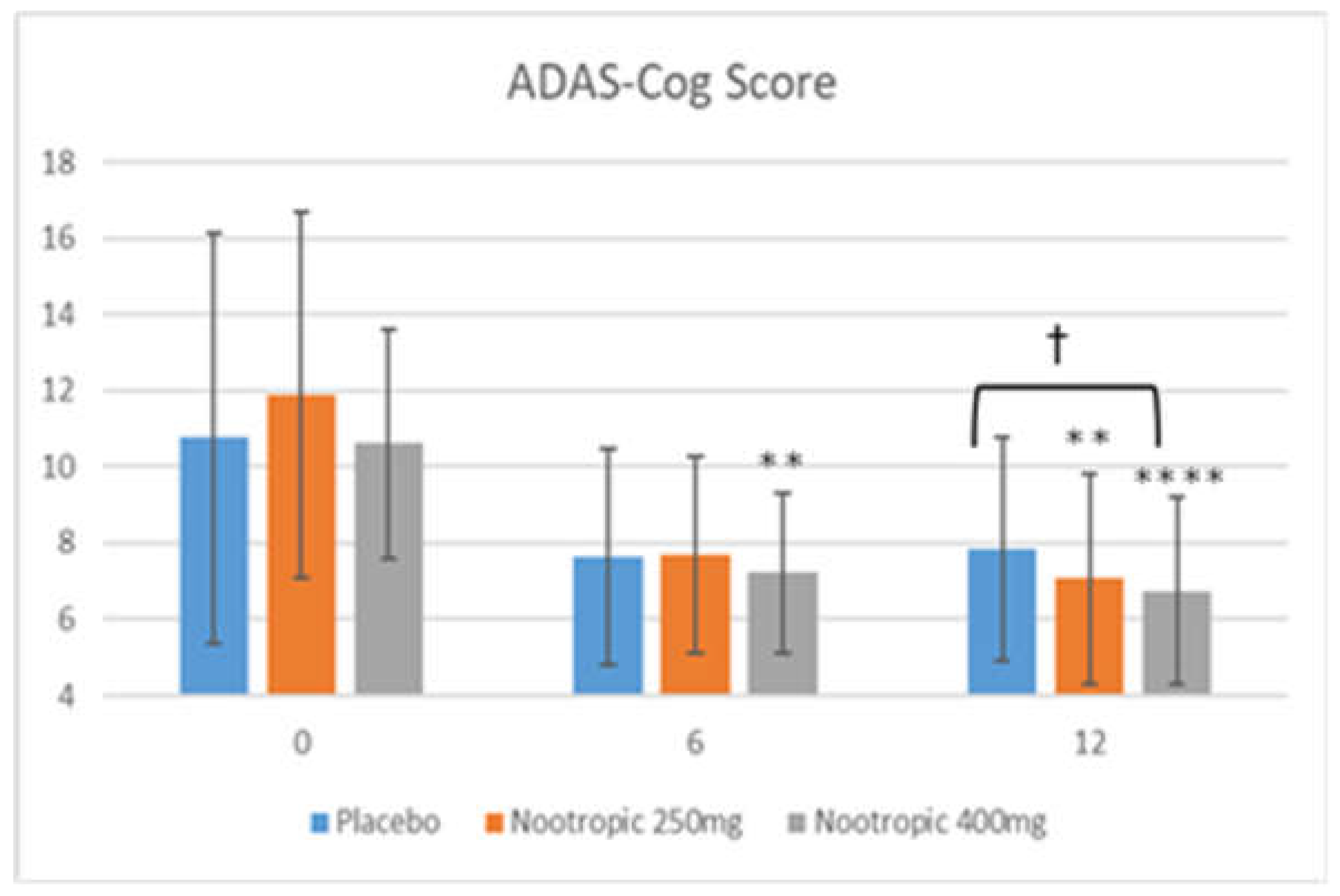
The ADAS-Cog test was also assessed at baseline, 6 weeks and 12 weeks of intake (Figure 9). At baseline, it was observed that the subjects were healthy at the cognitive levels, with average values below 17 (values above 17 indicate cognitive impairment) [18]. Intragroup analysis revealed that the placebo group did not present any significant differences with respect to baseline. On the other hand, the experimental product, at both doses, revealed significant improvements. Specifically, the 250mg dose revealed a significant improvement compared to baseline at 12 weeks of intake, while the 400mg dose significantly improved ADAS-Cog scores at both 6 and 12 weeks. With respect to the intergroup analysis, at week 12 it was observed that the groups consuming the experimental product presented significantly lower results than the placebo. No significant difference was observed comparing the 250mg and 400mg dose. Therefore, this suggests that the product at either dose can help improve this test after 12 weeks of intake.
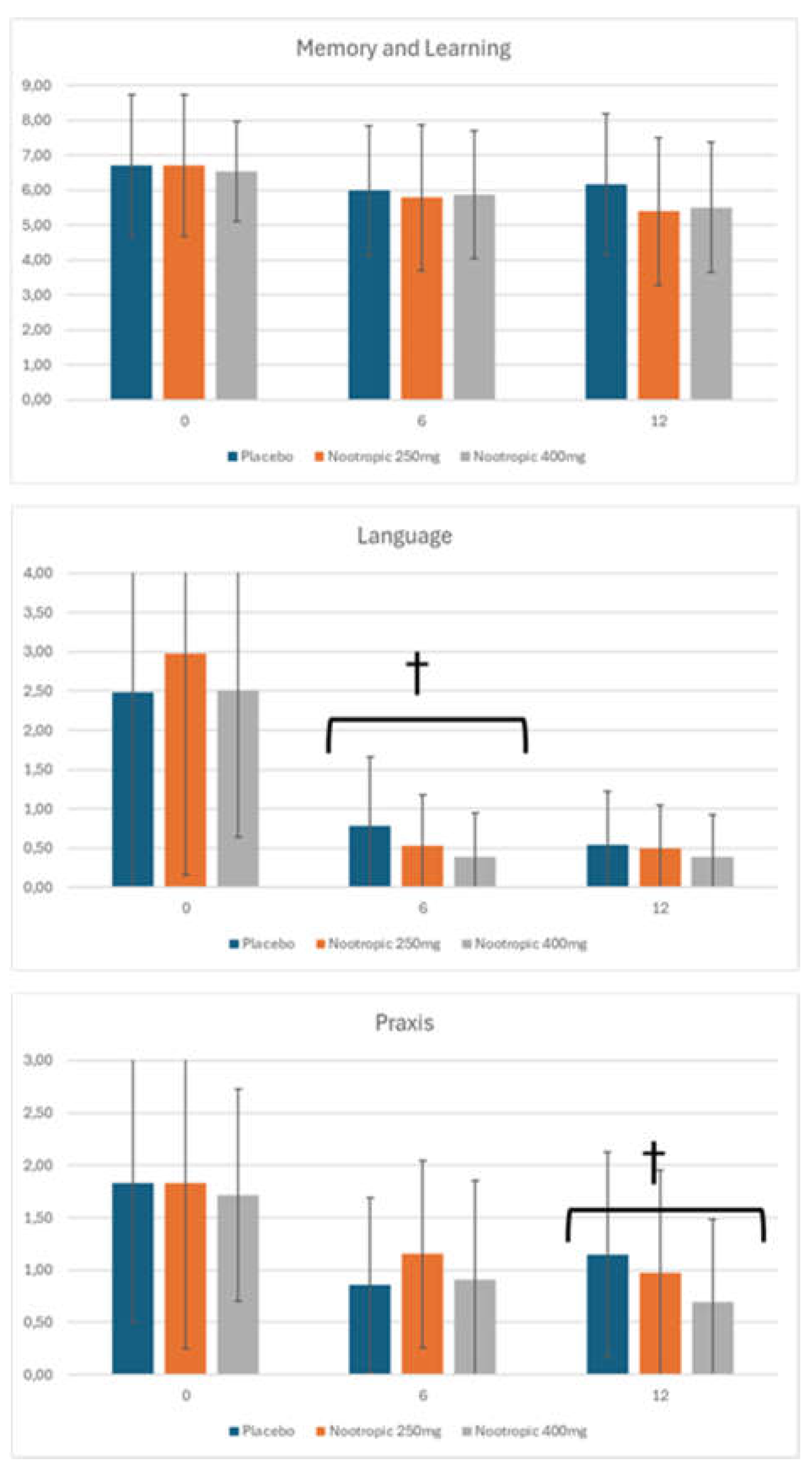
The ADAS-Cog questionnaire analyzes 3 types of memory, subdivided into a total of 11 items: memory and new learning (word recall, orientation, word recognition, remembering test instructions), language (commands, spoken language ability, naming objects, word-finding difficulty, comprehension) and praxis (constructional and ideational).
When analyzing the 3 types of memory, it was revealed that the product significantly improved language skills (after 6 weeks of intake) and praxis (after 12 weeks of intake), when comparing to the placebo group (Figure 10). No significant effect vs. placebo was detected in the memory and learning category.
Analysis of each individual item was also performed (Table 3). Briefly, there were several items where all study groups significantly improved throughout time, with no difference among the groups: orientation, spoken language ability, word-finding difficulty, comprehension, and naming objects/fingers. Also, there were certain items where the experimental groups improved throughout time, but the placebo group remained unchanged: word recall (both 250mg and 400mg at week 6, only 400mg at week 12), word recognition (250mg only, week 6), and commands (both doses at week 6, only 250mg at week 12).
The items with intergroup differences were: word recall (both 250mg and 400mg at week 12) and commands (both 250mg and 400mg at week 6). Also, a significant improvement was observed in constructional praxis at week 12 when comparing the nootropic at the 400mg dose vs 250mg.
Therefore, overall the results of the ADAS-Cog test reveals that both 250mg and 400mg contribute to significantly improve the global score after 12 weeks of intake, compared to placebo. Compared to baseline, both doses revealed significant improvements, with a dose-dependent response. Regarding the types of memory, both doses seem to significantly improve Language (at week 12) and Praxis (at week 6), compared to placebo. As for the analysis of the individual items, both doses contribute to improve the word recall item of the Memory and Learning category (at week 12), and the command item in the Language category (at week 6) when comparing to placebo. Word recall (both doses), word recognition (250mg only) and commands (both doses) significantly improved versus baseline values in the experimental groups.

4. Discussion

In this study, we assessed the antioxidant and protective effects of 9 different botanical extracts on human Neural Stem Cells (NSC), using the ROS/RNS antioxidant assay. Additionally, we also evaluated the protective capacity of different products measuring gene expression regulation after H2O2-induced toxicity in NSC.
The botanical extracts chosen for the analysis was based on a bibliographical review of potential candidates, where both preclinical and clinical studies have been performed in patients with dementia as well as healthy older adults. Both Vaccinium myrtillus (bilberry) and Vaccinium angustifolium (blueberry) were selected because they are rich in anthocyanins, which have been demonstrated to display anti-inflammatory and neuroprotective properties [19,20]. Specifically, bilberry anthocyanins have been shown to help reduce inflammation in aging rats [21]. Blueberries have been tested in a clinical setting on several occasions, with significant improvements observed in cognitive performance, execution and greater brain activity [22,23,24,25]. Bacopa monnieri (bacopa) contains bacosides, which have been shown to possess anti-inflammatory and antioxidant properties in the brain [26]. Clinical trials in patients with Alzheimer´s disease revealed significant results in various aspects of the MMSE [27], while studies in healthy adults revealed more limited results [28,29,30,31,32]. Salvia officinalis (sage) may not be as well known as other popular nootropics such as the bacopa or ginkgo biloba, but there is previous evidence that its components can act upon the brain through several mechanisms, including antioxidant/anti-inflammatory effects, cholinergic activity, trophic factor release and on amyloid-beta [33]. A few clinical studies have shown positive results in cognitive health [34–36and mood both in patients with Alzheimer´s disease and healthy older adults [34,35,36]. Crocus sativa stigmas (saffron) contain safranal and crocins [37], and is suggested to exert its effect on the brain through its antioxidant properties. Several clinical studies have also been conducted in both patients with Alzheimer´s disease and healthy adults, with results in both cognitive health and mood [38,39,40,41]. Rhodiola rosea (arctic root) is considered an adaptogen and is suggested to possess neuroprotective properties [42,43]. Clinical results with this botanical have shown positive effects related to mood and mental performance [44,45,46]. Ginkgo biloba is one of the most widely known and studied botanical extracts for cognitive health, and meta-analysis and systemic reviews revealed that it could be effective to improve memory, concentration, fatigue, stress and mood [47,48]. The same can be said with Panax ginseng [48,49]. Lastly, Centella asiatica (gotu kola) contains asiaticosides, which has been shown to possess neuroprotective properties mainly by reducing amyloid-beta deposition [50]. Clinical trials are less abundant with gotu kola despite positive results observed in healthy older adults [51].
The in vitro results indicated that only blueberry, sage and arctic root revealed a significant antioxidant effect, while the other extracts either did not have an effect or were even pro-oxidant (such as saffron and ginkgo). As for trophic factor release, sage was the only extract to increase the expression of all the factors analyzed: Nrf2, NGF and VEGF. For this reason, Salvia officinalis, or sage, standardized at 4% rosmarinic acid, was chosen as the main ingredient for a nootropic formula.
After selecting Salvia officinalis as the candidate botanical extract, tests were conducted analyzing sage and in combination with rutin, a polyphenol with known nootropic properties [14,16]. The results reveal an enhanced effect in increasing BDNF production when combining sage with rutin. BDNF is considered one of the most important molecules implicated in memory [52]. Studies revealed that low BDNF levels could be a biomarker for early dementia diagnosis, especially Alzheimer´s disease [53]. Increasing BDNF expression is one of the emerging therapeutic strategies for dementia [54]. In this respect, a botanical extract that increases BDNF may be a plausible solution to delay the progression in older adults with predementia. Studies have shown that compounds present in sage can increase BDNF expression [55].
The botanical blend also contributed to increase AChE inhibition in hippocampal cells, which is a potential biomarker for dementia. One of the suggested hypotheses for Alzheimer´s disease onset is due to the low levels of neurotransmitters such as acetylcholine, which causes memory loss and cognitive decline [56]. Cholinesterases, particularly acetylcholinesterase (AChE), breaks down cholinesterase and subsequently inhibits neurotransmission in cholinergic neurons. There are numerous natural botanicals that could potentially contribute to improve Alzheimer´s disease progress, including by inhibiting AChE [57].
There are several studies suggesting that components present in Salvia officinalis may help lower AChE activity [58,59,60,61]. This includes in vitro and animal studies, and several components, including rosmarinic acid, have been identified to be involved in this AChE inhibition. This inhibiting effect has been clinically shown to help improve memory, anxiety and mood in older adults [62,63]. However, the studies demonstrated the short-term effect of the extract in the few hours after consumption, and a longer exposure to the extract was not assessed.
Once the final formula was defined, a clinical trial in healthy older adults was conducted using 2 different doses, 250mg and 400mg, and compared to a placebo. Two questionnaires were conducted in the clinical study: MoCA and ADAS-Cog. The results of the MoCA test revealed significant improvements with the 400mg dose when comparing the intermediate measurement (week 6) with the end of the study (week 12). When analyzing the 7 individual categories, both doses contribute to improve short term memory recall, while abstract reasoning seemed to improve only in the 400mg dose.
As for the ADAS-Cog test, the results revealed that both doses significantly improved the global score after 12 weeks of intake, compared to placebo. Intragroup analysis revealed significant improvements only in the experimental product, with earlier significant results observed at the higher dose. Regarding the types of memory, both doses seem to significantly improve Language (at week 12) and Praxis (at week 6), compared to placebo. As for the analysis of the individual items, both doses contributed to improve Word Recall and Commands compared to both baseline values and vs placebo, with Word Recognition also detected in the 250mg dose only when compared to baseline.
Overall, the results of the clinical trial suggests that the combination of sage and rutin may help improve short-term memory recall. In this test, the subjects must perform two learning trials and then try to recall them after 5 minutes. A significant improvement in this test may suggest improved learning skills, with increased focus and concentration. Memory recall is usually one of the first traits where impairment appears while aging [64]. This result may indicate improvements in memory retention and increased focus and concentration. Interestingly, a significant improvement in abstract reasoning was also detected. Abstract reasoning generally decreases in older age, as opposed to other forms of creativity which seem to maintain stable [65]. Finally, the results in the ADAS-Cog suggest improvements in language and praxis, which are more related to long-term memory skills. As ADAS-Cog is generally reserved for later stages of dementia, individuals with mild cognitive impairment generally score at ceiling (a score of 0 in the ADAS-Cog) on 8 of the 11 times tested [66].

5. Conclusions

The analysis of various botanical extracts on their effectiveness in reducing ROS/RNS, enhancing cell survival and increasing the expression of several trophic factors in neural stem cells submitted to oxidative stress revealed that Salvia officinalis extract was the most efficient of the extracts analyzed. This extract, when combined with rutin, revealed additional benefits. The use of this blend in a clinical setting revealed significant improvements in certain memory capabilities in older adults with mild cognitive impairment.

Author Contributions

Conceptualization, J.J. and N.C.; methodology, J.J. and J.L.M.; software, J.M. and P.N.; validation, J.J., N.C., and J.L.M.; formal analysis, J.L.M. and J.M.; investigation, J.L.M. and J.M.; resources, J.J. and J.L.M.; data curation, J.L.M., J.M. and P.N.; writing—original draft preparation, J.J.; writing—review and editing, N.C., P.N. and J.L.M.; visualization, J.J. and J.L.M.; supervision, J.J. and J.L.M.; project administration, J.J. and J.L.M.; funding acquisition, J.J. All authors have read and agreed to the published version of the manuscript.

Funding

This research received no external funding.

Institutional Review Board Statement

The study was conducted in accordance with the Declaration of Helsinki and approved by the Institutional Review Board (or Ethics Committee) of Bionos Biotech (protocol code 0058-2023, 18/07/2023).

Informed Consent Statement

Informed consent was obtained from all subjects involved in the study.

Data Availability Statement

Data may be available upon request to the corresponding author.

Acknowledgments

We wish to thank the technical support received from Monteloeder and Bionos Biotech.

Conflicts of Interest

The authors declare no conflicts of interest.

References

  1. Giurgea, C.; Salama, M. Nootropic Drugs. Prog. Neuro-Psychopharmacol. 1977, 1, 235–247. [Google Scholar] [CrossRef]
  2. Vyas, S.; Kothari, S.; Kachhwaha, S. Nootropic Medicinal Plants: Therapeutic Alternatives for Alzheimer’s Disease. J. Herb. Med. 2019, 17, 100291. [Google Scholar] [CrossRef]
  3. Dormehl, I.C.; Jordaan, B.; Oliver, D.W.; Croft, S. SPECT Monitoring of Improved Cerebral Blood Flow During Long-Term Treatment of Elderly Patients with Nootropic Drugs. Clin. Nucl. Med. 1999, 24, 29–34. [Google Scholar] [CrossRef] [PubMed]
  4. Malík, M.; Tlustoš, P. Nootropic Herbs, Shrubs, and Trees as Potential Cognitive Enhancers. Plants (Basel), 2023, 12, 1364. [Google Scholar] [CrossRef] [PubMed]
  5. Kamkaew, N.; Norman Scholfield, C.; Ingkaninan, K.; Taepavarapruk, N.; Chootip, K. Bacopa Monnieri Increases Cerebral Blood Flow in Rat Independent of Blood Pressure. Phytother. Res. 2013, 27, 135–138. [Google Scholar] [CrossRef] [PubMed]
  6. Veerendra Kumar, M.H.; Gupta, Y.K. Effect of Different Extracts of Centella Asiatica on Cognition and Markers of Oxidative Stress in Rats. J. Ethnopharmacol. 2002, 79, 253–260. [Google Scholar] [CrossRef]
  7. Kim, D.H.; Kim, D.W.; Jung, B.H.; Lee, J.H.; Lee, H.; Hwang, G.S.; Kang, K.S.; Lee, J.W. Ginsenoside Rb2 Suppresses the Glutamate-Mediated Oxidative Stress and Neuronal Cell Death in HT22 Cells. J. Ginseng. Res. 2019, 43, 326–334. [Google Scholar] [CrossRef]
  8. Yonekura, L.; Martins, C.A.; Sampaio, G.R.; Monteiro, M.P.; César, L.A.M.; Mioto, B.M.; Mori, C.S.; Mendes, T.M.N.; Ribeiro, M.L.; Arçari, D.P.; et al. Bioavailability of Catechins from Guaraná (Paullinia cupana) and its Effect on Antioxidant Enzymes and other Oxidative Stress Markers in Healthy Human Subjects. Food Funct. 2016, 7, 2970–2978. [Google Scholar] [CrossRef]
  9. Singh, M.; Murthy, V.; Ramassamy, C. Modulation of Hydrogen Peroxide and Acrolein-Induced Oxidative Stress, Mitochondrial Dysfunctions and Redox Regulated Pathways by the Bacopa monniera Extract: Potential Implication in Alzheimer’s Disease. J. Alzheimer’s Dis. 2010, 21, 229–247. [Google Scholar] [CrossRef]
  10. Mori, K.; Obara, Y.; Hirota, M.; Azumi, Y.; Kinugasa, S.; Inatomi, S.; et al. Nerve Growth Factor-Inducing Activity of Hericium Erinaceus In 1321n1 Human Astrocytoma Cells. Biol. Pharm. Bull. 2008, 31, 1727–1732. [Google Scholar] [CrossRef]
  11. Sumiyoshi, E.; Matsuzaki, K.; Sugimoto, N.; Tanabe, Y.; Hará, T.; Katakura, M.; et al. Sub-Chronic Consumption of Dark Chocolate Enhances Cognitive Function and Releases Nerve Growth Factors: a Parallel-Group Randomized Trial. Nutrients, 2019, 11, 2800. [Google Scholar] [CrossRef] [PubMed]
  12. Decroix, L.; Tonoli, C.; Soares, D.D.; Tagougui, S.; Heyman, E.; Meeusen, R. Acute Cocoa Flavanol Improves Cerebral Oxygenation Without Enhancing Executive Function at Rest or After Exercise. Appl. Physiol. Nutr. Metab. 2016, 41, 1225–1232. [Google Scholar] [CrossRef] [PubMed]
  13. Malík, M.; Tlustoš, P. Nootropics as Cognitive Enhancers: Types, Dosage and Side Effects of Smart Drugs. Nutrients. 2022, 14, 3367. [Google Scholar] [CrossRef] [PubMed]
  14. Dubey, S.; Ganeshpurkar, A.; Bansal, D.; Dubey, N. Protective Effect of Rutin on Impairment of Cognitive Functions Due to Antiepileptic Drugs on Zebrafish Model. Indian J. Pharmacol. 2015, 47, 86–89. [Google Scholar]
  15. Bekinschtein, P.; Cammarota, M.; Katche, C.; Slipczuk, L.; Rossato, J.I.; Goldin, A.; et al. BDNF is Essential to Promote Persistence of Long-Term Memory Storage. Proc. Natl. Acad. Sci. United States Am. 2008, 105, 2711–2716. [Google Scholar] [CrossRef]
  16. Enogieru, A.B.; Haylett, W.; Hiss, D.C.; Bardien, S.; Ekpo, O.E. Rutin as a Potent Antioxidant: Implications for Neurodegenerative Disorders. Oxid. Med. Cell Longev. 2018, 2018, 624101. [Google Scholar] [CrossRef]
  17. Nyenhuis, D.L.; Reckow, J. Office- And Bedside-Based Screening for Cognitive Impairment and the Dementias: Which Tools to Use, Interpreting the Results, and What are the Next Steps? Clin. Geriatr. Med. 2023, 39, 15–25. [Google Scholar] [CrossRef]
  18. Kueper, J.K.; Speechley, M.; Montero-Odasso, M. The Alzheimer’s Disease Assessment Scale-Cognitive Subscale (ADAS-Cog): Modifications and Responsiveness in Pre-Dementia Populations. a Narrative Review. J. Alzheimers Dis. 2018, 63, 423–444. [Google Scholar] [CrossRef]
  19. Zhang, J.; Wu, J.; Liu, F.; Tong, L.; Chen, Z.; Chen, J.; He, H.; Xu, R.; Ma, Y.; Huang, C. Neuroprotective Effects of Anthocyanins and its Major Component Cyanidin-3-O-Glucoside (C3G) in the Central Nervous System: an Outlined Review. Eur. J. Pharmacol. 2019, 452, 858. [Google Scholar] [CrossRef]
  20. Zafra-Stone, S.; Yasmin, T.; Bagchi, M.; Chatterjee, A.; Vinson, J.A.; Bagchi, D. Berry Anthocyanins as Novel Antioxidants in Human Health and Disease Prevention. Mol. Nutr. Food Res. 2007, 51, 675–683. [Google Scholar] [CrossRef]
  21. Li, J.; Wu, T.; Li, N.; Wang, X.; Chen, G.; Lyu, X. Bilberry Anthocyanin Extract Promotes Intestinal Barrier Function and Inhibits Digestive Enzyme Activity by Regulating the Gut Microbiota in Aging Rats. Food Funct. 2019, 10, 333–343. [Google Scholar] [CrossRef] [PubMed]
  22. Krikorian, R.; Shidler, M.D.; Nash, T.A.; Kalt, W.; Vinqvist-Tymchuk, M.R.; Shukitt-Hale, B.; Joseph, J.A. Blueberry Supplementation Improves Memory in Older Adults. J. Agric. Food Chem. 2010, 58, 3996–4000. [Google Scholar] [CrossRef] [PubMed]
  23. Miller, M.G.; Hamilton, D.A.; Joseph, J.A.; Shukitt-Hale, B. Dietary Blueberry Improves Cognition Among Older Adults in a Randomized, Double-Blind, Placebo-Controlled Trial. Eur. J. Nutr. 2018, 57, 1169–1180. [Google Scholar] [CrossRef] [PubMed]
  24. Mcnamara, R.K.; Kalt, W.; Shidler, M.D.; McDonald, J.; Summer, S.S.; Stein, A.L.; Stover, A.N.; Krikorian, R. Cognitive Response to Fish Oil, Blueberry, and Combined Supplementation in Older Adults with Subjective Cognitive Impairment. Neurobiol. Aging. 2018, 64, 147–156. [Google Scholar] [CrossRef] [PubMed]
  25. Bowtell, J.L.; Aboo-Bakkar, Z.; Conway, M.E.; Adlam, A.L.R.; Fulford, J. Enhanced Task-Related Brain Activation and Resting Perfusion in Healthy Older Adults After Chronic Blueberry Supplementation. Appl. Physiol. Nutr. Metab. 2017, 42, 773–779. [Google Scholar] [CrossRef]
  26. Abdul-Manap, A.S.; Vijayabalan, S.; Madhavan, P.; Chia, Y.Y.; Arya, A.; Wong, E.H. , et al. Bacopa monnieri, a Neuroprotective Lead in Alzheimer Disease: a Review on its Properties, Mechanisms of Action, and Preclinical and Clinical Studies. Drug Target Insights. 2019, 13, 1177392819866412. [Google Scholar] [CrossRef]
  27. Goswami, S.; Saoji, A.; Kumar, N.; Thawani, V.; Tiwari, M.; Thawani, M. Effect of Bacopa monnieri on Cognitive Functions in Alzheimer’s Disease Patients. Int. J. Collaborative Res. Intern. Med. Public Health. 2011, 3, 285–93. [Google Scholar]
  28. Kumar, N.; Abichandani, L.G.; Thawani, V.; Gharpure, K.J.; Naidu, M.U.; Venkat-Ramana, G. Efficacy of Standardized Extract of Bacopa Monnieri (Bacognize®) on Cognitive Functions of Medical Students: a Six-Week, Randomized Placebo-Controlled Trial. Evid. Based Complement. Alternat. Med. 2016, 2016, 4103423. [Google Scholar] [CrossRef]
  29. Calabrese, C.; Gregory, W.L.; Leo, M.; Kraemer, D.; Bone, K.; Oken, B. Effects of a Standardized Bacopa monnieri Extract o Cognitive Performance, Anxiety, and Depression in the Elderly: a Randomized, Double-Blind, Placebo-Controlled Trial. J. Altern. Complement. Med. 2008, 14, 707–713. [Google Scholar] [CrossRef]
  30. Stough, C.; Lloyd, J.; Clarke, J.; Downey, L.A.; Hutchison, C.W.; Rodgers, T.; et al. The Chronic Effects of an Extract of Bacopa monniera (Brahmi) on Cognitive Function in Healthy Human Subjects. Psychopharmacology. 2001, 156, 481–484. [Google Scholar] [CrossRef]
  31. Stough, C.; Downey, L.A.; Lloyd, J.; Silber, B.; Redman, S.; Hutchison, C.; et al. Examining the Nootropic Effects of a Special Extract of Bacopa monnieri on Human Cognitive Functioning: 90 Day Double-Blind Placebo-Controlled Randomized Trial. Phytother. Res. 2008, 22, 1629–1634. [Google Scholar] [CrossRef] [PubMed]
  32. Morgan, A.; Stevens, J. Does Bacopa monnieri Improve Memory Performance in Older Persons? Results of a Randomized, Placebo-Controlled, Double-Blind Trial. J. Altern. Complement. Med. 2010, 16, 753–759. [Google Scholar] [CrossRef] [PubMed]
  33. Lopresti, A.L. Salvia (Sage): a Review of its Potential Cognitive-Enhancing and Protective Effects. Drugs R.D. 2017, 17, 53–64. [Google Scholar] [CrossRef] [PubMed]
  34. Kennedy, D.; Pace, S.; Haskell, C.F.; Okello, E.J.; Milne, A.; Scholey, A. Effects of Cholinesterase Inhibiting Sage (Salvia Officinalis) on Mood, Anxiety and Performance on a Psychological Stressor Battery. Neuropsychopharmacology. 2006, 31, 845–852. [Google Scholar] [CrossRef] [PubMed]
  35. Akhondzadeh, S.; Noroozian, M.; Mohammadi, M.; Ohadinia, S.; Jamshidi, A.H.; Khani, M. Salvia Officinalis Extract in the Treatment of Patients with Mild to Moderate Alzheimer’s Disease: a Double Blind, Randomized and Placebo-Controlled Trial. J. Clin. Pharm. Ther. 2003, 28, 53–59. [Google Scholar] [CrossRef]
  36. Scholey, A.; Tildesley, N.T.J.; Ballard, C.; Wesnes, K.; Tasker, A.; Perry, E.K.; Kennedy, D. An Extract of Salvia (Sage) with Anticholinesterase Properties Improves Memory and Attention in Healthy Older Volunteers. Psychopharmacology. 2008, 198, 127–139. [Google Scholar] [CrossRef]
  37. Christodoulou, E.; Kadoglou, N.P.; Kostomitsopoulos, N.; Valsami, G. Saffron: A Natural Product with Potential Pharmaceutical Applications. J. Pharm. Pharmacol. 2015, 67, 1634–1649. [Google Scholar] [CrossRef]
  38. Akhondzadeh, S.; Sabet, M.S.; Harirchian, M.H.; Togha, M.; Cheraghmakani, H.; Razeghi, S.; et al. Saffron in the Treatment of Patients with Mild to Moderate Alzheimer’s Disease: a 16-Week, Randomized And Placebo-Controlled Trial. J. Clin. Pharm. Ther. 2010, 35, 581–588. [Google Scholar] [CrossRef]
  39. Akhondzadeh, S.; Sabet, M.S.; Harirchian, M.H.; Togha, M.; Cheraghmakani, H.; Razeghi, S; et al. A 22-Week, Multicenter, Randomized, Double-Blind Controlled Trial of Crocus Sativus in the Treatment of Mild-to-Moderate Alzheimer’s Disease. Psychopharmacology. 2010, 207, 637–643. [Google Scholar] [CrossRef]
  40. Farokhnia, M.; Sabet, M.S.; Iranpour, N.; Gougol, A.; Yekehtaz, H.; Alimardani, R.; et al. Comparing the Efficacy and Safety of Crocus sativus L. with Memantine in Patients with Moderate to Severe Alzheimer’s Disease: a Double-Blind Randomized Clinical Trial. Hum Psychopharmacol. 2014, 29, 351–359. [Google Scholar] [CrossRef]
  41. Tsolaki, M.; Karathanasi, E.; Lazarou, I.; Dovas, K.; Verykouki, E.; Karacostas, A.; et al. Efficacy and Safety of Crocus sativus L. in Patients with Mild Cognitive Impairment: One Year Single-Blind Randomized, with Parallel Groups, Clinical Trial. J. Alzheimers. Dis. 2016, 54, 129–133. [Google Scholar] [CrossRef] [PubMed]
  42. Nabavi, S.F.; Braidy, N.; Orhan, I.E.; Badiee, A.; Daglia, M.; Nabavi, S.M. Rhodiola rosea L. and Alzheimer’s Disease: from Farm to Pharmacy. Phytother. Res. 2016, 30, 532–539. [Google Scholar] [CrossRef] [PubMed]
  43. Jowko, E.; Sadowski, J.; Dlugolecka, B.; Gierczuk, D.; Opaszowski, B.; Cieslinski, I. Effects of Rhodiola rosea Supplementation on Mental Performance, Physical Capacity, and Oxidative Stress Biomarkers in Healthy Men. J. Sport Health. Sci. 2018, 7, 473–480. [Google Scholar] [CrossRef]
  44. Darbinyan, V.; Kteyan, A.; Panossian, A.; Gabrielian, E.; Wikman, G.; Wagner, H. Rhodiola rosea in Stress Induced Fatigue--a Double Blind Cross-Over Study of a Standardized Extract Shr-5 with a Repeated Low-Dose Regimen on the Mental Performance of Healthy Physicians During Night Duty. Phytomedicine. 2000, 7, 365–371. [Google Scholar] [CrossRef] [PubMed]
  45. Cropley, M.; Banks, A.P.; Boyle, J. The Effects of Rhodiola rosea L. Extract on Anxiety, Stress, Cognition and Other Mood Symptoms. Phytother. Res. 2015, 29, 1934–1939. [Google Scholar] [CrossRef]
  46. Fintelmann, V.; Gruenwald, J. Efficacy and Tolerability of a Rhodiola rosea Extract in Adults with Physical and Cognitive Deficiencies. Adv. Ther. 2007, 24, 929–939. [Google Scholar] [CrossRef]
  47. Oken, B.S.; Storzbach, D.M.; Kaye, J.A. The Efficacy of Ginkgo Biloba on Cognitive Function in Alzheimer Disease. Arch. Neurol. 1998, 55, 1409–1415. [Google Scholar] [CrossRef]
  48. Ernst, E. The Risk-Benefit Profile of Commonly Used Herbal Therapies: Ginkgo, St. John’s Wort, Ginseng, Echinacea, Saw Palmetto, and Kava. Ann. Intern. Med. 2002, 136, 42–53. [Google Scholar] [CrossRef]
  49. Liu, L.; Zhang, C.S.; Zhang, A.L.; Cai, Y.; Xue, C.C. The Efficacy and Safety of Chinese Herbal Medicine for Mild Cognitive Impairment: a Systematic Review and Meta-Analysis of Randomized Placebo-Controlled Trials. Front. Pharmacol. 2024, 15, 1341074. [Google Scholar] [CrossRef]
  50. Chen, C.L.; Tsai, W.H.; Chen, C.J.; Pan, T.M. Centella asiatica Extract Protects Against Amyloid Β1–40-Induced Neurotoxicity in Neuronal Cells by Activating the Antioxidative Defence System. J. Tradit. Complement. Med. 2016, 6, 362–369. [Google Scholar] [CrossRef]
  51. Wattanathorn, J.; Mator, L.; Muchimapura, S.; Tongun, T.; Pasuriwong, O.; Piyawatkul, N.; Yimtae, K.; Sripanidkulchai, B.; Singkhoraard, J. Positive Modulation of Cognition and Mood in the Healthy Elderly Volunteer Following the Administration of Centella asiatica. J. Ethnopharmacol. 2008, 116, 325–332. [Google Scholar] [CrossRef] [PubMed]
  52. Bekinschtein, P.; Cammarota, M.; Medina, J.H. BDNF and Memory Processing. Neuropharmacology. 2014, 76 Pt C, 677–683. [Google Scholar] [CrossRef]
  53. Mori, Y.; Tsuji, M.; Oguchi, T.; Kasuga, K.; Kimura, A.; Futamura, A.; Sugimoto, A.; Kasai, H.; Kuroda, T.; Yano, S.; et al. Serum BDNF as a Potential Biomarker of Alzheimer’s Disease: Verification Through Assessment of Serum, Cerebrospinal Fluid, and Medial Temporal Lobe Atrophy. Front. Neurol. 2021, 12, 653267. [Google Scholar] [CrossRef] [PubMed]
  54. Ortega, A.; Chernicki, B.; Ou, G.; Parmar, M.S. From Lab Bench to Hope: Emerging Gene Therapies in Clinical Trials for Alzheimer’s Disease. Mol Neurobiol. 2024. [CrossRef] [PubMed]
  55. Chiang, N.; Ray, S.; Lomax, J.; Goertzen, S.; Komarnytsky, S.; Ho, C.T.; Munafo, J.P. Modulation of Brain-Derived Neurotrophic Factor (BDNF) Signaling Pathway by Culinary Sage (Salvia officinalis L. ). Int J Mol Sci. 2021, 22, 7382. [Google Scholar] [CrossRef]
  56. Mesulam, M.M. The Cholinergic Innervation of the Human Cerebral Cortex. Prog. Brain Res. 2004; 145, 67–78. [Google Scholar]
  57. Tripathi, P.N.; Lodhi, A.; Rai, S.N.; Nandi, N.K.; Dumoga, S.; Yadav, P.; Tiwari, A.K.; Singh, S.K.; El-Shorbagi, A.A.; Chaudhary, S. Review of Pharmacotherapeutic Targets in Alzheimer’s Disease and its Management using Traditional Medicinal Plants. Degener. Neurol. Neuromuscul. Dis. 2024, 14, 47–74. [Google Scholar] [CrossRef]
  58. Smach, M.A.; Hafsa, J.; Charfeddine, B.; Dridi, H.; Limem, K. Effects of Sage Extract on Memory Performance in Mice and Acetylcholinesterase Activity. Ann. Pharm. Fr. 2015, 73, 281–288. [Google Scholar] [CrossRef]
  59. Sallam, A.; Mira, A.; Ashour, A.; Shimizu, K. Acetylcholine Esterase Inhibitors and Melanin Synthesis Inhibitors from Salvia officinalis. Phytomedicine. 2016, 23, 1005–1011. [Google Scholar] [CrossRef]
  60. Merad, M.; Soufi, W.; Ghalem, S.; Boukli, F.; Baig, M.H.; Ahmad, K.; et al. Molecular Interaction of Acetylcholinesterase with Carnosic Acid Derivatives: a Neuroinformatics Study. CNS Neurol. Disord. Drug Targets. 2014, 13, 440–446. [Google Scholar] [CrossRef]
  61. Marcelo, F.; Días, C.; Martins, A.; Madeira, P.J.; Jorge, T.; Florencio, M.H.; et al. Molecular Recognition of Rosmarinic Acid from Salvia sclareoides Extracts by Acetylcholinesterase: a New Binding Site Detected by NMR Spectroscopy. Chemistry. 2013, 19, 6641–6649. [Google Scholar] [CrossRef]
  62. Kennedy, D.O.; Pace, S.; Haskell, C.; Okello, E.J.; Milne, A.; Scholey, A.B. Effects of Cholinesterase Inhibiting Sage (Salvia officinalis) on Mood, Anxiety and Performance on a Psychological Stressor Battery. Neuropsychopharmacology. 2006, 31, 845–852. [Google Scholar] [CrossRef] [PubMed]
  63. Scholey, A.B.; Tildesley, N.T.; Ballard, C.G.; Wesnes, K.A.; Tasker, A.; Perry, E.K.; et al. An Extract of Salvia (Sage) with Anticholinesterase Properties Improves Memory and Attention in Healthy Older Volunteers. Psychopharmacology. 2008, 198, 127–139. [Google Scholar] [CrossRef] [PubMed]
  64. Henneges, C.; Reed, C.; Chen, Y.F.; Dell’Agnello, G.; Lebrec, J. Describing the Sequence of Cognitive Decline in Alzheimer’s Disease Patients: Results from an Observational Study. J. Alzheimers Dis. 2016, 52, 1065–1080. [Google Scholar] [CrossRef] [PubMed]
  65. Ross, S.D.; Lachmann, T.; Jaarsveld, S.; Riedel-Heller, S.G.; Rodriguez, F.S. Creativity Across the Lifespan: Changes with Age and with Dementia. BMC Geriatr. 2023, 23, 160, Erratum In BMC Geriatr. 2023, 23, 307. [Google Scholar] [CrossRef]
  66. Grundman, M.; Petersen, R.C.; Ferris, S.H.; Thomas, R.G.; Aisen, P.S.; Bennett, D.A.; et al. Mild Cognitive Impairment can be Distinguished from Alzheimer Disease and Normal Aging for Clinical Trials. Arch Neurol. 2004, 61, 59–66. [Google Scholar] [CrossRef]
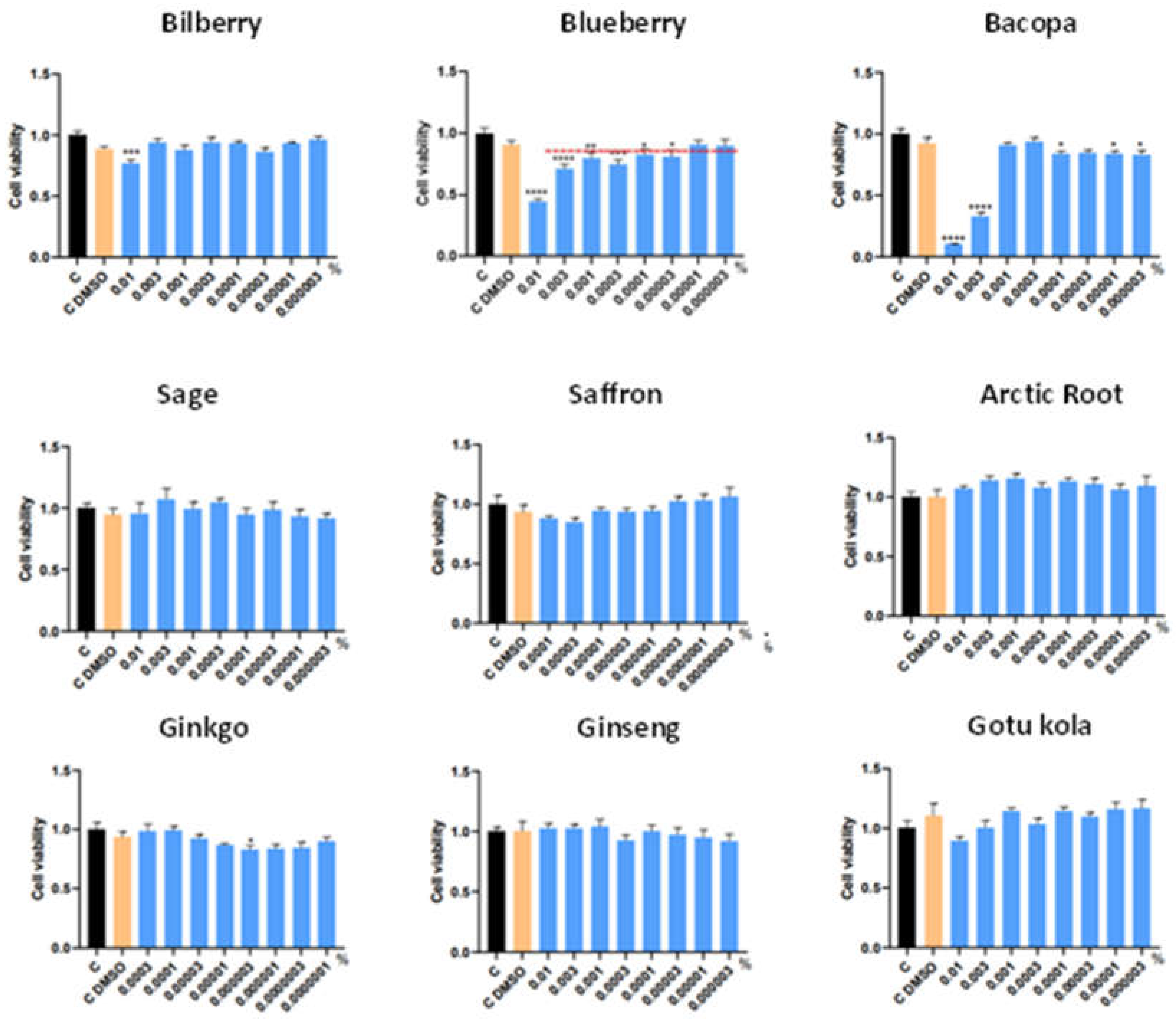
Figure 1. MTT analysis in neural stem cells and in presence of the botanical extracts, using DMSO as control. * p<0.05, ** p<0.01, *** p<0.005.
Preprints 119337 g001
Figure 2. ROS analysis of botanical extracts in neural stem cells exposed to H2O2 and in the presence of the botanical extracts. * p<0.05, ** p<0.01, *** p<0.005, **** p<0.001.
Preprints 119337 g002
Figure 3. RNS analysis in neural stem cells exposed to H2O2 and in the presence of the botanical extracts. * p<0.05, ** p<0.01, *** p<0.005, **** p<0.001.
Preprints 119337 g003
Figure 4. Caspase-9 gene expression analysis in neural stem cells exposed to H2O2 and in the presence of the botanical extracts. * p<0.05, ** p<0.01, *** p<0.005, **** p<0.001.
Preprints 119337 g004
Figure 5. Expression levels of trophic factors NRF2, NGF and VEGF in neural stem cells exposed to H2O2 and in the presence of the botanical extracts. * p<0.05, ** p<0.01, *** p<0.005, **** p<0.001.
Preprints 119337 g005aPreprints 119337 g005bPreprints 119337 g005c
Figure 6. Expression of BDNF in NSC cultured in the presence of sage or sage/rutin extract. ** p<0.01.
Preprints 119337 g006
Figure 7. AChE activity in hippocampal cell cultures, in the presence of sage/rutin at different concentrations. * p<0.05, ***p<0.005.
Preprints 119337 g007
Figure 8. MoCA general score of the healthy volunteers. * p<0.05.
Preprints 119337 g008
Figure 9. ADAS-Cog Scores. † p<0.05 vs. placebo.
Preprints 119337 g009
Figure 10. ADAS-Cog subscores. † p<0.05 vs. placebo.
Preprints 119337 g010
Table 1. Summary of results observed of neural stem cells submitted to oxidative stress and exposed to the various botanical extracts. Values indicate the concentrations where significant values were detected, whereas NS indicates not significant. P-ox: pro-oxidant effect, A-ox: antioxidant effect. * p<0.05, ** p<0.01, *** p<0.005, **** p<0.001.
Botanical Extract ROS RNS NRF2 NGF VEGF
Bilberry NS 0.0001% (*) NS 0.001% (*)
0.0001% (**)
NS
Blueberry 0.001% (*) 0.001%(****)
0.0001% (**)
NS 0.001% (*) NS
Bacopa NS NS NS 0.0001% (*) NS
Sage 0.01% (****) 0.01% (***)
0.0001% (*)
0.001% (***)
0.01% (***)
0.0001% (****) 0.01% (*)
0.001% (*)
Saffron P-ox:
0.0001% (*)
0.00005%(**)
NS NS 0.00005% (*) NS
Arctic Root 0.01% (***) 0.01% (*)
0.001% (*)
NS NS NS
Ginkgo P-ox: 0.0001%(***)
A-ox: 0.00005%(**)
0.00005%(****) NS 0.0001% (**) NS
Ginseng 0.01% (**)
0.0001% (**)
NS NS 0.0001% (**)
0.001% (*)
0.01% (***)
NS
Gotu kola NS NS NS 0.0001% (*) NS
Table 2. MoCA subscore assessment. *p<0.05, **p<0.01, ***p<0.005, †p<0.05 vs. placebo.
MoCA subscores Week 0 Week 6 Week 12
Short-term memory recall Placebo: 1.64 +/- 1.40
Nootropic 250mg: 2.10 +/- 1.47
Nootropic 400mg: 1.67 +/- 1.04
Placebo: 1.90 +/- 1.26
Nootropic 250mg: 2.78 +/- 1.43*
Nootropic 400mg: 1.77 +/- 1.48
Placebo: 2.14 +/- 1.27
Nootropic 250mg: 2.83 +/- 1.32***†
Nootropic 400mg: 2.51 +/- 1.24***
Visuospatial memory Placebo: 2.76 +/- 0.37
Nootropic 250mg: 2.83 +/- 0.30
Nootropic 400mg: 2.87 +/- 0.23
Placebo: 2.88 +/- 0.22 ***
Nootropic 250mg: 2.88 +/- 0.22**
Nootropic 400mg: 2.92 +/- 0.14
Placebo: 2.98 +/- 1.05 ***
Nootropic 250mg: 2.88 +/- 0.22***
Nootropic 400mg: 2.92 +/- 0.14***
Executive function Placebo: 3.55 +/- 0.99
Nootropic 250mg: 3.83 +/- 0.90
Nootropic 400mg: 3.44 +/- 0.97
Placebo: 4.26 +/- 0.74 ***
Nootropic 250mg: 4.20 +/- 0.68***
Nootropic 400mg: 4.23 +/- 0.63***
Placebo: 4.00 +/- 0.71 ***
Nootropic 250mg: 4.28 +/- 0.73***
Nootropic 400mg: 4.31 +/- 0.75***
Attention, concentration Placebo: 4.67 +/- 1.19
Nootropic 250mg: 4.70 +/- 1.16
Nootropic 400mg: 4.87 +/- 1.00
Placebo: 4.71 +/- 1.09
Nootropic 250mg: 4.70 +/- 1.06
Nootropic 400mg: 4.64 +/- 1.23
Placebo: 4.88 +/- 1.05
Nootropic 250mg: 4.93 +/- 0.95
Nootropic 400mg: 4.82 +/- 1.15
Language Placebo: 2.45 +/- 0.63
Nootropic 250mg: 2.55 +/- 0.56
Nootropic 400mg: 2.41 +/- 0.60
Placebo: 2.69 +/- 0.46 *
Nootropic 250mg: 2.68 +/- 0.49*
Nootropic 400mg: 2.46 +/- 0.55
Placebo: 2.60 +/- 0.52
Nootropic 250mg: 2.55 +/- 0.54
Nootropic 400mg: 2.44 +/- 0.64
Abstract reasoning Placebo: 1.71 +/- 0.42
Nootropic 250mg: 1.75 +/- 0.39
Nootropic 400mg: 1.77 +/- 0.36
Placebo: 1.88 +/- 0.21 ***
Nootropic 250mg: 1.88 +/- 0.23***
Nootropic 400mg: 1.95 +/- 0.10***
Placebo: 1.81 +/- 0.31
Nootropic 250mg: 1.85 +/- 0.26
Nootropic 400mg: 1.92 +/- 0.14***
Orientation in time and place Placebo: 5.98 +/- 0.05
Nootropic 250mg: 5.88 +/- 0.22
Nootropic 400mg: 5.95 +/- 0.10
Placebo: 5.95 +/- 0.09 ***
Nootropic 250mg: 5.98 +/- 0.05***
Nootropic 400mg: 6.00 +/- 0.00***
Placebo: 5.98 +/- 0.05***
Nootropic 250mg: 5.98 +/- 0.05***
Nootropic 400mg: 5.97 +/- 0.05***
Table 3. Average values obtained by the individual items of the ADAS-Cog test. *p<0.05, **p<0.01, ***p<0.005, †p<0.05 vs. placebo, †††p<0.005 vs. placebo, •p<0.05 nootropic 400mg vs 250mg.
ADAS-Cog Item Week 0 Week 6 Week 12
Word Recall Placebo: 4.28 +/- 1.05
Nootropic 250mg: 4.34 +/- 0.83
Nootropic 400mg: 4.41 +/- 0.78
Placebo: 3.91 +/- 1.07
Nootropic 250mg: 3.68 +/- 0.94*
Nootropic 400mg: 3.82 +/- 0.99*
Placebo: 4.06 +/- 0.93
Nootropic 250mg: 3.81 +/- 1.26†††
Nootropic 400mg: 3.66 +/- 1.07**†††
Orientation Placebo: 0.19 +/- 0.34
Nootropic 250mg: 0.53 +/- 0.17
Nootropic 400mg: 0.17 +/- 0.31
Placebo: 0.00 +/- 0.00***
Nootropic 250mg: 0.00 +/- 0.00***
Nootropic 400mg: 0.03 +/- 0.05***
Placebo: 0.02 +/- 0.05***
Nootropic 250mg: 0.05 +/- 0.09***
Nootropic 400mg: 0.03 +/- 0.05***
Word Recognition Placebo: 1.45 +/- 0.79
Nootropic 250mg: 1.31 +/- 0.77
Nootropic 400mg: 1.28 +/- 0.69
Placebo: 1.63 +/- 1.16
Nootropic 250mg: 1.59 +/- 0.95*
Nootropic 400mg: 1.56 +/- 0.11
Placebo: 1.45 +/- 1.10
Nootropic 250mg: 1.18 +/- 0.88
Nootropic 400mg: 1.23 +/- 0.79
Remembering Test Instructions Placebo: 0.69 +/- 0.72
Nootropic 250mg: 0.78 +/- 0.85
Nootropic 400mg: 0.59 +/- 0.69
Placebo: 0.37 +/- 0.55
Nootropic 250mg: 0.63 +/- 0.81
Nootropic 400mg: 0.46 +/- 0.64
Placebo: 0.57 +/- 0.71
Nootropic 250mg: 0.55 +/- 0.81
Nootropic 400mg: 0.53 +/- 0.72
Commands Placebo: 0.19 +/- 0.34
Nootropic 250mg: 0.35 +/- 0.52
Nootropic 400mg: 0.15 +/- 0.25
Placebo: 0.26 +/- 0.41
Nootropic 250mg: 0.10 +/- 0.18*†
Nootropic 400mg: 0.10 +/- 0.18†
Placebo: 0.12 +/- 0.21
Nootropic 250mg: 0.05 +/- 0.10**
Nootropic 400mg: 0.21 +/- 0.33*
Spoken Language Ability Placebo: 0.29 +/- 0.48
Nootropic 250mg: 0.35 +/- 0.58
Nootropic 400mg: 0.37 +/- 0.57
Placebo: 0.00 +/- 0.00***
Nootropic 250mg: 0.00 +/- 0.00***
Nootropic 400mg: 0.00 +/- 0.00***
Placebo: 0.00 +/- 0.00***
Nootropic 250mg: 0.00 +/- 0.00***
Nootropic 400mg: 0.00 +/- 0.00***
Word-finding Difficulty Placebo: 0.66 +/- 0.96
Nootropic 250mg: 0.65 +/- 0.88
Nootropic 400mg: 0.73 +/- 0.93
Placebo: 0.00 +/- 0.00***
Nootropic 250mg: 0.00 +/- 0.00***
Nootropic 400mg: 0.00 +/- 0.00***
Placebo: 0.00 +/- 0.00***
Nootropic 250mg: 0.05 +/- 0.1***
Nootropic 400mg: 0.03 +/- 0.05***
Comprehension Placebo: 0.36 +/- 0.56
Nootropic 250mg: 0.48 +/- 0.76
Nootropic 400mg: 0.44 +/- 0.64
Placebo: 0.00 +/- 0.00***
Nootropic 250mg: 0.00 +/- 0.00***
Nootropic 400mg: 0.00 +/- 0.00***
Placebo: 0.00 +/- 0.00***
Nootropic 250mg: 0.03 +/- 0.05***
Nootropic 400mg: 0.00 +/- 0.00***
Naming objects/ fingers Placebo: 0.95 +/- 1.09
Nootropic 250mg: 1.15 +/- 1.01
Nootropic 400mg: 0.88 +/- 0.81
Placebo: 0.50 +/- 0.69***
Nootropic 250mg: 0.43 +/- 0.60***
Nootropic 400mg: 0.30 +/-0.48***
Placebo: 0.40 +/- 0.58***
Nootropic 250mg: 0.39 +/- 0.48***
Nootropic 400mg: 0.18 +/- 0.31***
Constructional Praxis Placebo: 1.31 +/- 0.95
Nootropic 250mg: 1.30 +/- 1.11
Nootropic 400mg: 1.39 +/- 0.71
Placebo: 0.55 +/- 0.69***
Nootropic 250mg: 0.73 +/- 0.65***
Nootropic 400mg: 0.50 +/- 0.58***
Placebo: 0.62 +/- 0.68***
Nootropic 250mg: 0.47 +/- 0.65***
Nootropic 400mg: 0.41 +/- 0.55***•
Ideational Praxis Placebo: 0.48 +/- 0.68
Nootropic 250mg: 0.53 +/- 0.76
Nootropic 400mg: 0.37 +/- 0.55
Placebo: 0.29 +/- 0.44
Nootropic 250mg: 0.43 +/- 0.55
Nootropic 400mg: 0.45 +/- 0.59
Placebo: 0.55 +/- 0.63
Nootropic 250mg: 0.53 +/- 0.64
Nootropic 400mg: 0.31 +/- 0.47
Disclaimer/Publisher’s Note: The statements, opinions and data contained in all publications are solely those of the individual author(s) and contributor(s) and not of MDPI and/or the editor(s). MDPI and/or the editor(s) disclaim responsibility for any injury to people or property resulting from any ideas, methods, instructions or products referred to in the content.
Copyright: This open access article is published under a Creative Commons CC BY 4.0 license, which permit the free download, distribution, and reuse, provided that the author and preprint are cited in any reuse.
Alerts
Prerpints.org logo

Preprints.org is a free preprint server supported by MDPI in Basel, Switzerland.

Subscribe

© 2025 MDPI (Basel, Switzerland) unless otherwise stated