Preprint
Article

Peptide-Based Rapid and Selective Detection of Mercury in Aqueous Samples with Micro-Volume Glass Capillary Fluorometer

Altmetrics

Downloads

47

Views

21

Comments

0

A peer-reviewed article of this preprint also exists.

Submitted:

26 September 2024

Posted:

29 September 2024

You are already at the latest version

Alerts
Abstract
Mercury, a toxic heavy metal produced through both natural and anthropogenic processes, is found in all major Earth’s systems. Mercury's bioaccumulation characteristics in the human body have a significant impact on the liver, kidneys, brain, and muscles. In order to detect Hg2+ ions, a highly sensitive and specific fluorescent biosensor has been developed using a novel, modified seven amino acid peptide, FY7. The tyrosine ring in the FY7 peptide sequence forms a 2:1 complex with Hg2+ ions that are present in the water-based sample. As a result, the peptide's fluorescence emission decreases with higher concentrations of Hg2+. The FY7 peptide's performance was tested in the presence of Hg2+ ions and other metal ions, revealing its sensitivity and stability despite high concentrations. Conformational changes to the FY7 structure were confirmed by FTIR studies. Simultaneously, we designed a miniaturized setup to support an in-house-developed micro-volume capillary container for volume fluorometry measurements. We compared and verified the results from the micro-volume system with those from the commercial setup. The micro-volume capillary system accommodated only 2.9 µL of sample volume, allowing for rapid, sensitive, and selective detection of toxic mercury (II) ions ranging from 0.02 µM.
Keywords: 
Subject: Environmental and Earth Sciences  -   Water Science and Technology

1. Introduction

Mercury is present in the environment in a variety of forms, including elemental mercury and as a component of inorganic or organic compounds. Although elemental Hg(0) is not particularly toxic, it is easily oxidized to Hg2+, which is both highly reactive and highly toxic [1,2,3]. Despite stringent guidelines and regulations mercury is released to the environment through various industrial processes and uncontrolled waste disposal [4]. Exposure to mercury, primarily through ingestion and inhalation, remains a persistent concern. By consuming contaminated food or water living organisms accumulate mercury in their tissues over time. Difficulty in eliminating inorganic Hg compounds further strengthen this accumulation, leading to dangerously elevated mercury levels. Bioaccumulated mercury ions hinder functionality of proteins and enzymes by binding to their active sites or essential cofactors, such as thiol groups [5,6]. In result metabolic pathways and cellular functions are disrupted. Prolonged mercury exposure may weaken the immune system, making organisms more susceptible to infections and diseases [7,8]. Additionally, mercury generates reactive oxygen species (ROS) [9,10], which induces oxidative damage to lipids, proteins, and DNA. Mercury exposition is also associated with cardiovascular diseases, including heart attacks and epilepsy, neurological diseases like Parkinson’s and Alzheimer’s [11,12,13] as well as vision loss and potential fatality [14,15]. Furthermore, mercury can impair reproductive health, leading to developmental abnormalities, adverse effects on offspring, and reduced fertility [16,17]. Mercury is an environmentally persistent substance that resists degradation and remains in ecosystems for extended periods as it is continuously cycling through air, water, and soil. The toxicity varies with dosage and exposure time, therefore the development of new, rapid and precise detection methods is crucial to mitigate any health risks. Current detection methods of Hg2+ are atomic absorption spectrometry (AAS), inductively coupled plasma (ICP), and atomic fluorescence spectrometry (AFS) [18,19,20]. However, these conventional methods are often unsuitable for routine analysis because they require a significant time investment, complex sample preparation and specific, bulky instruments. It is essential to develop effective methods for the rapid, highly selective, and appropriate preliminary screening and field analysis of Hg2+. Among optical detection techniques used in general for heavy metal ions, it is essential to underscore quick and simple volume fluorometry methods. Fluorometry techniques significantly shorten the time necessary for measurements, producing nearly real-time results [21]. A variety of fluorometry techniques were successfully employed to detect extremely low concentrations of heavy metal ions with the use of specific fluorescent probes [22,23,24,25,26].These days, sensors that incorporate peptide motifs are starting to show promise as an alternative due to their simplicity in synthesis through 9-fluorenylmethoxycarbonyl (Fmoc) solid-phase peptide synthesis (SPPS) [27,28] and ability to be selective for metal ions.
This work propose a new, custom designed fluorescent probe FITC labelled FY7 peptide, meant for sensitive and rapid detection of Hg2+ ions in water samples. Fluorescein isothiocyanate (FITC) was chosen as the fluorescent moiety for FY7 labeling due to its rapid reactivity with primary amines and high quantum efficiency. FITC’s isothiocyanate group forms a stable thiourea bond with amine groups, allowing for efficient and stable conjugation. Additionally, its high quantum yield ensures strong fluorescence, making it an excellent choice for sensitive detection and imaging applications. [29,30]. FITC typically exhibits strong fluorescence around 520 nm when excited at an appropriate wavelength, usually around 490 nm [31]. The FY7 peptide is designed to generate changes in its fluorescence response when it binds to Hg2+ ions. In water-based samples, this response can be employed to detect and quantify the presence of Hg2+ ions. FY7 peptide’s behavior was investigated to evaluate its potential for bio-sensing applications and environmental monitoring. Peptide’s fluorescence response and selectivity were measured when exposed to a series of low concentrations of Hg2+ ions, as well as in the presence of seven other, different metal ions, using a commercial microplate spectrofluorometer. The proposed FY7 sensor exhibited exceptional water solubility, selectivity, and sensitivity. As a low-cost alternative to professional setups, we designed a custom, simple volume fluorometry setup employing glass capillary as a container. Capillaries were used in numerous studies and presented as a viable and straightforward alternative to well-established containers, such as well-plates or cuvettes, but offering a much lower sample volume for single measurement [32,33,34]. Here, FY7 peptide was investigated with capillary that required only 2.9 µL of sample volume. Comparing the results between high-end fluorometry device and custom setup almost identical responses were observed, highlighting the robustness of synthetized FY7 peptide. Application of the FY7 peptide in a cost-efficient, miniaturized optical setup showcases a promising advancement in fluorescence measurement technology to date.

2. Materials and Methods

2.1. Materials and Reagents

Fmoc-Rink Amide AM Resin (0.7 mmol/g) was purchasedbbb from Iris Biotech GmbH. Fmoc protected amino acids, Fmoc-βAla-OH Fmoc-Tyr(tBu)-OH, Fmoc-Lys(Boc)-OH, Fmoc-Ala-OH, Fmoc-Ser(tBu)-OH, Fmoc-Leu-OH, Fmoc-Leu-OH and Fmoc-Thr(tBu)-OH were purchased from CSBio (Shanghai) Ltd. Fluosescein-5-isothiocyanate FITC Isomer I (FITC), Oxyma pure, N,N’-Diisopropylcarbodiimide (DIC), N,N-diisopropylethylamine (DIEA), Piperidine, 99%, extra pure, Triisopropylsilane (TIS), 4-Morpholineethanesulfonic acid sodium salt (MES), Pb(NO3)2 and MnCl2 were obtained from Sigma-Aldrich. N,N-Dimethylformamide (DMF), 99.8% and Trifluoroacetic Acid (TFA) for synthesis, Chromium standard solution, Cadmium standard solution, Hg(NO3)2, ZnSO4, NaCl and KCl were gained from VWR International, LLC. Ethyl dieter was collected from POCH S.A.
D2O, 99.8 % was purchased from Thermo Scientific Chemicals. The Acetonitrile HPLC gradient grade, Acetonitrile hypergrade for LC-MS LiChrosolv®, water (LC-MS LiChrosolv®), trifluoracetic acid for the HPLC and formic acid (FA) (LC-MS LiChropur™) were purchased from Sigma-Aldrich. Doubly distilled water (Hydrolab-Reference purified) with conductivity not exceeding 0.05 µS/cm was used. All other chemicals used were of analytical reagent grade unless otherwise noted.

2.2. Peptide Synthesis and Characterization

The FY7 peptide with sequence of FITC-βAla-Tyr-Lys-Ala-Ser-Leu-Ile-Thr-NH2 was synthesized on Rink Amide resin (0,1 mmol) by microwave-assisted Fmoc solid-phase peptide synthesis (SPPS) [[i]]. Peptide chain elongation was carried out using an Initiator+ Alstra™ (Biotage, Sweden) automated microwave peptide synthesizer. Couplings were performed twice for 5 min at 75 °C using Fmoc-amino acid (5 equiv.), DIC (5 equiv.), and Oxyma (5 equiv.) in DMF. The Fmoc group was removed using a 20% piperidine solution in DMF at room temperature (1 x 3 min., 1 x 10 min.). FITC was introduced at the N-terminal end of the peptide in a separate step. For this purpose, FITC (3 equiv.) and DIEA (6 equiv.) were added to the peptidyl resin in the dark for 24 h. FITC-labeled peptide was cleaved from Rink Amide resin using the mixture cleavage solution TFA/TIS/H2O (95:2,5:2,5) for 2 h, precipitated with anhydrous, cold diethyl ether, and lyophilized. The obtained modified peptide was characterized using an analytical reverse-phase HPLC Shimadzu system (Prominence-i LC-2030C Plus, Shimadzu, Japan) with a Jupiter 4 µm Proteo, 90Å, 4,6 x 250 mm column, with UV detection at λ=224 nm, using the linear gradient method from 5 to 95% solvent B for 60 min at a flow rate of 1 mL/min., where solvent A was water and B was acetonitrile as eluents containing 0.1 % TFA. ESI MS in positive ion mode (+) was performed using a single quadrupole mass spectrometer (LCMS 2020 Shimadzu, Japan). Isocratic elution, 60% B, where eluent A consisted of water and 0.1% formic acid (LCMS grade) and eluent B consisted of acetonitrile (LCMS grade) containing 0.1% formic acid as eluents containing 0.1% formic acid (FA), at a flow rate of 1.5 ml/min. FITC-labeled peptide characterization: yellowish solid; Synthetic yield: 89%; HPLC purity > 90%; Rt 23,017 min.; ESI MS of peptide calculated value: 1253.43 (g/mol); Observed value: [M+2H]2+ m/z = 628.3.

2.3. General Fluorescence Measurements

We prepared all the metal ion solutions from Hg(NO3)2, Pb(NO3)2, MnCl2, ZnSO4, KCl, NaCl, Cd2+, and Cr3+ standard solutions in double-distilled water at a concentration of 10 mM. A stock solution of the peptide (10 mM) was prepared in double-distilled water and acetonitrile (9:1, v/v). Fluorescence spectra were measured using Multimode Microplate Reader Synergy H1MG (BioTek Instruments, United States) at an excitation wavelength of 480 nm on F-bottom 96-well plate with a working volume per well of 200 µL (Greiner Bio-one International GmbH, Austria).

2.4. Fluorescence Response of FY7 Peptide for Hg2+ Ions. Metal selectivity and Cross-Reactivity Studies

The fluorescent selection ability of FY7 to 8 metal ions (Hg2+, Pb2+, Mn2+, Zn2+, Cd2+, Cr3+, Na+ and K+) was investigated in MES buffer solutions (50 mM, pH 5,65). Separate solutions of each metal ion prepared at varying concentrations (0, 0.02, 0.05, 0.1, 0.5, 1, 2, 3, 5, and 10 µM) were individually added to the 5 μM FY7 solutions. The fluorescence spectra of the FY7 solutions with different concentrations of each metal ion were recorded.
A fluorescence titration experiment was used to investigate the binding properties and interactions of fluorescent molecules, such as FY7 with Hg2+ ions. For this purpose, the different concentrations of Hg2+ ion solutions (0, 0.02, 0.05, 0.1, 0.5, 1, 2, 3, 5, 10 µM) were added to the 5 μM FY7 in MES buffer solutions (50 mM, pH 5,65) and fluorescence spectra of the FY7 solutions were measured.
Cross-reactivity studies. To ensure this specificity, it is essential to test the response of the FY7 peptide to its target ion, Hg2;⁺, in the presence of other potentially interfering metal ions. The fluorescence responses of FY7 peptide (5 μM) to Hg2+ (0.5 equiv.) ions in the presence of seven different metal ions in MES buffer solutions (50 mM, pH 5,65) were measured both separately and in a mixture of all tested ions.

2.5. Characterization of the Peptide-Metal Ion Complexation

To determine the binding stoichiometry of FY7 peptide and Hg2+ ions the Job’s plot was performed. Fluorescence emission intensity, which is the total concentration of FY7 (0–5 μM) and Hg2+ ions (5–0 μM), was used to probe the Job’s plot, with a constant concentration of 5 μM. The measurements were performed in MES buffer solutions with a pH of 5.65 and a concentration of 50 mM. The standard deviation was determined by repeating each pro-portional concentration three times.
FTIR spectra of the free FY7 and FY7−Hg2+ complex in MES buffer solutions (50 mM, pD 5,65). The Invenio-R FTIR spectrometer (Bruker, Germany) was employed to measure the samples, which was equipped with an MTC detector that was cooled with liquid nitrogen. The cuvette was composed of two CaF2 windows that were separated by PTFE spacers with a thickness of approximately 90 μm. The temperature of 25 °C was maintained by mounting the cuvette within a thermostated housing. The spectra were acquired with a spectral resolution of 1 cm-1 and a total of 256 scans. The Savitzky-Golay method was employed to calculate the second derivatives, which involved 21 data points and a polynomial of degree 3. All solutions were prepared using heavy water (D2O). After 24 hours of incubation, the FY7−Hg2+ complex, buffer components, and dry FY7 peptide were suspended in D2O and lyophilized. The spectrometer was continuously purified with dry nitrogen during the measurements to reduce the impact of water vapor on the peptide amide bands. The water vapor correction was conducted in accordance with the algorithm and protocol recommended by Bruździak [35]. The algorithm and measurement protocol implemented guaranteed the effortless and nearly complete elimination of water vapor interference.

2.6. Low-Volume Fluorescence Measurements

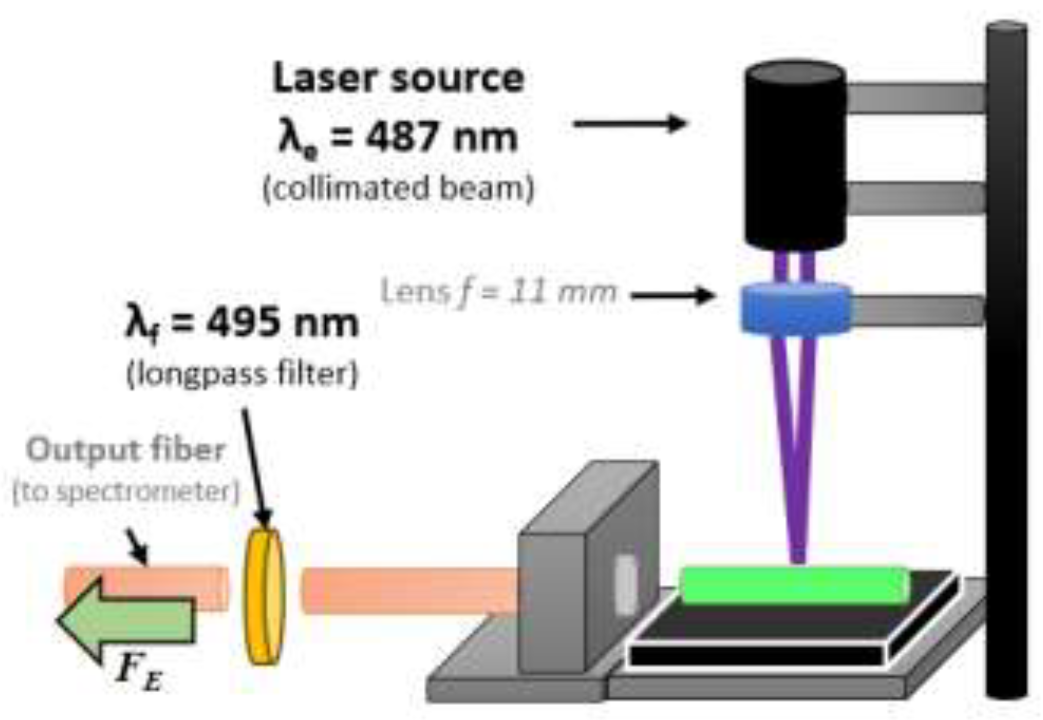
For low-volume measurements, a custom benchtop setup was designed. This setup includes a λL = 487 nm laser source (Lambdawave, with build- in collimating lenses), an aspherical lens with a focal length f = 11 mm (C340TMD-A, Thorlabs Inc., USA) for focusing the beam on a sample container, and a pair of optical fibers equipped with λF = 495 nm optical longpass filter. An optical detector (USB4000 spectrometer, Ocean Optics) collects spectra in the 200-900 nm wavelength range. The setup was assembled on an optical breadboard and secured with stands, rails, posts, and a 3-axis micro stage (Thorlabs Inc.) to ensure better alignment of the elements. The container used was a 15 mm long fused silica capillary with outer and inner diameters of 1000 and 500 μm, respectively. These capillaries accomodate a total volume of VC = 2.9 μL of the sample and were filled via capillary action by simply submerging one end in the selected liquid.
Optical measurements were performed in dark conditions at a room temperature of approximately 210C. The USB4000 spectrometer was set to an integration time of 12 ms. The measurement and data processing procedure began with collecting a blank spectrum (FB) when the capillary was filled only with the buffer solution. Subsequently, after filling with a sample and exposing it to the laser beam, the emission fluorescence spectrum (FE) was registered. The difference between these two measurements (ΔF=FE-FB) was used to identify the sample’s emission spectra. The USB4000 was set to average five repeatedly registered FE. The analysis involved tracing the peptide’s emission at λem, specified as the fluorescence intensity at the emission maximum IF = ΔF(λem).

3. Results and Discussion

In order to develop peptide-based micro-volume glass capillary fluorometer for rapid and selective, detection of mercury in aqueous samples a metal-binding peptide identified through phage display biopanning [36] was used. Affinity of the FY7 sequence for heavy metal ions was tested.

3.1. FY7 Peptide Synthesis and Characterization

Metal ion-binding peptide was synthesized by Fmoc/tBu microwave-assisted solid phase peptide synthesis [27].
FITC was introduced at the N−terminal side of the peptide in this study, preceded by a linker. In a microwave-assisted automated SPPS step, the β-Ala linker was incorporated into the growing peptide chain. The utilization of Fmoc-β-AlaOH as a linker in SPPS is a strategic advantage in the synthesis of FITC−modified peptides, ensuring the production of functionally intact, high-purity FITC−labeled peptides that are suitable for a variety of applications. The separation of the bulky fluorophore from the bioactive peptide sequence and the prevention of side reactions during the TFA−mediated cleavage process in solid phase peptide synthesis can be achieved by employing Fmoc-β-Ala-OH as a linker. The Fmoc-β-Ala-OH linker is a non-α amino acid, which means that it has a distinct structural configuration from standard α-amino acids. This distinction enables the spatial separation of the peptide chain from the fluorophore (FITC). Furthermore, the β-Ala linker can function as a protective spacer, thereby reducing the interaction between the reactive FITC group and the peptide backbone during the TFA cleavage process. This mitigates the likelihood of adverse reactions, including the unintended degradation of the modified peptide chain.[37]. As a consequence, the fluorescent peptide was designed and produced with a high purity (>90%) and yield (89%) through TFA-mediated cleavage.

3.2. FY7 Peptide Metal Selectivity and Cross-Reactivity Studies

Conducted fluorescence measurements verified that presented novel, FITC-labeled FY7 peptide can be employed as a biosensor that is both highly sensitive and specific to the Hg2+ ions. Initially, we investigated the affinity of the synthetic FY7 peptide for specific metal ions. In order to ascertain the selectivity of the FITC−labeled peptide (5 µM) toward eight distinct metal ions (Hg2+, Pb2+, Mn2+, Zn2+, Cd2+, Cr3+, Na+, and K+) in 50 mM MES buffer solutions at pH 5.65, the fluorescence spectra were analyzed. The FY7 probe exhibited a sensitive response exclusively to Hg2+ ions when compared to the fluorescence response of selected metal ions at three concentrations (0.5, 5.0 and 10 µM) (Figure 1). When the FY7 peptide was treated with the other 7 metal ions, no such effect was observed, which was approximately 4.3 times less than in the case of Hg2+. The interaction with mercury ions resulted in a substantial reduction in fluorescence intensity, as evidenced by the fluorescence quenching rate of approximately 48% for Hg2+. FY7 demonstrates a high level of selectivity toward Hg2+ ions, as evidenced by the selectivity experiment results.
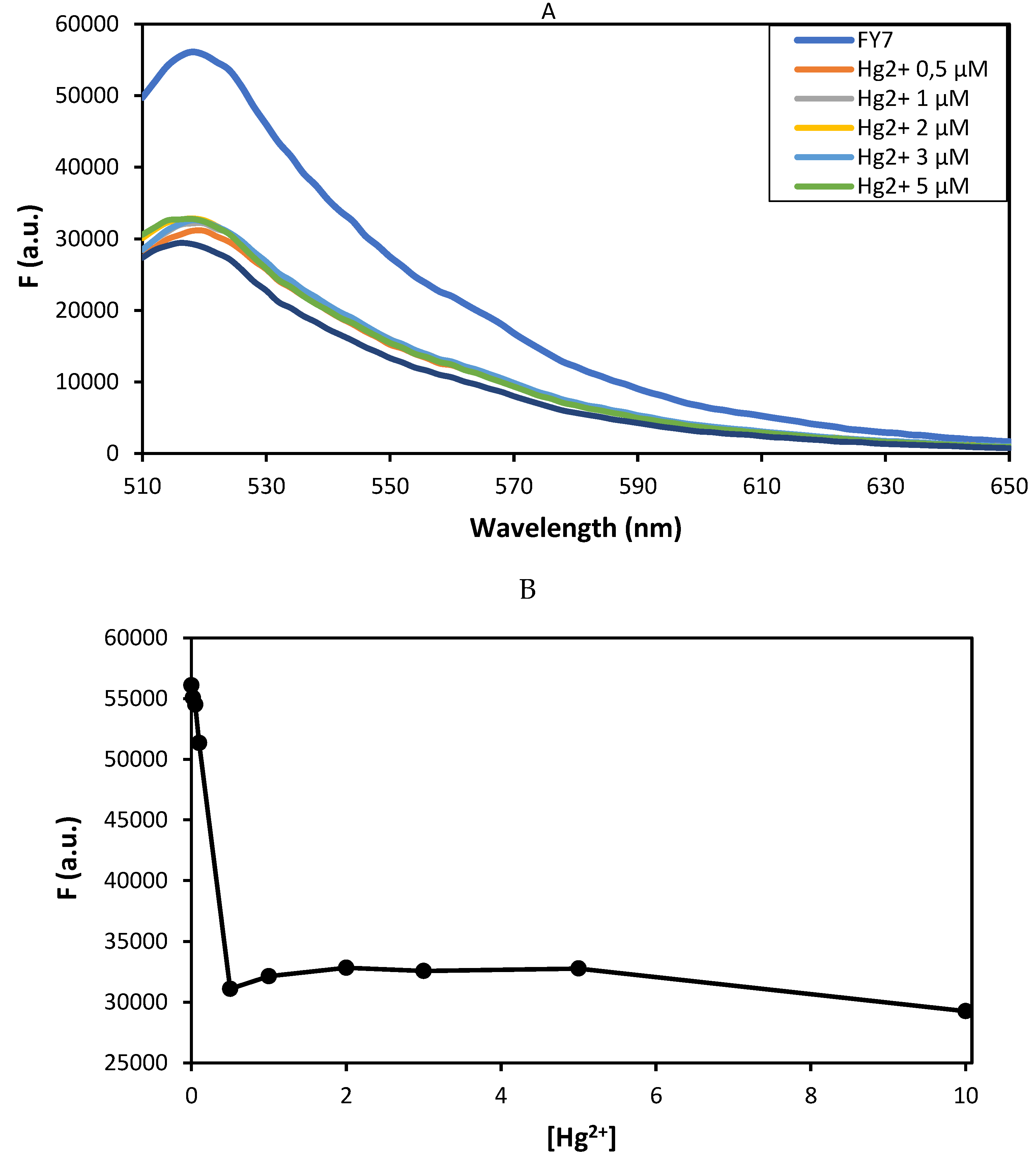
Upon interaction with Hg2+, the fluorescence spectra of the FY7 peptide at varying concentrations of Hg2+ ions in MES buffer solution (pH 5.65) exhibited substantial changes (Figure 2). A prominent fluorescence peak at approximately 518 nm is observed in the free FITC−labeled peptide, FY7, in the absence of mercury (II) ions, as illustrated in Figure 2A. the fluorescence intensity of FY7 decreases significantly upon the addition of Hg2+ ions. The fluorescence titration spectra of FY7 (5 µM) with Hg2+ demonstrated that the fluorescence intensity of FY7 was significantly reduced when the peptide-Hg2+ complex was formed. Based on the turn-off response, Hg2+ is the sole metal ion that diminishes the fluorescence intensity of the FY7 sample. The interaction between the labeled peptide and Hg2+ ions was demonstrated by the decrease in fluorescence intensity. The photoinduced electron transfer (PET) effect resulted in a decrease in fluorescence intensity as a result of the complex formation between the FY7 peptide and Hg2+ ions. This phenomenon arises when the excited fluorophore (FITC) transfers an electron to the Hg2+ ion, which is a strong electron acceptor due to its high electron affinity. This process results in non-radiative relaxation and fluorescence quenching.
The addition of 0−0.5 µM Hg2+.ions to the peptide caused a gradual decrease in its fluorescence intensity (Figure 2A). Furthermore, a quenching rate of approximately 48% was observed at a higher concentration of Hg2+ (ranging from 0.5−10 µM) when a significant turn-off response was observed (Figure 2B). It is intriguing that the titration curve achieved a stable plateau with the addition of only 0.1 equivalent of Hg2+ ions.
The Hg2+ ion in water systems can be rapidly and consistently detected by the FY7 peptide. Furthermore, the fluorescent quenching effect (turn-off response) demonstrates that the FITC-labeled peptide possesses exceptional selectivity and sensitivity toward Hg2+ ions.
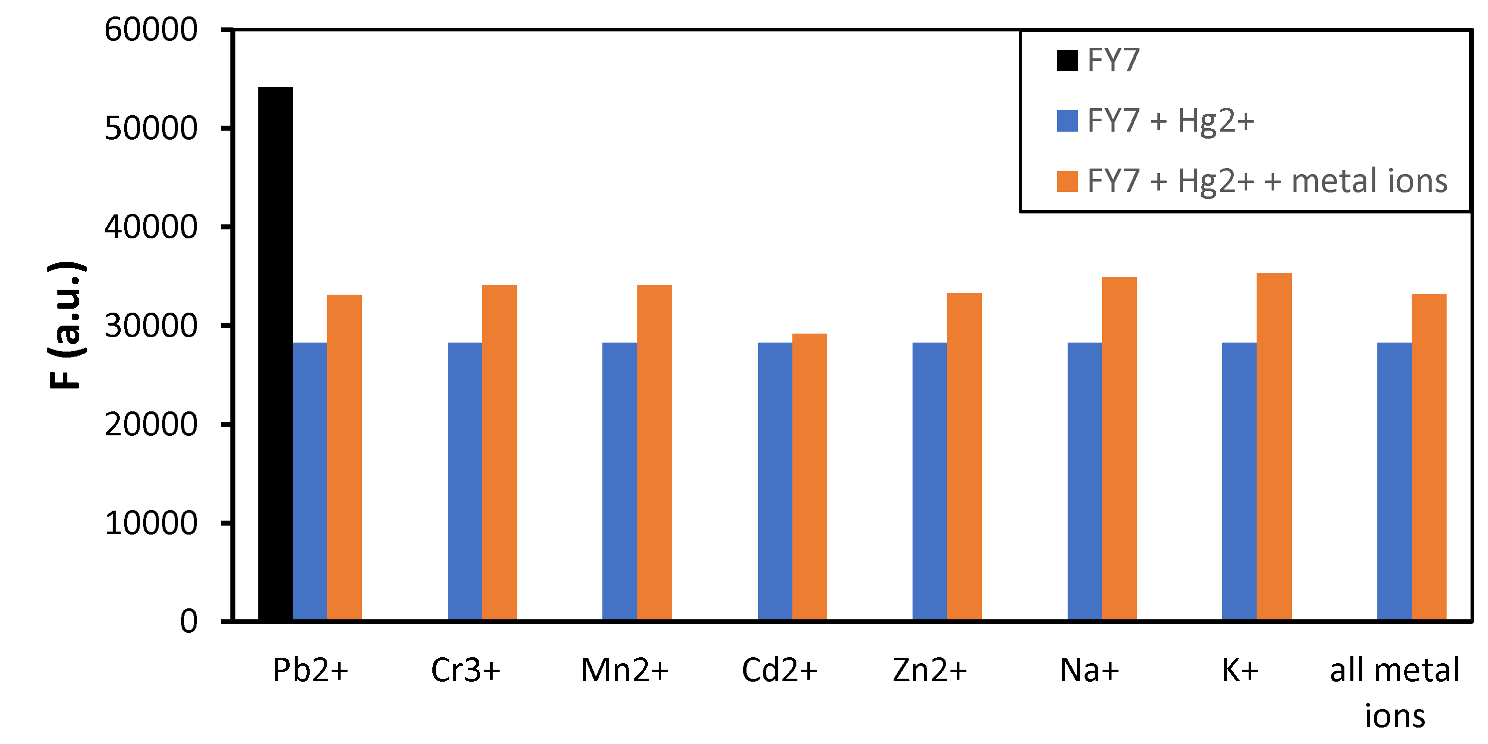
The fluorescence signal’s interpretation may be influenced by the peptide FY7’s response to multiple metal ions, which is referred to as cross-reactivity. In order to confirm the peptide biosensor’s affinity for mercury (II) ions, the FY7 probe was introduced into a mixture of Hg2+ ions (0.5 equiv.) and other 7 metal ions (5.0 equiv.) one at a time, and the resulting fluorescence emission spectra were analyzed. The fluorescence response of the FY7 was not affected by the presence of other divalent metal ions, such as Zn2+, Mn2+, and Cd2+, or trivalent chromium ions, as illustrated in Figure 3. Likewise, the presence of strong Lewis acid metal ions, such as Pb2+, did not interfere with the detection of Hg2+ ions, as they did not compete for binding to the peptide biosensor. Furthermore, the fluorescence emission of the peptide−Hg2+ complex was not significantly reduced by the simultaneous introduction of all the analyzed ions into the sample containing the FY7 peptide probe and Hg2+ ions. This properties is essential for the development of selective biosensors that can accurately identify and measure specific metal ions in complex mixtures.
The results indicate that the fluorescence can be regulated by an ON−OFF switch, which is consistent with the FITC−labeled peptide biosensor’s potent chelating ability toward Hg2+ ions. It is imperative to emphasize that the FY7 peptide maintains a high level of responsiveness to Hg2+ ions, even in the presence of high concentrations of other competitive ions.

3.3. Molecular Characterisation of FY7 and Hg2+ Ions Interation

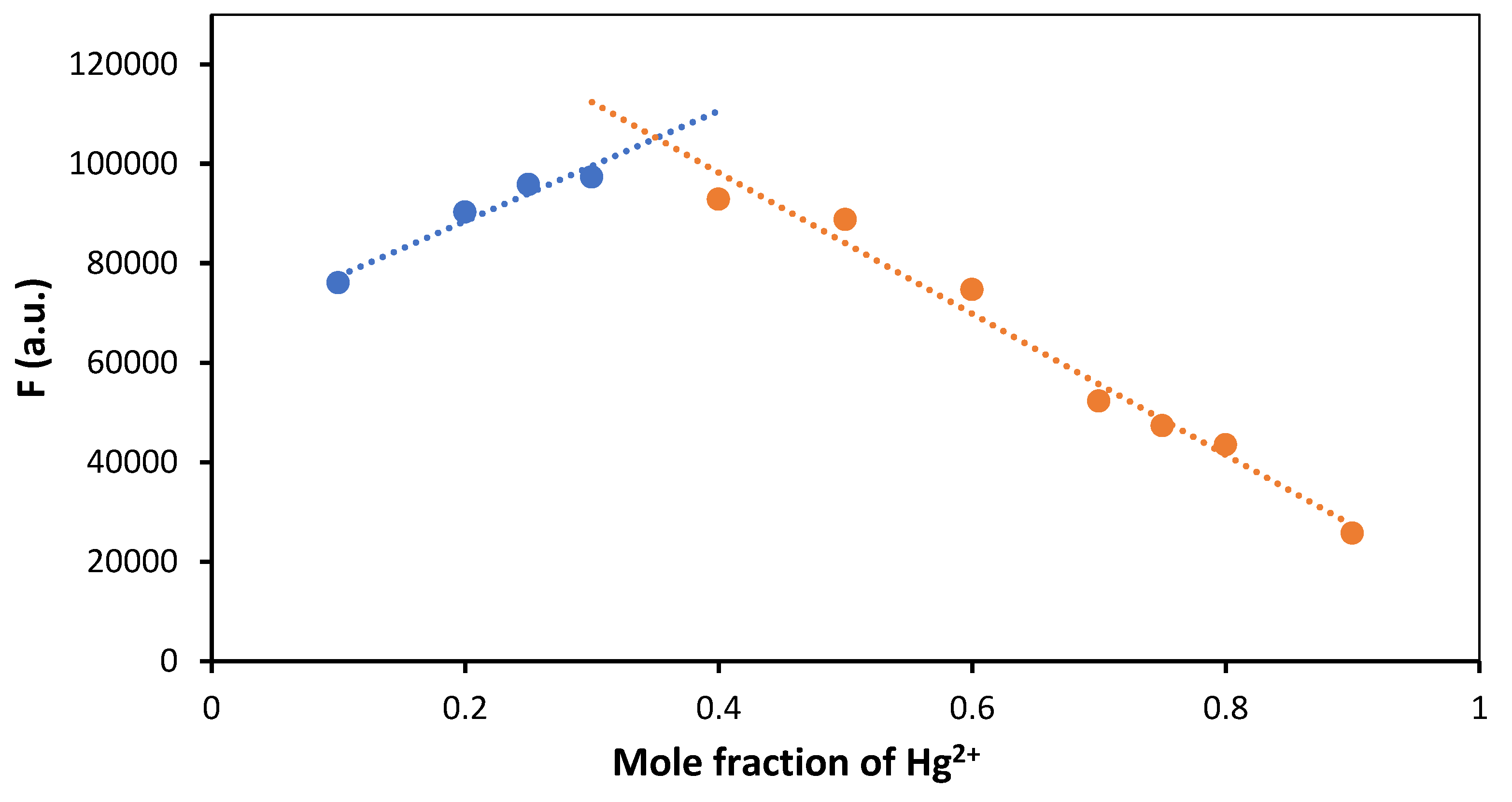
The stoichiometry of a complex formed between two interacting species, such as a peptide and metal ions, can be determined using the Job’s plot, which is also referred to as the continuous variation method. It encompasses the preparation of a sequence of solutions in which the mole fractions of the two reactants (in this instance, the FY7 peptide and Hg2;⁺ ions) are varied, while the total molar concentration of the two reactants remains constant. The mole fraction of one of the components is plotted against the fluorescence response. The stoichiometry of the complex is determined by the point at which the response is at its maximum. A Job’s plot experiment was conducted to determine the stoichiometry of the interaction between the FY7 peptide and Hg2+ in MES buffer (50 mM, pH 5.65). The Job’s plot of fluorescence intensity, which is influenced by the complex formation, was plotted as a function of the mole fractions of Hg2+ (Figure 4). The stoichiometry of the complex formed between the FY7 peptide and Hg2;⁺ ion is determined to be 2:1 based on the Job’s plot, which shows the maximum fluorescence intensity at a mole fraction of 0.35 for Hg2;⁺. This implies that one Hg2;⁺ ion is bound by two FY7 peptides.
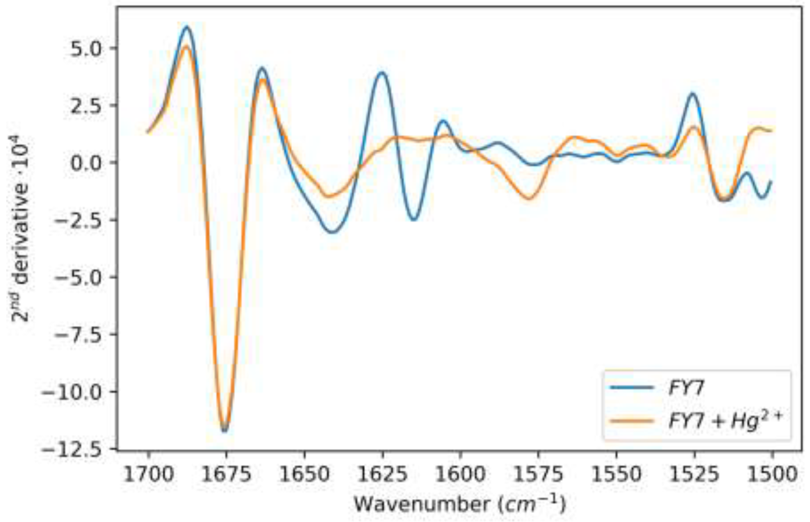
In order to determine which chemical groups of the FY7 peptide are crucial for the interaction with Hg2+ (Figure 5), an FTIR experiment was conducted. FTIR can be used to obtain information regarding the molecular vibrations and structures of the peptide and functional groups. D2O, as a solvent, enables the identification of amide bands in the peptide, which are directly associated with potential structural modifications that may result from interactions with mercury ions. The FTIR analysis yielded valuable insights into the involvement of specific functional groups within the peptide in the complexation process with Hg2;⁺. The amide band region of the spectra of the peptide and peptide-metal complexes was virtually unaffected by the buffer solution, which contained acetonitrile, salts, and buffer components. The asymmetric stretching of the carboxyl group of trifluoroacetate (TFA) in D2O is responsible for the strong peak at 1675 cm–1 [38,39]. The presence of this band is readily apparent in spectra that contain peptides; however, it is absent from spectra obtained from buffers, D2O, D2O-acetonitrile mixtures, and other sources. We opted not to subtract it, as it could introduce subtraction bands that are easily misinterpreted. Nevertheless, we calculated the second spectra derivatives to improve the sub-band changes in the complex amide I’ band. Significant absorption was observed only in the peptide and TFA, the remaining solute utilized in peptide synthesis, following this preparation.
The FTIR spectrum of free FY7 (blue line) is significantly different from that of FY7 saturated with Hg2+ ions (orange line) (Figure 5). The primary backbone band (1670–1625 cm–1) expands, but its overall shape does not suggest the formation of any new secondary structures, such as aggregates or intermolecular β-sheets [40]. Nevertheless, the binding of Hg2+ may induce a shift in the clearly separated bands that correspond to amino acid side chains. The v(CC) and in-plane δ(CH) vibrations of the Tyrosine ring are responsible for a signal at approximately 1615 cm–1 in the free FY7 peptide. Upon the formation of the complex, the intensity of this band experienced a substantial decrease and may have shifted to approximately 1575 cm–1. This suggests that the ring was directly involved in the binding interaction with the metal ions, potentially involving the π-orbitals of the ring in the complexation. The minor changes in other bands that are characteristic of the Tyrosine side chain, particularly C−OD (C−O- ) is not anticipated in the specified pH or pD range), indicate that the Hg2+ binding is indeed achieved through the ring orbitals, rather than the C−OD group. There is still a question as to whether the FY7−Hg2+ interaction involves a single Tyr ring as the base and the remaining peptide acting as a tail over the base with a trapped ion, or if one Hg2+ ion coordinates with two Tyrosine rings from two FY7 molecules through a π-stacking or sandwich interaction. The latter scenario is strongly supported by the complex’s 2:1 stoichiometry. It showed that the Hg2+ ion induces the formation of a new coordination envi-ronment within the peptide, with the tyrosine residue being the primary component. The aromatic ring of the tyrosine residues is directly involved in the binding of the Hg2;⁺ ions, as indicated by the observed spectral changes. The FY7−Hg2+ interaction is best described by the model in which one Hg2+ ion coordinates with two tyrosine rings from two separate FY7 molecules through a π-stacking or sandwich interaction, based on the stoichiometric evidence and the likely stability conferred by π-π interactions.

3.4. Low-Volume Miniaturized Optical Nano-Biosensor for the Rapid, Sensitive, and Selective Detection of Toxic Mercury in Aqueous Samples based on FY7 Peptide

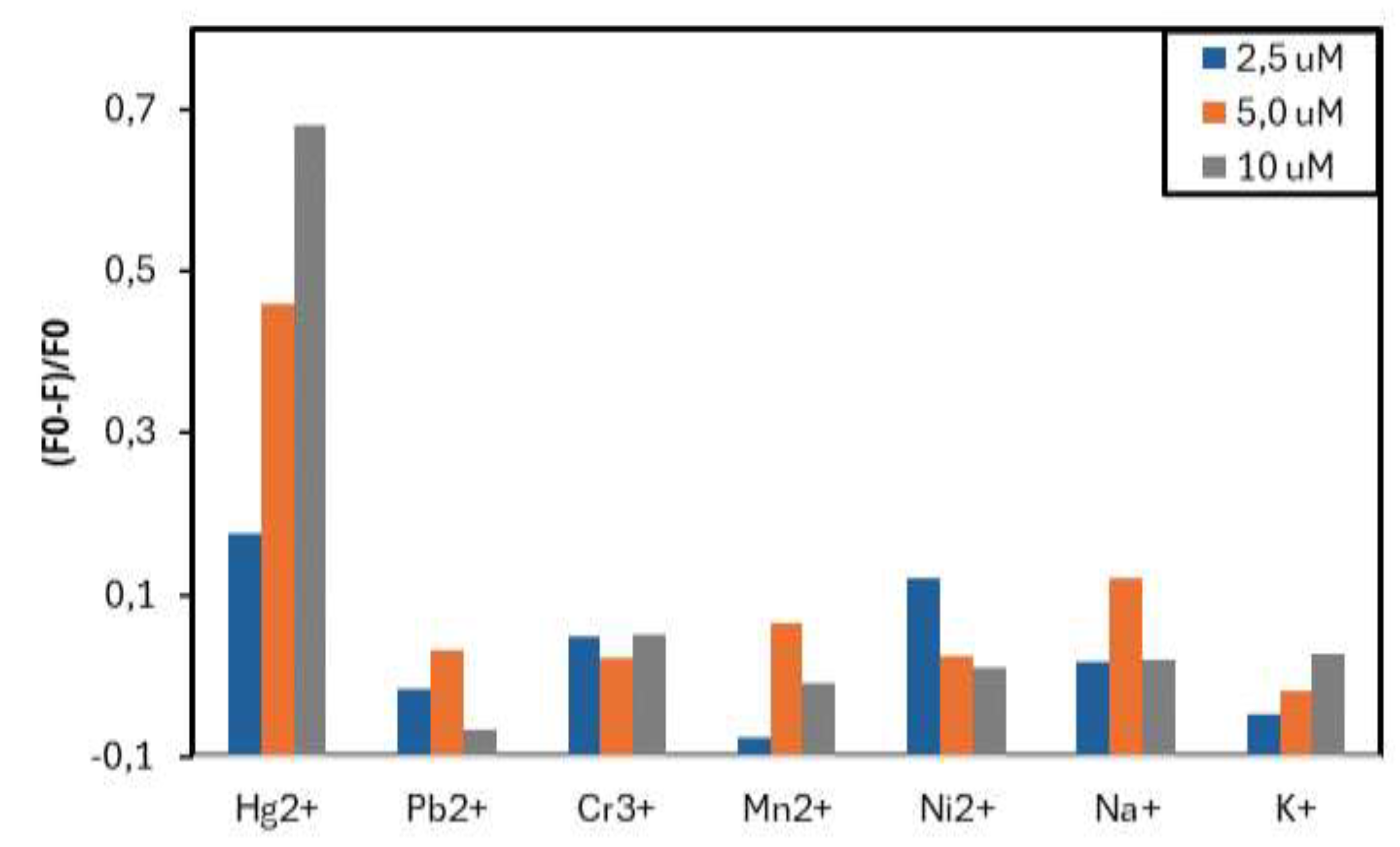
The fluorescence response of FY7 peptide was measured in the custom, low-cost glass capillary setup. Results achieved for H1MG spectrofluorometer had been considered as reference and used as a benchmark for this custom, miniaturized setup (Figure 6). As a first step, similiarly as for H1MG, FY7 selectivity toward distinct metal ions was assessed. After initiall tests, peptide concentration when using miniaturized setup was increased to 50 µM (from 5 µM in H1MG) in order to achieve a more robust dynamic response within the limited parameters of small USB4000 spectrometer. Similiarly to prior tests the FY7 peptide in capillaries was analyzed in a 50 mM MES buffer solution at pH 5.65, but with modified set of seven distinct metal ions (Hg2;⁺, Pb2;⁺, Mn2;⁺, Ni2;⁺, Cr3;⁺, Na⁺, and K⁺). In the commercial setup utilized ion solutions that were in the 0.02 - 10 µM range, while in the low-cost setup concentrations tested were 0.1, 0.25, 0.5, 1, 2.5, 5, and 10 µM. With the inclusion of nickel, six of the metal ions that were chosen remained unchanged from the H1MG commercial setup. Despite lower dynamic range of the low-cost setup, FY7 response for distinct metal ions was matching the results from H1MG (Figure 7).
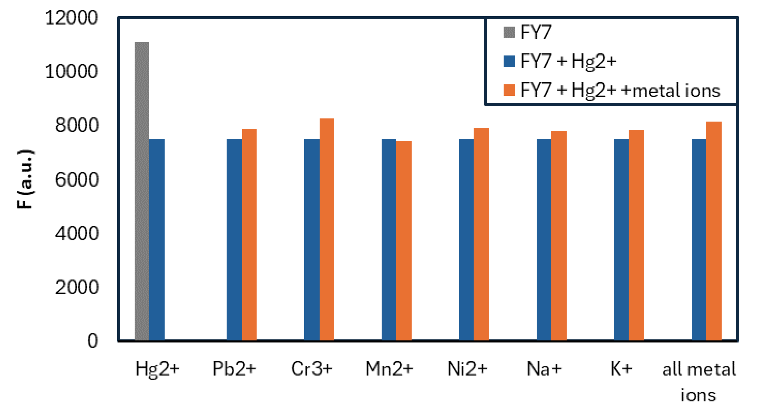
Next, the FY7 peptide’s response to Hg2+ ions in the additional presence of the seven metal ions mentioned was assessed. Similiarly to prior test concentrations of HY7 were 0.1, 0.25, 0.5, 1, 2.5, 5, and 10 µM, while matching test for the commercial setup were prepared in the 0.02-10 µM range (Figure 8).
Obtained results with low-volume setup are in accordance with the commercial setup, as the FY7 peptide demonstrated a sensitive response exclusively to Hg2;⁺ ions. The observed fluorescence emission was sliglthy higher, up to 65% in the presence of Hg2;⁺ ions, while the fluorescence signal reduction for the remaining six metal ions, including Ni2;⁺, was less than 10%, again matching the results from H1MG. Both tests demonstrated the robustness of the FY7 peptide, as well as confirmed the potential of the low-volume measurements, as the sample volume used per one measurement was 69 times lower. Miniaturized capillary setup was verified as a suitable alternative to commercial device, and in addition it offers flexibilty to be usead as a fully mobile POC (point−of−care) device.

4. Conclusions

Finally, a FY7 phage display-derived peptide was employed for Hg2+ ion detection using a low-cost, low-volume capillary setup. With sample volume of only 2.9 µL per measurement and simple optical equipment, the FY7 peptide produced comparable results to those obtained from a professional spectrofluorometer. Low-volume measurements in the presence of other metal ions confirmed FY7’s robustness, specificity and high proficiency in conformational changes. Notably, the combination of the low-cost capillary system with the synthetized, efficient FY7 peptide presents a costeffective and strong alternative to conventional commercial spectrofluorometers. This straightforward setup is affordable, easy to use, highly sensitive, and well-suited for portable measurements. The FY7 peptide, when used with such system, offers flexibility and is suitable for a wide range of applications, including environmental monitoring of mercury (II) ions.

5. Patents

The results of this work were included in the patent application number PL 449082 entitled “Synthetic 7-amino acid peptide linked by a beta-alanine linker with a FITC fluorophore as a nanobiosensor for the detection of toxic mercury (II) ions.”

Author Contributions

Conceptualization, M.S., E.P., and B.G.; methodology, M.S., P.B., M.J., E.P.; validation, M.S., E.P.; formal analysis, M.S. P.B; investigation, M.S., E.P.; data curation, M.S.; writing—original draft preparation, M.S.; writing—review and editing, B.G., E.P., M.J.; visualization, B.G., M.S.; supervision, M.O., M.Ś..; project administration, M.O., D.N..; funding acquisition, M.O., D.N. All authors have read and agreed to the published version of the manuscript.-

Funding

This work was financially supported by the Warsaw University of Technology and The National Centre for Research and Development under the III program TECHMATSTRATEG—Strategic research and development program “Modern material technologies—TECHMATSTRATEG” no. TECHMATSTRATEG-III/0042/2019-00 and acronym ASTACUS, “Biopolymer materials with chemically and genetically programmed heavy metals selectivity for new generation of ultra-sensitive biosensors”

Data Availability Statement

All research data will be made available upon request.

Conflicts of Interest

The authors declare no conflicts of interest.

References

  1. Song, S.; Li, Y.; Liu, Q.S.; Wang, H.; Li, P.; Shi, J.; Hu, L.; Zhang, H.; Liu, Y.; Li, K.; Zhao, X.; Cai, Z. Interaction of mercury ion (Hg2+) with blood and cytotoxicity attenuation by serum albumin binding. J. Hazard. Mater. 2021, 412, 125158. [Google Scholar] [CrossRef] [PubMed]
  2. Park, J.M.; Smith, J.C. Modeling mercury in proteins. Methods Enzymol. 2016, 578, 103–122. [Google Scholar] [CrossRef]
  3. Bjørklund, C.; Dadar, M.; Mutter, J.; Aaseth, J. The toxicology of mercury: Current research and emerging trends. Environ. Res. 2017, 159, 545–554. [Google Scholar] [CrossRef] [PubMed]
  4. Streets, D.G.; Horowitz, H.M.; . Jacob, D.J.; Lu, Z.; Levin, L.; Schure, A.F.; Sunderland, E.M. Total Mercury Released to the Environment by Human Activities. Environ. Sci. Technol. 2017, 51, 5969–5977. [Google Scholar] [CrossRef]
  5. Ajsuvakova, O.P; Tinkov, A.A.; Aschner, M.; Rocha, J.B.T.; Michalke, B.; Skalnaya, M.G.; Skalny, A.V.; Butnariu, M.; Dadar, M.; Sarac, I.; Aaseth, J.; Bjørklund, G. Sulfhydryl groups as targets of mercury toxicity. Coord. Chem. Rev. 2020, 417, 213343. [Google Scholar] [CrossRef] [PubMed]
  6. Rupa, S.A.; Patwary, M.A.M.; Matin, M.M.; Ghann, W.E.; Uddin, J.; Kazi, M. Interaction of mercury species with proteins: towards possible mechanism of mercurial toxicology. Toxicol. Res. (Camb) 2023, 12, 355–368. [Google Scholar] [CrossRef] [PubMed]
  7. Clarkson, T.W.; Magos, L.; Myers, G.J. The toxicology of mercury-current exposures and clinical manifestations. N. Engl. J. Med. 2003, 349, 1731–1737. [Google Scholar] [CrossRef]
  8. Khan, F.; Momtaz, S.; Abdollahi, M. The relationship between mercury exposure and epigenetic alterations regarding human health, risk assessment and diagnostic strategies. J. Trace Elem. Med. Biol. 2019, 52, 37–47. [Google Scholar] [CrossRef]
  9. Flores-Cáceres, M.L.; Ortega-Villasante, C.; Carril, P.; Sobrino-Plata, J.; Hernández, L.E. The Early Oxidative Stress Induced by Mercury and Cadmium Is Modulated by Ethylene in Medicago sativa Seedlings. Antioxidants 2023, 12, 551. [Google Scholar] [CrossRef]
  10. Ciacci, C.; Betti, M.; Abramovich, S.; Cavaliere, M.; Frontalini, F. Mercury-Induced Oxidative Stress Response in Benthic Foraminifera: An In Vivo Experiment on Amphistegina lessonii. Biology (Basel) 2022, 11, 960. [Google Scholar] [CrossRef]
  11. Pyatha, S.; Kim, H.; Lee, D.; Kim, K. Association between Heavy Metal Exposure and Parkinson’s Disease: A Review of the Mechanisms Related to Oxidative Stress. Antioxidants (Basel) 2022, 11, 2467. [Google Scholar] [CrossRef]
  12. Doroszkiewicz, J.; Farhan, J.A.; Mroczko, J.; Winkel, I.; Perkowski, M.; Mroczko, B. Common and Trace Metals in Alzheimer’s and Parkinson’s Diseases. Int. J. Mol. Sci. 2023, 24, 15721. [Google Scholar] [CrossRef] [PubMed]
  13. Wallin, C.; Friedemann, M.; Sholts, S.B.; Noormägi, A.; Svantesson, T.; Jarvet, J.; Roos, P.M.; Palumaa, P.; Gräslund, A.; Wärmländer, S.K.T.S. ; Mercury and Alzheimer’s Disease: Hg(II) Ions Display Specific Binding to the Amyloid-β Peptide and Hinder Its Fibrillization. Biomolecules 2019, 44. [Google Scholar] [CrossRef] [PubMed]
  14. Kaur, B.; Gupta, A.; Kaur, N. A novel, anthracene-based naked eye probe for detecting Hg2+ ions in aqueous as well as solid state media. Microchem. J. 2020, 153, 104508. [Google Scholar] [CrossRef]
  15. Naija,A. ; Yalcin, H.C.; Evaluation of cadmium and mercury on cardiovascular and neurological systems: Effects on humans and fish. Toxicol. Rep. 2023, 10, 498–508. [Google Scholar] [CrossRef] [PubMed]
  16. Bjørklund, G.; Chirumbolo, S.; Dadar, M.; Pivina, L.; Lindh, U.; Butnariu, M. J. Aaseth. Mercury exposure and its effects on fertility and pregnancy outcome. Basic Clin. Pharmacol. Toxicol. 2019, 125, 317–327. [Google Scholar] [CrossRef] [PubMed]
  17. Kumar, S.; Sharma, A.; Sedha, S. Occupational and environmental mercury exposure and human reproductive health - a review. J. Turk. Ger. Gynecol. Assoc. 2022, 23, 199–210. [Google Scholar] [CrossRef]
  18. Erxleben, H.; Ruzicka, J. Atomic Absorption Spectroscopy for Mercury, Automated by Sequential Injection and Miniaturized in Lab-on-Valve System. Anal. Chem. 2005, 77, 5124–5128. [Google Scholar] [CrossRef] [PubMed]
  19. Winter, M.; Lessmann, F.; Harth, V. A method for reliable quantification of mercury in occupational and environmental medical urine samples by inductively coupled plasma mass spectrometry. Anal. Methods 2023, 15, 2030–2038. [Google Scholar] [CrossRef] [PubMed]
  20. Li, Y.; Zhu, Z.; Zhen, H.; Jin, L.; Hu, S. Significant signal enhancement of dielectric barrier discharge plasma induced vapor generation by using non-ionic surfactants for determination of mercury and cadmium by atomic fluorescence spectrometry. J. Anal. At. Spectrom. 2016, 31, 383–389. [Google Scholar] [CrossRef]
  21. Komatsu, T.; Johnsson, K.; Okuno, H.; Bito, H.; Inoue, T.; Nagano, T.; Urano, Y. Real-Time Measurements of Protein Dynamics Using Fluorescence Activation-Coupled Protein Labeling Method. J. Am. Chem. Soc. 2011, 133, 17–6745. [Google Scholar] [CrossRef] [PubMed]
  22. Li, M.; Shi, Q.; Song,N. ;, Xiao, Y.;, Wang, L.; Chen, Z.; James, T.D. Current trends in the detection and removal of heavy metal ions using functional materials. Chem. Soc. Rev. 2023, 52, 5827–5860. [Google Scholar] [CrossRef] [PubMed]
  23. Mehta, P.K.; Jeon, J.; Ryu, K.; Park, S-H. ; Lee, K-H. Ratiometric fluorescent detection of lead ions in aquatic environment and living cells using a fluorescent peptide-based probe. J. Hazard. Mater. 2022, 427, 128161. [Google Scholar] [CrossRef]
  24. Gou, Y.; Hou, P.; Wang, O.; He, F.; Wang, P.; Yang, X. A novel AIE peptide-based fluorescent probe for highly selective detection of mercury(II) ions and its application in food samples and cell imaging. Microchem. J. 2023, 195, 109400. [Google Scholar] [CrossRef]
  25. Pang, X.; Gao, L.; Feng, H.; Li, X.; Kong, J.; Li, L. A peptide-based multifunctional fluorescent probe for Cu2+, Hg2+ and biothiols. New J. Chem. 2018, 42, 15770–15777. [Google Scholar] [CrossRef]
  26. Yu, H.; Ryu, K.; Park, J.; Subedi, S.; Lee, K-H. Design and synthesis of fluorescent peptide-based probes with aggregation-induced emission characteristic for detecting CH3Hg+ and Hg2+ in aqueous environment: Tuning fluorescent detection for CH3Hg+ by replacing peptide receptors. Dyes Pigm. 2022, 204, 110461. [Google Scholar] [CrossRef]
  27. Merrifield, R.B. Solid Phase Peptide Synthesis. I. The Synthesis of a Tetrapeptide. J. Am. Chem. Soc. 1963, 85(14), 2149–2154. [Google Scholar] [CrossRef]
  28. Fields, G. B.; Noble, R. L. Solid phase peptide synthesis utilizing 9-fluorenylmethoxycarbonyl amino acids. Intern. J Pep. and Prot. Res. 1990, 35, 161–214. [Google Scholar] [CrossRef] [PubMed]
  29. Brelje, T.C.; Wessendorf, M.W.; Sorenson, R. L. Multicolor Laser Scanning Confocal Immunofluorescence Microscopy: Practical Application and Limitations Methods. Cell Biol. 2002, 70, 70,165–249. [Google Scholar] [CrossRef]
  30. Hermanson, G.T. Fluorescent Probes. In: Bioconjugate Techniques, Third Edition, Academic Press, New York, 2013, 395-463. [CrossRef]
  31. Zhou, H.; Gao, T.; Liu, Y.; Wu, Y.; Fang, Y.; Wang, B.; Xu, B. Targeted fluorescent imaging of a novel FITC-labeled PSMA ligand in prostate cancer. Amino Acids 2022, 54, 147–155. [Google Scholar] [CrossRef] [PubMed]
  32. Li, Y-S. ; Ju, X.; Gao, X-F.; Zhao, Y-Y.; Wu, Y-F. Immobilization enzyme fluorescence capillary analysis for determination of lactic acid. Anal. Chim. Acta 2008, 610, 249–256. [Google Scholar] [CrossRef] [PubMed]
  33. Li, Y-S. ; Li, O-J.; Gao, X-F. A novel immobilization fluorescence capillary analysis method and its applications. Analyst 2020, 145, 1980–1996. [Google Scholar] [CrossRef] [PubMed]
  34. Pituła, E.; Janik, M.; Sikora, J.; Kasztelanic, R.; Stępniewski, G.; Gong, Y.; Olszewski, M.; Buczyński, B.; Koba, M.; Śmietana, M. Glass capillary systems for micro-volume fluorometry. Measurement 2025, 240, 15569. [Google Scholar] [CrossRef]
  35. Bruździak, P. Vapor correction of FTIR spectra – A simple automatic least squares approach. Acta A Mol. Biomol. Spectrosc. 2019, 223, 117373. [Google Scholar] [CrossRef]
  36. Yang, T.; . Zhang, X-Y. ; Zhang, X-X.; Chen, M-L.; Wang, J-H. Chromium(III) Binding Phage Screening for the Selective Adsorption of Cr(III) and Chromium Speciation. ACS Appl. Mater. Interfaces. 2015, 7, 21287–21294. [Google Scholar] [CrossRef]
  37. Jullian, M.; Hernandez, A.; Maurras, A.; Puget, K.; Amblard, M.; Martinez, J.; Subra, G. N-terminus FITC labeling of peptides on solid support: the truth behind the spacer. Tetrahedron Lett. 2009, 50, 260–263. [Google Scholar] [CrossRef]
  38. Kuroda, D.G.; Vorobyev, D.Y.; Hochstrasser, R.M. Ultrafast relaxation and 2D IR of the aqueous trifluorocarboxylate ion. J. Chem. Phys. 2010, 132, 044501. [Google Scholar] [CrossRef]
  39. Lotze, S.; Bakker, H. J. Structure and dynamics of a salt-bridge model system in water and DMSO. J. Chem. Phys. 2015, 142, 212436. [Google Scholar] [CrossRef]
  40. Barth, A. Infrared spectroscopy of proteins. Biochim. Biophys. Acta 2007, 1767, 1073–1101. [Google Scholar] [CrossRef]
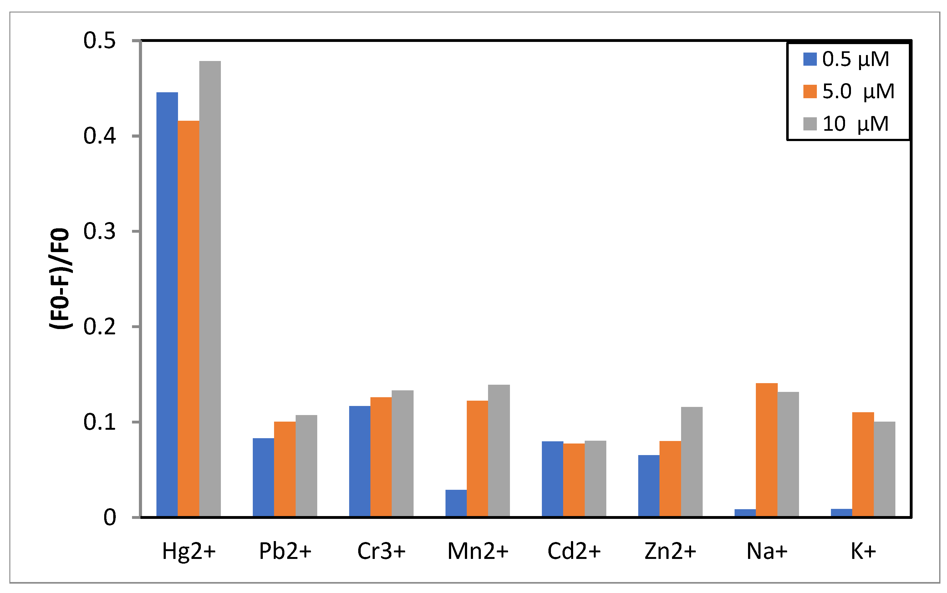
Figure 1. The selectivity of FY7 peptide (5 µM) for Hg2+ ions in MES buffer solutions (50 mM, pH 5.65). Concentration of all metal ions were at 0.5, 5.0 and 10 µM. F0 and F were the fluorescence intensities of FY7 in the absence and presence of metal ions, respectively.
Preprints 119435 g001
Figure 2. (A) Fluorescence emission spectra of FY7 (5 µM) upon the addition of Hg2+ ions (0-10 µM) in MES buffer solutions (50 mM, pH 5.65). (B) Plots of fluorescence intesity of FY7 as a function of Hg2+ ions concentration (µM).
Preprints 119435 g002
Figure 3. Fluorescence response of FY7 (5 µM) to Hg2+ (0.5 equiv.) in the presence of various metal ions (5 equiv.) in MES buffer solutions (50 mM, pH 5.65).
Preprints 119435 g003
Figure 4. Job’s plot for determining the stoichiometry of FY7 and Hg2+ in MES buffer solutions (50 mM, pH 5,65), the total concentration of FY7 and Hg2+ was 5 µM.
Preprints 119435 g004
Figure 5. Second derivatives of FTIR spectra of FY7 peptide (blue line) and FY7-Hg2+ complex (orange line). Please see description in text.
Preprints 119435 g005
Figure 6. Schematic representation of an experimental setyp for micro-volume capillary measurements.
Preprints 119435 g006
Figure 7. The selectivity of FY7 peptide (5 µM) for Hg2+ ions in MES buffer solutions (50 mM, pH 5.65) measured in low-volume fluorescence setup. Concentration of all metal ions were at 0.5, 5.0 and 10 µM . F0 and F were the fluorescence intensities of FY7 in the absence and presence of metal ions, respectively.
Preprints 119435 g007
Figure 8. Fluorescence response of FY7 (50 µM) in the presence of various metal ions (10 µM) in MES buffer solutions (50 mM, pH 5.65) measured in low-volume fluorescence setup.
Preprints 119435 g008
Disclaimer/Publisher’s Note: The statements, opinions and data contained in all publications are solely those of the individual author(s) and contributor(s) and not of MDPI and/or the editor(s). MDPI and/or the editor(s) disclaim responsibility for any injury to people or property resulting from any ideas, methods, instructions or products referred to in the content.
Copyright: This open access article is published under a Creative Commons CC BY 4.0 license, which permit the free download, distribution, and reuse, provided that the author and preprint are cited in any reuse.
Prerpints.org logo

Preprints.org is a free preprint server supported by MDPI in Basel, Switzerland.

Subscribe

© 2024 MDPI (Basel, Switzerland) unless otherwise stated