Preprint
Article

Translational Research on Azacitidine Post- Remission Therapy of Acute Myeloid Leukemia in Elderly Patients (QOL-ONE Trans-2)

Altmetrics

Downloads

71

Views

48

Comments

0

A peer-reviewed article of this preprint also exists.

Submitted:

27 September 2024

Posted:

30 September 2024

You are already at the latest version

Alerts
Abstract
Achieving complete remission (CR) is crucial for acute myeloid leukemia (AML) patients undergoing curative therapy, but relapse often occurs within months, highlighting the need for strategies to prolong leukemia-free survival (DFS). Our phase III study compared the efficacy and safety of azacitidine (AZA) versus best supportive care (BSC) in elderly AML patients who achieved CR after intensive induction and consolidation therapy. This ancillary study (QOL-ONE Trans-2) evaluated biological changes in bone marrow using Next Generation Sequencing (NGS). We analyzed baseline, randomization, and 6-month post-remission samples from 24 patients (median age 71, 12 males). High-throughput NGS targeted 350 myeloid malignancy-related genes, considering variants with a Variant Allele Frequency (VAF) ≥ 4%. At diagnosis, all patients had 5 to 17 (median 10) mutations, with DNMT3A (42%), NPM1 (33%), and TET2 (33%) being most frequent. FANCA mutations in 4 patients were linked to a higher relapse risk (HR 4.96, p=0.02) for DFS at both 2 and 5 years. Further HLA-specific NGS analyses are ongoing to confirm these results and their therapeutic implications.
Keywords: 
Subject: Medicine and Pharmacology  -   Oncology and Oncogenics

1. Introduction

The achievement of complete remission (CR) is an important milestone for patients with acute myeloid leukemia (AML) undergoing curative-intent therapy [1]. Age, comorbidities, and biological aspects of leukemia in older adults [2] affect the ability of such patients to tolerate treatment and contribute to worse outcomes with lower rates of CR compared to younger patients [3,4]. Independent of their age, virtually all patients who achieve remission with induction therapy for AML will relapse within months, unless additional therapy is given [5]. Consequently, there has been long-standing interest in the use of lower-intensity maintenance therapies after completion of the intensive treatment phase, to prolong the duration of remission and increase survival and the likelihood of cure [6,7,8].
Azacitidine (AZA) is a hypomethylating agent with well-known antileukemic activity, widely used alone or in association with other drugs for the frontline treatment of AML patients unfit for intensive chemotherapy, due to its favourable safety profile [9,10,11,12,13].
A recent report tested AZA as maintenance therapy for 1-year treatment after CR achievement in elderly AML patients, showing an advantage in prolonging relapse-free survival (RFS), but not overall survival (OS) [14]. Moreover, in a placebo-controlled randomized clinical trial, which evaluated oral AZA formulation (CC-486) as a maintenance therapy in patients aged >55 years following induction, there were significantly longer OS and RFS in those receiving the investigational product [15].
The aim of the “A Randomized Study to Evaluate the Efficacy of 5-Aza for Post-Remission Therapy of Acute Myeloid Leukemia in Elderly Patients (QoLESS AZA-AMLE)” randomized phase 3 trial was to test the efficacy and safety of long-term AZA maintenance compared to placebo in AML elderly patients who achieved first CR after a homogeneous intensive induction and consolidation phase [16]. The trial evaluated the efficacy of subcutaneous AZA post-remission therapy vs. best supportive care (BSC) in elderly AML patients. The primary endpoint was the difference in disease-free survival (DFS) from CR to relapse/death. Patients with newly diagnosed AML aged ≥61 years received 2 courses of induction chemotherapy (“3+7” daunorubicin and cytarabine) followed by consolidation (cytarabine). At CR, 54 patients were randomized (1:1) to receive BSC or AZA. At 2 years, median DFS was 6.0 (95% CI:0.2–11.7) months for patients receiving BSC) vs. 10.8 months (95% CI:1.9–19.6, p=0.20) months for AZA. At 5 years, DFS was 6.0 (95% CI:0.2–11.7) months in the BSC arm vs. 10.8 (95% CI:1.9–19.6 p=0.23) months in the AZA arm. Significant benefit was afforded by AZA on DFS at 2 and 5 years in patients aged >68 years (HR=0.34, 95% CI:0.13–0.90, p=0.030 and HR=0.37, 95% CI:0.15–0.93, p=0.034, respectively). No deaths occurred prior to leukemic relapse. Neutropenia was the most frequent adverse event. There were no differences in patient-reported outcome measures between study arms. In conclusion, AZA post-remission therapy is feasible, safe, and favourable, particularly in AML patients aged >68 years.
We report on the ancillary “Translational study, research on Azacitidine Post-Remission Therapy of Acute Myeloid Leukemia in Elderly Patients (QOL-ONE Trans-2)”, to evaluate biological changes through Next Generation Sequencing (NGS) using a custom genomic panel, courtesy of Prof. Seishi Ogawa (University of Kyoto, Japan), for all subjects with available vials.

2. Results

2.1. Description of the Study Cohort

In this manuscript, we present NGS data in randomized patients performed at disease diagnosis (baseline visit), at CR post induction chemotherapy (randomization visit) and at 6 months post-randomization.
Bone marrow samples were available for 63 patients at baseline, however due to time-related preservation defects, biological samples for 10 patients were not evaluable.
Baseline characteristics of the 53 evaluable patients are presented in Table 1.
All 53 patients presented mutations at diagnosis with a range of 3 to 19 (median 10) simultaneous mutations, the most frequent being DNMT3A (38 %), TET2 (28 %), NPM1 (23%) and DST (23%).
After induction chemotherapy, 29 patients did not achieve CR, therefore the bone marrow samples of post induction therapy phase were not collected.
The sample data of the 24 patients who had reached CR and were randomized in 5-AZA arm (11 patients) or in the BSC arm (13 patients) are here reported. In Table 2, baseline characteristics of the 24 patients in CR are shown.
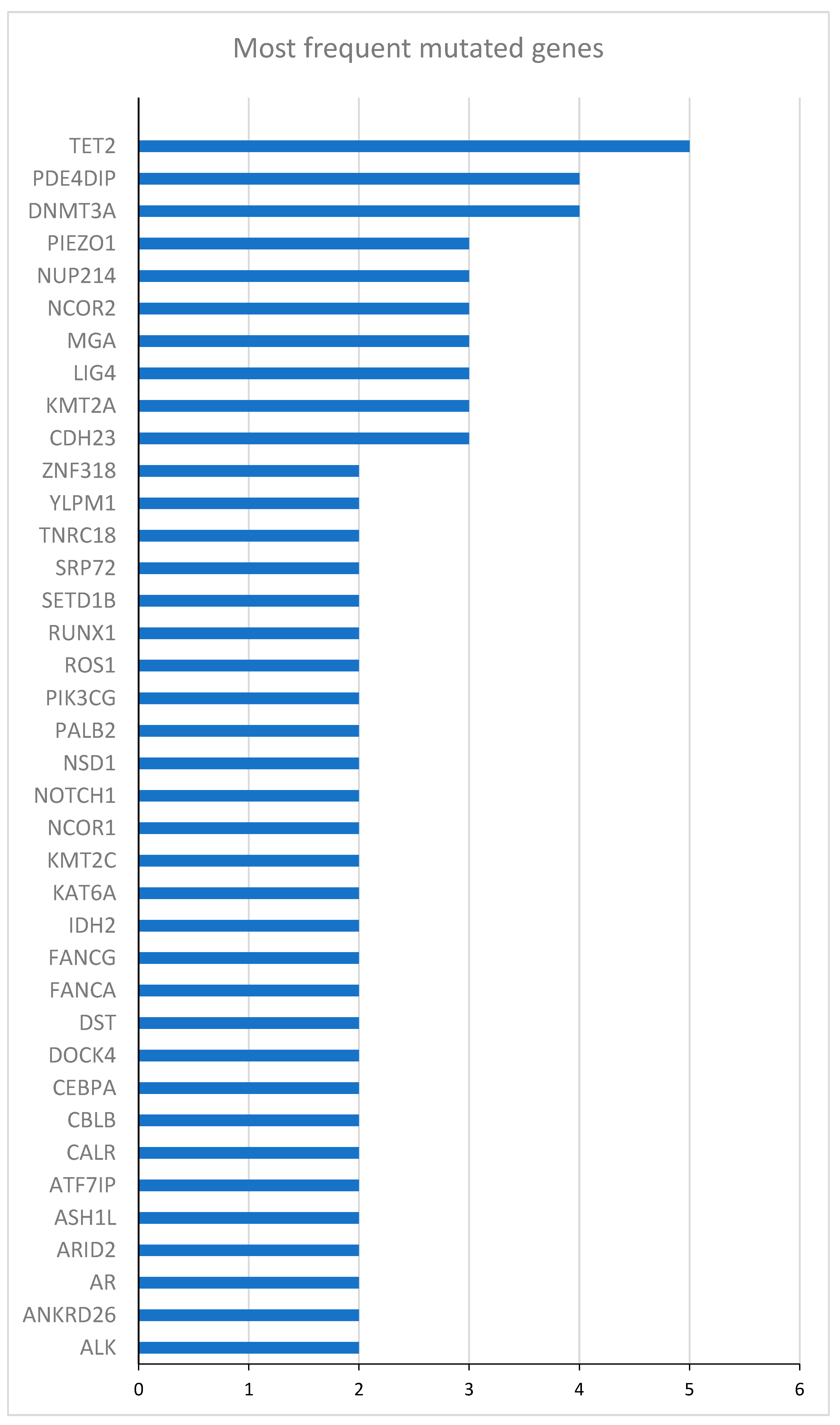
All 24 patients presented mutations at diagnosis with a range of 5 to 17 (median 10) simultaneous mutations, the most frequent being DNMT3A (42%), NPM1 (33%) and TET2 (33%). In Figure 1, the most frequent mutated genes at diagnosis are represented.
The following gene mutations occurred once only: ABL1, ACIN1, ADA2, ALAS2, ANKRD26, ARID1A, ARID2, ASXL1, ATG2B, BCORL1, CALR, CBFA2T3, CBLB, CDC25C, CDKN2B, CHM, CTCF, DAZAP1, DCAF8L1, DIS3, DYNC2H1, ELANE, FANCD2, FANCG, FANCM, FBXW7, FMC1, FMC1_LUC7L2, GIGYF2, HCFC1, HCN1, HSPA9, IDH1, JAK1, KDM5A, KDM6A, KLF1, KMT2D ,KRAS, LIN28A, MBD4, MBNL1, MDM2, MET, MYC, MZF1, NFE2, NIPBL, NOL3, NTRK3, NXF1, PALB2, PARN, PDGFRA, PDGFRB, PIK3CG, PRF1, PRPF8, PTPN11, PUS1, PXDNL, ROBO1, ROS1, RTEL1, RUNX1T1, SAMD9, SF1, SF3A1, SLIT3, SMC3, SNX13, SRCAP, SRP54, SRP72, SRSF2, STAG1, SYK, TLR2, TNRC18, VEGFA, ZBTB7A, ZEB2, ZFPM1

2.2. Primary Endpoints

2.2.1. Effect Modifications by Gene Mutations at AML Diagnosis

The effect modification by gene mutations at AML diagnosis on the relationship between AZA versus BSC and relapse were evaluated at 2 and 5 years.
The relationship between gene mutations at diagnosis and DFS at 2 and 5 years for all patients, independently of allocation arm, was evaluated with Kaplan-Meier analysis and univariate COX regression.
In Table 3 and Table 4, hazard ratio (HR) and 95% CI of relapse at 2 and 5 years respectively of most frequently occurring mutations are shown.

2.2.2. Effect Modifications by Gene Mutations at Randomization

Apart from a significant effect modification by FANCA (p=0.02 for both 2 and 5 years), no other effect modification by gene mutations at random (MRD) on the relationship between AZA versus BSC and relapse in elderly AML patients receiving induction chemotherapy followed by post-remission BSC vs AZA maintenance was observed at 2 and 5 years (p=0.14 to p=0.98). In Figure 2 the most frequent mutated genes at randomization are represented.

3. Discussion

Post-transplant relapse poses a significant challenge in treating myeloid malignancies such as myelodysplastic syndromes (MDS) and AML, with low survival rates despite various interventions[17,18].
In the present analysis, samples from 24 patients revealed 5-17 mutations each (median 10), with DNMT3A, NPM1, and TET2 being most common. Only FANCA (mutated in 4 patients) correlated with higher relapse risk (HR 4.96).
The role of genetic mutations in AML prognosis and treatment response is a crucial area of research. The current study highlights the prognostic significance of specific gene mutations, particularly FANCA, in predicting DFS and relapse in elderly patients who achieved CR after intensive induction and consolidation therapy.
Mutations in the FANCA gene were associated with a significantly increased HR of relapse, indicating a poorer prognosis for patients with these mutations. This finding aligns with previous studies that have demonstrated the involvement of FANCA and other Fanconi anemia (FA) genes in hematologic malignancies. In addition, emerging lines of evidence indicate that the FA pathway constitutes a general surveillance mechanism for the genome by protecting against a variety of DNA replication stresses[19]. Consequently, studies have been undertaken to improve our understanding of DNA repair signaling that is regulated by the FA pathway, and the potential role of DNA lesions underlying the FA pathophysiology for the treatment of FA and FA-associated cancers.
Pawlikowska and colleagues demonstrated that loss of the Fanconi anemia pathway, known to control genetic instability, promotes the expansion of leukemic cells carrying oncogenic mutations rather than mutation formation[20]. Furthermore, in a study by D'Andrea and colleagues the role of the FA pathway in DNA repair mechanisms was emphasized, suggesting that mutations in FANCA could impair the repair of DNA crosslinks, thereby contributing to leukemogenesis[21]. Furthermore, Voso et al. observed that FA gene mutations, including FANCA, were prevalent in therapy-related myeloid neoplasms, highlighting their potential role in the pathogenesis of secondary AML[22].
The association of FANCA mutations with poor prognosis in AML has also been corroborated by other studies. Zhang et al. identified FANCA mutations as a common event in AML patients and linked these mutations with adverse outcomes, particularly in terms of DFS and overall survival (OS). Similarly, Tischkowitz et al. (2015) found that germline mutations in FANCA were associated with increased AML risk, further highlighting the gene's relevance in the disease's etiology and progression. However, in a separate study by Chang et al. compared to the FA wild-type group, a decrease in the expression of FNACD2, FANCI, and RAD51C was observed in the FA mutation group. Interestingly, the FA mutation group exhibited a more favorable clinical overall survival prognosis[23].
However, the frequency of FANCA mutations and their prognostic impact can vary across studies. In the current study, FANCA mutations were present in 4 out of 24 patients (approximately 17%), which is consistent with some reports but higher than others. This suggests potential variability due to differences in patient demographics or study methodologies. Reinforcing this idea, Steinberg-Shemer, performed a characterization and genotype-phenotype correlation of patients with FA in a multi-ethnic population. Patients with FANCA mutations developed cancer at a significantly older age compared to patients with mutations in other Fanconi genes, however, overall survival was not found to be dependent on the causative gene[24].
A phase II trial explored AZA in 39 patients with persistent disease or early relapse post-HCT, finding a 30% response rate, including three CR[25]. The most commonly mutated genes at the time of relapse included TP53 (48%), TET2 (33%) and DNMT3A (14%). Mutations in TP53 were significantly associated with poor responsiveness to AZA (OR 3.08, 95%CI: 1.1-9.0; P=0.04] and inferior survival; HR 3.04, 95% CI: 1.3-5.8; P=0.02]. We also observed a high number of mutations for the TET2 gene (10 mutations), however, the risk of 2-and 5- year DFS was not statistically significant (p=0.73 and p=0.52 respectively)
Our findings on FANCA mutations suggest potential markers for tailoring post-remission therapy to prolong DFS.
Overall, these studies contribute valuable insights into the genetic factors influencing treatment outcomes in AML/MDS, advocating for personalized approaches based on mutational profiles to enhance therapeutic strategies and improve patient prognosis.
The differential impact of FANCA mutations on AML prognosis highlights the importance of personalized medicine in this field. Patients with FANCA mutations may benefit from more aggressive monitoring and potentially alternative therapeutic strategies to mitigate the higher risk of relapse.
Further research is warranted to validate these findings and explore their underlying mechanisms. Specifically, additional studies should investigate the functional consequences of FANCA mutations in AML cells and their interactions with the bone marrow microenvironment. The use of larger patient cohorts and diverse populations will also be essential to generalize these results.

3.1. Study Limitations

Our analysis was based on a small number of patients (24 patients were included) with wide heterogeneity of mutations evaluated. A wide range of 5 to 17 simultaneous mutations per patient may potentially complicate the analysis and interpretation of the specific impact of a given gene. Data were collected at baseline, randomization, and 6 months post-remission, potentially missing long-term effects. Results may not be applicable to younger AML patients due to the specific age group studied (median age 71). These limitations suggest that while the findings are promising, they should be interpreted cautiously and validated in larger, more diverse cohorts.

3.2. Conclusion

The present study adds valuable insights into the prognostic significance of FANCA mutations in elderly AML patients. The association of FANCA mutations with increased relapse risk with improved DFS highlights the potential for these genetic markers to inform treatment decisions and risk stratification. Future research should aim to elucidate the mechanisms underlying these associations and explore their implications for personalized AML therapy.

4. Materials and Methods

4.1. Study Design and Patient Population

The study was performed on available biological samples collected at baseline, randomization, and 6-month post remission. For study design and patient population refer to the published report [16].

4.2. Inclusion and Exclusion Criteria

Samples of patients that have been enrolled in the Azacitidine Post-Remission Therapy for Elderly Patients with AML: A Randomized Phase-3 Trial [16] are included if they have a baseline (at diagnosis) bone marrow sample available. The lack of an available baseline bone marrow sample is an exclusion criterion for the present study.

4.3. Study Endpoints

The primary endpoints of the study were to evaluate how gene mutations at AML diagnosis modify the effect of AZA versus BSC on relapse, and how gene mutations at randomization (minimal residual disease, MRD) modify the effect of AZA versus BSC on relapse in elderly AML patients who received induction chemotherapy followed by either post-remission BSC or AZA maintenance.

4.4. Study Procedures

Next Generation Sequencing (NGS) assessment was performed on bone marrow samples from patients enrolled in the study by the Medical Genetics Unit, Grande Ospedale Metropolitano Bianchi Melacrino Morelli, Reggio Calabria, Italy. Samples were serially collected at different time-points as required by the protocol. The genomic DNA was extracted from bone marrow (preserved in DMSO/Trizol or pellet) with a Trizol/chloroform method or QIAmp DNA Mini Kit (QIAamp DNA Mini and Blood Mini Handbook: www.qiagen.com/HB-0329) following the manufacturer’s instructions and stored at -80°C until the time of use.
DNA was subjected to high throughput NGS using genes commonly mutated in myeloid malignancies and prepared with a home-set of genes for Illumina (see Supplementary Materials, courtesy of Prof. Seishi Ogawa, Kyoto, Japan).
Targeted sequencing was performed using a custom DNA bait library (Sure Select; Agilent Technology) as previously described[26].
Sequencing libraries were generated according to an Illumina paired-end library protocol. The targets were subjected to massive sequencing using Hiseq 2000 (Illumina), with sufficient read coverage. Only variants with high-quality reads, were considered. Variants were annotated using Agilent Technologies Alissa Interpret v5.4.2, Platform data set was 44_1, RefSeq Transcripts v205, Genome build GRCh37.p13, Database of functional predictions for non-synonymous SNPs was dbNSFP, dbSNP build 151, NCBI ClinVar 2022-12.
A Variant Allele Frequency (VAF) ≥ 4% was considered an appropriate threshold for minimal burden of clonality to be reported.

4.5. Statistical Analysis

Data were summarized as mean and standard deviation, median and interquartile range, or absolute frequency and percentage, as appropriate.
The effect modification by gene mutations at AML diagnosis and at randomization on the relationship between AZA versus BSC and relapse was investigated by Cox proportional hazard model and by applying the standard linear combination method. In Cox models, data were expressed as hazard ratios (HRs), 95% confidence intervals and p-values. In these models, potential confounders were taken into account. All calculations were performed using the SPSS version 13 for Windows software.

Supplementary Materials

The following supporting information can be downloaded at the website of this paper posted on Preprints.org, Supplementary Materials: Home-set of genes for Illumina

Author Contributions

E.N.O. conceived and designed the study. All authors participated in the collection of data. E.N.O., G.I. and G.T. participated in data analysis and interpretation. E.N.O., G.I., G.T. and C.M. were involved in the preparation of the manuscript. All authors have read and agreed to the published version of the manuscript.

Funding

Unconditional funding support for the undertaking of this trial was provided by BMS.

Institutional Review Board Statement

Ethics committee approval was obtained, and this study complies with the ethical standards laid down in the 1975 Declaration of Helsinki and was reg-istered at clinicaltrials.gov (NCT05188326) and in the EU Clinical Trials Register (2010-019710-24).

Informed Consent Statement

Written informed consent was obtained from all patients.

Data Availability Statement

Supplementary Material accompanies this paper. Raw data are available upon request from the corresponding author (ENO).

Acknowledgments

The trial described in this manuscript was sponsored and funded by Associ-azione QOL-ONE, a non-profit organization. Celgene donated the investigational product for evaluation in the trial, and provided partial and unconditional funding to Associazione QOL-ONE. All authors had full and independent access to all the data and vouch for the accuracy and completeness of the reported data and the fidelity of the trial to the protocol. Editorial assistance for the preparation of this manuscript was provided by Colin Egan (CE Medical Writing SRLS, Pisa, Italy).

Conflicts of Interest

E.N.O. reports honoraria from advisory boards from Alexion, Bristol-Myers Squibb, Celgene, Daiichi-Sankyo, Novartis, and Janssen, and consultancy fees from Alexion, Bristol-Myers Squibb, and Daiichi-Sankyo. A.C. reports honoraria (consultancy, advisory role, and/or travel support) from AbbVie, Astellas, Janssen, Jazz, Celgene, Gilead, Pfizer, Incyte, and Amgen outside the submitted work. G.A.P. has received honoraria for Advisory Boards/Meetings as speaker from Abbvie, AOP Orphan, AstraZeneca, Beigene, Bristol-Myers Squibb, GSK, Incyte, MorphoSys, Novartis; Support for attending meetings and travel from Abbvie, AOP Orphan, Beigene, Johnson & Johnson, Novartis and Stemline, Menarini. G.R. has been a consultant for AbbVie, AstraZeneca, BeiGene, and Janssen; at the time of the study was affiliated with Fondazione IRCCS Ca' Granda Ospedale Maggiore, University of Milan , while he is currently an employee of AstraZeneca. G.T. reports consultancy honoraria from Amgen, Biotest, Abbvie, Janssen-Cilag, Fresnius MC, and QOL-ONE. C.A. reports honoraria for participation in advisory board meetings for Abbvie, Amgen, and Novartis. L.M.A.M. has received support for attending meetings from Abbvie. R.L. reports honoraria from Bristol-Myers Squibb and Novartis. The remaining authors declare no competing financial interests.
Previous Publication: Presented in abstract form at the 65th American Society of Hematology Meeting (Blood, 2023, 142 (Supplement 1): 6046, URL: https://ashpublications.org/blood/article/142/Supplement%201/6046/505530/Novel-Gene-Mutations-Associated-with-Disease-Free.

References

  1. Döhner, H.; Estey, E.; Grimwade, D.; Amadori, S.; Appelbaum, F.R.; Büchner, T.; Dombret, H.; Ebert, B.L.; Fenaux, P.; Larson, R.A.; et al. Diagnosis and Management of AML in Adults: 2017 ELN Recommendations from an International Expert Panel. Blood 2017, 129, 424–447. [CrossRef]
  2. Hernandez-Valladares, M.; Aasebø, E.; Berven, F.; Selheim, F.; Bruserud, Ø. Biological Characteristics of Aging in Human Acute Myeloid Leukemia Cells: The Possible Importance of Aldehyde Dehydrogenase, the Cytoskeleton and Altered Transcriptional Regulation. Aging 2020, 12, 24734–24777. [CrossRef]
  3. Ferrara, F. Conventional Chemotherapy or Hypomethylating Agents for Older Patients with Acute Myeloid Leukaemia? Hematol. Oncol. 2014, 32, 1–9. [CrossRef]
  4. Webster, J.A.; Pratz, K.W. Acute Myeloid Leukemia in the Elderly: Therapeutic Options and Choice. Leuk. Lymphoma 2018, 59, 274–287. [CrossRef]
  5. Medeiros, B.C.; Chan, S.M.; Daver, N.G.; Jonas, B.A.; Pollyea, D.A. Optimizing Survival Outcomes with Post-Remission Therapy in Acute Myeloid Leukemia. Am. J. Hematol. 2019, 94, 803–811. [CrossRef]
  6. Molica, M.; Breccia, M.; Foa, R.; Jabbour, E.; Kadia, T.M. Maintenance Therapy in AML: The Past, the Present and the Future. Am. J. Hematol. 2019, 94, 1254–1265. [CrossRef]
  7. de Lima, M.; Roboz, G.J.; Platzbecker, U.; Craddock, C.; Ossenkoppele, G. AML and the Art of Remission Maintenance. Blood Rev. 2021, 49, 100829. [CrossRef]
  8. Reville, P.K.; Kadia, T.M. Maintenance Therapy in AML. Front. Oncol. 2020, 10. [CrossRef]
  9. Ramos, F.; Thépot, S.; Pleyer, L.; Maurillo, L.; Itzykson, R.; Bargay, J.; Stauder, R.; Venditti, A.; Seegers, V.; Martínez-Robles, V.; et al. Azacitidine Frontline Therapy for Unfit Acute Myeloid Leukemia Patients: Clinical Use and Outcome Prediction. Leuk. Res. 2015, 39, 296–306. [CrossRef]
  10. Dombret, H.; Seymour, J.F.; Butrym, A.; Wierzbowska, A.; Selleslag, D.; Jang, J.H.; Kumar, R.; Cavenagh, J.; Schuh, A.C.; Candoni, A.; et al. International Phase 3 Study of Azacitidine vs Conventional Care Regimens in Older Patients with Newly Diagnosed AML with >30% Blasts. Blood 2015, 126, 291–299. [CrossRef]
  11. Molica, M.; Mazzone, C.; Niscola, P.; Carmosino, I.; Di Veroli, A.; De Gregoris, C.; Bonanni, F.; Perrone, S.; Cenfra, N.; Fianchi, L.; et al. Identification of Predictive Factors for Overall Survival and Response during Hypomethylating Treatment in Very Elderly (≥75 Years) Acute Myeloid Leukemia Patients: A Multicenter Real-Life Experience. Cancers 2022, 14, 4897. [CrossRef]
  12. DiNardo, C.D.; Jonas, B.A.; Pullarkat, V.; Thirman, M.J.; Garcia, J.S.; Wei, A.H.; Konopleva, M.; Döhner, H.; Letai, A.; Fenaux, P.; et al. Azacitidine and Venetoclax in Previously Untreated Acute Myeloid Leukemia. N. Engl. J. Med. 2020, 383, 617–629. [CrossRef]
  13. Garcia-Manero, G.; Döhner, H.; Wei, A.H.; Torre, I.L.; Skikne, B.; Beach, C.L.; Santini, V. Oral Azacitidine (CC-486) for the Treatment of Myeloid Malignancies. Clin. Lymphoma Myeloma Leuk. 2022, 22, 236–250. [CrossRef]
  14. Huls, G.; Chitu, D.A.; Havelange, V.; Jongen-Lavrencic, M.; van de Loosdrecht, A.A.; Biemond, B.J.; Sinnige, H.; Hodossy, B.; Graux, C.; Kooy, R. van M.; et al. Azacitidine Maintenance after Intensive Chemotherapy Improves DFS in Older AML Patients. Blood 2019, 133, 1457–1464. [CrossRef]
  15. Wei, A.H.; Döhner, H.; Pocock, C.; Montesinos, P.; Afanasyev, B.; Dombret, H.; Ravandi, F.; Sayar, H.; Jang, J.-H.; Porkka, K.; et al. Oral Azacitidine Maintenance Therapy for Acute Myeloid Leukemia in First Remission. N. Engl. J. Med. 2020, 383, 2526–2537. [CrossRef]
  16. Oliva, E.N.; Candoni, A.; Salutari, P.; Palumbo, G.A.; Reda, G.; Iannì, G.; Tripepi, G.; Cuzzola, M.; Capelli, D.; Mammì, C.; et al. Azacitidine Post-Remission Therapy for Elderly Patients with AML: A Randomized Phase-3 Trial (QoLESS AZA-AMLE). Cancers 2023, 15, 2441. [CrossRef]
  17. Sauerer, T.; Velázquez, G.F.; Schmid, C. Relapse of Acute Myeloid Leukemia after Allogeneic Stem Cell Transplantation: Immune Escape Mechanisms and Current Implications for Therapy. Mol. Cancer 2023, 22, 180. [CrossRef]
  18. Mina, A.; Greenberg, P.L.; Deeg, H.J. How I Reduce and Treat Posttransplant Relapse of MDS. Blood 2024, 143, 1344–1354. [CrossRef]
  19. Rageul, J.; Kim, H. Fanconi Anemia and the Underlying Causes of Genomic Instability. Environ. Mol. Mutagen. 2020, 61, 693–708. [CrossRef]
  20. Pawlikowska, P.; Delestré, L.; Gregoricchio, S.; Oppezzo, A.; Esposito, M.; Diop, M.B.; Rosselli, F.; Guillouf, C. FANCA Deficiency Promotes Leukaemic Progression by Allowing the Emergence of Cells Carrying Oncogenic Driver Mutations. Oncogene 2023, 42, 2764–2775. [CrossRef]
  21. D’Andrea, A.D. Susceptibility Pathways in Fanconi’s Anemia and Breast Cancer. N. Engl. J. Med. 2010, 362, 1909–1919. [CrossRef]
  22. Voso, M.T.; Fabiani, E.; Zang, Z.; Fianchi, L.; Falconi, G.; Padella, A.; Martini, M.; Li Zhang, S.; Santangelo, R.; Larocca, L.M.; et al. Fanconi Anemia Gene Variants in Therapy-Related Myeloid Neoplasms. Blood Cancer J. 2015, 5, e323. [CrossRef]
  23. Chang, L.; Zhang, L.; Zhao, B.; Cheng, X.; Wan, Y.; Zhang, R.; Yuan, W.; Gao, X.; Zhu, X. Mutation Spectrum, Expression Profiling, and Prognosis Evaluation of Fanconi Anemia Signaling Pathway Genes for 4259 Patients with Myelodysplastic Syndromes or Acute Myeloid Leukemia. BMC Med. Genomics 2023, 16, 290. [CrossRef]
  24. Steinberg-Shemer, O.; Goldberg, T.A.; Yacobovich, J.; Levin, C.; Koren, A.; Revel-Vilk, S.; Ben-Ami, T.; Kuperman, A.A.; Zemer, V.S.; Toren, A.; et al. Characterization and Genotype-Phenotype Correlation of Patients with Fanconi Anemia in a Multi-Ethnic Population. Haematologica 2020, 105, 1825–1834. [CrossRef]
  25. Woo, J.; Howard, N.P.; Storer, B.E.; Fang, M.; Yeung, C.C.; Scott, B.L.; Deeg, H.J. Mutational Analysis in Serial Marrow Samples during Azacitidine Treatment in Patients with Post-Transplant Relapse of Acute Myeloid Leukemia or Myelodysplastic Syndromes. Haematologica 2017, 102, e216–e218. [CrossRef]
  26. Nagata, Y.; Makishima, H.; Kerr, C.M.; Przychodzen, B.P.; Aly, M.; Goyal, A.; Awada, H.; Asad, M.F.; Kuzmanovic, T.; Suzuki, H.; et al. Invariant Patterns of Clonal Succession Determine Specific Clinical Features of Myelodysplastic Syndromes. Nat. Commun. 2019, 10, 5386. [CrossRef]
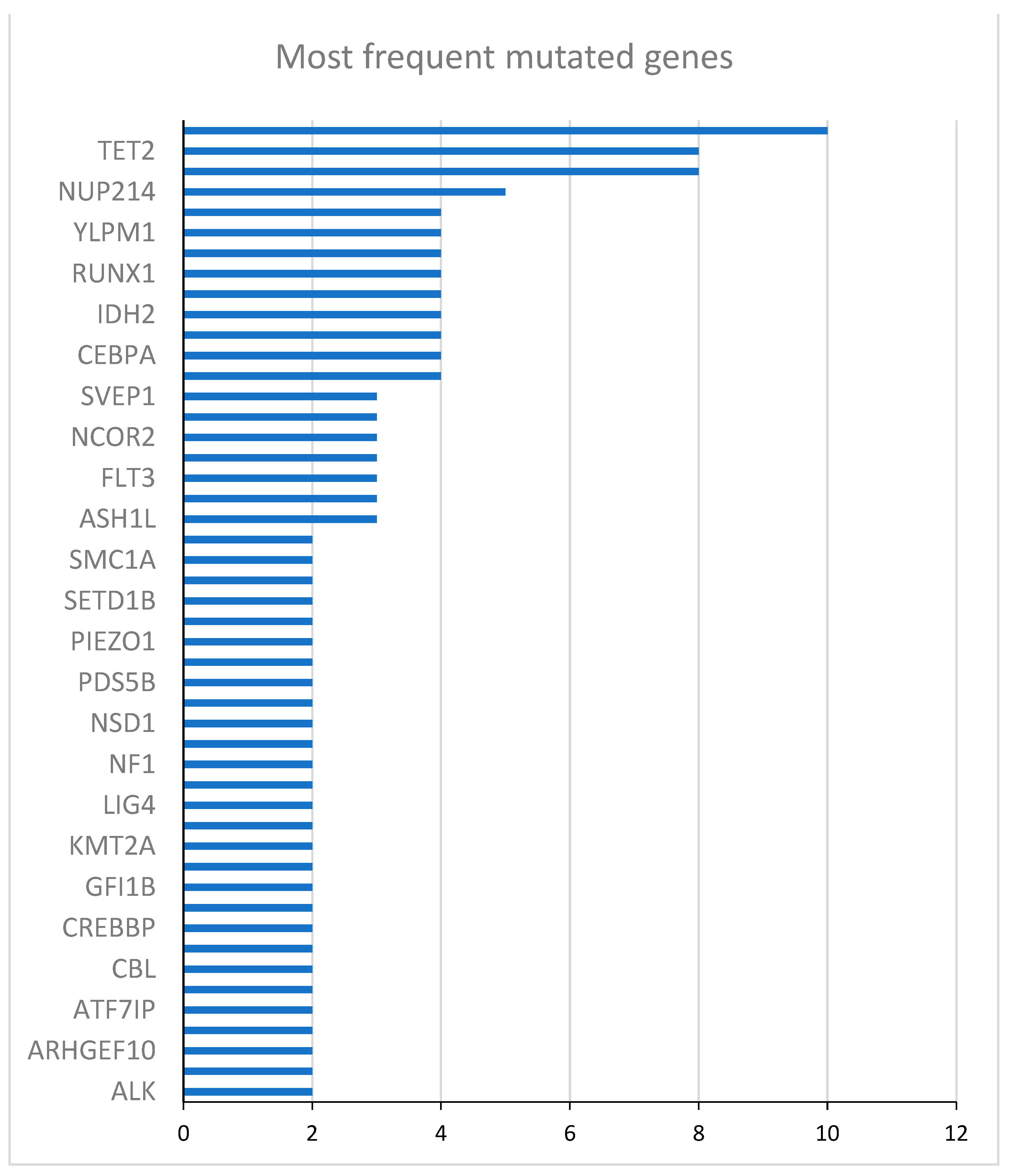
Figure 1. Mutated genes at diagnosis of randomized patients.
Preprints 119559 g001
Figure 2. Mutated genes at randomization.
Preprints 119559 g002
Table 1. Baseline patient characteristics.
 Characteristics N= 53
Age, median years (IQR) 71 (66 - 74)
Male, n (%) 28 (52.8)
AML de novo, n (%) 46 (86.8)
Hemoglobin (Hb), mean g/dL (±SD) 9.4±1.6
White blood cell (WBC) × 103, median (IQR) 15.6 (2.8 – 37.8)
Platelet (PLT) × 103, median (IQR) 61.5 (27.0 – 85.0)
WHO Classification, n (%)
        AML with minimal differentiation 8 (15.1)
        Acute myelomonocytic leukemia 9 (17.0)
        AML with myelodysplasia-related changes 9 (17.0)
        AML with maturation 12 (22.6)
        Acute monoblastic and monocytic leukemia 5 (9.4)
        AML without maturation 5 (9.4)
        AML with recurrent genetic abnormalities 3 (5.7)
        Therapy-related myeloid neoplasms 1 (1.9)
        Acute erythroid leukemia 1 (1.9)
Cytogenetic risk profile, n (%)
        Intermediate 40 (75.5)
        Poor 7 (13.2)
        Not evaluable 6 (12.3)
AML = acute myeloid leukemia, WHO = World Health Organization.
Table 2. Baseline characteristics of randomized CR patients.
 Characteristics 5-Aza
(n=11)
BSC
(n=13)
All patients (n=24)
Age, median years (IQR) 70 (66-75) 73 (65-74) 71 (65-74)
Male, n (%) 6 (54.5) 6 (46) 12 (50)
AML de novo, n (%) 9 (82) 13 (100.0) 22 (92)
Hemoglobin (Hb), mean g/dL (±SD) 9.3±0.9 9.4±1.4 9.4±1.2
White blood cell (WBC) × 103, median (IQR) 3.1 (1.7 - 40.2) 17.1 (2.7 - 25.1) 15.6 (1.8 - 28.9)
Platelet (PLT) × 103, median (IQR) 43 (26 - 63) 29 (22-71) 41 (24 - 65)
WHO Classification, n (%)
        AML with minimal differentiation 1 (9.1) 2 (15.4) 3 (12.5)
        Acute myelomonocytic leukemia 3 (27.3) 2 (15.4) 5 (20.8)
        AML with myelodysplasia-related changes 2 (18.2) 1 (7.7) 3 (12.5)
        AML with maturation 2 (18.2) 3 (23.1) 5 (20.8)
        Acute monoblastic and monocytic leukemia 1 (9.1) 3 (23.1) 4 (16.6)
        AML without maturation 1 (9.1) - 1 (4.2)
        AML with recurrent genetic abnormalities - 1 (7.7) 1 (4.2)
        Therapy-related myeloid neoplasms - 1 (7.7) 1 (4.2)
        Acute erythroid leukemia 1 (9.1) - 1 (4.2)
Cytogenetic risk profile, n (%)
        Intermediate 8 (72.7) 11(84.6) 19 (79.2)
        Poor 1 (9.1) 2 (15.4) 3 (12.5)
        Not evaluable 2 (18.2) - 2 (8.3)
AML = acute myeloid leukemia, WHO = World Health Organization.
Table 3. 2-year DFS.
Gene Mutated patients (N) Unmutated patients (N) HR (95% CI) p-value
DNMT3A 10 14 0.45 (0.15 - 1.30) 0.14
TET2 8 16 1.20 (0.44 - 3.27) 0.73
NPM1 8 16 0.47 (0.15 - 1.48) 0.17
NUP214 5 19 0.72 (0.23 - 2.23) 0.56
ZNF318 4 20 1.92 (0.62 - 5.95) 0.28
YLPM1 4 20 0.37 (0.85 - 1.65) 0.19
WT1 4 20 1.39 (0.39 - 4.88) 0.61
RUNX1 4 20 0.34 (0.15 - 2.85) 0.57
NCOR1 4 20 0.34 (0.08 - 1.50) 0.15
IDH2 4 20 0.52 (0.12 - 2.31) 0.39
FANCA 4 20 4.96 (1.34 - 18.35) 0.02
CEBPA 4 20 1.01 (0.29 - 3.53) 0.98
BCOR 4 20 1.29 (0.29 - 5.74) 0.74
CI = confidence interval, DFS = disease-free survival, HR = hazard ratio
Table 4. 5-year DFS.
Gene Mutated patients (N) Unmutated patients (N) HR (95% CI) p-value
DNMT3A 10 14 0.53 (0.19 - 1.43) 0.21
TET2 8 16 1.38 (0.53 - 3.57) 0.52
NPM1 8 16 0.53 (0.19 -1.62) 0.28
NUP214 5 19 0.69 (0.22 - 2.11) 0.51
ZNF318 4 20 1.92 (0.62 - 5.95) 0.26
YLPM1 4 20 0.58 (0.16 - 2.01) 0.39
WT1 4 20 1.26 (0.36 -4.38) 0.72
RUNX1 4 20 0.60 (0.14 - 2.63) 0.50
NCOR1 4 20 0.49 (0.14 - 1.74) 0.27
IDH2 4 20 0.49 (0.11 - 2.15) 0.34
FANCA 4 20 4.96 (1.34 - 18.35) 0.02
CEBPA 4 20 0.93 (0.27 - 3.24) 0.91
BCOR 4 20 0.74 (0.29 - 5.74) 0.74
CI = confidence interval, DFS = disease-free survival, HR = hazard ratio
Disclaimer/Publisher’s Note: The statements, opinions and data contained in all publications are solely those of the individual author(s) and contributor(s) and not of MDPI and/or the editor(s). MDPI and/or the editor(s) disclaim responsibility for any injury to people or property resulting from any ideas, methods, instructions or products referred to in the content.
Copyright: This open access article is published under a Creative Commons CC BY 4.0 license, which permit the free download, distribution, and reuse, provided that the author and preprint are cited in any reuse.
Prerpints.org logo

Preprints.org is a free preprint server supported by MDPI in Basel, Switzerland.

Subscribe

© 2024 MDPI (Basel, Switzerland) unless otherwise stated