1. Introduction
Parkinson´s disease (PD) is the fastest-growing human neurological disease due to an aging population, with a prediction to affect almost 2.5 million people in Europe by 2040 [
1]. It is characterized by the loss of dopaminergic neurons in the substantia nigra pars compacta and the presence of Lewy bodies [
2]. The loss of dopaminergic neurons is considered to be the reason for the motor symptoms (tremor, rigidity, and slowness with walking), but there are several non-motor symptoms that frequently occur and may precede the onset of the motor symptoms. Although most of the PD cases are sporadic, around 10% have a familial background. So far 20 disease-associated genes have been identified, including mutations in LRRK2, SNCA, PINK1, Parkin, and GBA [
3]. The G2019S mutation in LRRK2 (leucine-rich repeat kinase 2) accounts for 5-13% of the familial cases [
4]. This mutation results in excessive activation of the kinase domain, and the clinical pathophysiology closely mimics that of sporadic cases. LRRK2 has been shown to be expressed and is known to be localized in mitochondria, endoplasmic reticulum, Golgi apparatus, endolysosomal system, and synaptic vesicles. The physiological function of LRRK2 has been linked to neurite outgrowth, vesicle trafficking, regulation of the autophagy pathway, cytoskeleton dynamics, mitochondrial function, mRNA translation, and the immune system.
While several molecular mechanisms are identified in PD, including α-synuclein pathology, neuroinflammation, mitochondrial dysfunction, and impaired protein degradation, the exact cause remains unknown [
5]. Furthermore, disruption of the blood-brain barrier (BBB) has been identified in PD patients and in animal models [
6]. The BBB is a semipermeable membrane that separates the central nervous system from the periphery. The BBB is mainly formed by brain endothelial cells (ECs), pericytes, and astrocytes that, together with neurons and microglia, present a functional unit called the neurovascular unit. The BBB maintains the brain homeostasis, regulates delivery of oxygen and important nutrients to the brain, protects the brain from changes in the periphery, and removes carbon dioxide and toxic metabolites from the brain. Altogether, proper function of the BBB is crucial for maintenance of healthy brain tissue.
Previous studies have identified decreased P-glycoprotein expression, increased leakiness of the BBB in basal ganglia, accumulation of serum proteins, and EC degeneration in PD patients [
7,
8,
9,
10,
11,
12,
13]. 6-hydroxydopamine (6-OHDA) and 1-methyl-4-phenyl-1,2,3,6-tetrahydropyridine (MPTP) animal models have identified BBB leakage, increased P-gp immunoreactivity, and infiltration of immune cells [
14,
15,
16,
17]. Moreover, activation of astrocytes in PD can enhance release of pro-inflammatory cytokines, increasing neuronal death and affecting the function of the BBB [
18]. Although BBB impairment is recognized in PD, there have been limited investigations on this topic, and the underlying mechanism is incompletely understood.
Here, we report for the first time how the LRRK2 G2019S mutation affects human induced pluripotent stem cell (hiPSC)-derived ECs. The impact was examined at the transcriptome and functional levels under basal and inflammatory-exposed conditions.
3. Discussion
In the past, PD research concentrated mainly on neurons; but a current interest in studying other types of brain cells has shifted this viewpoint. While the involvement of astrocytes [
20,
21] and microglia [
22] in PD pathogenesis is now well-established, there have been limited research focused on investigating the role of ECs, or the BBB, in this context. A recent study using single cell RNA sequencing from PD and healthy individuals suggested that ECs may have a significant impact on cell-cell communication in PD. The study identified ECs as the core cell cluster involved in intracellular communications [
23]. Nevertheless, the effect of PD-related mutations on EC function remains unexplored.
Although the overall impact of the LRRK2 mutation on the ECs transcriptome was moderate, several noteworthy genes were identified. Kruppel-like factor 4 (KLF4) and maternally expressed gene 3 (MEG3) were downregulated in PD ECs. KLF4 has an important role in regulating several biological processes, including embryogenesis, proliferation, differentiation, neuroinflammation, oxidative stress, and apoptosis [
24]. In ECs, KLF4 has been shown to promote vascular integrity and maintain vascular health [
25]. In stroke models, KLF4 has a protective effect on microvascular ECs [
26,
27]. In addition, KLF4 has been shown to regulate EC activation after pro-inflammatory stimuli. Moreover, the overexpression of KLF4 induces the expression of several anti-inflammatory and anti-thrombotic factors [
28]. In contrast to ECs, knockdown of KLF4 has shown beneficial effects on neuronal survival in several PD models [
24]. This indicates that KLF4 has different roles in various cell types and downregulation might have a negative effect on LRRK2 mutated ECs.
MEG3 is a long noncoding RNA, and its role has been studied especially in cancer. Recent studies have identified MEG3 levels as being altered in PD patients. Two studies have reported decreased levels in plasma, and one has increased [
29,
30,
31]. Huang et al. also studied the effect of MEG3 in SH-SY5Y cells treated with MPP+ [
29]. Overexpression of MEG3 increased LRRK2 expression, improved cell viability, and decreased apoptosis, indicating a connection between MEG3 and LRRK2. In ECs, MEG3 has been shown to prevent vascular EC senescence, control VEGFA-mediated angiogenesis, and regulate proliferation and migration [
32,
33,
34]. Knockdown of MEG3 in HUVECs decreased cell viability, increased apoptotic cell rate, and impaired migration function [
35]. In addition, MEG3 knockdown has shown to induce cellular senescence, which is characterized by increased senescence-associated β–galactosidase activity, increased levels of endogenous superoxide, impaired mitochondrial structure and function, and impaired autophagy [
36]. Taken together, these studies indicate that MEG3 has an important role in EC function and that the loss of expression in PD cells could be linked to pathogenesis.
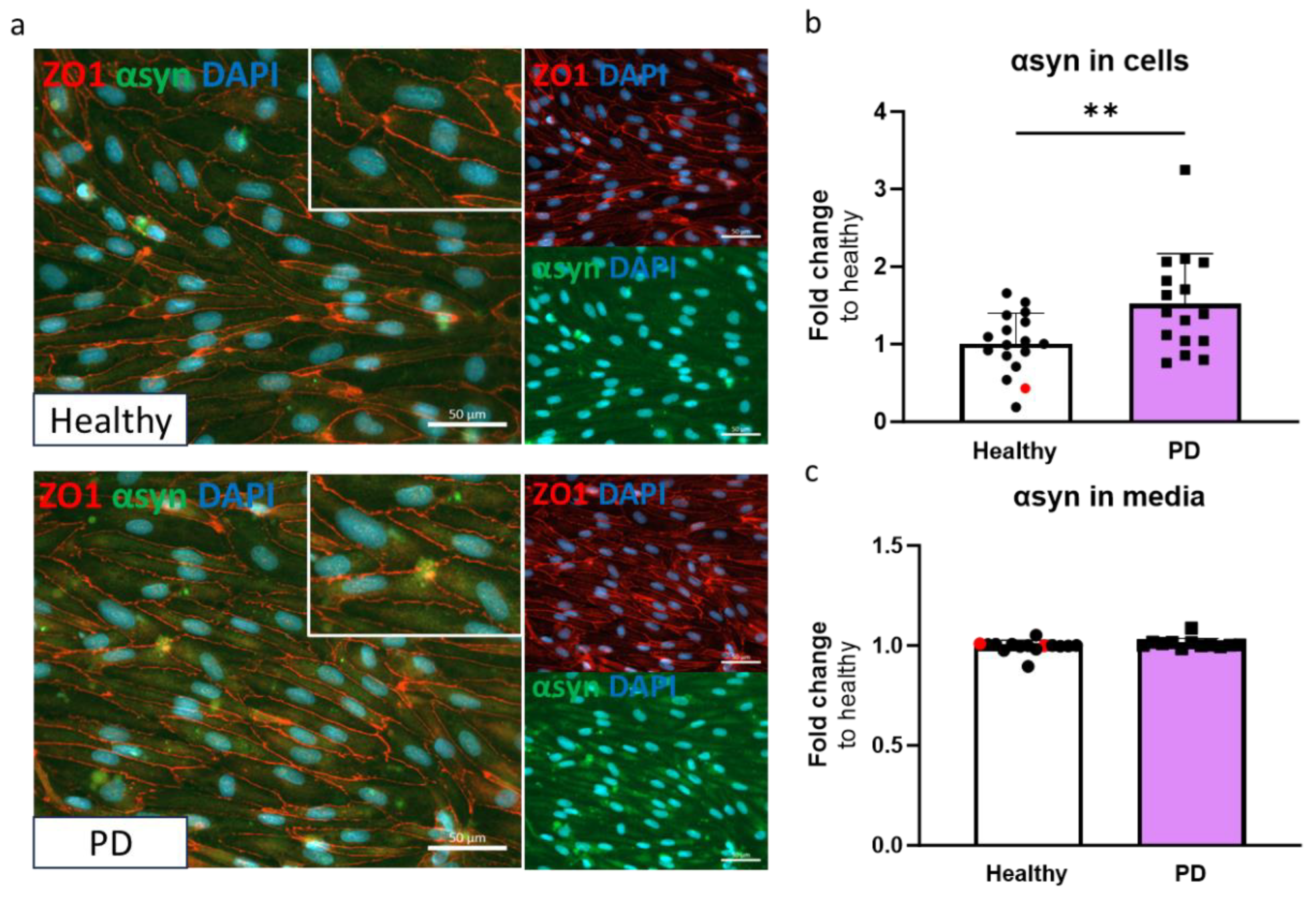
Accumulation of α-synuclein is one of the hallmarks of PD. α-synuclein is a small 14 kDa protein highly expressed in neurons but also expressed in peripheral cells and ECs [
6]. It can be transported bi-directionally across the BBB. Higher plasma levels of exosomal α-synuclein have been found in PD patients and post-mortem studies have associated α-synuclein aggregates with endothelial degeneration and decreased P-gp expression [
9,
37,
38]. Our findings demonstrate that LRRK2 mutant ECs have higher levels of α-synuclein, but the released levels remained the same compared to healthy cells. This could indicate failure in exporting or degradation of α-synuclein in PD ECs, although this would require more extensive studies.
The transcriptomics analysis revealed alterations in pathways related to unsaturated fatty acids and their biosynthesis in PD ECs. Fatty acids have several important roles in the body, as they serve as a source of energy, are major components of cell membranes, and regulate inflammatory responses [
39]. Excessive intake of fatty acids is associated with obesity, energy overload, and increased brain inflammation. Polyunsaturated fatty acids can exert a direct cytotoxic effect on α-synuclein. Polyunsaturated fatty acids can bind to α-synuclein and promote oligomerization, and high concentrations of DHA increase intracellular accumulation of soluble and insoluble α-synuclein and neuronal injury in A53T mice [
40,
41,
42]. In addition, a higher intake of arachidonic acid has also been linked to an increased risk of PD pathogenesis [
43]. Besides fatty acids themselves, fatty acid binding proteins (FABPs) 3 and 5 have been linked to α-synuclein accumulation and aggregation, and mitochondrial dysfunction, and increased apoptosis [
39]. The RNA expression of both genes was elevated in PD ECs after TNFα exposure. Increased expression of FABPs combined with increased fatty acid biosynthesis might exacerbate the toxic effect of α-synuclein in ECs.
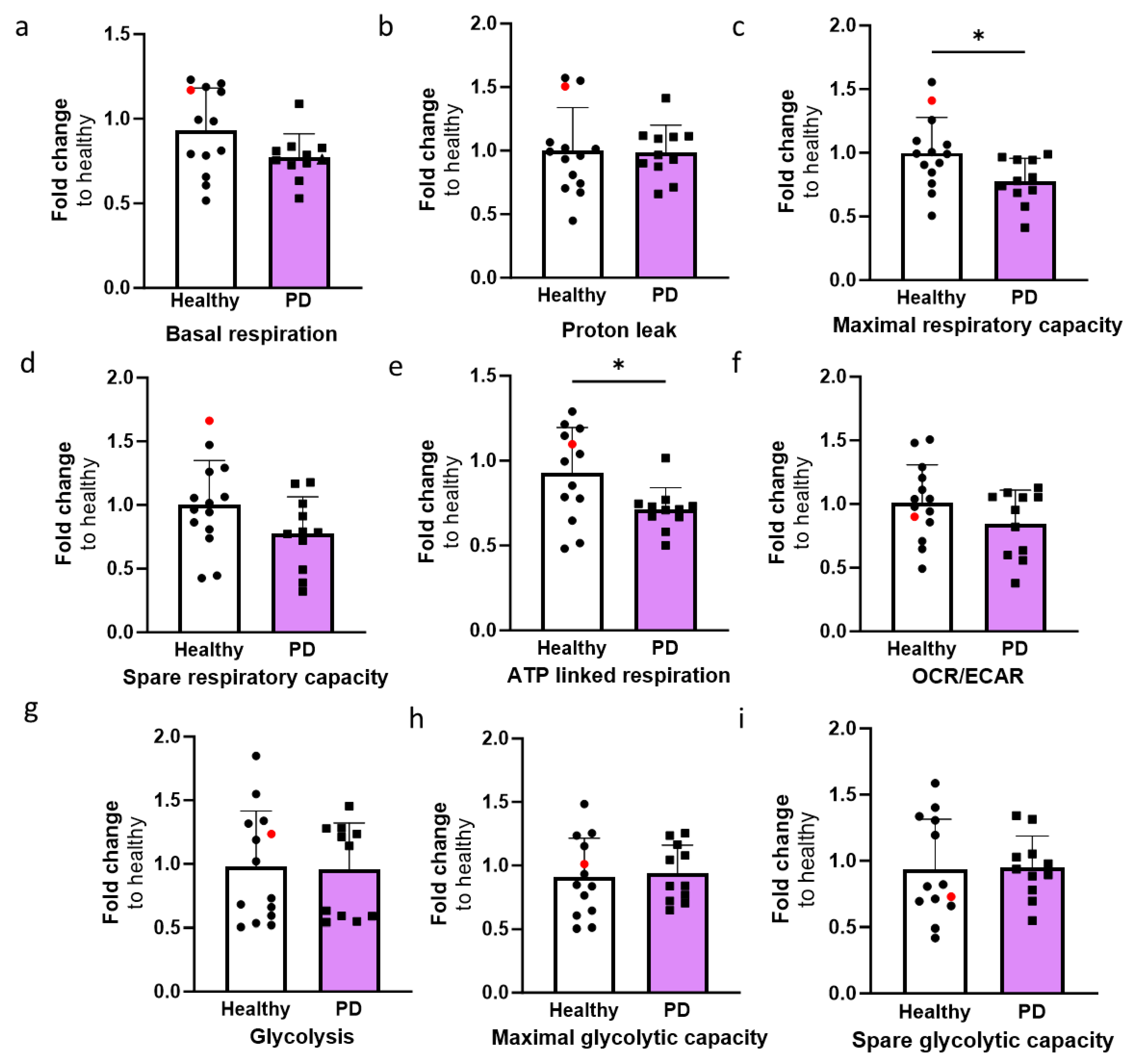
Mitochondrial dysfunction is one of the key mechanisms linked to PD pathogenesis. Higher oxidative stress, reduced mitochondria membrane potential, decreased ATP production, mitochondria DNA damage, elongated mitochondria, mitochondrial fragmentation and mitophagy have been observed in postmortem human tissues and animal and cellular models of LRRK2 [
44]. PD ECs with the LRRK2 mutation showed altered mitochondrial function, including decreased maximal respiration and ATP-linked respiration. Alterations in oxygen consumption have been found in other cell types, including hiPSC-derived neurons [
45] and astrocytes [
20] which carry the LRRK2 G2019S mutation. This could indicate a similar effect of the mutation, regardless of the cell type.
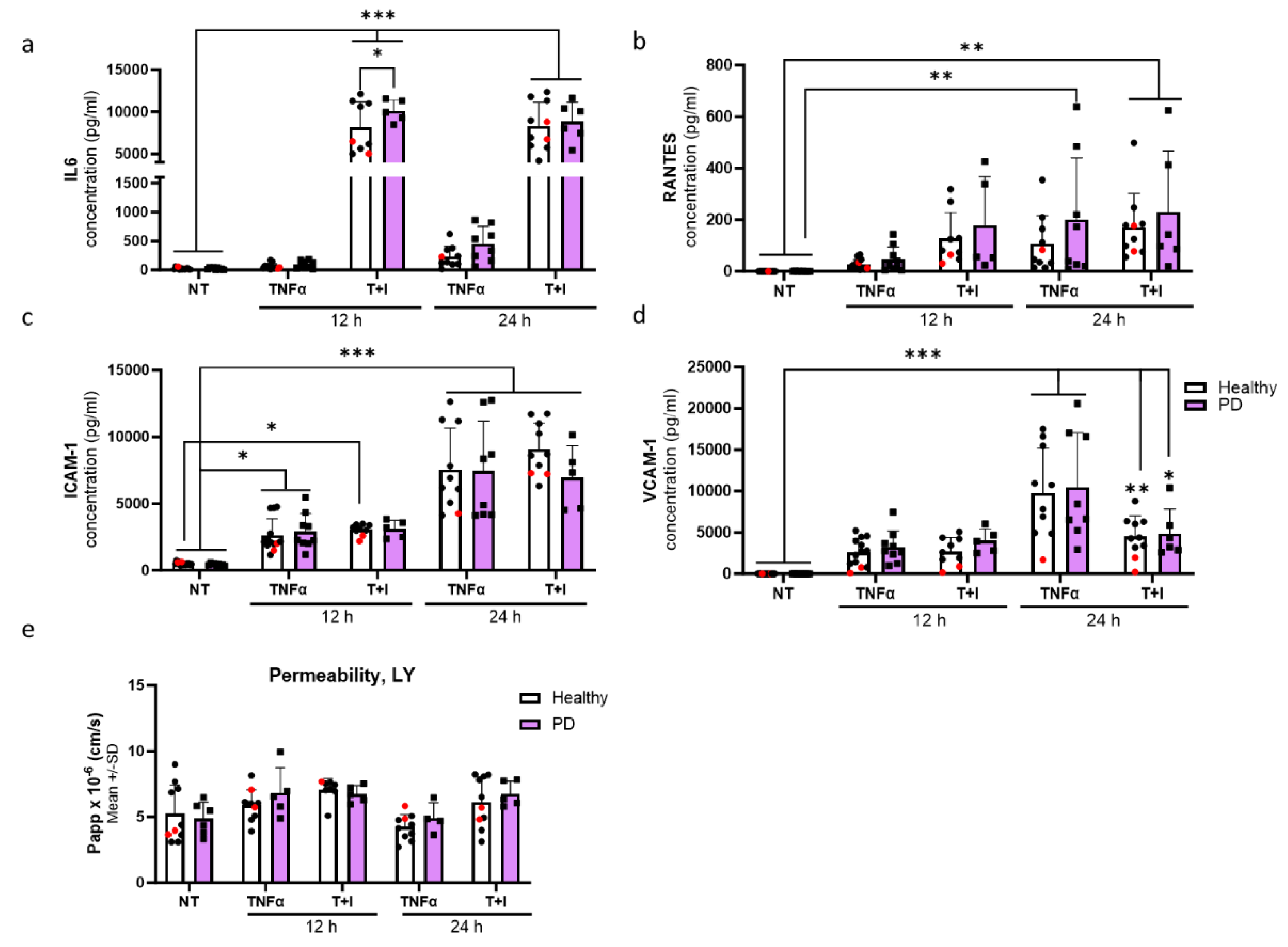
One common hallmark of several neurodegenerative diseases, including PD, is neuroinflammation. The effect of inflammatory stimuli on PD ECs was studied on a transcriptomic and functional level. While TNFα+IL-1β exposure elicited a more pronounced response in both healthy and PD ECs compared to TNFα alone, the combination did not cause a substantial genotypic difference in transcriptomics. This could suggest that PD ECs have an altered response only to specific stimuli. At the functional level, the LRRK2 mutation did not cause major differences. ECs responded to inflammatory exposures by increasing the release of cytokines, but the variation across different cell lines was substantial. This could suggest that the response is rather individual than LRRK2-related. Although we did detect increased IL6 secretion after 12 h TNFα+IL-1β exposure, elevated levels of IL6 have been detected in PD patients and linked to disease mortality [
46,
47,
48,
49].
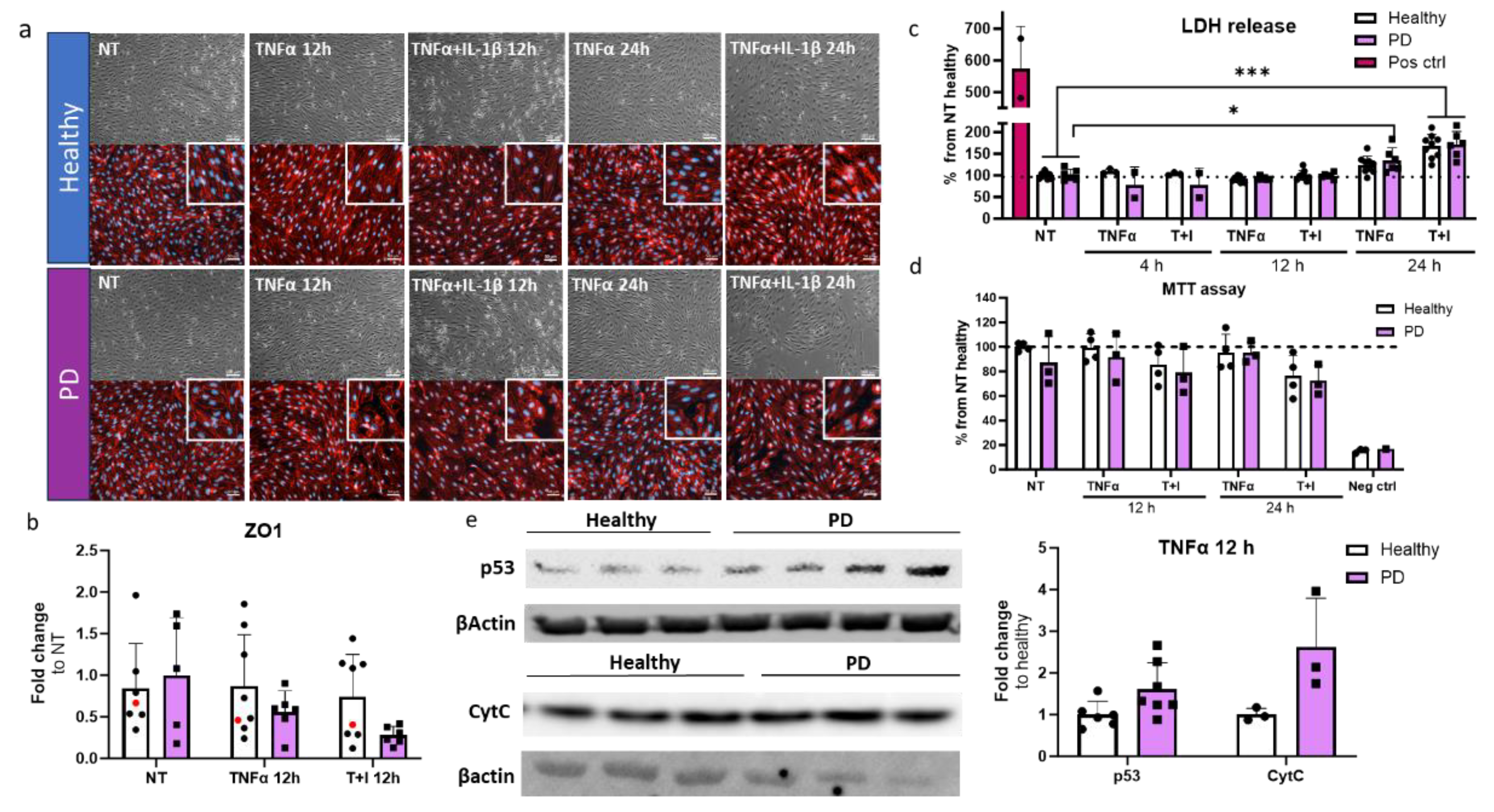
Upon longer inflammatory exposure, the cell death was increased in ECs. This was seen in both healthy and PD ECs after 24 h of combination exposure, but already in PD ECs after 24 h of TNFα exposure. We also detected elevated levels of p53 and cytochrome C in PD ECs after 12 h TNFα exposure. The p53 signaling pathway was also linked to upregulated genes in PD ECs after 4 h TNFα exposure. P53 is involved in several processes which are related to PD, like neuronal oxidative stress, apoptosis, and abnormal protein aggregation [
50]. Moreover, high expression of p53 has been found in PD cell models and in the substantia nigra region in PD patients and animal models [
51,
52,
53]. In addition, p53 can induce apoptosis through mitochondrial cytochrome C release and activation of caspases [
54]. Interestingly, MEG3 has been shown to regulate p53 signaling, but the response seems to be cell specific. For example, in neurons, MEG3 activates p53 and mediates neuronal death in stroke [
55], whereas in cardiac fibroblasts, MEG3 does not alter the p53 response [
56]. The role of MEG3 and p53 in HUVECs was studied by Ali et al. [
57]. After 4 h TNFα exposure, MEG3 knockdown induced p53 activation by increasing the expression of p53 target genes, promoting apoptosis, and inhibiting proliferation. Whether LRRK2 mutation is linked to MEG3 and p53 activation is still unclear and would need further studies.
Limitations of the study
This study focused on the LRRK2 G2019S mutation in ECs only. It is important to keep in mind that these results represent LRRK2 related cases and may not directly translate into sporadic ones. We haven´t checked additional mutations from the LRRK2 mutated cells and there is a small possibility that additional mutations or risk variants might influence the results. Although the dysfunction of the BBB and ECs is recognized in PD, its role probably is not as substantial as, for example, in Alzheimer's disease, and individual variation might play a part. Our results showed variation between healthy and PD individuals especially in transcriptomics and inflammatory responses. Of course, a low number of cell lines could mask the differences and ideally, more lines should be included. One way to reduce the variation is to use isogenic lines. We included one isogenic line, but this was absent from the transcriptomic studies. It would be important to repeat the RNA sequencing with additional lines and isogenic controls. Additionally, we used only 2D monoculture, and the remaining BBB cell types were missing. The BBB is a multicellular unit, including astrocytes and pericytes, and their presence could change the properties of ECs and thus also the effect of the LRRK2 mutation. Within this study, we used a protocol for deriving vascular ECs as opposed to brain ECs. Because of this, some BBB specific changes might have been overlooked in our study. Given our emphasis on inflammation, we aimed to ensure that cells respond to inflammatory stimuli. While there are existing protocols published for obtaining brain-like ECs, these protocols have major limitations. It has been shown that brain-like EC protocols produce cells with more epithelial cell phenotype and lack the EC-specific response to inflammatory stimuli [
58,
59,
60]. Furthermore, one common feature of all hiPSC-ECs is the absence of P-glycoprotein expression and more importantly, its functionality [
61]. Similarly, we observed very low levels in our cells. Decreased expression of P-glycoprotein has been linked to PD and investigation of this would require a different model system. Advances in different 3D models, like organ-on-chips or vascularized brain organoids, could provide more accurate models in the future.
4. Materials and Methods
4.1. Cell Culture
Human iPSCs (hiPSC) lines used in this study included six healthy controls, three lines with G2019S mutation in the LRRK2 gene and one isogenic line (
Table S1). Healthy lines 4-6 and PD LRRK2 line 1 were generated and characterized in the Stem Cell Laboratory, at the University of Eastern Finland [
62]. Healthy lines 1-3 were obtained from Takara Bio (Y00275, Y00305, and Y00325) and PD LRRK2 lines 2 and 3 and isogenic line from NIBSC.
4.2. Endothelial Cell Differentiation
Two differentiation protocols were used in this study (referred to as protocols 1 and 2). Detailed information of lines and protocols used in different experiments are listed in
Table S2. In both protocols, ECs are differentiated first into EC progenitors and then vascular ECs with small differences in small molecules. Protocol 1 was more efficient and for that reason it was chosen for most of the experiments. Protocol 2 was used to measure α-synuclein levels with ELISA and metabolic function with Seahorse assay.
For protocol 1, ECs were differentiated according to the protocol published by Harding et al. 2017 with minor modifications. To start the differentiation, hiPSCs were plated to Matrigel-coated dishes at a density of 15-25 k/cm2 in E8 media with 5 µM of ROCK inhibitor (Y-27632 dihydrochloride, Selleckchem). The next day (day 1), the media was changed to Stemdiff APEL2 media (Stemcell Technologies) supplemented with 6 µM CHIR-99021 (Cayman). On day 3, the media was changed to APEL2 media with an additional 25 ng/ml of BMP4 (Peprotech), 10 ng/ml of FGF (Peprotech), and 50 ng/ml of VEGF (Peprotech). Cells were cultured in this medium for three days, changing media on day 5. On day 6, ECs were sorted by magnetic separation (Miltenyi Biotec) using CD144 microbeads according to the manufacture`s protocol. CD144 positive cells were plated on Matrigel-coated dishes in Endothelial Growth Media MV2 (ECGM-MV2, PromoCell) with an additional 20 ng/ml of VEGF for the first passage. ECs were expanded, and cells in passage three were used for the experiments.
For protocol 2, ECs were differentiated using a protocol published by Patch et al. [
63]. Briefly, the hiPSCs were dissociated using EDTA (Invitrogen) and seeded at a density of 300 000 cells/35 mm plate in E8 medium supplemented with ROCK inhibitor. The next day, the induction of the lateral mesoderm was started by replacing the media with N2B27 medium supplemented with 8 µM CHIR-99021 and 25 ng/ml BMP4 and continued for three days. The EC induction was started by replacing the N2B27 media with StemPro-34 SFM medium (Life Technologies) supplemented with 200 ng/ml VEGF and 2 µM Forskolin (Sigma). The cells were incubated in StemPro medium for two to four days, changing the medium every day. When the areas of differentiation were visible, the ECs were sorted by magnetic separation using CD144 microbeads according to the manufacture`s protocol. CD144 positive cells were plated on 6 cm Matrigel-coated plates in StemPro medium supplemented with 20 ng/ml VEGF.
4.3. Immunocytochemistry
ECs were fixed with 4 % formaldehyde (VWR) for 15 min in room temperature (RT) or methanol (VWR) for 15 min at 4°C followed by washes with PBS. For intracellular antigens, the ECs were permeabilized with 0.2 % Triton X-100 in PBS (Sigma) for 20 min at RT. The cells were blocked with 5 % normal goat serum (NGS, Vector) or 10 % horse serum (HS, Gibco) in PBS at RT for 1 h and then incubated with primary antibodies in 5 % NGS or 10 % HS at 4 °C overnight. For unconjugated antibodies, the cells were incubated in 5 % NGS or 10 % HS in PBS containing a secondary antibody for 1 h at RT. The cells were washed with PBS, and the nuclei were visualized with DAPI (ThermoFisher). The cells were imaged with the Zen Observer Z1 or Zen Imager AX10. The antibodies used in this study are listed in
Table S3.
4.4. RT-qPCR
RNA was extracted using the RNA easy MiniKit (Qiagen) and RNA concentration was measured with NanoDrop or DS-11 FX Spectrophotometer/Fluorometer (Denovix). 500-1000 ng of the RNA were converted to cDNA (Maxima Reverse Transcriptase, ThermoFisher). RT-qPCR was conducted using the Maxima probe/ROX qPCR master mix (ThermoFisher) to quantify the relative expression of genes of interest utilizing TaqMan assays (
Table S4) with Step One Plus (Applied Biosciences). The Ct mean value was normalized to internal Ct mean value of β-actin, and the relative expression is presented as a fold change compared to the control group
4.5. α-Synuclein ELISA
The amount of endogenous α-synuclein was determined from ECs using a human α-synuclein ELISA kit (Invitrogen and Biolegend). For the assay, cells were lysed with M-PER (Invitrogen) or cell extraction buffer (Invitrogen) and diluted 1 to 10 in reagent diluent. The luminescence or absorbance 450 nm was measured with PerkinElmer`s VICTOR2 multilabel plate reader. The α-synuclein concentration from the cells was standardized to total protein concentration using the Pierce™ BCA Protein Assay Kit (ThermoFisher). Media samples were collected from ECs cultured for three days without media change. Samples were stored at -70 °C before analysis. Human α-synuclein ELISA kit (AssayGene) was used to measure the α-synuclein levels from media. The luminescence or absorbance at 450 nm was measured with PerkinElmer`s VICTOR2 multilabel plate reader.
4.6. Seahorse
The oxygen consumption rate (OCR) and the extracellular acidification rate (ECAR), indicators of mitochondrial respiration and glycolysis, respectively, were measured in real time from live cells with the Seahorse Extracellular Flux (XF) 24 or 96 Analyzer. First, the ECs were plated on Matrigel-coated plates in StemPro34 or ECGM-MV2 medium depending on protocol used and incubated overnight. Before the assay, the medium was changed to Seahorse XF Assay medium (Seahorse DMEM supplemented with 2 mM Glutamax) and incubated for 1 h at 37 °C without CO2 before adding the compounds. The final concentrations of D-glucose and sodium pyruvate were 10 mM, and 0.5 mM, respectively. For oligomycin, FCCP, antimycin, and rotenone the concentrations were 1.0 µM. The assay was performed as follows: 2 min mix time, 2 min wait time, and 3 min measure cycle. After the assay, the results were normalized to protein concentrations using Pierce™ BCA Protein Assay Kit (ThermoFisher). The results were analyzed with Wave program. Wells with negative values or aberrant OCR or ECAR patterns were excluded from each of the individual experiments.
4.7. Inflmammatory Exposures
To assess the inflammatory response, ECs were exposed to TNFα or a combination of TNFα (Peprotech) and IL-1β (Peprotech). Detailed information on duration, concentrations, and assays are listed in
Table S5. Briefly, ECs were plated in ECGM-MV2 medium, and the next day, the media was changed to human Endothelial Serum Free Media (ESFM, Gibco) with additional 1X B27 (Gibco) and 10 ng/ml FGF (Peprotech). The following day, we initiated inflammatory exposures using the same media as described above.
4.8. Total RNA Sequencing
Cells were lysed on ice (Buffer RLT + β-mercaptoethanol 10 µl/ml), and RNA was extracted directly after lysis with the Qiagen RNeasy Mini Kit (Qiagen) with DNase I digestion (Qiagen). RNA concentration was measured with DS-11 FX Spectrophotometer/Fluorometer (Denovix). 1-4 µg of RNA were sent for library preparation and sequencing (performed by Azenta). Quality was evaluated with FastQC, and the mean quality score for the samples was 35.30. The library preparation workflow included ribosomal RNA depletion, RNA fragmentation and random priming, cDNA synthesis, end repair, 5´ phosphorylation and dA-tailing, adaptor ligation and PCR enrichment. Sequencing was done with Illumina NovaSeq, PE 2x150. Sequence reads were trimmed by using Trimmomatic v.0.36 to remove possible adaptor sequences and nucleotides of poor quality. Trimmed reads were mapped to the Homo sapiens GRCh38 reference genome available on ENSEMBL using the STAR aligner v.2.5.2b. Unique gene hit counts were calculated by using featureCounts from the Subread package v.1.5.2 and used for downstream differential expression analysis (DESeq2). The Wald test was used to generate p-values and log2 fold changes, and the Benjamini-Hochberg for adjusted p-values. Genes with a p-value < 0.05 and absolute log2 fold change > 1 were called as differentially expressed genes (DEGs) for healthy vs. PD LRRK2 and genes with an adjusted p-value < 0.05 and absolute log2 fold change > 1 when comparing exposures within the group. Pathway enrichment analysis (KEGG and gene ontology biological processes, GO BP) was conducted on DEGs using Enrichr.
4.9. LDH Cytotoxicity Test
To assess the toxicity of TNFα and IL-1β exposures, the release of LDH was measured from ECs media samples at different time points (
Table S5) using the CyQuant LDH Cytotoxicity assay (Invitrogen). The samples were analyzed with a Wallac Victor2 1420 Multilabel Counter (Perkin Elmer).
4.10. MTT Assay
ECs were plated to 96-well plate and treated as previously described (
Table S5). After inflammatory exposure, a part of the cells was exposed to 1 % Triton-X for 5 min to induce cell death. This was used as a negative control. Then fresh media was changed with an additional 1.2 mM of 3-(4,5-Dimethylthiazol-2-yl)-2,5-diphenyltetrazolium bromide (MTT) (VWR) and cells were incubated for 2 h at 37 °C. After the incubation, media was removed, and DMSO was added to the cells and incubated at RT overnight. The next day, 100 µl of sample was transferred to a new 96-well plate and absorbance was measured at 595 nm with PerkinElmer`s Victor2 multilabel plate reader.
4.11. Western Blotting
Protein was extracted from ECs using in-house made RIPA buffer (50 mM Tris, 1% Triton-X 100, 0.5% Sodium deoxycholate, 0.1% SDS, 150 mM NaCl) with Pierce™ protease inhibitor (ThermoFisher). Total protein amount was measured with Pierce™ BCA Protein Assay Kit (ThermoFisher). Samples were mixed with Laemmli sample buffer (60 mM Tris, 10% glycerol, 2% SDS, 1% Bromophenol blue, with 5% 2-Mercaptoethanol added just before use) and incubated at 95°C for 5 min. 15 µg (P53, Cytochrome C) or 30 µg (LRRK2) of protein was resolved with 12 % gels (P53, Cytochrome C) or 4%-12% gradient gel in Tris-Glycine running buffer.
Proteins were transferred to 0.2 µm PVDF pre-cut membrane transfer pack (Bio-rad) and transferred with Trans-Blot Turbo® transfer system (Bio-Rad, Hercules, CA, USA). Membranes were blocked in 5% Milk powder in TBST (20 mM Trish, 150 mM NaCl, 0.1% Tween-20, pH 7.6) and the membranes were incubated overnight at 4°C with primary antibodies diluted in 5% BSA in TBST (1:1000): p53, (1:1000): Cytochrome C, (1:10,000): LRRK2. Membranes were washed with TBST and incubated with HRP-conjugated antibodies for 2 hours at room temperature. After washing, detection was performed with SuperSignal™ West Pico PLUS Chemiluminescent Substrate (ThermoFisher) and imaged with ChemiDoc™ MP Imaging System (Bio-Rad, Hercules, CA, USA). For normalization, membranes were incubated with anti β-actin primary antibody (1:1000) diluted in 5% BSA in TBST overnight at 4°C. Cy3-conjugated secondary antibody was diluted in 1:1000 in TBST and incubated for 2 h at room temperature. After washing, the membrane was imaged with the same equipment as previously. The intensity of the target bands was quantified with ImageLab (version 5.1, Bio-Rad Laboratories). Detailed information about antibodies is found in
Table S3.
4.12. Quantification of Immunofluorescence
ECs were fixed and stained as previously described. Images were taken with Zen Imager AX10 and minimum of two images per sample. Fluorescence was analyzed with Fiji (ImageJ). IntDen of each sample was normalized to number and intensity of DAPI.
4.13. Cytometric Bead Assay
Smount of released cytokines was measured with the cytometric bead array (CBA) Human Soluble Protein Master Buffer Kit (BD). Before analysis, media samples from non-exposed and exposed samples were collected and stored at -70 °C. Capture beads used were: Human IL-6, Human RANTES, Human ICAM1, Human VCAM1. The samples were analyzed with CytoflexS (Becman Coultier). At least 300 events per cytokine were measured. Excitation was done with 638 nm red laser, and the bead clusters were detected with 660/20 BP (APC) and 780/60 BP filters (APC-A750). Cytokine reporter PE was excited with 561 nm yellow laser and filter 585/42 BP was used for detection. Data were analyzed using FCAP Array 2.0 (SoftFlow, Hungary) and cytokine concentrations were calculated by regression analysis from known standard concentrations.
4.14. Permeability Assay
ECs were plated to Matrigel-coated 24 well inserts (0.4 µm pore size, Sarstedt) at a density of 100 k/cm2 in ECGM-MV2. The next day, the media was changed to human Endothelial Serum Free Media (ESFM, Gibco) with additional 1X B27 (Gibco) and 10 ng/ml FGF (Peprotech). The following day, media was refreshed, or inflammatory exposures were started (
Table S5). Permeability was measured with 4 kDa FITC Dextran (Merck) and Lucifer yellow (LY, Merck) from unexposed ECs. LY was used to measure permeability from inflammatory stimulated ECs. 150 µl of dextran (500 µg/ml) or LY (100 µg/ml) solution was added to the apical side and 800 µl of fresh media to basolateral side. Basal ESFM was used for the assay. Samples were collected from the wells at timepoints of 20, 40 and 60 min and the volume was replaced with fresh media. The fluorescence values were measured with PerkinElmer`s Victor2 multilabel plate reader. The background was reduced, and corrected fluorescence values were calculated. Standards were used to calculate the accumulated amounts over time and plotted against time. The apperent permeability coefficient (Papp) was calculated according to the equation (1), where dQ/dt (µg/s) is the dextran/LY flux (liner range) across the barrier, A is the insert membrane area (cm2) and C0 is the initial dextran/LY concentration (µg/cm3).
4.15. Data Analysis
The data were analyzed with GraphPad Prism (version 10.0.3). When comparing healthy vs. PD LRRK2, unpaired t-test was used. When comparing exposures, a two-way ANOVA with Tukey´s multiple comparison test was used. Levels of significance *p < 0.05; **p < 0.01; ***p < 0.001 were used. Outliers were tested with the GraphPad Grubbs' test.
The visualization of RNA transcriptomic data was done by using a free online platform for data analysis and visualization,
https://www.bioinformatics.com.cn/en. Images were modified with Inkscape 1.2.
Figure 1.
Differentiation and characterization of hiPSC-derived endothelial cells. (a) A schematic illustration of the differentiation of hiPSC-ECs. HiPSCs are differentiated first to mesoderm and then vascular progenitors. VE cadherin (CD144) positive cells are MACS separated and ECs are expanded before experiments. (b) Representative bright field images of different stages of EC differentiation. Scale bar 50 µm. (c) Relative gene expression of CDH5, CLDN5, OCLN, and TJP1 in ECs compared to CD144 negative cells from MACS separation. n= 2 (MACS neg), 3 (healthy), 3 (PD LRRK2) (d) Representative fluorescent images of ECs stained with CD31, ZO1, VE cadherin, and claudin 5. Nuclei stained with DAPI. Scale bar 50 µm. (e) Permeability (Papp) of 4 kDa Dextran and Lucifer yellow. n= 8-14 (healthy), 1-2 (isogenic), 6-13 (PD LRRK2) from four (4kDa Dextran) or two (Lucifer yellow) independent experiments. (F) Western blot analysis of LRRK2 (230 kDa). Normalization to β-actin (42 kDa) protein levels. Representative blots and quantification of the data. n=8 (healthy), 2 (isogenic), 8 (PD LRRK2). All data shown as mean +/-SD.
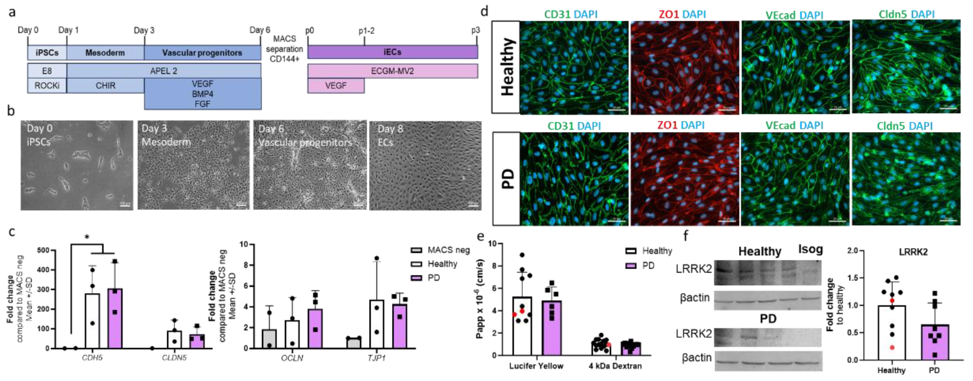
Figure 2.
RNA expression analysis of hiPSC-derived endothelial cells from healthy donors and PD patients. (a) Volcano plot of up- and down-regulated DEGs between healthy and PD ECs based on p-value (<0.05) and absolute log2 Fold >1. (b) Cluster heatmap of the top 30 DEGs between healthy and PD ECs based on the p-value (<0.05). (c) Gene expression levels of POU5F1 and SOX-2 in healthy and PD ECs compared to hiPSCs, assessed by qPCR. n= 2 (hiPSCs), n=6 (healthy), n=5 (PD LRRK2), two-way ANOVA. (d) Gene expression of KLF4 and MEG3 in healthy and PD ECs quantified with qPCR compared to healthy. KLF4 n=7 (healthy), n=6 (PD LRRK2), MEG3 n=6 (healthy), n=5 (PD LRRK2). Unpaired t-test. (e) KEGG pathway of up- and down-regulated genes in PD ECs compared to healthy. (f) Heatmap showing genes related to arachidonic acid metabolism in healthy and PD ECs. (g) GO pathway of up- and down-regulated genes in PD ECs compared to healthy. (h) GO biological processes related to vascular or EC function of up- and down-regulated genes in PD ECs compared to healthy. (i) Heatmap showing genes related to Blood vessel morphogenesis and vascular transport/transport across BBB in healthy and PD ECs. KEGG, the Kyoto Encyclopedia of Genes and Genomes; GO BP, gene ontology biological processes; PD1-3, patients carrying a mutation in LRRK2 gene (G2019S.) Data shown as mean +/-SD.
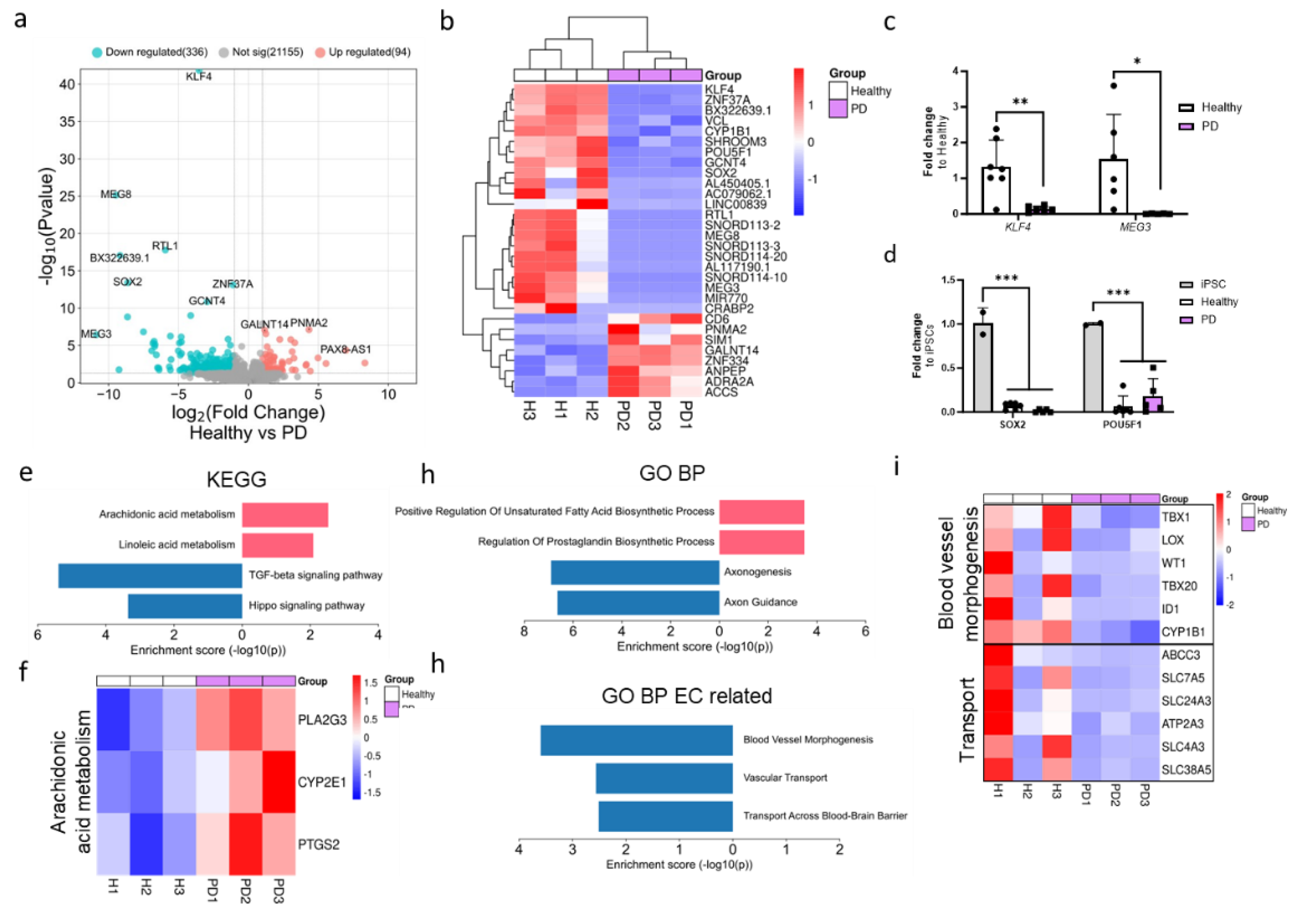
Figure 3.
Alpha-synuclein protein expression is increased in LRRK2 mutant endothelial cells. (a) Representative fluorescent images of ECs stained with ZO1 and α-synuclein (αsyn). Nuclei stained with DAPI. Scale bar 50 µm. (b) Protein expression of α-synuclein in ECs measured with ELISA. Data shown as fold change to healthy + isogenic (marked as red) and results are normalized to total protein amount. n= 16 (healthy), 1 (isogenic), 14 (PD LRRK2), from five independent experiments. (c) Released amount of α-synuclein in media measured with ELISA. Data shown as fold change to healthy + isogenic. n= 14 (healthy), 2 (isogenic), 12 (PD LRRK2) from five independent experiments. Data shown as mean +/- SD. Unpaired t-test, ** p< 0.01.
Figure 4.
The altered metabolic profile in PD endothelial cells. Oxygen consumption rate (OCR) and extracellular acidification rate (ECAR) were measured with Seahorse XF assay. (a-e) Calculated OCR values of basal respiration, proton leak, maximal respiration, spare respiratory capacity, and ATP linked respiration, respectively. (f) OCR/ECAR ratio from basal level. (g-i) Calculated ECAR values of glycolysis, maximal glycolytic capacity, and spare glycolytic capacity, respectively. Data shown as fold change to healthy + isogenic. n= 11-12 (healthy), 1 (isogenic), 10-11 (PD LRRK2) from five independent experiments. Data shown as mean +/- SD, Unpaired t-test, * p< 0.05.
Figure 5.
Transcriptional profile of PD endothelial cells after inflammatory stimuli. (a)Volcano plot of up- and down-regulated DEGs between healthy and PD ECs after TNFα exposure based on p-value (<0.05) and absolute log2 Fold >1. (b) Gene expression of MEG3 in healthy and PD ECs quantified with qPCR. n=5 (healthy), 6 (PD LRRK2). Two independent experiments. (c) KEGG and GO pathways of up- and down-regulated DEGs in PD ECs compared to healthy. (d) Heatmap showing genes related to p53 signalling pathway and steroid biosynthesis in healthy and PD ECs. (e) Volcano plot of up- and down-regulated DEGs between healthy and PD ECs after TNFα+IL-1β exposure based on p-value (<0.05) and absolute log2 Fold >1. (f) Gene expression of MEG3 in healthy and PD ECs. n=6 (healthy), 6 (PD LRRK2). Two independent experiments. (g) KEGG and GO pathways of up- and down-regulated genes in PD ECs compared to healthy. (h) Venn diagram of DEGs between healthy and PD ECs in unexposed, TNFα and TNFα+IL-1β exposed conditions. All exposures lasted for 4 h and the concentrations were: 10 ng/ml TNFα or 10 ng/ml TNFα+ IL-1β. Data shown as mean +/-SD, n= 3, unpaired t-test ** p< 0.01.
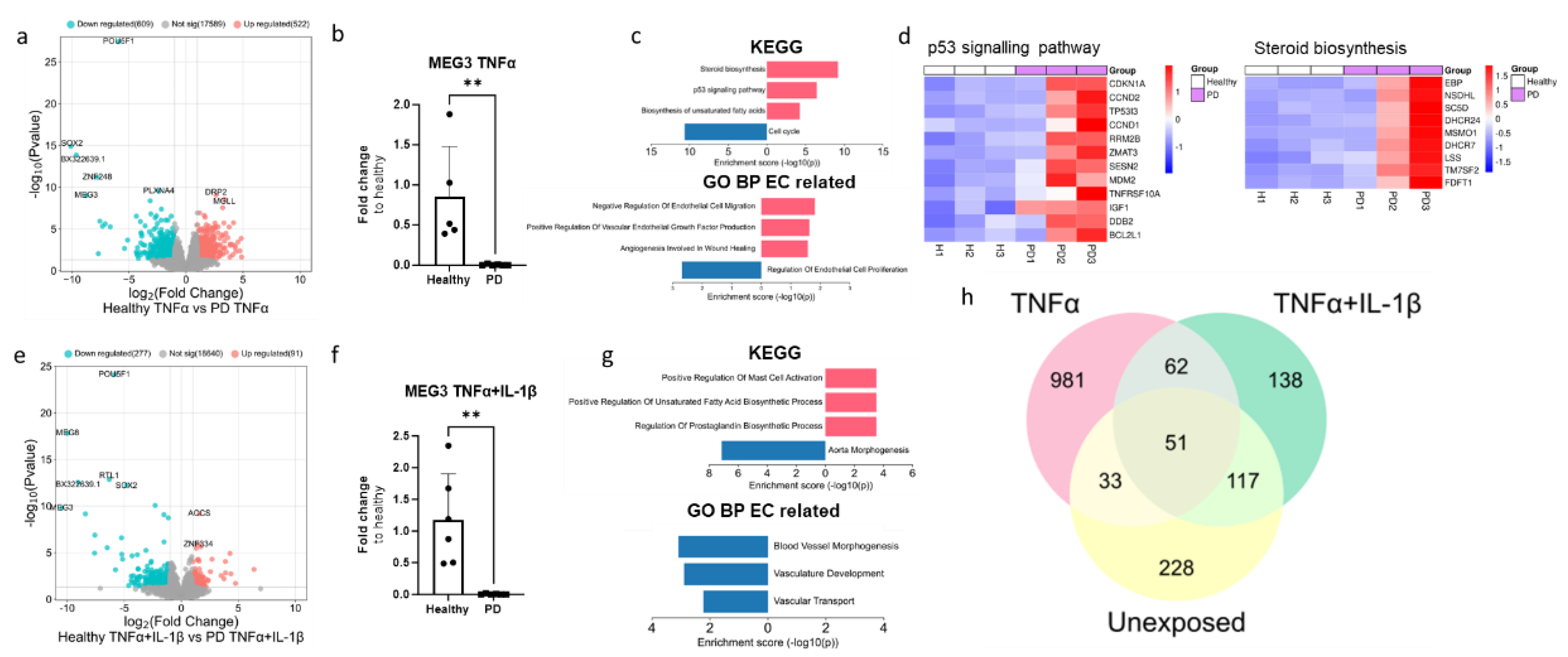
Figure 6.
Cell viability and cytotoxicity in endothelial cells after inflammatory stimuli. (a) Representative bright field (upper panel) and immunofluorescence images stained for ZO1 (lower panel) of ECs, non-exposed, 12 h TNFα and TNFα+IL-1β exposed, 24 h TNFα and TNFα+IL-1β exposed. Scale bar 50 µm. (b) Quantification of the immunocytochemistry. Fluorescence normalized to DAPI. n=6-7 (Healthy), 1 (isogenic),5-6 (PD LRRK2) from two independent experiments. (c) LDH release from ECs of non-exposed and after 4, 12 and 24 h TNFα and TNF+IL-1β (TI) exposure. n= 3-9 (healthy), 2-6 (PD LRRK2) from two independent experiments. (d) MTT assay of non-treated and 12 and 24 h TNFα and TNF+IL-1β treated ECs. n= 2-4 (healthy), 1-3 (PD LRRK2. (e) Western blot analysis of p53 and Cytochrome C (CytC) in healthy and PD EC normalized to β-actin protein levels. Representative images and quantification of the data. n=3-6 (healthy), 3-7 (PD LRRK2). Data shown as mean +/-SD. Two-way ANOVA with Tukey´s multiple comparison test, p*<0.05, p**<0.01, p***<0.001.
