1. Introduction
Gamma brain waves, typically characterized by frequencies ranging from approximately 30 to 100 Hz, are closely associated with high-level cognitive functions, including attention, memory, and consciousness [
1]. Alterations in gamma rhythms can profoundly impact brain function and overall mental health [
2]. Elevated gamma activity has been linked to enhanced cognitive processes [
3] such as improved focus, faster information processing, and better working memory[
4], which are beneficial across various contexts, from academic performance to rapid decision-making in high-stress environments.
On the other hand, disruptions in gamma wave activity have been implicated in several neurological and psychiatric disorders. For instance, abnormal gamma rhythms have been observed in individuals with schizophrenia, Alzheimer’s disease [
2], and autism spectrum disorder [
5], highlighting the critical role these waves play in maintaining cognitive coherence and neural synchrony. In schizophrenia, reduced gamma activity is believed to contribute to the cognitive deficits and sensory processing abnormalities that characterize the disorder[
6].
Therapeutic interventions targeting gamma brain wave activity, such as neurofeedback, transcranial alternating current stimulation (tACS) [
7], transcranial magnetic stimulation (TMS) [
8], and specific pharmacological agents, show potential for treating conditions associated with gamma wave dysregulation. More recently, emerging treatments like the Radio Electric Asymmetric Conveyer (REAC) brain wave optimization (BWO-G) have been developed to enhance gamma activity.
The REAC Neuro Psycho Physical Optimization [
9] - Gamma Brain Wave Optimization (NPPO BWO-G) protocol was designed to optimize gamma brain wave generation, which is associated with heightened cognitive activity and motor strategies. This neuromodulation protocol aims to restore neuronal allostasis, thereby improving overall functionality.
The treatment is administered using an Asymmetric Conveyer Probe (ACP) applied to the cervico-brachial area, connected to the REAC device (BENE Mod. 110, ASMED SRL, Scandicci, Italy). This procedure is entirely non-invasive, with no subjective sensation experienced by the patient. Each session lasts approximately five minutes, totaling 18 sessions over six days, with three sessions per day. An interval of at least one hour was maintained between each session to ensure adequate treatment spacing.
This approach could offer novel methods for enhancing cognitive performance in healthy individuals and rehabilitating cognitive functions in those with neurological impairments. Understanding and modulating gamma brain rhythms thus represents a promising avenue for advancing clinical practice and cognitive enhancement strategies.
In this manuscript, we present the results from a case report who underwent REAC NPPO BWO-G treatment, with quantitative electroencephalography (qEEG) monitoring performed before and after treatment.
2. Case Presentation
The subject of this case study is a 16-year-old female with a history of premature birth at 34 weeks, early childhood irritability, and resistance to the school environment. By age four, she required surgical interventions due to recurrent middle ear infections. Emotional trauma at age 11, including the death of a pet, the loss of a grandmother, and her parents' divorce, contributed to the onset of depressive symptoms, gender dysphoria, and social withdrawal.
The patient demonstrated exceptional musical talent, particularly in playing the piano and cello, yet struggled with heightened auditory sensitivity, especially to certain 'wet' noises. Over four years, her weight increased by 30 kg, and she displayed significant aversion to recalling her childhood experiences. Despite counseling from ages 4 to 10 and again at age 14, her psychological symptoms persisted.
3. Results
3.1. Therapeutic Assessment and Intervention
Before starting the REAC NPPO BWO-G treatment, a comprehensive QEEG analysis was performed [
10], which revealed altered brainwave activity with pronounced peaks in alpha rhythm frequencies in the occipital and right posterior temporal regions. The REAC NPPO BWO-G protocol was then administered using an Asymmetric Conveyer Probe (ACP) applied to the cervico-brachial area. Each session lasted approximately five minutes, with a total of 18 sessions conducted over six days, at a rate of three sessions per day. Each session was spaced at least one hour apart to ensure adequate intervals between treatments.
Post-treatment QEEG analyses indicated a marked reduction in delta and theta rhythms in the temporal areas, accompanied by a decrease in the alpha rhythm in the occipital region. These findings corresponded with significant clinical improvements in anxiety, mood, cognitive clarity, and social engagement.
3.1.1. EEG Power Spectra Analysis
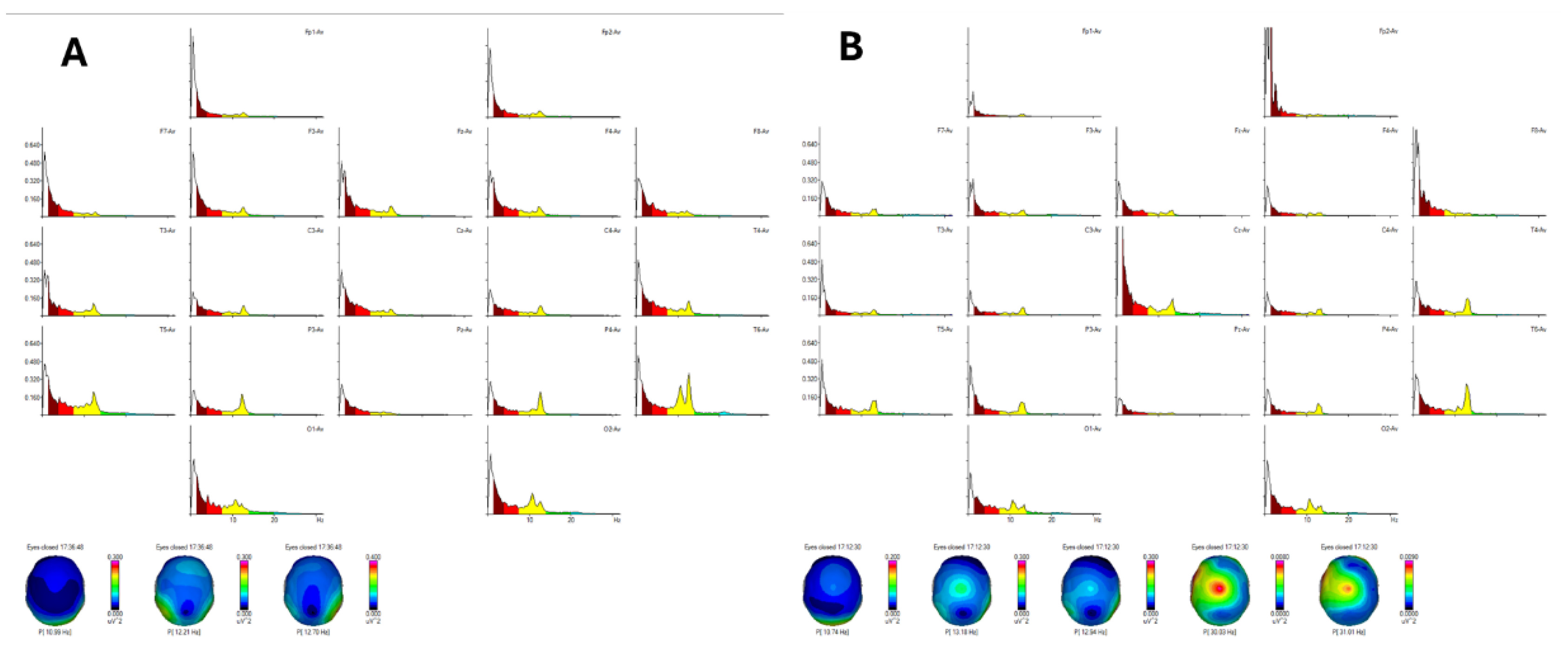
The initial EEG power spectra analysis [
11] revealed a peak in alpha rhythm frequency at 10.99 Hz in the occipital region and 12.70 Hz in the right posterior temporal areas, with delta and theta rhythms being diffusely distributed. Post-treatment EEG analysis demonstrated a significant change, with the posterior alpha rhythm frequency decreasing to 10.74 Hz. This reduction correlated clinically with improvements in anxiety and cognitive clarity observed in the patient. Additionally, gamma rhythm activity emerged in the central region, extending bilaterally across both hemispheres, suggesting enhanced sensory-motor integration and cognitive coherence.
Figure 1.
EEG Power Spectra Analysis (Fragment: Eyes closed 17:36:48, Offset: 0.00 s, Length: 299.98 s, Number of epochs 144).
Figure 1A: Demonstrates the pre-treatment peak in alpha rhythm frequency at 10.99 Hz in the occipital region and 12.70 Hz in the right posterior temporal areas, with diffuse delta and theta rhythm distribution.
Figure 1B: Shows the post-treatment decrease in alpha rhythm to 10.74 Hz, correlating with reduced anxiety and enhanced mental clarity. The emergence of gamma rhythm activity in the central region is indicative of improved cognitive function.
3.1.2. Comparative Analysis of EEG Power Spectra for Band Ranges
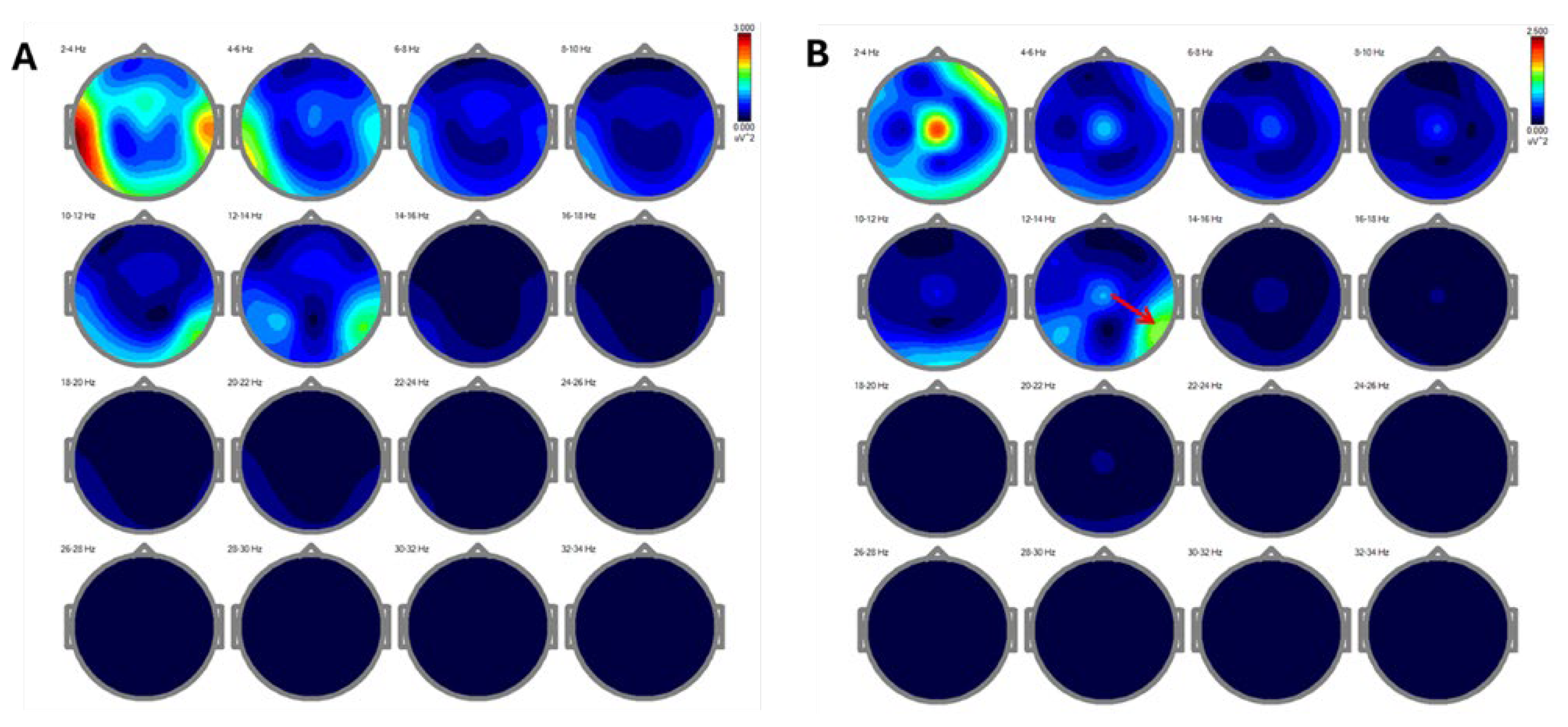
The comparative analysis of EEG power spectra across different frequency bands (delta, theta, alpha, beta, and gamma) [
12] revealed substantial changes post-treatment. Initially, a notable presence of delta rhythms was observed in the temporal and mid-frontal regions (
Figure 2A). Following REAC NPPO BWO-G treatment, there was a marked reduction of slow rhythms in these areas, with an evident shift toward faster alpha and gamma rhythms, particularly in the central and parietal regions (
Figure 2B). This transition is consistent with improved cognitive processing and mental clarity.
3.1.3. Independent Component Analysis – winEEG
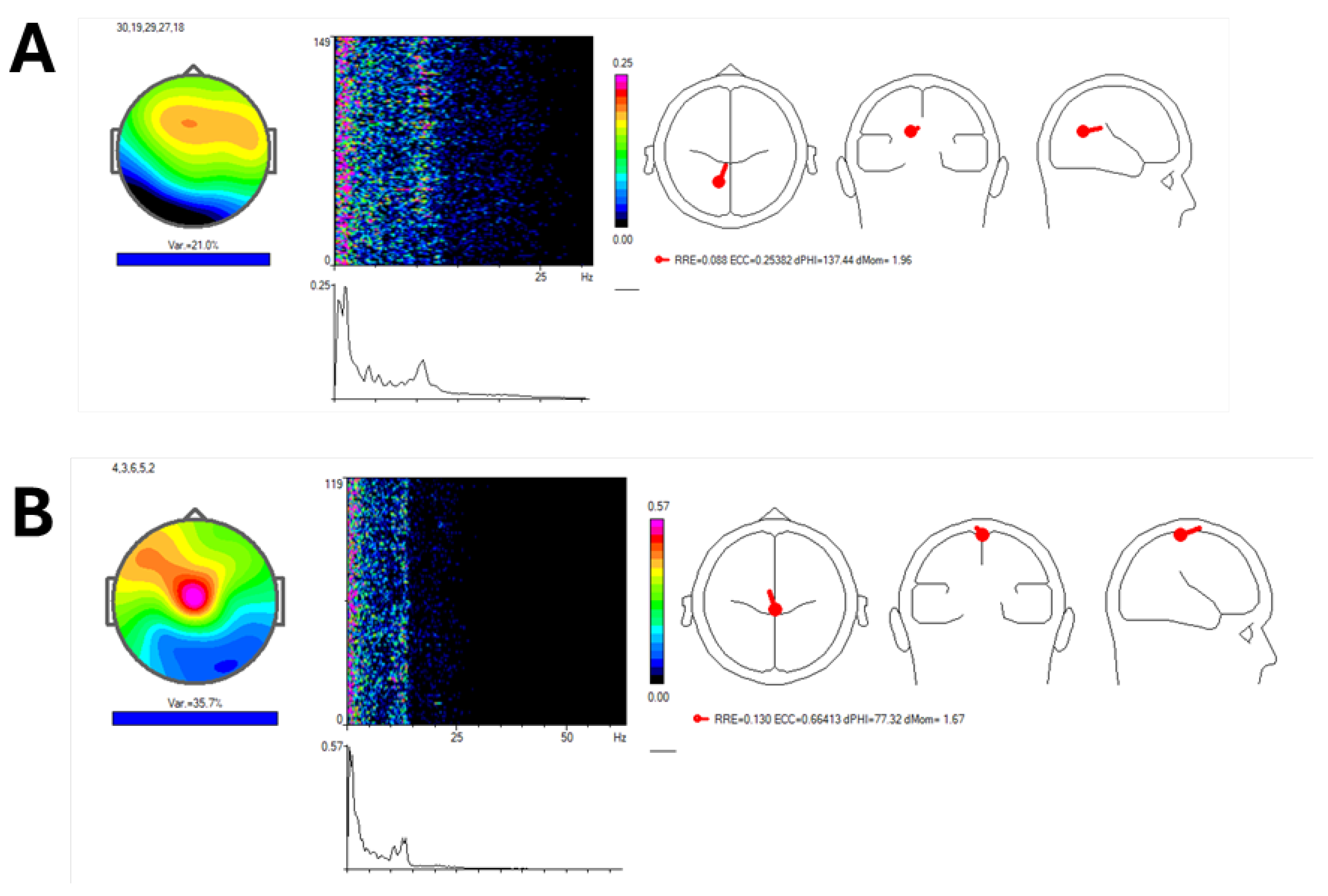
The Independent Component Analysis (ICA) performed using winEEG [
13] showed frequency peaks pre-treatment ranging from 1.95 Hz to 10.74–12.45 Hz (
Figure 3A). Post-treatment, frequency peaks shifted to a range of 1.71 Hz to 13.43 Hz, indicating a broader and more balanced distribution of brainwave frequencies (
Figure 3B). This change signifies improved cognitive flexibility and processing speed, which aligns with the patient's reported improvements in mood, anxiety, and cognitive clarity.
3.1.4. sLORETA Analysis
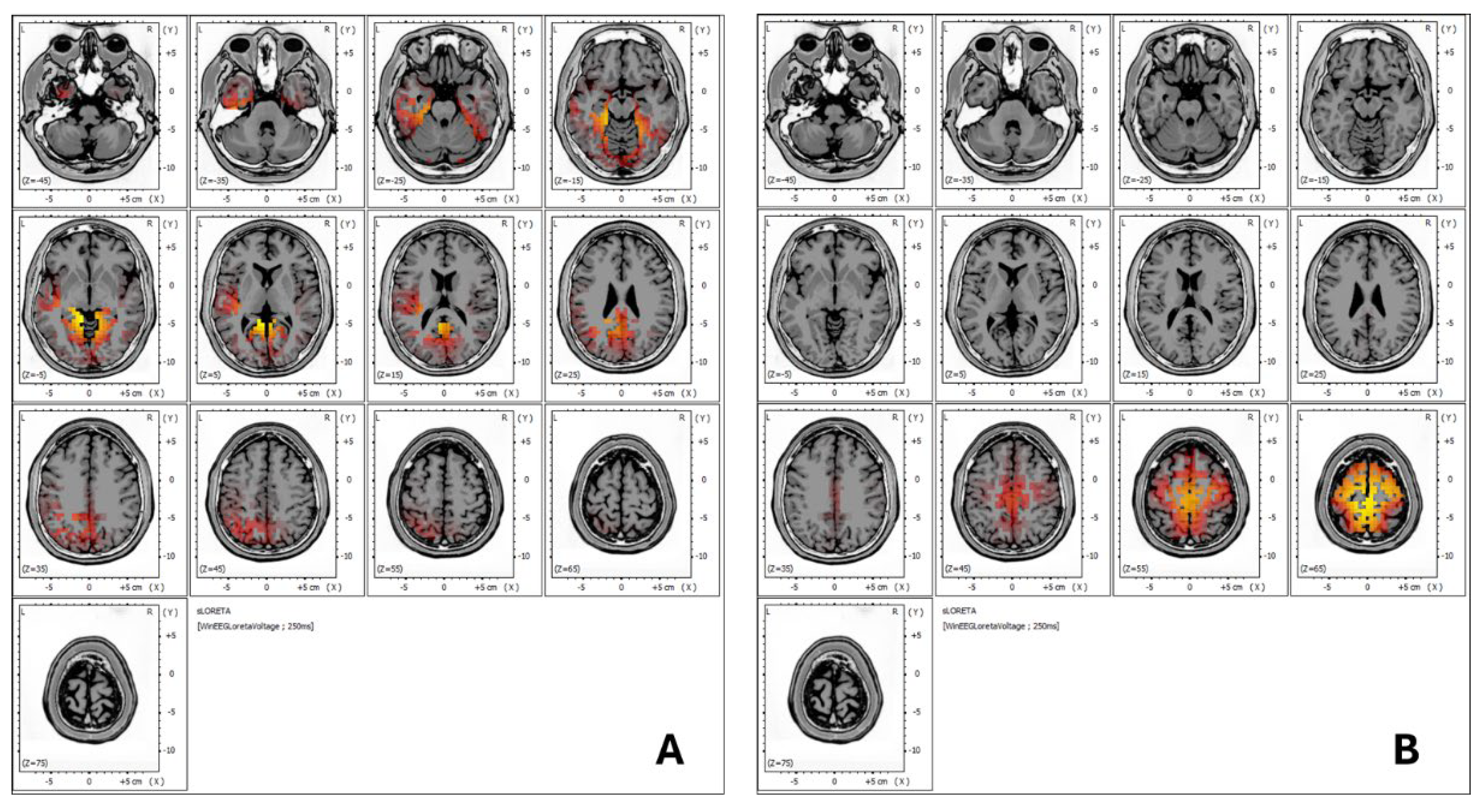
sLORETA (standardized Low-Resolution Brain Electromagnetic Tomography) analysis is a neuroimaging technique used to estimate the three-dimensional distribution of brain wave activity within different regions of the brain [
14]. By providing a spatial representation of electrical activity, sLORETA allows researchers and clinicians to identify specific brain areas involved in various cognitive, sensory, and emotional processes. This technique offers valuable insights into how different brain regions are activated under certain conditions or in response to treatment.
In
Figure 4, the sLORETA analysis demonstrates notable changes in brainwave activity before and after treatment.
Figure 4A shows that, prior to treatment, the patient exhibited brainwave activity across a range of frequencies, from slower waves (delta and theta) to faster frequencies, including an alpha rhythm at 12.45 Hz. These were localized in Brodmann areas 30, 19, 29, 27, and 18, which are linked to functions such as emotional regulation, receptive language, auditory processing, semantic analysis, visual information processing, and sentence formation.
After the treatment, as shown in
Figure 4B, there was a more extensive distribution of frequencies across the prefrontal cortex, motor regions, and both the anterior and posterior cingulate gyrus. Notably, the slow delta and theta rhythms previously present in the temporal areas (Brodmann areas 21 and 42) were no longer observed. Clinically, this change correlated with improved tolerance to wet noises, which allowed the patient to integrate more comfortably into public environments, such as restaurants, indicating enhanced auditory processing and emotional regulation.
4. Discussion
This case report demonstrates the potential of REAC NPPO BWO-G in achieving significant cognitive and emotional improvements in an adolescent with complex neuropsychological challenges, such as emotional trauma and gender dysphoria. The therapeutic effects were substantiated by changes in brainwave patterns, as revealed by qEEG analysis. These findings suggest that modulating gamma brain wave activity is key to enhancing cognitive coherence, emotional regulation, and sensory processing. This non-invasive treatment, which achieves brain modulation without discomfort, presents a promising alternative to existing neuromodulation methods.
Comparison with Other Neuromodulation Techniques
Several neuromodulation techniques, including neurofeedback, transcranial alternating current stimulation (tACS), and transcranial magnetic stimulation (TMS), have been explored for their ability to influence brain wave activity and improve cognitive and emotional functioning. These techniques have shown efficacy in enhancing gamma wave activity and alleviating symptoms in conditions such as schizophrenia[
15], autism[
16], and Alzheimer's disease[
17,
18]. However, the mechanisms through which they affect brain wave patterns and their effects on qEEG vary significantly compared to REAC NPPO BWO-G.
Neurofeedback allows individuals to self-regulate their brain wave activity by receiving real-time feedback on brainwave frequencies [
19], typically aiming to increase gamma activity over time. While effective, it often requires prolonged training sessions and active patient participation, which may not be feasible for everyone [
20]. Furthermore, qEEG changes with neurofeedback tend to manifest more gradually[
21], in contrast to the rapid improvements observed with REAC NPPO BWO-G, as evidenced in this case study.
tACS and TMS stimulate specific brain regions to influence gamma rhythms [
22,
23]. These techniques have been investigated for their role in treating neuropsychiatric disorders, where they are found to effectively increase gamma oscillations. However, these methods may involve discomfort, such as scalp irritation or mild side effects, which can reduce their appeal for certain patients [
24]. Moreover, qEEG changes resulting from tACS and TMS tend to be localized to the specific areas targeted by the stimulation, limiting their broader impact on brain wave activity[
25].
In contrast, REAC NPPO BWO-G induces a more extensive modulation of brain wave activity without requiring patient engagement or invasive procedures. This technique restores neuronal allostasis, resulting in marked qEEG changes—such as the reduction in delta and theta rhythms and an increase in alpha and gamma rhythms—within a relatively short time frame of 18 sessions over six days. These qEEG improvements correlated with the patient’s clinical progress, including enhanced cognitive clarity, emotional regulation, and sensory tolerance. The speed of these changes sets REAC NPPO BWO-G apart from other methods, offering a more efficient treatment option.
Clinical Relevance of qEEG Findings
The pre-treatment qEEG in this case revealed pronounced peaks in alpha rhythms, alongside diffuse delta and theta rhythms, in the occipital and right posterior temporal regions. Such patterns are commonly associated with dysregulated brain wave activity in individuals with neuropsychological disorders [
26,
27]. After REAC NPPO BWO-G treatment, a substantial shift was observed in the qEEG analysis, with a reduction in slower delta and theta rhythms and an increase in faster alpha and gamma waves, particularly in the central and parietal regions. These changes reflect enhanced cognitive processing, emotional stability, and improved sensory integration [28-30].
Compared to other neuromodulation techniques, the effects of REAC NPPO BWO-G on qEEG appear more widespread. For instance, TMS typically focuses on stimulating specific brain areas associated with cognitive and emotional functions [
31], and while it can yield positive outcomes, the qEEG effects are often confined to those specific regions. In contrast, REAC NPPO BWO-G demonstrates broader changes in brain wave activity, suggesting it influences multiple interconnected brain networks simultaneously, which may lead to more comprehensive therapeutic effects.
Addressing Gaps in Knowledge:
While this case study provides promising evidence for the efficacy of REAC NPPO BWO-G, further research is needed to confirm its long-term effects and broader applicability. Larger-scale, controlled studies could help clarify how this treatment compares to other neuromodulation techniques across a wider range of neuropsychological conditions. Moreover, the present study demonstrates significant changes in brain wave activity and clinical outcomes. This case report, along with other studies, has shown that these changes result from the optimization of endogenous bioelectrical activity, which is specifically targeted by the various therapeutic protocols of REAC technology.
Advancing the Field
This case study highlights the value of REAC NPPO BWO-G as a non-invasive, effective, and rapid method for modulating brain wave activity, supported by significant changes in qEEG within a short duration. In comparison to other neuromodulation techniques, REAC NPPO BWO-G offers several advantages, including ease of use, accessibility, and broader modulation of brain activity. The findings suggest that REAC NPPO BWO-G could be a promising tool for enhancing cognitive and emotional resilience, particularly in patients with complex neuropsychological challenges.
5. Conclusions
The integration of EEG Power Spectra, Comparative Analysis [
32], Independent Component Analysis[
33], and sLORETA[
34] findings in this case demonstrates the potential of REAC NPPO BWO-G to induce significant changes in brainwave dynamics, particularly in enhancing gamma rhythms and reducing slower delta and theta rhythms. These neurophysiological changes were accompanied by marked clinical improvements in cognitive clarity, emotional regulation, and sensory processing, supporting the hypothesis that non-invasive modulation of gamma activity can lead to broader neuropsychological resilience.
Compared to other neuromodulation techniques, REAC NPPO BWO-G offers distinct advantages in terms of non-invasiveness, ease of application, and comprehensive modulation of brain activity, as observed through qEEG changes. While these findings are promising, further research is needed to explore the long-term effects and broader applicability of this approach across different neuropsychological conditions and populations. This case suggests that REAC NPPO BWO-G could be an effective treatment for individuals with complex psychosocial challenges, but larger-scale studies are necessary to validate its therapeutic potential.
Author Contributions
The following statements should be used “Conceptualization, V.M., A.R., S.R., and V.F.; methodology, V.M., A.R.; validation, V.M., A.R., S.R., and V.F.; formal analysis, V.M., A.R., S.R., and V.F.; data curation, V.M., A.R., S.R., and V.F.; writing—original draft preparation, V.M., A.R., S.R., and V.F.; writing—review and editing, V.M., A.R., S.R., and V.F.; visualization, V.M., A.R.; supervision, S.R., V.F.; project administration, S.R., V.F. .All authors have read and agreed to the published version of the manuscript.
Funding
This research received no external funding.
Institutional Review Board Statement
Not applicable.
Informed Consent Statement
Informed consent was obtained from the patient involved in the study.
Data Availability Statement
All data are included in the manuscript.
Conflicts of Interest
S.R. and V.F. are the authors of the REAC patent. A.R. is the daughter of SR and V.F., V.M. declare no conflict of interest.
References
- Braboszcz, C.; Cahn, B.R.; Levy, J.; Fernandez, M.; Delorme, A. Increased Gamma Brainwave Amplitude Compared to Control in Three Different Meditation Traditions. PLoS One 2017, 12, e0170647. [Google Scholar] [CrossRef] [PubMed]
- Guan, A.; Wang, S.; Huang, A.; Qiu, C.; Li, Y.; Li, X.; Wang, J.; Wang, Q.; Deng, B. The role of gamma oscillations in central nervous system diseases: Mechanism and treatment. Front Cell Neurosci 2022, 16, 962957. [Google Scholar] [CrossRef] [PubMed]
- Benussi, A.; Cantoni, V.; Grassi, M.; Brechet, L.; Michel, C.M.; Datta, A.; Thomas, C.; Gazzina, S.; Cotelli, M.S.; Bianchi, M.; et al. Increasing Brain Gamma Activity Improves Episodic Memory and Restores Cholinergic Dysfunction in Alzheimer's Disease. Ann Neurol 2022, 92, 322–334. [Google Scholar] [CrossRef] [PubMed]
- Miller, E.K.; Lundqvist, M.; Bastos, A.M. Working Memory 2.0. Neuron 2018, 100, 463–475. [Google Scholar] [CrossRef] [PubMed]
- Kayarian, F.B.; Jannati, A.; Rotenberg, A.; Santarnecchi, E. Targeting Gamma-Related Pathophysiology in Autism Spectrum Disorder Using Transcranial Electrical Stimulation: Opportunities and Challenges. Autism Res 2020, 13, 1051–1071. [Google Scholar] [CrossRef]
- Tanaka-Koshiyama, K.; Koshiyama, D.; Miyakoshi, M.; Joshi, Y.B.; Molina, J.L.; Sprock, J.; Braff, D.L.; Light, G.A. Abnormal Spontaneous Gamma Power Is Associated With Verbal Learning and Memory Dysfunction in Schizophrenia. Front Psychiatry 2020, 11, 832. [Google Scholar] [CrossRef]
- De Paolis, M.L.; Paoletti, I.; Zaccone, C.; Capone, F.; D'Amelio, M.; Krashia, P. Transcranial alternating current stimulation (tACS) at gamma frequency: an up-and-coming tool to modify the progression of Alzheimer's Disease. Transl Neurodegener 2024, 13, 33. [Google Scholar] [CrossRef]
- Maiella, M.; Casula, E.P.; Borghi, I.; Assogna, M.; D'Acunto, A.; Pezzopane, V.; Mencarelli, L.; Rocchi, L.; Pellicciari, M.C.; Koch, G. Simultaneous transcranial electrical and magnetic stimulation boost gamma oscillations in the dorsolateral prefrontal cortex. Sci Rep 2022, 12, 19391. [Google Scholar] [CrossRef]
- Rinaldi, A.; Marins Martins, M.C.; De Almeida Martins Oliveira, A.C.; Rinaldi, S.; Fontani, V. Improving Functional Abilities in Children and Adolescents with Autism Spectrum Disorder Using Non-Invasive REAC Neuro Psycho Physical Optimization Treatments: A PEDI-CAT Study. J Pers Med 2023, 13. [Google Scholar] [CrossRef]
- Kopanska, M.; Ochojska, D.; Dejnowicz-Velitchkov, A.; Banas-Zabczyk, A. Quantitative Electroencephalography (QEEG) as an Innovative Diagnostic Tool in Mental Disorders. Int J Environ Res Public Health 2022, 19. [Google Scholar] [CrossRef]
- Liu, S.; Shen, J.; Li, Y.; Wang, J.; Wang, J.; Xu, J.; Wang, Q.; Chen, R. EEG Power Spectral Analysis of Abnormal Cortical Activations During REM/NREM Sleep in Obstructive Sleep Apnea. Front Neurol 2021, 12, 643855. [Google Scholar] [CrossRef] [PubMed]
- Kim, D.-W.; Im, C.-H. EEG Spectral Analysis. In Computational EEG Analysis: Methods and Applications, Im, C.-H., Ed.; Springer: Singapore, 2018; pp. 35–53. [Google Scholar]
- Marcu, G.M.; Szekely-Copindean, R.D.; Zagrean, A.M. Shared Psychophysiological Electroencephalographic Features in Maltreated Adolescent Siblings and Twins: A Case Report. Cureus 2024, 16, e63269. [Google Scholar] [CrossRef] [PubMed]
- Higuchi, Y.; Odagiri, S.; Tateno, T.; Suzuki, M.; Takahashi, T. Resting-state electroencephalogram in drug-free subjects with at-risk mental states who later developed psychosis: a low-resolution electromagnetic tomography analysis. Front Hum Neurosci 2024, 18, 1449820. [Google Scholar] [CrossRef] [PubMed]
- Cole, J.C.; Green Bernacki, C.; Helmer, A.; Pinninti, N.; O'Reardon J, P. Efficacy of Transcranial Magnetic Stimulation (TMS) in the Treatment of Schizophrenia: A Review of the Literature to Date. Innovations in clinical neuroscience 2015, 12, 12–19. [Google Scholar]
- Casanova, M.F.; Shaban, M.; Ghazal, M.; El-Baz, A.S.; Casanova, E.L.; Opris, I.; Sokhadze, E.M. Effects of Transcranial Magnetic Stimulation Therapy on Evoked and Induced Gamma Oscillations in Children with Autism Spectrum Disorder. Brain Sci 2020, 10. [Google Scholar] [CrossRef]
- Traikapi, A.; Konstantinou, N. Gamma Oscillations in Alzheimer's Disease and Their Potential Therapeutic Role. Front Syst Neurosci 2021, 15, 782399. [Google Scholar] [CrossRef]
- Benussi, A.; Cantoni, V.; Cotelli, M.S.; Cotelli, M.; Brattini, C.; Datta, A.; Thomas, C.; Santarnecchi, E.; Pascual-Leone, A.; Borroni, B. Exposure to gamma tACS in Alzheimer's disease: A randomized, double-blind, sham-controlled, crossover, pilot study. Brain Stimul 2021, 14, 531–540. [Google Scholar] [CrossRef]
- Marzbani, H.; Marateb, H.R.; Mansourian, M. Neurofeedback: A Comprehensive Review on System Design, Methodology and Clinical Applications. Basic Clin Neurosci 2016, 7, 143–158. [Google Scholar] [CrossRef]
- Babaskina, L.; Afanasyeva, N.; Semyonkina, M.; Myasnyankina, O.; Sushko, N. Effectiveness of Neurofeedback Training for Patients with Personality Disorders: A Systematic Review. Iran J Psychiatry 2023, 18, 352–361. [Google Scholar] [CrossRef]
- Rance, M.; Walsh, C.; Sukhodolsky, D.G.; Pittman, B.; Qiu, M.; Kichuk, S.A.; Wasylink, S.; Koller, W.N.; Bloch, M.; Gruner, P.; et al. Time course of clinical change following neurofeedback. Neuroimage 2018, 181, 807–813. [Google Scholar] [CrossRef]
- Nissim, N.R.; Pham, D.V.H.; Poddar, T.; Blutt, E.; Hamilton, R.H. The impact of gamma transcranial alternating current stimulation (tACS) on cognitive and memory processes in patients with mild cognitive impairment or Alzheimer's disease: A literature review. Brain Stimul 2023, 16, 748–755. [Google Scholar] [CrossRef] [PubMed]
- Siebner, H.R.; Funke, K.; Aberra, A.S.; Antal, A.; Bestmann, S.; Chen, R.; Classen, J.; Davare, M.; Di Lazzaro, V.; Fox, P.T.; et al. Transcranial magnetic stimulation of the brain: What is stimulated? - A consensus and critical position paper. Clin Neurophysiol 2022, 140, 59–97. [Google Scholar] [CrossRef] [PubMed]
- Holmes, N.P.; Meteyard, L. Subjective Discomfort of TMS Predicts Reaction Times Differences in Published Studies. Front Psychol 2018, 9, 1989. [Google Scholar] [CrossRef] [PubMed]
- Radecke, J.O.; Fiene, M.; Misselhorn, J.; Herrmann, C.S.; Engel, A.K.; Wolters, C.H.; Schneider, T.R. Personalized alpha-tACS targeting left posterior parietal cortex modulates visuo-spatial attention and posterior evoked EEG activity. Brain Stimul 2023, 16, 1047–1061. [Google Scholar] [CrossRef]
- Zhou, P.; Wu, Q.; Zhan, L.; Guo, Z.; Wang, C.; Wang, S.; Yang, Q.; Lin, J.; Zhang, F.; Liu, L.; et al. Alpha peak activity in resting-state EEG is associated with depressive score. Front Neurosci 2023, 17, 1057908. [Google Scholar] [CrossRef]
- He, X.; Zhang, Y.; Chen, J.; Xie, C.; Gan, R.; Wang, L.; Wang, L. Changes in theta activities in the left posterior temporal region, left occipital region and right frontal region related to mild cognitive impairment in Parkinson's disease patients. Int J Neurosci 2017, 127, 66–72. [Google Scholar] [CrossRef]
- Rathee, S.; Bhatia, D.; Punia, V.; Singh, R. Peak Alpha Frequency in Relation to Cognitive Performance. J Neurosci Rural Pract 2020, 11, 416–419. [Google Scholar] [CrossRef]
- Attar, E.T. Review of electroencephalography signals approaches for mental stress assessment. Neurosciences (Riyadh) 2022, 27, 209–215. [Google Scholar] [CrossRef]
- Ronconi, L.; Busch, N.A.; Melcher, D. Alpha-band sensory entrainment alters the duration of temporal windows in visual perception. Sci Rep 2018, 8, 11810. [Google Scholar] [CrossRef]
- Sack, A.T.; Paneva, J.; Kuthe, T.; Dijkstra, E.; Zwienenberg, L.; Arns, M.; Schuhmann, T. Target Engagement and Brain State Dependence of Transcranial Magnetic Stimulation: Implications for Clinical Practice. Biol Psychiatry 2024, 95, 536–544. [Google Scholar] [CrossRef]
- Jeong, H.T.; Youn, Y.C.; Sung, H.H.; Kim, S.Y. Power Spectral Changes of Quantitative EEG in the Subjective Cognitive Decline: Comparison of Community Normal Control Groups. Neuropsychiatr Dis Treat 2021, 17, 2783–2790. [Google Scholar] [CrossRef]
- Lisha, S.; Ying, L.; Beadle, P.J. Independent component analysis of EEG signals. In Proceedings of the Proceedings of 2005 IEEE International Workshop on VLSI Design and Video Technology, 28-30 May 2005; pp. 219–222. [Google Scholar]
- Nash, K.; Leota, J.; Kleinert, T.; Hayward, D.A. Anxiety disrupts performance monitoring: integrating behavioral, event-related potential, EEG microstate, and sLORETA evidence. Cereb Cortex 2023, 33, 3787–3802. [Google Scholar] [CrossRef]
|
Disclaimer/Publisher’s Note: The statements, opinions and data contained in all publications are solely those of the individual author(s) and contributor(s) and not of MDPI and/or the editor(s). MDPI and/or the editor(s) disclaim responsibility for any injury to people or property resulting from any ideas, methods, instructions or products referred to in the content. |
© 2024 by the authors. Licensee MDPI, Basel, Switzerland. This article is an open access article distributed under the terms and conditions of the Creative Commons Attribution (CC BY) license (http://creativecommons.org/licenses/by/4.0/).


