1. Introduction
Consumer demand in the modern world leans towards products with natural preservatives. Thus, food science and technology researchers have shown a sharp increase in interest in the development and investigation of natural compounds’ properties [
1,
2]. Simultaneously, the increasing focus on sustainability and the mindset of the circular economy are leading to the transition from synthetic food additives to the use of natural compounds with antioxidant and antimicrobial effects to tackle microbiological growth and inhibit enzymatic reactions in food [
3,
4,
5,
6,
7]. Essential oils (EOs) and their derivatives have extensively been investigated due to their antioxidant and antimicrobial properties, along with their claim to be Generally Regarded as Safe (GRAS). It has also been reported that the EO market is estimated at almost USD 24 billion and it is projected to grow at an annual rate of 7.6% until 2027 [
8]. EOs and their derivatives have been analyzed for their chemical composition [
9]. EO’s ability to linger oxidative reactions or inhibit the formation of free radicals and microbial growth is well-documented, suggesting their potential use as alternatives to chemical preservatives [
10,
11,
12].
EOs and their derivatives are known for their volatility and are produced by a variety of plants [
13,
14,
15,
16]. These oils are primarily composed of compounds such as terpenes, phenylpropanoids, aldehydes, esters, alcohols, and ketones [
17,
18]. Due to their diverse chemical composition, EOs and their derivatives do not have a single defined mechanism of action against microbial cells. One commonly observed effect is cell membrane disruption, affecting proton pumps and leading to ATP depletion [
19,
20].
Monoterpenoid derivatives and phenylpropenes derived from EOs, such as citral (C
10H
16O), and carvacrol (C
10H
14O), are recognized for their potent bioactive properties [
21,
22]. Each of these compounds has well-documented antioxidant and antimicrobial properties, as supported by numerous studies [
22,
23]. Citral (CT), a compound formed by a mixture of neral and geranial isomers, is a linear monoterpene aldehyde that constitutes more than 85% of lemongrass EOs. This compound is primarily sourced from citrus fruits such as limes and lemons. Recognized for its citrus-based flavor, citral has been widely used in various consumer goods. The FDA has designated citral as Generally Recognized as Safe (GRAS), highlighting its extensive application in the industry [
24]. Carvacrol (CV) is primarily sourced from oregano and thyme. During its metabolic pathway, hydroxycarvacrols are produced, which can target bacterial cell membranes and ATPase functions. Additionally, these hydroxycarvacrols may contribute hydrogen atoms to stabilize free radicals [
25].
However, the direct application of EOs and their derivatives in foods is not suggested due to their potential to adversely affect flavors, aromas, and color, thereby deteriorating the sensory characteristics and making food products undesirable in certain context [
26]. Additionally, the high concentration of bioactive compounds in EOs increases the likelihood and severity of toxicity or allergic reactions, necessitating strict control over their use to ensure consumer safety[
27]. To address these challenges, alternative methods such as nanoencapsulation have been developed, leading to the synthesis of essential oil derivative nanoformulations [
22,
26,
28,
29,
30].
Nanoencapsulation of bioactive compounds produced by self-assembly of food-derived proteins and polysaccharides provides the possibility to create nanostructures that have various characteristics, such as biodegradability, biocompatibility, effectively controlled release, and site-specific delivery, combining the advantages of both biopolymers [
31,
32]. Such nanostructures can improve certain sensory characteristics, such as masking unpleasant flavors. They can also enhance the bioaccessibility and bioavailability of substances, prevent oxidative reactions that can degrade their bioactivity, and regulate the release of bioactive agents. Additionally, biopolymer based nanogels can improve the quality, stability, and bioactivity of substances by taking advantage of their reduced size at the nanoscale [
33,
34].
Biopolymer-based nanogels are considered as highly desirable nanocarriers due to their high water stability, biocompatibility, biodegradability, well-defined structure, and versatile applications in the fields of food, pharmaceuticals, and biomedicine. [
35] These nanogels have the potential to gradually and specifically release the encapsulated compound [
36]. Nanogels provide some unique benefits among the available nano delivery systems including higher loading capacity of bioactive compounds because of their property to form a 3d matrix [
37,
38]. For food industry applications, they can be produced from GRAS materials and, therefore, they have the potential to be secure, non-immunogenic, and fully/semi biodegradable. Nanogels allow the grouping of several bioactive ingredients in the same formula. They are capable of encapsulating both hydrophobic and hydrophilic compounds [
36,
37,
39].
Shen et al., [
40] developed a double-network emulsion gel containing probiotics using a food-grade complex of whey protein concentrate, xanthan gum and kappa carrageenan. The gel’s stability and gel-like properties were enhanced by the presence of xanthan gum and kappa carrageenan. The encapsulated probiotics showed improved viability during storage and maintained their effectiveness in balancing intestinal flora and inhibiting harmful bacteria. This suggests the potential of using food-grade biopolymers and nonthermal processing methods for functional food delivery systems. Avallone et al. [
41] developed a gelling formulation using xanthan gum and kappa carrageenan to replace animal gelatin. The combination of these two hydrocolloids offered a more stable gel network with enhanced viscoelastic properties. It was observed that the synergistic effect between xanthan gum and kappa carrageenan significantly increased the gel strength, with the complex modulus (∣G
*∣) improving as the xanthan gum concentration increased. The researchers explained that this synergy enhances the formation of double helices in the kappa carrageenan gel network, leading to a stronger gel. Xanthan gum reduces the number of accessible water molecules, promoting easier self-association of kappa carrageenan helices and the formation of junction zones, which contribute to improved mechanical properties. The gelation temperature remained relatively constant, ensuring the gelation process was not hindered. This combination showed promise as a green alternative to gelatin, providing strong, stable hydrogels suitable for various applications, including 3D food printing.
Xanthan gum (XG) is a high molecular weight, anionic exopolysaccharide that is produced through the aerobic fermentation of sugars by the bacterium
Xanthomonas campestris [
42]. Structurally, it features a β-(1→4) linked glucose backbone with charged trisaccharide sidechains. Known for being non-toxic, hydrophilic, and biodegradable, XG is extensively utilized in the food, pharmaceutical, and cosmetic sectors due to its outstanding rheological properties. It dissolves readily in both cold and hot water, hydrates swiftly, and generates high viscosity even at low concentrations [
43]. In the pharmaceutical industry, XG is employed as a controlled release agent in solid dosage forms and functions as a thickening, suspending, and stabilizing agent in liquid formulations [
44].
Kappa carrageenan (KC) is an anionic polysaccharide extracted from red edible seaweeds, specifically from the order Gigartinales. It is composed of alternating 1,3-linked galactose 4-sulfate and 1,4-linked 3,6-anhydro-D-galactose residues. κ-carrageenan is most sensitive to potassium (K+) ions, which enable it to form rigid, thermally reversible aqueous gels [
45,
46,
47]. Known for its high gel-forming ability and thermoreversible gelation properties, κ-carrageenan forms strong, stable gels upon cooling and melts when reheated [
41]. It is widely used in the food industry to improve texture, stability, and viscosity in products like dairy, meat, and plant-based foods. Additionally, κ-carrageenan’s interactions with proteins enhance the rheological properties of food systems, making it valuable in creating desirable textures and structures in various applications [
47].
Chalkidiki green table olives constitute a traditional product registered in the Commission Implementing Regulation (EU) No 426/2012 as a Protected Destination of Origin (PDO) product [
48]. The total production of such a finished Chalkidiki table olive product exceeds 145,000 tons. Notwithstanding the fact that olive oil is a common practice to preserve table olives, according to Eurostat [
49], in January 2024 the olive oil price surged by 50% higher than in January 2023 due to the depreciation, making it a not-so-beneficial option. Various studies have focused on flavoring olive oil with herbs and essential oils, or oleogels demonstrating significant shelf-life extension and consumer acceptance. However, there is limited research available to investigate the shelf-life of table olives with polysaccharide-based nanogels enhanced with essential oils.
To the best of our knowledge, the current study is the first to develop, characterize, and apply innovative and edible nanogels to extend the shelf life of table olives. The novelty of the current study is justified by the following objectives:
Development and characterization of Nanogels (NGs): To develop polysaccharide-based nanogels using xanthan gum and kappa carrageenan, enhanced with derivatives of essential oils.
Enhanced Functionality: To enhance the functionality of the novel nanogel with EO derivatives, including CV and CT, and to examine their potential synergistic effects.
Application on PDO Olives: To apply the developed nanogels as a coating on Chalkidiki traditional green PDO table olives and to evaluate their effectiveness in extending shelf life.
2. Results and Discussion
2.1. Particle Size-Z Potential Dynamic Light Scattering (DLS) and Scanning Electron Microscopy (SEM) Images Characterization of NGs Coatings
The determined zeta potential values of all obtained nanogel coatings are shown in
Table 1.
The results from the data in the table demonstrate that the coatings are negatively charged with high zeta potential (>|30| mV) values, confirming the stability of the nanogels. The values range between -35.28 and -45.72 mV. The most noticeable difference is that, while the zeta potential for individual derivative oils nanogels demonstrate higher values, although the nanogels mixtures do not have statistically significantly lower zeta potential at the 5% level. The anionic character of xanthan gum and kappa-carrageenan, combined with the negative ions on the surface of the oil droplets, explains the negative charge of the nanogels [
50]. Moreover, the presence of functional groups as found in essential oils, such as hydroxyl (–OH) and carboxyl (C=O) groups seem to affect the zeta potential [
51]. It is suggested that the hydroxyl groups of carvacrol form stronger hydrogen bonds and electrostatic interactions with the nanogel surface, resulting in more negative zeta potential values. On the other hand, zeta potential value of CT nanogel is lower than that of CV nanogel zeta potential value implying a weaker interaction between CT molecule and polysaccharide matrix. It is suggested that CV’s higher polarity and potential for stronger electrostatic repulsion and ion adsorption contribute to these variations.
The zeta potential values reported here are consistent with those observed in recent studies on EO based nanogels, which highlight similar trends in stability and charge characteristics [
52,
53,
54,
55,
56,
57].
In
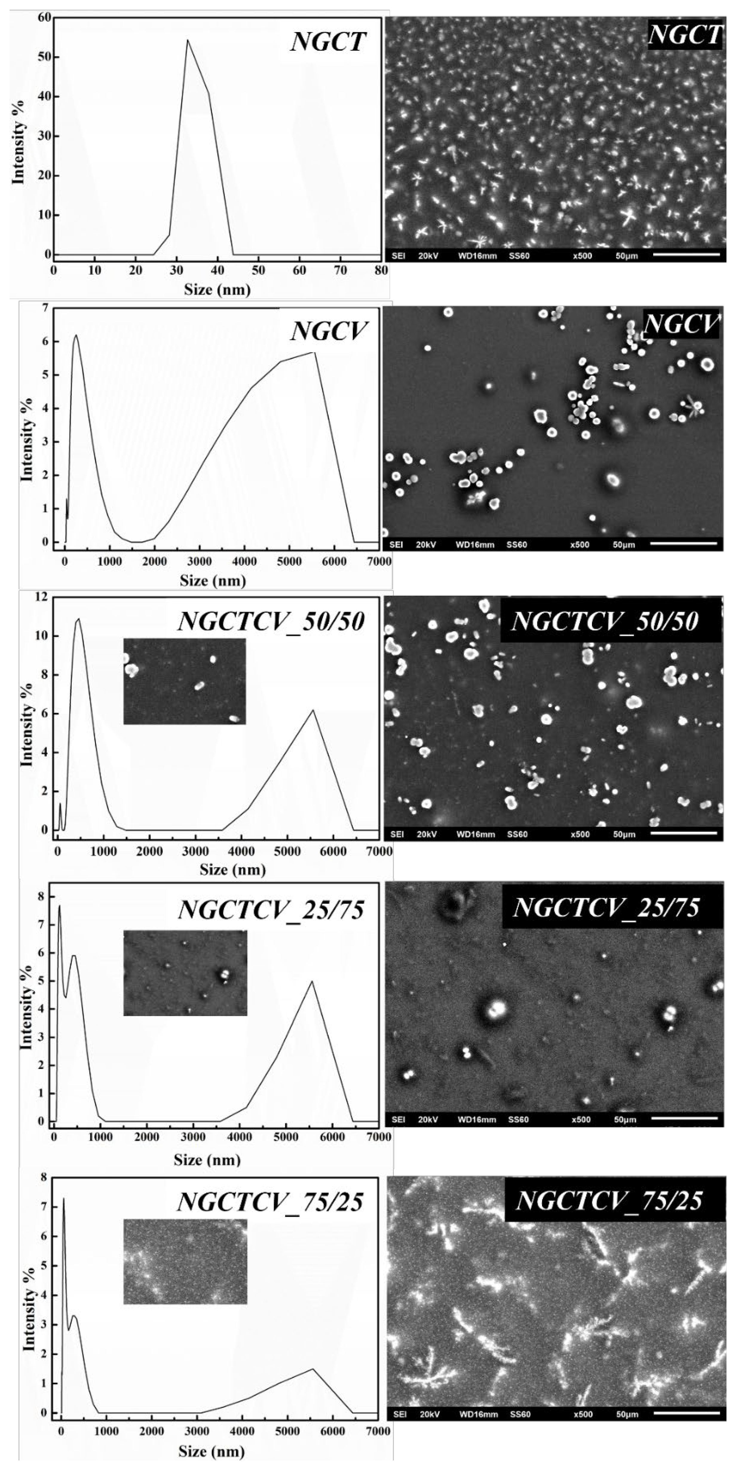
Figure 1 there are shown the DLS recorded data (in the left part) and the SEM images for all obtained NGs.
As it is observed in
Figure 1 (left part) NGCT sample showed an average particle size of ~34 nm (~100 %). The NGCV sample showed an average particle size of ~310 nm (~70 %) and of 4300 nm (~24 %). The DLS data of the samples NGCTCV_50/50, NGCTCV_25/75 and NGCTCV_75/25 show a major population of the particles between 140 nm and 480 nm and the rest in the range of μm. As it is observed in the right part of
Figure 1 SEM images (see right part in
Figure 1) agree with the data provided by DLS measurements
2.2. Antioxidant Activity EC50
Table 2 enlists the average EC
50 values derived from the DPPH assay for the examined nanogel treatments.
From the EC
50 values listed in
Table 2 for all obtained nanogels it demonstrated that NGCV exhibited the highest free radical antioxidant activity and NGCT the lowest one. All nanogels containing mixtures of CV and CT exhibited lower antioxidant activity than the NGCV and did not indicate any synergistic effect. In all NGCTCV samples, it is obtained that the higher of CV concentration the higher of obtained antioxidant activity or the lower of CT concentration the higher of obtained antioxidant activity. The results presented here are similar to that presented by Al-Mansori et al., [
58] which claimed that no synergistic antioxidant activity of thymol and carvacrol mixture was observed.
Moreover, various studies are in accordance with these results showing a higher efficacy of CV formulations against CT ones [
59,
60,
61,
62,
63,
64].
2.3. Antibacterial Activity of Nanogels
2.3.1. Agar Diffusion Zone
In the
Table 3 are presented the obtained antibacterial activity of the examined nanogel samples against two bacterial strains,
Escherichia coli and
Staphylococcus aureus, measured by the agar diffusion method. The measured zones of inhibition, expressed in millimeters (mm), indicate the effectiveness of each treatment. The findings, expressed as the average ± standard deviation (SD), are outlined in
Table 3.
Figure S1 displays representative images of the observed diffusion zones for all tested nanogel derivatives.
Overall, the analysis of the provided data indicated that the most important finding is that the mixtures of CT and CV in all ratios exhibited higher antibacterial activity than the individual nanogels. Among these, the 50:50 ratio (NGCTCV50:50) demonstrated the largest and most significant (p<0.05) inhibition zones against both bacterial strains.
To begin with, S aureus, all nanogel samples exhibited substantial antibacterial activity. NGCTCV50:50 exhibited the largest inhibition zone of 20.06 mm, approximately one-third larger than the 15 mm zone observed for NGCV and almost double the 11 mm zone for NGCT (p<0.05). NGCTCV25:75 and NGCTCV75:25 also showed considerable activity with inhibition zones of 16.5 mm and 18.34 mm, respectively, both surpassing the individual nanogels NGCV and NGCT.
For S. aureus, the antibacterial activity was generally lower than for E. coli. NGCTCV50:50 again showed the largest inhibition zone of 15 mm, which was 25% larger than the 12 mm zone observed for NGCV and almost double the 9 mm zone for NGCT. Other mixture ratios, such as NGCTCV25:75 and NGCTCV75:25, had inhibition zones of 12.34 mm and 12.05 mm, respectively, still surpassing the individual nanogels.
There is a noticeable difference in the antibacterial activity against the two examined strains. All nanogel samples exhibited higher antimicrobial activity against E. coli than S. aureus. NGCTCV50:50 has an inhibition zone of 20.06 mm for E. coli O157:H7, which is approximately one-third larger than the 15 mm zone for S. aureus. Similarly, the NGCV exhibited a 15 mm inhibition zone for E. coli O157:H, which is 25% larger than the 12 mm zone for S. aureus. NGCT showed an 11 mm zone for E. coli, which is over 20% larger than the 9 mm zone for S. aureus. These comparisons highlight that the synergistic effect of citral and carvacrol, particularly in a 50:50 ratio, significantly enhances antibacterial activity, especially against E. coli.
Various studies have claimed that there is a synergistic effect of CT and CV against several bacteria such as
L. monocytogenes,
L. innocua [
65] and
C. sakazakii [
66]. According to the study conducted by Silva-Angulo et al., [
65] CT and CV may have synergistic effects due to their targeting of different sites in bacterial cells. It has been suggested by the researchers that CV increases the outer membrane permeability, allowing CT to enter the cytoplasm and interact with proteins and nucleic acids. Combining these substances could improve CT’s and CV’s solubility, and enhance their antibacterial effect [
67]. Further investigation is needed to understand the molecular action of individual or combined treatments with natural substances. Overall, the synergistic effect of CV and CT is for the first time reported in such xanthan-carragenan based nanogels.
2.3.2. MIC, MBC, FIC
Table 4 provides the MIC, MBC, and FIC results for the examined nanogel samples against
Escherichia coli O157:H7 and
Staphylococcus aureus. The MIC and MBC values reported in μg/mL, indicate the lowest concentration of nanogel required to inhibit the growth of each bacterial strain, and the FIC values indicate the effectiveness of each derivative, as well as the synergistic effects of combined treatments.
The most essential finding from
Table 4 is that the mixtures of CT and CV exhibited significantly enhanced antibacterial activity due to their synergy or additive effect compared to individual derivative compound nanogels. Specifically, the NGCTCV25:75 and NGCTCV75:25 mixtures demonstrated the highest synergistic effects against
E. coli O157:H7. Specifically, the NGCTCV25:75 and NGCTCV75:25 mixtures showed the highest synergistic effects against
E. coli O157:H7, with MIC and MBC values of <125 μg and FIC of 0.375. Similarly, the NGCTCV50:50 mixture demonstrated the highest antimicrobial activity against
S. aureus, with MIC and MBC values of <125 μg and FIC of 0.375, indicating a synergistic effect. This highlights that specific ratios of CT and CV not only enhance antibacterial efficacy but also work synergistically to inhibit and or kill bacterial strains more effectively than the individual components alone.
Regarding E. coli O157:H7, the MIC and MBC values indicate that NGCTCV25:75 and NGCTCV75:25 were the most effective, with both achieving inhibition and bactericidal action at concentrations of <125 μg. The 50:50 mixture (NGCTCV50:50) showed MIC and MBC values of <250 μg, which is still more effective than the individual nanogels NGCT and NGCV, with MIC and MBC values of <1000 μg and <500 μg, respectively. The FIC values for these combinations highlight significant synergy (FIC < 0.5) for NGCTCV25:75 and NGCTCV75:25, with a FIC value of 0.375, indicating a strong synergistic effect. The NGCTCV50:50 mixture showed an additive effect (FIC < 1.0) with a FIC value of 0.75.
NGCTCV50:50 exhibited the most potent antibacterial activity with MIC and MBC values of <125 μg, significantly lower than the individual nanogels NGCT and NGCV, which had MIC and MBC values of <1000 μg and <500 μg, respectively. NGCTCV25:75 and NGCTCV75:25 had MIC and MBC values of <250 μg, showing notable efficacy. The FIC values for these combinations showed significant synergy for NGCTCV50:50, with an FIC of 0.375, and additive effects for NGCTCV25:75 and NGCTCV75:25, with FIC values of 0.75.
Comparing the two bacterial strains, E. coli O157 exhibited greater susceptibility to the nanogel treatments than S. aureus. The MIC and MBC values for E. coli were consistently lower, indicating higher effectiveness at lower concentrations. For instance, the MIC and MBC values for NGCTCV25:75 and NGCTCV75:25 were <125 μg for E. coli, compared to <250 μg for S. aureus. The FIC values also highlight stronger synergistic effects against E. coli (FIC of 0.375) compared to the additive effects against S. aureus (FIC of 0.75). These findings emphasize that the combinations of CT and CV, particularly in certain ratios, provide enhanced antibacterial activity and synergy, especially against E. coli O157.
Similar to our findings have been found for the additive effect of CT and CV against
E. coli [
68]. Kim et al. [
69] found that CV and CT can control
Listeria growth at 25% of their MICs. Zanini et al. [
70] suggested CT and CV could enhance antibacterial activity. Synergistic effects of cinnamaldehyde and thymol, carvacrol and eugenol, and thymol and eugenol have also been reported [
71]. Cao et al., [
66] reported FICI index of the synergistic combination is 0.5, with citral and carvacrol at a concentration of 1/4 MIC.
2.3.3. Time-Killing Assay
The time-kill assay was used to confirm the synergistic effect of CT and CV on Staphylococcus aureus and Escherichia coli. This method provided detailed insights into the antibacterial efficacy of various nanogel samples over a 24-hour period, measured in log10 CFU/mL.
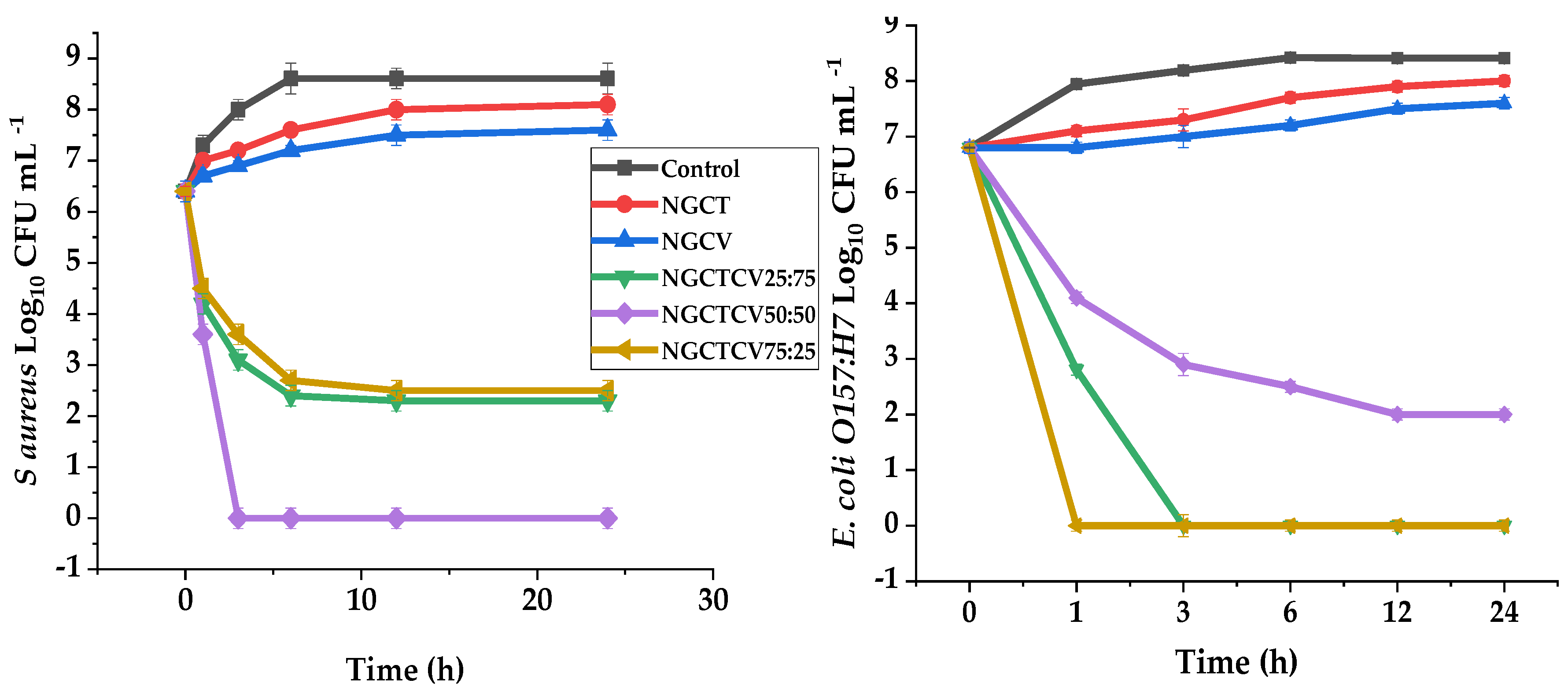
The most noticeable observation in
Figure 2 is the superior efficacy of the NGCTCV 50:50 mixture, which completely eradicated
E. coli by the 3rd hour and maintained this level up to the 24th hour. Additionally, for
S. aureus, this mixture reduced the bacterial count to 0 log10 CFU/mL by the 3rd hour and sustained this reduction throughout the testing period. This indicates a remarkable synergistic effect when CT and CV are combined in equal proportions.
Analyzing the nanogels, NGCTCV 50:50 showed a 2 log reduction (p<0.05) in S. aureus within the first hour, leading to a total population loss. For E. coli, the NGCTCV 75:25 mixture also resulted in a complete eradication (p<0.05) by the first hour, demonstrating its strong antibacterial activity. Comparatively, NGCT and NGCV were less effective, with NGCT only reducing S. aureus by about 0.5 log and NGCV achieving a similar reduction for both bacteria. The NGCTCV 25:75 mixture achieved a significant (p<0.05) 4.1 log reduction for E. coli within the first hour and maintained a steady low count, indicating its potent antibacterial effect.
Comparing the nanogels’ performance in both bacteria, the mixtures generally exhibited stronger antibacterial effects against E. coli than S. aureus. NGCTCV 50:50 eradicated E. coli by the 3rd hour, while achieving complete reduction of S. aureus took slightly longer. The NGCTCV 75:25 mixture demonstrated complete eradication of E. coli from the first hour, whereas for S. aureus, the same mixture only reduced the population to about 2.5 log10 CFU/mL (p<0.05) by the 24th hour. This indicates that E. coli O157:H7 is more susceptible to the synergistic effects of citral and carvacrol than S. aureus.
The results highlight the enhanced efficacy of mixture derivative compound nanogels against the individual components. The synergistic effect of CT and CV, particularly in the 50:50 and 75:25 ratios, significantly improves antibacterial activity, leading to complete eradication or substantial reduction of bacterial populations. This indicates the potential of these combined nanogels as powerful antibacterial agents, offering greater effectiveness than individual citral or carvacrol nanogels.
Silva-Angulo et al., [
65] found that CV and CT combined at 25% MIC effectively inhibited
Listeria growth. Similarly, the combination at 1/4 MIC completely inhibited
C. sakazakii growth after 8 hours, and at 1/2 MIC, completely killed cells after 1 hour. These results confirm that carvacrol and citral have a synergistic inhibitory effect on
C. sakazakii [
66].
2.4. Application of Obtained Nanogels in Olives Preservation
2.4.1. Yeasts & Mold and LAB Bacteria LOG (CFU/gr-1)
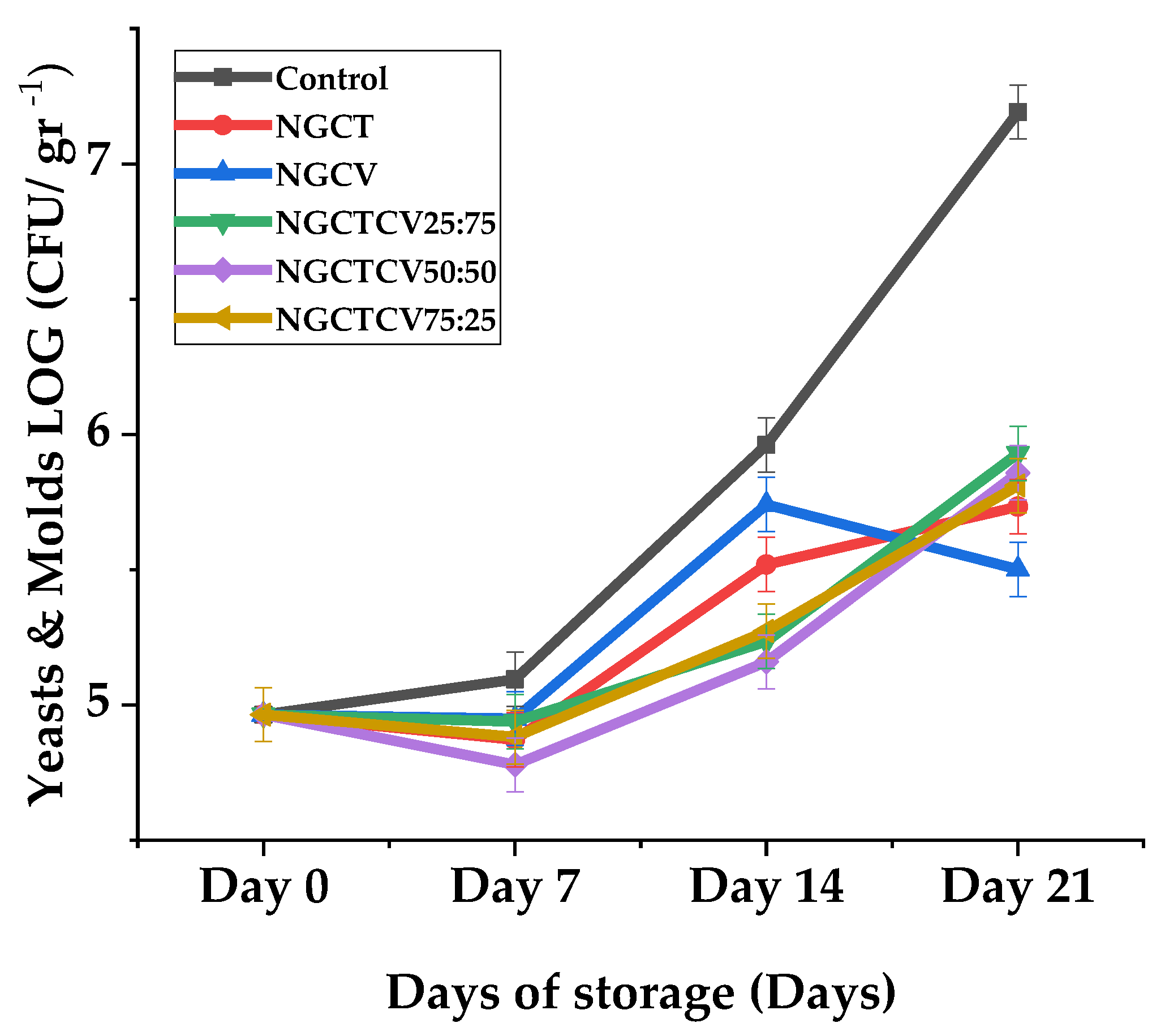
Figure 3 illustrates the evaluation of yeast and mold population on olives during the 21-day storage period which was conducted to confirm the synergistic effect of CT and CV in preserving Chalkidiki green table olives.
The most important observation is the superior performance of the NGCTCV 50:50 mixture, which consistently showed lower yeast populations compared to other treatments and the control. On Day 21, the yeast population in olives treated with NGCTCV 50:50 was 5.86 log10 CFU/gr-1 significantly lower than the 7.19 log10 CFU/gr-1 observed in the control sample, indicating a strong preservative effect.
Analyzing the nanogels, NGCTCV 50:50 exhibited a yeast population reduction of approximately 1.33 log10 CFU/gr-1 compared to the control by Day 21. This represents a nearly two-fold reduction in yeast population. Similarly, NGCTCV 25:75 and NGCTCV 75:25 also showed substantial reductions, with yeast populations of 5.93 and 5.81 log10 CFU/mL respectively on Day 21, indicating effective preservation but slightly less so than the 50:50 mixture. In contrast, the individual nanogels NGCT and NGCV showed less effectiveness, with yeast populations of 5.73 and 5.50 log10 CFU/mL, respectively, by Day 21.
Comparing the nanogels’ performance over the storage period, the combined nanogels consistently outperformed the individual components NGCT and NGCV. Specifically, on Day 14, the NGCTCV 50:50 mixture had a yeast population of 5.16 log10 CFU/mL, compared to 5.52 for NGCT and 5.74 for NGCV. This pattern continued through Day 21, where the NGCTCV 50:50 mixture maintained the lowest yeast population among all treatments.
The results from pairwise comparisons further reinforce these findings. For example, on Day 7, the yeast population for NGCTCV 50:50 was significantly lower than NGCV, NGCTCV 25:75, and the control (p<0.05). By Day 14, the NGCTCV 50:50 mixture continued to show significant reductions compared to NGCV and the control, with p-values of 0.039 and <0.001, respectively. On Day 21, the comparison between NGCV and NGCTCV 50:50 revealed a significant difference (p<0.05) (see
Table S5).
These results indicate that the mixture derivative compound nanogels, particularly the NGCTCV 50:50 ratio, provide enhanced preservative effects against yeast compared to individual citral or carvacrol nanogels. The synergistic effect of these mixtures not only effectively reduces yeast populations but also maintains this reduction consistently over the storage period, demonstrating their potential as powerful preservative agents for olives.
In
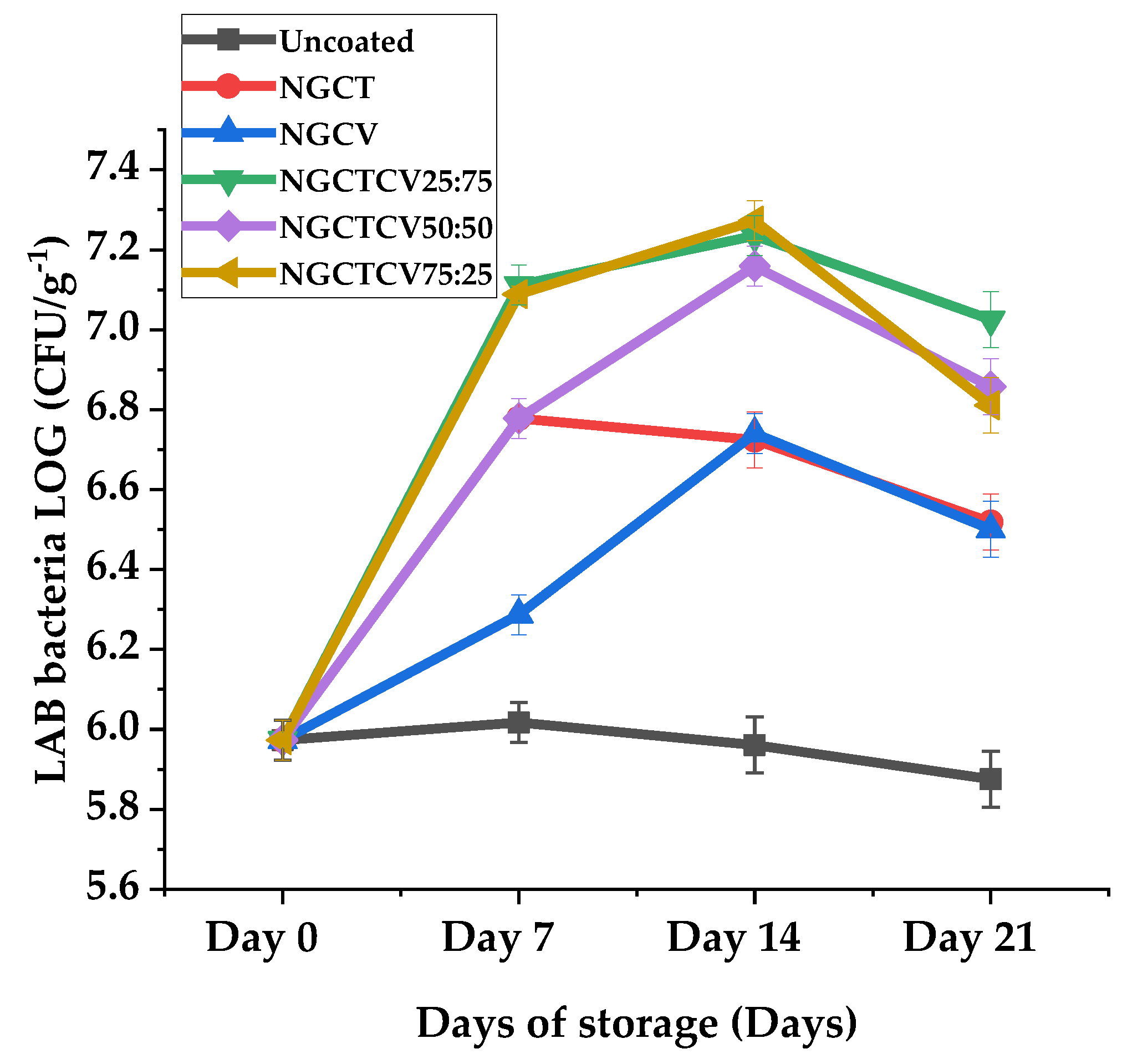
Figure 4 are plotted the LAB growth mean values of olives during 21-day storage period. The evaluation of LAB bacteria population on olives during the 21-day storage period reveals some statistically significant trends that highlight the effects of different nanogel treatments. The most noticeable difference is that certain mixtures of CT and CV significantly increased the LAB population on olives, contrary to the expected antibacterial activity.
On day 7, the control sample had a LAB population of 6.0 log10 CFU/mL, whereas the NGCTCV 25:75 and NGCTCV 75:25 mixtures showed higher populations of 7.1 log10 CFU/mL, which is almost two-fold higher than the control. The NGCTCV 50:50 mixture also had a LAB population of 6.8 log10 CFU/mL, significantly higher than the control, demonstrating a similar trend. These differences were statistically significant at the 5% level (p < 0.05), indicating a clear increase in LAB population due to the nanogel treatments (see
Table S6).
By day 14, the control’s LAB population decreased to 5.5 log10 CFU/mL, while NGCTCV mixtures continued to show higher LAB counts. NGCTCV 25:75 and NGCTCV 50:50 both reached 7.2 log10 CFU/mL, and NGCTCV 75:25 reached 7.3 log10 CFU/mL, indicating more than a two-fold increase compared to the control. These differences were also statistically significant at the 5% level, reinforcing the observation that the nanogel mixtures sustain higher LAB populations over time.
By day 21, the LAB population in the control sample further decreased to 5.1 log10 CFU/mL. Notwithstanding, the NGCTCV mixtures maintained higher populations: NGCTCV 25:75 had 7.0 log10 CFU/mL, NGCTCV 50:50 had 6.9 log10 CFU/mL, and NGCTCV 75:25 had 6.8 log10 CFU/mL. This represents nearly a two-fold increase compared to the control. The observed differences were statistically significant at the 5% level, confirming the sustained higher LAB populations in the presence of nanogel mixtures.
These statistically significant trends suggest that while nanogel mixtures, may control the growth of yeast and mold populations, they do not seem to affect the fermentation process of LAB bacteria differently than expected, indicating potential interactions that warrant further investigation.
The breakdown of polysaccharides by lactic acid bacteria relies on different hydrolases. In fermented foods, this polysaccharide degradation supplies energy to the lactic acid bacteria and produces various beneficial substances for humans [
72]. Papapostolou et al., [
73] reported that, while lactic acid bacteria population on green table fermented olives were not affected by use of essential oils during storage, yeasts and mold population experienced a more controlled increase compared to the control sample. These results indicate potential use of probiotic microorganisms could be used in the future.
2.4.2. Weight Loss Analysis
Table 5 represents the results from the percentage weight loss analysis of CChalkidiki green table olives over a storage period of 21 days.
The most noticeable observation is that nanogels effectively managed to control the weight loss of olives over the 21-day of storage period.
The results on day 7 indicate that all nanogel treatments significantly reduced weight loss compared to the uncoated olives. NGCT and the mixtures exhibited the lowest reductions, similar to NGCTCV 50:50. Statistically, significant differences were noted between uncoated olives and those coated with NGCV (p = 0.012), NGCT (p = 0.037), and NGCTCV 75:25 (p = 0.037), indicating more effective treatments.
By day 14, among the nanogel coatings, NGCT, NGCTCV at ratios 25:75 and 75:25 demonstrated the most effective control, with the mixtures generally performing like the individual essential oil coatings. Statistically significant differences were observed when comparing the uncoated olives to those coated with NGCTCV 25:75 (p = 0.010), NGCTCV 50:50 (p = 0.012), and NGCTCV 75:25 (p = 0.010).
Finally, on day 21, the observed trend continued, with the nanogel formulations NGCTCV at ratios 25:75 and 50:50 to demonstrate the most effective control overweight loss. Although the individual essential oil nanogels exhibited considerable efficacy, the nanogel mixtures, particularly NGCTCV 25:75, outperformed them. Statistically significant differences were identified between the uncoated olives and those treated with NGCV (p = 0.045), NGCTCV 25:75 (p = 0.007), NGCTCV 50:50 (p = 0.009), and NGCTCV 75:25 (p = 0.011) but also between NGCTCV 25:75 and NGCV (p = 0.045).
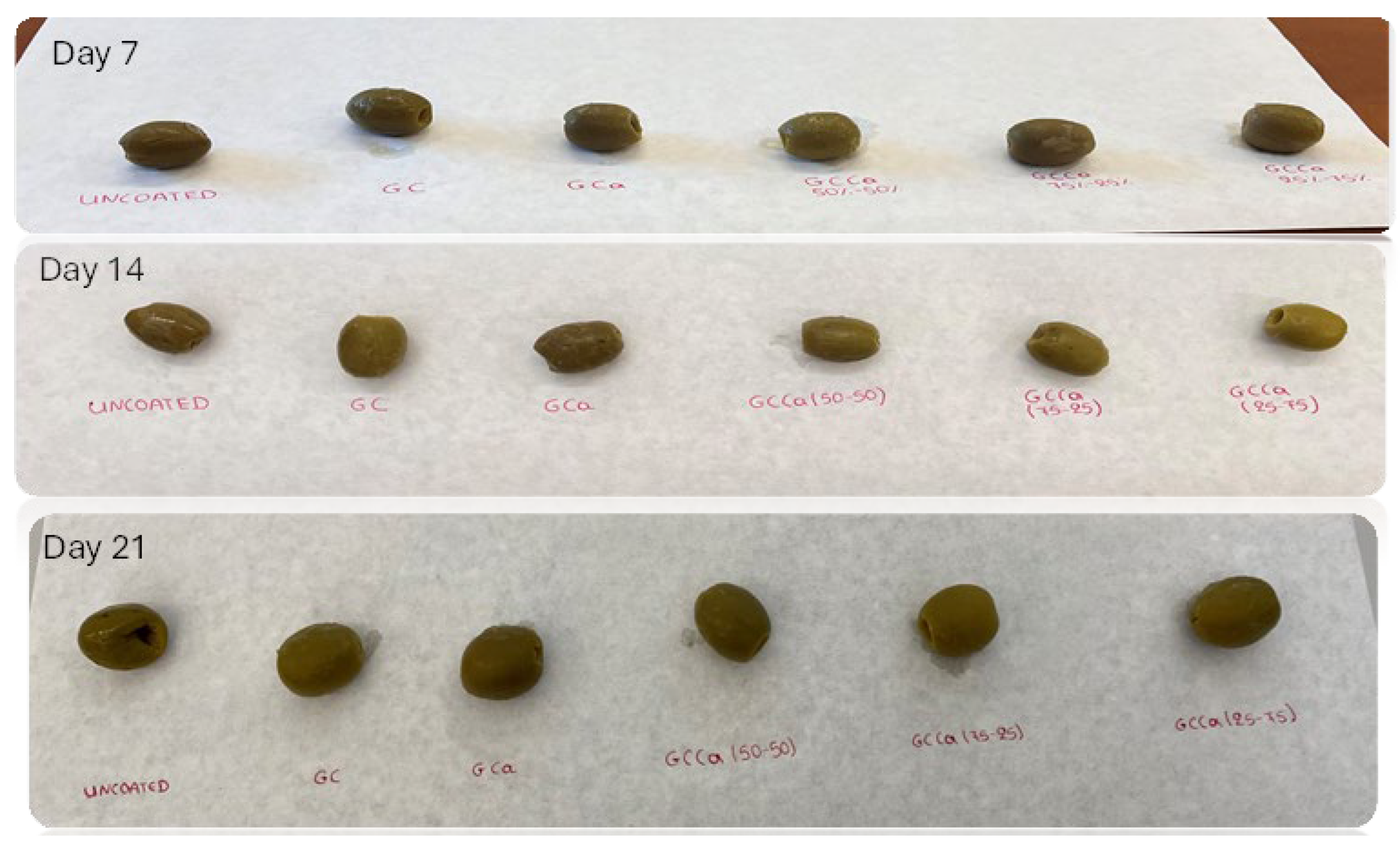
Figure 5 visualizes the effect of novel essential oil nanogel coatings applied on Days 7, 14, and 21. The mixture of CT and CV exhibited the highest efficacy compared to the individual components, particularly at ratios of NGCTCV 50:50 and 25:75. It is evident that the Chalkidiki green table olives coated with these nanogels showed no skin cracks, and their color remained unchanged throughout the examined period. However, olives treated with carvacrol nanogel experienced skin shrinkage, and their color darkened as early as Day 14.
These findings underscore the superior efficacy of the nanogel coatings, especially the CT-CV mixtures, in managing weight loss in green table olives over the 21-day period.
According to Priya et al., [
74] claim in their study that polysaccharide coatings may prevent the loss of volatile compounds present in food products, maintain fruit firmness and reduces the weight loss to some extent. Moisture loss can be minimized by the application of coating, which acts as a barrier. To prevent deterioration of fruits and vegetables, a coating must provide at least a minimal level of permeability barrier against water vapor. Since, they lose freshness upon the loss of water. Ali et al., [
75] reported in their study on the application of carboxymethyl cellulose coatings on mango fruits that they managed to control the weight loss, as well as maintain color and firmness. Similarly Kerdchoechuen et al., [
76] also found that starch-based edible coatings on minimally processed pummelo effectively manage weight loss during storage, with cassava starch showing superior results in preserving the fruit’s quality and appearance.
Martillanes et al., [
77] in their study found that incorporating natural antioxidants, such as polyphenols and essential oils, into absorbent pads effectively controls moisture, prevents spoilage, and enhances the safety of food products.
2.4.3. pH Analysis
Table 6 represents the results from the pH analysis of Chalkidiki green table olives over a storage period of 21 days.
The most noticeable observation is that olives coated with nanogels during the first 7 days of the storage period exhibited a significant fall in pH values. The most dramatic pH decreases were observed in the mixtures NGCTCV 50:50 and NGCTCV 25:75, which had pH values of 3.77 and 3.79, respectively. Statistically significant differences were noted between uncoated olives and those coated with NGCTCV 50:50 (p = 0.037), NGCTCV 25:75 (p = 0.005), and NGCT (p = 0.005). Furthermore, by day 14, the pH levels of all samples began to increase, with uncoated olives showing the highest pH rise to 4.65. Among the nanogel coatings, NGCTCV 75:25 and NGCT continued to demonstrate lower values than those of the uncoated olives. Statistically significant differences were observed between uncoated olives and those coated with NGCTCV 50:50 (p = 0.005), NGCTCV 25:75 (p = 0.037), and NGCTCV 75:25 (p = 0.037).
Moreover, on Day 21, the trend continued with the nanogel formulations NGCTCV 50:50 and NGCTCV 75:25 demonstrating the most effective control over pH levels. Although the individual essential oil nanogels also showed efficacy, the nanogel mixtures, particularly NGCTCV 75:25, maintained a lower pH compared to the uncoated samples. Statistically significant differences were identified between the uncoated olives and those treated with NGCTCV 75:25 (p = 0.013), NGCTCV 50:50 (p = 0.034), and NGCV (p = 0.001), highlighting the superior performance of the nanogel mixtures.
The results indicate that the presence of polysaccharide edible nanogel coatings provided a nutrient source for lactic acid bacteria, leading to the production of acid and a subsequent lowering of pH values. The essential oils incorporated in the nanogels did not seem to tackle the growth of the bacterial population. Finally, nanogels containing mixtures of essential oils NGCTCV 50:50 and NGCTCV 75:25, were most effective in controlling pH levels over the storage period, enhancing the preservation and quality of the olives.
2.4.4. Lab Colorimetry
In
Table 7,
Table 8 and
Table 9 are demonstrated the results of the L*a*b* colorimetry analysis, which examined the discoloration of Chalkidiki green table olives coated with various nanogels over a 21-day storage period.
To begin with, the most prominent outcome is that all nanogel coatings on olives, except for NGCV, effectively controlled the discoloration of olives during the 21-day storage period. Indeed, the discoloration rate was slower, and the values of the L*a*b* parameters were higher for the coated olives compared to the uncoated ones.
Regarding the L* parameter, the decrease in values, indicating the darkening of olives over time, was most pronounced in NGCV-coated and uncoated olives. Statistically significant changes occurred between Days 7 and 21, and Days 14 and 21. The statistically significant differences observed between NGCV vs. NGCT (p = 0.026) and NGCV vs. NGCTCV 50:50 (p = 0.005) suggest that NGCV was less effective in preventing darkening compared to these other nanogel coatings. Similarly, the differences between NGCTCV 75:25 vs. NGCTCV 50:50 (p = 0.014) and Uncoated vs. NGCTCV 50:50 (p = 0.026) indicate that the essential oil mixtures, particularly NGCTCV 50:50, provided superior protection against darkening. NGCV demonstrated L* values similar to uncoated olives. This may occur as carvacrol is prone to oxidation when exposed to air and light, while NGCTCV 25:75 was the most effective in maintaining higher L* values.
With respect to the a* parameter, which reflects the shift toward red and indicates the loss of the green hue in olives, statistically significant changes were observed between Days 0 and 14, particularly in NGCTCV 25:75 (p = 0.005), NGCTCV 50:50 (p = 0.013), and NGCTCV 75:25 (p = 0.024) coatings. These mixtures effectively stabilized the a* values, reducing the loss of the green hue compared to NGCV and uncoated olives. Significant differences (p < 0.05) appeared between pairs like NGCTCV 50:50 vs. NGCV (p = 0.007), NGCTCV 25:75 vs. NGCV (p = 0.014), and Uncoated vs. NGCV (p = 0.013), with NGCTCV 25:75 showing the best control.
Furthermore, the b* parameter, representing the blue-yellow axis with higher values indicating a stronger yellow hue, showed significant changes (p < 0.05) between Days 7 and 21 for NGCV and NGCTCV-coated olives. NGCV exhibited a rapid decline in b* values, indicating a faster loss of the yellow hue, while NGCTCV mixtures retained the yellow color more effectively than NGCV and uncoated samples. Significant differences (p < 0.05) were found between pairs such as NGCT vs. NGCV (p = 0.003), NGCTCV 25:75 vs. NGCTCV 50:50 (p = 0.042), NGCTCV 25:75 vs. Uncoated (p = 0.009), and NGCTCV 75:25 vs. NGCTCV 50:50 (p = 0.047). NGCTCV 25:75 was particularly effective in maintaining higher b* values, preserving the yellow hue better than other coatings.
In conclusion, while NGCV-coated olives displayed discoloration trends similar to uncoated samples, the essential oil mixtures in the NGCTCV coatings proved to be more effective in controlling the discoloration rate across all L*a*b* parameters. Among the mixtures, NGCTCV 25:75 consistently outperformed others, demonstrating the best preservation of the color of Chalkidiki green table olives during the 21-day storage period. These findings highlight the superior effectiveness of essential oil mixtures in nanogels over individual oils in maintaining olive color.
2.4.5. Sensory Analysis
Table 10 enlists the sensory analysis results for the nanogel coatings on Chalkidiki green table olives, highlighting the effects of different treatments over a period of time.
The most noticeable difference is that while uncoated olives showed a marked decline in sensory qualities such as taste, odor, color, and texture, the olives treated with nanogel coatings retained significantly better sensory properties. In particular, the mixed nanogel coatings (NGCTCV) demonstrated the highest efficacy in preserving the olives’ overall sensory attributes throughout the examined period.
The mixed nanogels, especially in ratios of NGCTCV 50:50 and NGCTCV 25:75, outperformed both the individual oil-based nanogels NGCV and NGCT and the uncoated olives in nearly every parameter. Specifically, they maintained superior taste, odor, color, and texture with significantly higher scores. The results are significant at the 5% level, indicating that the use of mixed nanogel coatings can be an effective strategy for enhancing the shelf life and sensory appeal of table olives. The mixed nanogel coatings not only retained the olives’ freshness but also prevented skin cracking and color darkening, issues commonly observed in the uncoated olives and, to a lesser extent, in those coated with individual oils. Even though individual nanogel coatings NGCV and NGCT showed some level of improvement over the uncoated samples, they were less effective than the mixed nanogel coatings. For instance, olives coated with NGCV showed signs of skin shrinkage and a noticeable decline in sensory qualities, particularly after Day 7. NGCT performed slightly better than NGCV but still did not match the efficacy of the mixed formulations in terms of maintaining the desired sensory characteristics. The benefit of using the mixed nanogel coatings included better preservation of taste and odor, maintenance of the olives’ vibrant color, and prevention of textural degradation. However, a potential con could be the complexity of preparing the mixed formulations compared to single-component coatings. Uncoated olives, by contrast, suffered from rapid quality loss, including flavor deterioration, odor decline, and skin darkening, rendering them less desirable for consumption.
Notwithstanding the fact that in our previous study the essential oil nanoemulsions were proved to be an alternative solution to maintain the main sensory categories in meat [
22], it is first time reported that the mixture of the essential oils give an extra benefit on the examined aspects of sensory analysis on fermented food product such as Chalkidiki green table olives.
3. Materials and Methods
3.1. Materials
Citral (CAS No. 5392-40-5), carvacrol (CAS No. 499-75-2), Tween 80 (CAS No. 9005-65-6), xanthan gum (CAS No. 11138-66-2) , kappa-carrageenan (CAS No. 11114-20-8), were purchased from Sigma-Aldrich Co. (3050 Spruce Street, St. Louis, MO 63103, USA; Tel: 314-7715765).
Müeller–Hinton agar plates, Müeller–Hinton broth, Tryptic Soy Agar (TSA), De Man–Rogosa–Sharpe agar (MRS) and sterile swabs and forceps were also acquired from Sigma-Aldrich Co. (3050 Spruce Street, St. Louis, MO 63103, USA; 149 Tel.: 314-771-5765). Pure Gram-positive and Gram-negative bacterial culture of Staphylococcus aureus and E. coli O157:H7 were obtained from the Institute of Technology of Agricultural Products, ELGO151 DEMETER, Lykovryssi, Greece.
3.2. Preparation of NG Samples
The preparation of all nanogels carried out according to the methodology suggested by Bahloul et al., [
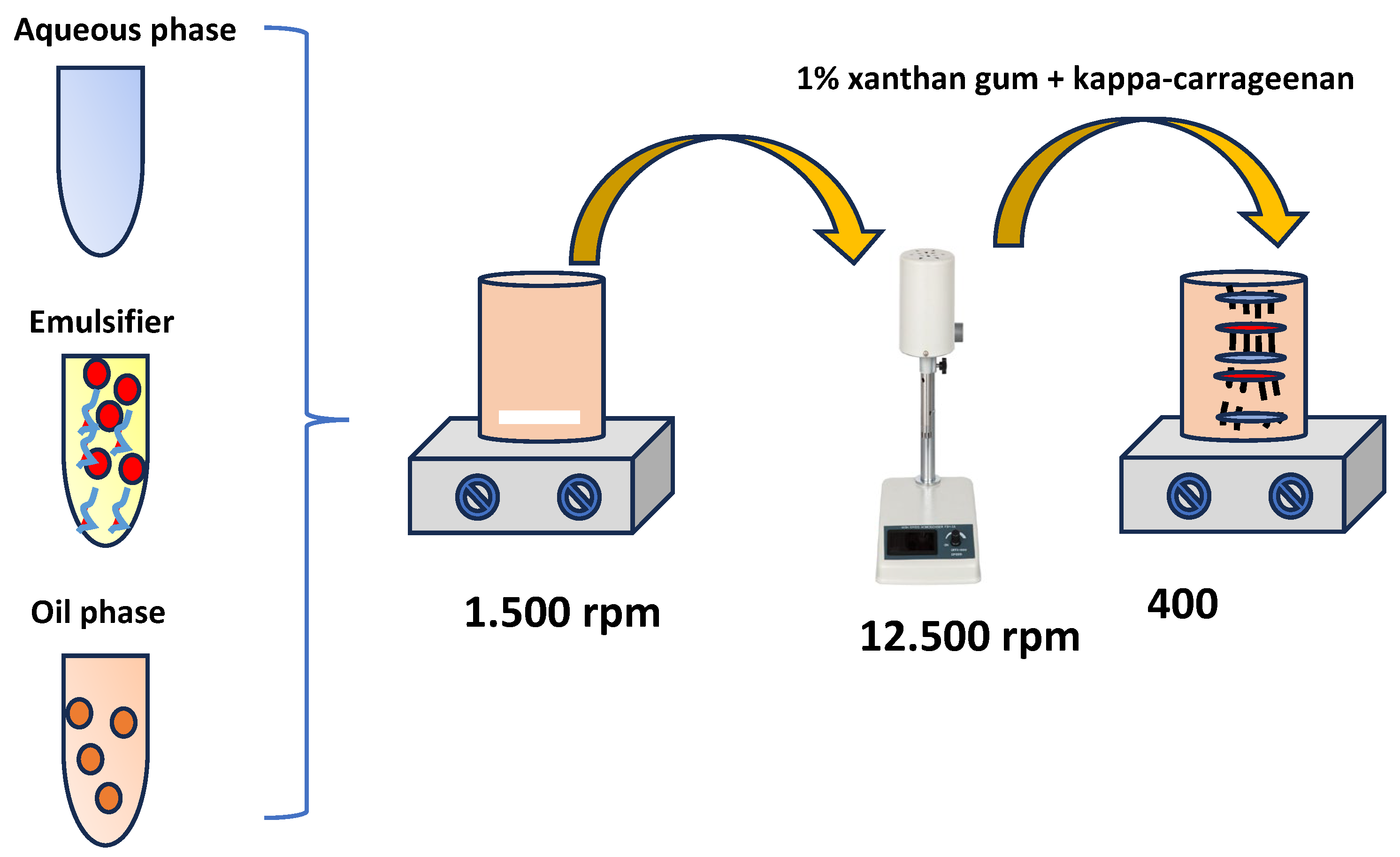
78] with some modification. In detail the preparation of nanogels commenced with the formulation of nanoemulsions by emulsifying the aqueous phase with the oil phase, which consisted of essential oil (EO) derivatives, utilizing Tween 80 as a surfactant. This process yielded a nanoemulsion with a final concentration of 2.5% solution. Five samples were prepared, including pure citral, carvacrol, and their mixtures at ratios of 25:75, 50:50, and 75:25. Emulsification was performed using a high-speed ultrasonic homogenizer operating at 15,000 rpm for 20 minutes, with the emulsions maintained in an ice bath to avoid thermal instability. The emulsions were subsequently stored at 4°C for 24 hours to further enhance stability. Following this, xanthan gum and kappa-carrageenan were incorporated at a concentration of 1%, and the mixture was homogenized by stirring at 400 rpm for over 6 hours leading to formulation of nanogels. In
Figure 6 the preparation steps are illustrated as they were followed step by step.
3.3. Physicochemical Characterization of Obtained NEs
3.3.1. Z- Potential Measuraments
A NanoBrook Omni series (Brookhaven Instruments Corporation, 750 Blue Point Road, Holtsville, NY, USA 11742) size and zeta potential analyzer equipped with a solvent-resistant electrode used to determine zeta potential of the NGs The NGs were diluted 100 times with deionized water to reduce the multiple scattering effect, and tested at 25 ◦C with an equilibrium of 120 s.
3.3.2. Dynamic Light Scattering (DLS)
Dynamic Light Scattering (DLS) analyses were performed utilizing a sophisticated dual-angle size and molecular analysis system (Zetasizer Nano ZS, Malvern, UK), functioning at a 633 nm wavelength with a helium-neon laser at the conventional 173° angle. A square-section glass cuvette facilitated these analyses. These procedures aimed to determine the solutions’ hydrodynamic diameter and polydispersity index at a controlled temperature of 37°C, employing the Einstein-Stokes equation for calculating the hydrodynamic radius (RH).
where k
B (J/K) is the Boltzmann constant, T (K) is the absolute temperature, η (N·s·m
−2) is the solvent’s dynamic viscosity, and D (m
2·s
−1) is the diffusion coefficient. The concentration of the solutions was approximately 0.1 wt%.3.3.2 Z-potential characterization of NEs
3.3.3. Transmission Electron Microscope (TEM) Images of NEs
The surface morphologies of the samples were observed using a JEOL JSM-6510 LV SEM Microscope (Ltd., Tokyo, Japan) equipped with an X-Act EDS detector from Oxford Instruments, Abingdon, Oxford shire, UK (an acceleration voltage of 20 kV was applied) with a possibility to function under low-vacuum conditions. Before examination, all samples were sputter-coated with gold/palladium for 45 s to prevent sample charging during observation with SEM.
3.4. Determination of Antioxidant Activity of NGs with 2,2-Diphenyl-1-Picrylhydrazyl (DPPH) Assay
The preparation of the DPPH ethanolic solution followed the methodology recommended by Kechagias et al., [
79]. A specific quantity of 30 ppm DPPH radical was dissolved in 250 mL of ethanol to create a 2.16 mM DPPH radical solution. This solution was mixed using a vortex while avoiding light exposure, resulting in a deep purple solution with a stable pH of approximately 7.02 ± 0.01. To ensure its stability, the solution was stored at 4°C for over 2 hours before being used in the experiment. Serial dilutions were then performed to generate a calibration curve.
To examine the correlation between DPPH radical absorbance and concentration, a calibration graph was plotted. The initial ethanol-based DPPH radical solution was serially diluted with ethanol to prepare five different concentrations ranging from 0 to 60 mg/L (ppm). The absorbance of these diluted DPPH radical solutions was measured at a wavelength of 517 nm using a Jasco V-530 UV/VIS Spectrometer [
79]. A linear calibration curve was constructed from the absorbance data collected, following a previously described method, enabling quantitative analysis of antioxidant capacity [
26].
To assess the concentration required to achieve 50% antioxidant effectiveness (EC
50) of the newly developed NG coatings NGCT, NGCV, NGCTCV (25:75, 50:50, 75:25), varying volumes of each coating (20, 40, 60, 80, and 100 μL) were incorporated, in triplicate, into a mixture containing 2.8 mL of a 30 ppm DPPH radical solution in ethanol and 0.2 mL of a sodium acetate buffer (CH
3COONa·3H
2O). A control sample consisting solely of the DPPH radical solution and buffer was also prepared for comparative analysis. After a one-hour reaction period, considered sufficient to reach equilibrium, absorbance levels were recorded at 517 nm for both the experimental and control coatings. Using a proper equation, the percentage of DPPH radical reduction at equilibrium for each coating was determined, allowing for the assessment of the antioxidant capacity of each sample.
The reduced percentage of remaining DPPH radical at equilibrium is inversely correlated with the increased antioxidant efficacy of the tested substance (Zaharioudakis et al., 2023). Based on this principle, the antioxidant activity percentages derived from the coatings NGCT, NGCV, NGCTCV (25:75, 50:50, 75:25) were graphically represented against the respective volumes of coatings used. The performance of these coatings was assessed using the equations derived from the graphs. By applying these linear equations, the EC50,DPPH values, which indicate the concentration at which 50% antioxidant activity is observed, were accurately calculated for each sample. This process allowed for the precise measurement of the antioxidant effectiveness and quantitative assessment of each nanogel.
3.5. Antibacterial Activity Test of NGs
3.5.1. Inhibition Zone Tests
The antimicrobial properties of the examined nanogels, including NGCT, NGCV, and NGCTCV (25:75, 50:50, 75:25), were assessed using the well diffusion method against two significant foodborne pathogens Escherichia coli O157:H7 and Staphylococcus aureus. Initially, the bacterial strains were cultivated in Müeller Hinton Broth at 37°C for 24 hours to reach a concentration of 107 to 108 CFU mL^-1. The bacterial cultures were then spread onto Müller-Hinton agar plates using sterile cotton swabs, with the plates rotated every 60 degrees to ensure even bacterial distribution. A sterile cork borer, sanitized with alcohol and flame, was used to create 6-mm wells in the agar, which were subsequently filled with 100 μL of each treatment. Following this, the agar plates were incubated overnight at 37°C. The effectiveness of the various treatments against the pathogens was assessed by measuring the sizes (in mm) of the clear zones surrounding each well using a digital caliper. These clear zones indicated regions where bacterial growth was inhibited by the nanogel coatings.
3.5.2. MIC and MBC Tests and FIC Index
The Minimum Inhibitory Concentration (MIC) is identified as the least quantity of an antimicrobial agent needed to prevent visible microbial growth, indicating bacteriostatic capabilities without detailed analysis of the microbial viability. Cultures of S. aureus, E. coli O157:H7 were grown in Müeller-Hinton broth to achieve a bacterial concentration of 106 CFU/mL. The antimicrobial efficacy of examined nanogels and their free EOs, was assessed across a range of concentrations: 1000, 500, 250, 125, 62.5, 31.25, 15.625 μg/mL. Later, serial dilutions of microbial cultures were prepared, and the antimicrobial agents were incorporated at each specified concentration for examination. The mixtures were then homogenized using a vortex mixer and incubated at 37 °C for a 24-hour period. The control sample consisted of the microbial cultures without any coating, and it was also evaluated at a fixed concentration of 250 μg/mL. The presence of turbidity was monitored as an indication of microbial proliferation, with further confirmation obtained via culturing and colony enumeration. The procedure was performed in triplicate to ensure the reliability of the results.
To further assess antimicrobial efficacy, the minimum bactericidal concentration (MBC) was determined by transferring 100 μL of the samples from the MIC test with no visible growth onto sterile Müeller-Hinton agar plates. These plates were incubated at 37 °C for 24 hours. After incubation, the plates were assessed for bacterial growth. Finally, the MBC was determined as the lowest concentration at which no colonies were observed. This test was performed in triplicate to ensure consistent and reliable results.
To evaluate the potential synergistic effect between CT and CV in combination against the examined pathogens, the Fractional Inhibitory Concentration (FIC) index was calculated. The FIC index for each combination was calculated using the formula:
If the FIC index is below 0.5, a synergistic effect is confirmed. If the FIC index ranges between 0.5 and 1.0, an additive effect is indicated. Finally, if the FIC index is above 1.0, an antagonistic effect occurs.
3.5.3. Time-Kill Kinetics Assay
Time-kill assays were carried out according to the method described by Boonyanugomol et al., [
80]. For these assays,
E. coli O157:H7 and
S. aureus strains were standardized to a concentration of 10
6 CFU/ml. The MBC lowest value of NGCTCV (25:75, 50:50, 75:25) were used in the experiments. At intervals of 1 to 24 hours, 50 μl samples were taken from the treated bacterial solutions. These samples were then subjected to ten-fold serial dilutions and plated on Mueller-Hinton Agar (MHA) for colony enumeration. A 0.5% v/v Tween 80 solution was employed as the control. The bacterial counts were plotted against time to create time-kill curves. All tests were performed in triplicate to ensure accuracy and reproducibility.
3.6. Application of NG as Active Coatings in Chalkidiki Green Table Olives
All the manufactured nanogel treatments were applied as active coatings to preserve green table olives by dipping method. Commercial Chalkidiki green table olives were purchased by a local market and stored at the laboratory. 50 grams of olives were weighted and dipped in 30mL of each nanogel treatment including NGCT, NGCV, NGCTCV(25:75, 50:50, 75:25) to ensure concistency across all samples. Afterwards, every olive sample was wrapped in a membrane and stored at ambient room temperature. During the storage period, various analyses were performed at regular intervals to assess the effectiveness of the applied treatments in preserving Chalkidiki green table olives (See
Figure S3). These analyses included pH measurements, Lab* colorimetry analysis, weight loss and sensory evaluations.
3.6.1. Physicochemical Properties of Chalkidiki Green Table Olives
The physicochemical properties of Chalkidiki green table olives coatings, including pH, color values (Lab), and weight loss, were analyzed at specific intervals: days 0, 7, 14, and 21.
pH Analysis
The pH values of the Chalkidiki green table olives coatings were measured using a portable pH meter fitted with a penetration electrode and a temperature sensor (pH-Star, Matthäus GmbH, Poettmes, Germany). Prior to each set of measurements, the pH meter was calibrated using pH standard solutions of 4.0 and 7.0, and temperature-adjusted to match the olive coating temperature of 20 °C. The entire procedure was conducted in triplicate, and for each treatment group, ten separate pH readings were taken to ensure accuracy and reliability, as per the methods (Janisch, Krischek, and Wicke 2011).
Weight Loss Analysis
The weight loss analysis was conducted by measuring the initial weight of the olive samples NGCT, NGCV, NGCTCV (25:75, 50:50, 75:25) at the start of the experiment (day 0) and at subsequent intervals on days 7, 14, and 21. At each sampling point, the weight loss was determined by subtracting the final weight of the olives from their initial weight on the respective days. The percentage difference in weight was calculated and interpreted as humidity loss, serving as a quality indicator to assess significant weight loss among the examined samples.
Lab* Analysis
The alterations in the CIELAB color parameters (L*, a*, and b*) of Chalkidiki green table olives over a period of 9 days under refrigerated storage were assessed using a LS171 colorimeter from Linshang Company. Prior to conducting the measurements, the colorimeter was calibrated with a white standard plate in accordance with methods cited in studies (Hernandez et al., 2019). Color evaluations were conducted directly on the surface of the olive coatings, with each treatment group comprising three separate portions. For each of these portions, five discrete readings were taken to capture a robust assessment of the color. The total color differences (
ΔE) were calculated using the equation (Roy Choudhury, 2015):
In this equation, L*0, a*0, b*0 denote the initial color parameters of the Chalkidiki green table olives at day 0 post-treatment. L*, a*, b* represent the respective color parameters at different time points during the 21-day refrigerated storage at 4 °C.
3.6.2. Evaluation of Bacterial Counts in Chalkidiki Green Table Olives
Chalkidiki green table olives were submitted to assessment for mesophilic bacteria, LAB bacteria and yeasts and moulds on days 0, 7, 14 and 21 during storage period at 4 °C. Initially a 10g Chalkidiki green table olives sample aseptically transferred to a stomacher bag, followed by homogenization with 90 mL of peptone water. Subsequently, a serial dilution (1:10 ratio) was performed.
For LAB bacteria, MRS agar was used, and 1 mL of the diluted sample was incorporated into the plates. The plates were then incubated anaerobically at 30 °C for 72 hours, following the guidelines of ISO 15214:1998. For the enumeration of yeasts and molds, Dichloran Rose Bengal Chloramphenicol (DRBC) agar was used. A 0.1 mL aliquot from the diluted samples was spread onto the DRBC agar plates, which were then incubated aerobically at 25 °C for 5 days, according to ISO 21527-1:2008.
3.6.3. Sensory Evaluation of Chalkidiki Green Table Olives
Sensory analysis was performed on Chalkidiki green table olives treated with nanogel coatings to evaluate key sensory characteristics, including taste, odor, color, texture, and overall acceptability. A panel of 15 trained assessors with expertise in food sensory evaluation participated in the assessment. The evaluation employed a 5-point hedonic scale, where a score of 5 indicated “like extremely” and a score of 1 indicated “dislike extremely.” An acceptability threshold score of 3.5 was used, as outlined by Hasani-Javanmardi et al. [
81].
Each treatment group, including individual nanogels (NGCV and NGCT), mixed nanogel coatings (NGCTCV 25:75, NGCTCV 50:50, and NGCTCV 75:25), and uncoated olives, was assessed at four different time points (Days 0, 7, 14, and 21). Each sample underwent triplicate assessments per replicate, resulting in a total of six evaluations for each treatment group at each time point. This allowed for a comprehensive analysis of the sensory attributes of the olives throughout the storage period. The results of this sensory evaluation provided insights into the effectiveness of the nanogel coatings in preserving the quality and sensory appeal of the olives over time.
3.7. Statistical Analysis
For the purposes of the statistical analysis, the average results of each experiment were recorded in triplicate. The software was used refers to IBM SPSS version 22 (IBM Armonk, NY, USA), the statistical analyses were performed. After checking for homogeneity of data, an alpha level of P < 0.05 and due to the low number of samples, Kruskal Wallis test was preferred as a tool for the examination of the significance of the resulting difference.
4. Discussion
In this study polysaccharide based nanogels enhanced with essential oil derivatives, individually and in mixtures of CT and CV were successfully developed and evaluated for their potential as natural preservatives for the shelf-life extension of fermented products such as Chalkidiki PDO green table olives.
The zeta potential results indicated a high stability of the nanogels, which constitute an ideal option as preservative agents. All nanogel coatings exhibited high negative values, indicating effective colloidal stability. Among them, the NGCV nanogel showed the highest zeta potential, likely due to the strong bonding and electrostatic interactions between the examined essential oil derivatives and the negative charged polysaccharides xanthan gum and kappa carrageenan and our results are in accordance to various studies [
12,
50,
52,
53,
54,
55,
56,
57].
Furthermore, the nanogels showed significant antioxidant activity, as examined by the DPPH. The NGCV nanogel outperformed the rest of the coatings, needing the lowest quantity to achieve a 50% radical reduction. The results are in accordance with other studies [
82]. However, the antioxidant activity of the nanogel mixtures did not demonstrate a significant synergistic effect, with the NGCTCV mixtures showing lower activity than NGCV individually and this result is in agreement with Al-Mansori et al.,[
58].
The antimicrobial preliminary tests results including MIC, MBC, FIC, and time-killing assay demonstrated that mixtures of CT and CV exhibit enhanced antibacterial activity compared to individual nanogels. In particular, the NGCTCV 50:50 mixture, showed bactericidal effect against
Escherichia coli O157:H7, while significantly reduced
Staphylococcus aureus populations within hours. FIC index showed the synergistic effect of NGCTCV 25:75 and 75:25 against
E. coli O157:H7 and the synergistic effect of NGCTCV 50:50 against
S. aureus. The synergistic effect of CT and CV is well-documented in several studies and is explained as result of different sites targeting within bacterial cells [
65]. Specifically, carvacrol increases the permeability of the bacterial membrane, allowing citral to penetrate more effectively into the cytoplasm, improving the overall antimicrobial effect. The bactericidal effect of the synergism is well documented in various studies against pathogens like
L. monocytogenes,
C. sakazakii, and
E. coli [
66,
67].
The application of nanogel coatings on Chalkidiki PDO green table olives effectively controlled the growth of yeast and molds during the 21-day storage period. Among the treatments, NGCTCV 50:50 was particularly effective in reducing yeast populations by 2log compared to uncoated olives by Day 21. This reduction in microbial growth underscores the preservative efficacy of the nanogel mixtures, likely resulting from the combined antimicrobial properties of CT and CV.
Importantly, the nanogels did not inhibit the growth of lactic acid bacteria (LAB), which are crucial for the fermentation process. On the contrary, the LAB populations increased significantly in the presence of the nanogel mixtures, particularly NGCTCV 25:75. A study conducted by Papastolopoulou et al., [
73] noted that indeed essential oil did not affect fermentation. This suggests that the polysaccharide 3d network in the nanogels may provide a nutrient source for LAB, promoting their activity while simultaneously essential oil derivatives inhibit the growth of harmful bacteria. The dual effect of the novel examined nanogels as natural preservative agents seem ideal for fermented products such as Chalkidiki PDO green table olives. To the best of our knowledge, this is the first time such a result has been reported, and further investigation is suggested.
Weight loss is a critical factor in maintaining the quality of Chalkidiki PDO green table olives during storage. The nanogel coatings, especially the NGCTCV 50:50 and NGCTCV 25:75 mixtures, were highly effective in controlling weight loss over the 21-day storage period. These coatings outperformed individual nanogels, suggesting a synergistic effect that enhances moisture retention. This finding is consistent with other studies demonstrating the effectiveness of polysaccharide and essential oil-based coatings in reducing weight loss in fruits and vegetables [
74,
75,
76,
77,
83]. By controlling moisture levels, these coatings help preserve the physical and textural quality of the olives, making them attractive to consumers.
The pH analysis further supported the effectiveness of the nanogels, particularly in maintaining a stable, low pH throughout the storage period. The initial pH drop observed in the first week indicates active LAB fermentation, which is beneficial for the preservation process. The NGCTCV 50:50 and NGCTCV 75:25 mixtures were particularly effective in maintaining lower pH levels, likely due to the LAB activity and controlling yeasts and molds populations. This stable pH environment is crucial for preventing the growth of pathogenic bacteria and ensuring the safety and quality of Chalkidiki PDO green table olives.
Color is a crucial quality parameter in the examined Chalkidiki PDO green table olives, and maintaining it during storage is critical for consumer acceptance. The Lab colorimetry analysis demonstrated that the essential oil mixtures, particularly NGCTCV 25:75 and NGCTCV 50:50, were significantly more effective in preserving the color of the olives than individual nanogels or uncoated samples. These mixtures effectively controlled the darkening of olives and retained the green and yellow hues, which are essential for the visual appeal of the product.
Finally, he mixed nanogel coatings, particularly NGCTCV 50:50 and NGCTCV 25:75, demonstrated superior preservation of sensory attributes such as taste, odor, color, and texture, outperforming both individual oil nanogels and uncoated olives. This study is the first to report that combining essential oils in a nanogel formulation provides additional benefits for maintaining sensory qualities in fermented food products like Chalkidiki green table olives.
5. Conclusion
This study explored the development, characterization, and application of novel polysaccharide based nanogels enhanced with essential oil derivatives CT and CV, examining a potential synergistic effect and finally their use as active edible coating on fermented products such as Chalkidiki PDO green table olives for shelf-life extension. Zeta potential measurements confirmed the physicochemical stability of the nanogels. Both DLS and SEM studies confirm that NGCT, NGCV samples showed an average particle size of ~34 and of ~310 nm (~70 %) nm (~100 %) correspondingly while the samples NGCTCV_50/50, NGCTCV_25/75 and NGCTCV_75/25 showed a major population of the particles between 140 nm and 480 nm and the rest in the range of μm. Preliminary tests indicated the high antioxidant profile of all those coatings. Furthermore, a significant bactericidal effect was demonstrated by nanogel mixtures, while FIC confirmed the synergistic effect of NGCTCV 50:50 against S. aureus, and NGCTCV 25:75 and NGCTCV 75:25 against E. coli O157:H7.
The application of nanogel coatings to Chalkidiki PDO green table olives, demonstrated the efficacy of NGCTCV 50:50 and NGCTCV 25:75 in reducing weight loss, maintaining stable pH levels, and preserving the visual quality of the olives over the 21-day storage period. These nanogels not only inhibited spoilage microorganisms but also did not affect the growth of beneficial lactic acid bacteria essential for fermentation. Notwithstanding the fact that Lab* colorimetry results showed that these nanogel formulations successfully prevented discoloration, maintaining the olives’ desired color characteristics, the mixtures seem to have statistically significant difference. Finally, the nanogels managed to extend olives’ shelf life for over 14 days. The mixed nanogel coatings, particularly NGCTCV 50:50 and 25:75, effectively preserved the sensory qualities of Chalkidiki green table olives, outperforming both individual nanogels and uncoated samples. Overall, this research highlights the advantages of polysaccharide based nanogels, particularly in the effective ratios of 50:50 and 25:75, for extending the shelf life of perishable products like Chalkidiki PDO green table olives. Future research should focus on the application of these nanogels to a broader range of food products and under varying storage conditions. Additionally, further investigation into the mode of action of the synergistic effects of CT and CV could provide deeper insights into optimizing these formulations.
Supplementary Materials
The following supporting information can be downloaded at the website of this paper posted on Preprints.org. Figure S1: Agar Diffusion zone of nanogels against pathogens A) E. coli O157:H7 and B) S. aureus; Figure S2: Application process of olives coatings on nanogels; Table S1: Statistical analysis (Kruskal-Wallis) of Zeta potential results title; Table S2: Statistical analysis (Kruskal-Wallis) for Antioxidant activity EC50 results; Table S3: Statistical analysis results (Kruskal-Wallis) for E. coli O157:H7 antibacterial activity tests; Table S4: Statistical analysis results (Kruskal-Wallis) for S. aureus antibacterial activity tests; Table S5: Statistical analysis results (Kruskal-Wallis) for Yeasts results; Table S6: Statistical analysis results (Kruskal-Wallis) for LAB results, Table S7: Statistical analysis results (Kruskal-Wallis) for weight loss results; Table S8: Statistical analysis results (Kruskal-Wallis) for weight loss results; Table S9: Statistical analysis results (Kruskal-Wallis) for pH results; Table S10: Statistical analysis results (Kruskal-Wallis) for pH results; Table S11: Statistical analysis results (Kruskal-Wallis) for L* parameter results; Table S12: Statistical analysis results (Kruskal-Wallis) for L* parameter results; Table S13. Statistical analysis results (Kruskal-Wallis) for a* parameter results; Table S14: Statistical analysis results (Kruskal-Wallis) for a* parameter results, Table S15: Statistical analysis results (Kruskal-Wallis) for b* parameter results; Table S16: Statistical analysis results (Kruskal-Wallis) for b* parameter results; Table S17: Statistical analysis results (Kruskal-Wallis) for sensory analysis results; Table S18: Statistical analysis results (Kruskal-Wallis) for sensory analysis results.
Author Contributions
Conceptualization—K.Z., C.E.S., and A.E.G.; data curation—K.Z., A.L; D.M., A.K.-M., A.E.G., and C.E.S.; formal analysis—K.Z., D.M., A.K.-M., A.E.G., and C.E.S.; investigation—K.Z., N.D.A., A.L., D.M., A.K.-M., and C.P.; methodology—K.Z., N.D.A., D.M., A.K-M., E.T., A.A., N.Z.,C.E.S.,; project administration—C.E.S. and A.E.G.; resources—K.Z., A.L., A.A., and N.E.Z.; software—K.Z., A.L., D.M., A.K-M., C.E.S., and A.E.G.; supervision—A.A., N.E.Z., N.D.A., C.P., C.E.S., and A.E.G.; validation—K.Z., N.D.A., A.L., D.M., A.,K-M.,N.E.Z., G.K., and C.P.; visualization—C.P., C.E.S., and A.E.G.; writing–original draft—K.Z., A.L., D.M., A. K-M., C.E.S., and A.E.G.; writing–review and editing—K.Z.,A.L., C.E.S., and A.E.G.; all authors have read and agreed to the published version of the manuscript.
Funding
This research received no external funding.
Institutional Review Board Statement
Not applicable.
Informed Consent Statement
Not applicable.
Data Availability Statement
The datasets generated for this study are available on request to the corresponding author.
Conflicts of Interest
The authors declare no conflicts of interest.
References
- Pandey, A.K.; Kumar, P.; Singh, P.; Tripathi, N.N.; Bajpai, V.K. Essential Oils: Sources of Antimicrobials and Food Preservatives. Front. Microbiol. 2017, 7. [Google Scholar] [CrossRef] [PubMed]
- Guzik, P.; Szymkowiak, A.; Kulawik, P.; Zając, M. Consumer Attitudes towards Food Preservation Methods. Foods 2022, 11, 1349. [Google Scholar] [CrossRef] [PubMed]
- Rabbi, M.F.; Amin, M.B. Circular Economy and Sustainable Practices in the Food Industry: A Comprehensive Bibliometric Analysis. Cleaner and Responsible Consumption 2024, 14, 100206. [Google Scholar] [CrossRef]
- Fassio, F.; Chirilli, C. The Circular Economy and the Food System: A Review of Principal Measuring Tools. Sustainability 2023, 15, 10179. [Google Scholar] [CrossRef]
- Hamam, M.; Chinnici, G.; Di Vita, G.; Pappalardo, G.; Pecorino, B.; Maesano, G.; D’Amico, M. Circular Economy Models in Agro-Food Systems: A Review. Sustainability 2021, 13, 3453. [Google Scholar] [CrossRef]
- Prasanna, S.; Verma, P.; Bodh, S. The Role of Food Industries in Sustainability Transition: A Review. Environ Dev Sustain 2024. [Google Scholar] [CrossRef]
- Baldwin, C.; Wilberforce, N.T. Sustainability Principles and Sustainable Innovation for Food Products. In Sustainability in the Food Industry; John Wiley & Sons, Ltd., 2009; pp. 239–249 ISBN 978-1-118-46758-9.
- Essential Oils Market Size, Share & Growth Report, 2030. Available online: https://www.grandviewresearch.com/industry-analysis/essential-oils-market (accessed on 7 February 2024).
- Rodilla, J.M.; Rosado, T.; Gallardo, E. Essential Oils: Chemistry and Food Applications. Foods 2024, 13, 1074. [Google Scholar] [CrossRef]
- Chen, X.; Shang, S.; Yan, F.; Jiang, H.; Zhao, G.; Tian, S.; Chen, R.; Chen, D.; Dang, Y. Antioxidant Activities of Essential Oils and Their Major Components in Scavenging Free Radicals, Inhibiting Lipid Oxidation and Reducing Cellular Oxidative Stress. Molecules 2023, 28, 4559. [Google Scholar] [CrossRef]
- de Sousa, D.P.; Damasceno, R.O.S.; Amorati, R.; Elshabrawy, H.A.; de Castro, R.D.; Bezerra, D.P.; Nunes, V.R.V.; Gomes, R.C.; Lima, T.C. Essential Oils: Chemistry and Pharmacological Activities. Biomolecules 2023, 13, 1144. [Google Scholar] [CrossRef]
- Guo, Y.; Pizzol, R.; Gabbanini, S.; Baschieri, A.; Amorati, R.; Valgimigli, L. Absolute Antioxidant Activity of Five Phenol-Rich Essential Oils. Molecules 2021, 26, 5237. [Google Scholar] [CrossRef]
- Bunse, M.; Daniels, R.; Gründemann, C.; Heilmann, J.; Kammerer, D.R.; Keusgen, M.; Lindequist, U.; Melzig, M.F.; Morlock, G.E.; Schulz, H.; et al. Essential Oils as Multicomponent Mixtures and Their Potential for Human Health and Well-Being. Front Pharmacol 2022, 13, 956541. [Google Scholar] [CrossRef] [PubMed]
- Mehdi, Y.; Létourneau-Montminy, M.-P.; Gaucher, M.-L.; Chorfi, Y.; Suresh, G.; Rouissi, T.; Brar, S.K.; Côté, C.; Ramirez, A.A.; Godbout, S. Use of Antibiotics in Broiler Production: Global Impacts and Alternatives. Animal Nutrition 2018, 4, 170–178. [Google Scholar] [CrossRef] [PubMed]
- Manzoor, A.; Yousuf, B.; Pandith, J.A.; Ahmad, S. Plant-Derived Active Substances Incorporated as Antioxidant, Antibacterial or Antifungal Components in Coatings/Films for Food Packaging Applications. Food Bioscience 2023, 53, 102717. [Google Scholar] [CrossRef]
- Lubbe, A.; Verpoorte, R. Cultivation of Medicinal and Aromatic Plants for Specialty Industrial Materials. Industrial Crops and Products 2011, 34, 785–801. [Google Scholar] [CrossRef]
- Angane, M.; Swift, S.; Huang, K.; Butts, C.A.; Quek, S.Y. Essential Oils and Their Major Components: An Updated Review on Antimicrobial Activities, Mechanism of Action and Their Potential Application in the Food Industry. Foods 2022, 11, 464. [Google Scholar] [CrossRef]
- Nychas, G.-J.E.; Tassou, C.C. PRESERVATIVES | Traditional Preservatives – Oils and Spices. In Encyclopedia of Food Microbiology (Second Edition); Batt, C.A., Tortorello, M.L., Eds.; Academic Press: Oxford, 2014; pp. 113–118. ISBN 978-0-12-384733-1. [Google Scholar]
- Abdi-Moghadam, Z.; Mazaheri, Y.; Rezagholizade-shirvan, A.; Mahmoudzadeh, M.; Sarafraz, M.; Mohtashami, M.; Shokri, S.; Ghasemi, A.; Nickfar, F.; Darroudi, M.; et al. The Significance of Essential Oils and Their Antifungal Properties in the Food Industry: A Systematic Review. Heliyon 2023, 9, e21386. [Google Scholar] [CrossRef]
- Tariq, S.; Wani, S.; Rasool, W.; Shafi, K.; Bhat, M.A.; Prabhakar, A.; Shalla, A.H.; Rather, M.A. A Comprehensive Review of the Antibacterial, Antifungal and Antiviral Potential of Essential Oils and Their Chemical Constituents against Drug-Resistant Microbial Pathogens. Microbial Pathogenesis 2019, 134, 103580. [Google Scholar] [CrossRef]
- Masyita, A.; Mustika Sari, R.; Dwi Astuti, A.; Yasir, B.; Rahma Rumata, N.; Emran, T.B.; Nainu, F.; Simal-Gandara, J. Terpenes and Terpenoids as Main Bioactive Compounds of Essential Oils, Their Roles in Human Health and Potential Application as Natural Food Preservatives. Food Chem X 2022, 13, 100217. [Google Scholar] [CrossRef]
- Zaharioudakis, K.; Salmas, C.E.; Andritsos, N.D.; Kollia, E.; Leontiou, A.; Karabagias, V.K.; Karydis-Messinis, A.; Moschovas, D.; Zafeiropoulos, N.E.; Avgeropoulos, A.; et al. Carvacrol, Citral, Eugenol and Cinnamaldehyde Casein Based Edible Nanoemulsions as Novel Sustainable Active Coatings for Fresh Pork Tenderloin Meat Preservation. Front. Food. Sci. Technol. 2024, 4. [Google Scholar] [CrossRef]
- Ebhohimen, I.E.; Okolie, N.P.; Okpeku, M.; Unweator, M.; Adeleke, V.T.; Edemhanria, L. Evaluation of the Antioxidant Properties of Carvacrol as a Prospective Replacement for Crude Essential Oils and Synthetic Antioxidants in Food Storage. Molecules 2023, 28, 1315. [Google Scholar] [CrossRef]
- Gutiérrez-Pacheco, M.M.; Torres-Moreno, H.; Flores-Lopez, M.L.; Velázquez Guadarrama, N.; Ayala-Zavala, J.F.; Ortega-Ramírez, L.A.; López-Romero, J.C. Mechanisms and Applications of Citral’s Antimicrobial Properties in Food Preservation and Pharmaceuticals Formulations. Antibiotics (Basel) 2023, 12, 1608. [Google Scholar] [CrossRef] [PubMed]
- Mączka, W.; Twardawska, M.; Grabarczyk, M.; Wińska, K. Carvacrol—A Natural Phenolic Compound with Antimicrobial Properties. Antibiotics (Basel) 2023, 12, 824. [Google Scholar] [CrossRef] [PubMed]
- Zaharioudakis, K.; Kollia, E.; Leontiou, A.; Moschovas, D.; Karydis-Messinis, A.; Avgeropoulos, A.; Zafeiropoulos, N.E.; Ragkava, E.; Kehayias, G.; Proestos, C.; et al. Carvacrol Microemulsion vs. Nanoemulsion as Novel Pork Minced Meat Active Coatings. Nanomaterials (Basel) 2023, 13, 3161. [Google Scholar] [CrossRef] [PubMed]
- Olaniran, A.F.; Adeyanju, A.A.; Olaniran, O.D.; Erinle, C.O.; Okonkwo, C.E.; Taiwo, A.E. Chapter 10 - Improvement of Food Aroma and Sensory Attributes of Processed Food Products Using Essential Oils/Boosting up the Organoleptic Properties and Nutritive of Different Food Products. In Applications of Essential Oils in the Food Industry; Adetunji, C.O., Sharifi-Rad, J., Eds.; Academic Press, 2024; pp. 107–116 ISBN 978-0-323-98340-2.
- Ferreira, C.D.; Nunes, I.L. Oil Nanoencapsulation: Development, Application, and Incorporation into the Food Market. Nanoscale Res Lett 2019, 14, 9. [Google Scholar] [CrossRef]
- Giannakas, A.E. Plant Extracts-Based Food Packaging Films. In Natural Materials for Food Packaging Application; John Wiley & Sons, Ltd., 2023; pp. 23–49 ISBN 978-3-527-83730-4.
- Giannakas, A.E. 7 - Bionanocomposites with Hybrid Nanomaterials for Food Packaging Applications. In Advances in Biocomposites and their Applications; Karak, N., Ed.; Woodhead Publishing Series in Composites Science and Engineering; Woodhead Publishing, 2024; pp. 201–225 ISBN 978-0-443-19074-2.
- Zhang, H.; Li, X.; Kang, H.; Peng, X. Antimicrobial and Antioxidant Effects of Edible Nanoemulsion Coating Based on Chitosan and Schizonepeta Tenuifolia Essential Oil in Fresh Pork. Journal of Food Processing and Preservation 2021, 45, e15909. [Google Scholar] [CrossRef]
- Pateiro, M.; Gómez, B.; Munekata, P.E.S.; Barba, F.J.; Putnik, P.; Kovačević, D.B.; Lorenzo, J.M. Nanoencapsulation of Promising Bioactive Compounds to Improve Their Absorption, Stability, Functionality and the Appearance of the Final Food Products. Molecules 2021, 26, 1547. [Google Scholar] [CrossRef]
- Bazana, M.T.; Codevilla, C.F.; de Menezes, C.R. Nanoencapsulation of Bioactive Compounds: Challenges and Perspectives. Current Opinion in Food Science 2019, 26, 47–56. [Google Scholar] [CrossRef]
- Chudasama, M.; Goyary, J. Nanostructured Materials in Food Science: Current Progress and Future Prospects. Next Materials 2024, 5, 100206. [Google Scholar] [CrossRef]
- Papagiannopoulos, A.; Sotiropoulos, K. Current Advances of Polysaccharide-Based Nanogels and Microgels in Food and Biomedical Sciences. Polymers 2022, 14, 813. [Google Scholar] [CrossRef]
- Silva, M.P.; Fabi, J.P. Food Biopolymers-Derived Nanogels for Encapsulation and Delivery of Biologically Active Compounds: A Perspective Review. Food Hydrocolloids for Health 2022, 2, 100079. [Google Scholar] [CrossRef]
- Yang, Z.; Xie, F.; Cai, J. Advances in the Delivery of Bioactive Compounds Using Starch-Based Nanogels: Formation Mechanism, Contemporary Challenges, and Prospects. Journal of Agriculture and Food Research 2024, 16, 101142. [Google Scholar] [CrossRef]
- Abedi, F.; Ghandforoushan, P.; Adeli, F.; Yousefnezhad, M.; Mohammadi, A.; Moghaddam, S.V.; Davaran, S. Development of Stimuli-Responsive Nanogels as Drug Carriers and Their Biomedical Application in 3D Printing. Materials Today Chemistry 2023, 29, 101372. [Google Scholar] [CrossRef]
- Fasolin, L.H.; Pereira, R.N.; Pinheiro, A.C.; Martins, J.T.; Andrade, C.C.P.; Ramos, O.L.; Vicente, A.A. Emergent Food Proteins – Towards Sustainability, Health and Innovation. Food Research International 2019, 125, 108586. [Google Scholar] [CrossRef] [PubMed]
- Shen, J.; Chen, Y.; Li, X.; Zhou, X.; Ding, Y. Enhanced Probiotic Viability in Innovative Double-Network Emulsion Gels: Synergistic Effects of the Whey Protein Concentrate-Xanthan Gum Complex and κ-Carrageenan. Int J Biol Macromol 2024, 270, 131758. [Google Scholar] [CrossRef] [PubMed]
- Avallone, P.R.; Russo Spena, S.; Acierno, S.; Esposito, M.G.; Sarrica, A.; Delmonte, M.; Pasquino, R.; Grizzuti, N. Thermorheological Behavior of κ-Carrageenan Hydrogels Modified with Xanthan Gum. Fluids 2023, 8, 119. [Google Scholar] [CrossRef]
- Froelich, A.; Jakubowska, E.; Jadach, B.; Gadziński, P.; Osmałek, T. Natural Gums in Drug-Loaded Micro- and Nanogels. Pharmaceutics 2023, 15, 759. [Google Scholar] [CrossRef]
- Petri, D.F.S. Xanthan Gum: A Versatile Biopolymer for Biomedical and Technological Applications. Journal of Applied Polymer Science 2015, 132. [Google Scholar] [CrossRef]
- Nsengiyumva, E.M.; Alexandridis, P. Xanthan Gum in Aqueous Solutions: Fundamentals and Applications. International Journal of Biological Macromolecules 2022, 216, 583–604. [Google Scholar] [CrossRef]
- Lei, Y.; Zhao, X.; Li, D.; Wang, L.; Wang, Y. Effects of κ-Carrageenan and Guar Gum on the Rheological Properties and Microstructure of Phycocyanin Gel. Foods 2022, 11, 734. [Google Scholar] [CrossRef]
- Rupert, R.; Rodrigues, K.F.; Thien, V.Y.; Yong, W.T.L. Carrageenan From Kappaphycus Alvarezii (Rhodophyta, Solieriaceae): Metabolism, Structure, Production, and Application. Front. Plant Sci. 2022, 13. [Google Scholar] [CrossRef]
- Souza, H.K.S.; Kraiem, W.; Ben Yahia, A.; Aschi, A.; Hilliou, L. From Seaweeds to Hydrogels: Recent Progress in Kappa-2 Carrageenans. Materials 2023, 16, 5387. [Google Scholar] [CrossRef] [PubMed]
-
Commission Implementing Regulation (EU) No 426/2012 of 22 May 2012 Entering a Name in the Register of Protected Designations of Origin and Protected Geographical Indications (Πράσινες Ελιές Χαλκιδικής (Prasines Elies Chalkidikis) (PDO)); 2012; Vol. 132;
- Eurostat, E. Price of Olive Oil up 50% in One Year Available online: https://ec.europa.eu/eurostat/web/products-eurostat-news/w/ddn-20240227-1.
- Guo, J.; Zhu, S.; Chen, P.; Liu, Z.; Lin, L.; Zhang, J. Effect of Physiological pH on the Molecular Characteristics, Rheological Behavior, and Molecular Dynamics of κ-Carrageenan/Casein. Front. Nutr. 2023, 10. [Google Scholar] [CrossRef] [PubMed]
- Perumal, A.B.; Li, X.; Su, Z.; He, Y. Preparation and Characterization of a Novel Green Tea Essential Oil Nanoemulsion and Its Antifungal Mechanism of Action against Magnaporthae Oryzae. Ultrasonics Sonochemistry 2021, 76, 105649. [Google Scholar] [CrossRef] [PubMed]
- Alqarni, M.H.; Foudah, A.I.; Aodah, A.H.; Alkholifi, F.K.; Salkini, M.A.; Alam, A. Caraway Nanoemulsion Gel: A Potential Antibacterial Treatment against Escherichia Coli and Staphylococcus Aureus. Gels 2023, 9, 193. [Google Scholar] [CrossRef] [PubMed]
- Aldawsari, M.F.; Foudah, A.I.; Rawat, P.; Alam, A.; Salkini, M.A. Nanogel-Based Delivery System for Lemongrass Essential Oil: A Promising Approach to Overcome Antibiotic Resistance in Pseudomonas Aeruginosa Infections. Gels 2023, 9, 741. [Google Scholar] [CrossRef]
- Ullah, N.; Amin, A.; Farid, A.; Selim, S.; Rashid, S.A.; Aziz, M.I.; Kamran, S.H.; Khan, M.A.; Rahim Khan, N.; Mashal, S.; et al. Development and Evaluation of Essential Oil-Based Nanoemulgel Formulation for the Treatment of Oral Bacterial Infections. Gels 2023, 9, 252. [Google Scholar] [CrossRef]
- Rahmatpour, A.; Alijani, N.; Mirkani, A. Supramolecular Self-Assembling Hydrogel Film Based on a Polymer Blend of Chitosan/Partially Hydrolyzed Polyacrylamide for Removing Cationic Dye from Water. Reactive and Functional Polymers 2023, 185, 105537. [Google Scholar] [CrossRef]
- Farooq, M.; Sagbas, S.; Sahiner, M.; Siddiq, M.; Turk, M.; Aktas, N.; Sahiner, N. Synthesis, Characterization and Modification of Gum Arabic Microgels for Hemocompatibility and Antimicrobial Studies. Carbohydrate Polymers 2017, 156, 380–389. [Google Scholar] [CrossRef]
- Al-Musawi, M.H.; Khoshkalampour, A.; Adnan Shaker Al-Naymi, H.; Farooq Shafeeq, Z.; Pourvatan Doust, S.; Ghorbani, M. Optimization and Characterization of Carrageenan/Gelatin-Based Nanogel Containing Ginger Essential Oil Enriched Electrospun Ethyl Cellulose/Casein Nanofibers. International Journal of Biological Macromolecules 2023, 248, 125969. [Google Scholar] [CrossRef]
- Al-Mansori, B.; El-Ageeli, W.H.; Alsagheer, S.H.; Ben-Khayal, F.A.F. Antioxidant Activity- Synergistic Effects of Thymol and Carvacrol. Al-Mukhtar Journal of Sciences 2020, 35, 185–194. [Google Scholar] [CrossRef]
- Rayan, M.; Abu-Farich, B.; Basha, W.; Rayan, A.; Abu-Lafi, S. Correlation between Antibacterial Activity and Free-Radical Scavenging: In-Vitro Evaluation of Polar/Non-Polar Extracts from 25 Plants. Processes 2020, 8, 117. [Google Scholar] [CrossRef]
- Habib, S.; Gupta, P.; Bhat, S.S.; Gupta, J. In Silico, in-Vitro and in Vivo Screening of Biological Activities of Citral. International Journal for Vitamin and Nutrition Research 2020. [Google Scholar] [CrossRef] [PubMed]
- Xu, X.L.; Zhu, Q.; Lv, H.; Wu, W.H.; Lv, M.; Liu, K.H. Isolation and Biological Activities of Citral from Sweet Orange Oil. Advanced Materials Research 2013, 750–752, 1621–1625. [Google Scholar] [CrossRef]
- Rojas-Armas, J.P.; Arroyo-Acevedo, J.L.; Palomino-Pacheco, M.; Herrera-Calderón, O.; Ortiz-Sánchez, J.M.; Rojas-Armas, A.; Calva, J.; Castro-Luna, A.; Hilario-Vargas, J. The Essential Oil of Cymbopogon Citratus Stapt and Carvacrol: An Approach of the Antitumor Effect on 7,12-Dimethylbenz-[α]-Anthracene (DMBA)-Induced Breast Cancer in Female Rats. Molecules 2020, 25, 3284. [Google Scholar] [CrossRef] [PubMed]
- Chroho, M.; Rouphael, Y.; Petropoulos, S.A.; Bouissane, L. Carvacrol and Thymol Content Affects the Antioxidant and Antibacterial Activity of Origanum Compactum and Thymus Zygis Essential Oils. Antibiotics 2024, 13, 139. [Google Scholar] [CrossRef]
- Mokhtari, R.; Kazemi Fard, M.; Rezaei, M.; Moftakharzadeh, S.A.; Mohseni, A. Antioxidant, Antimicrobial Activities, and Characterization of Phenolic Compounds of Thyme (Thymus Vulgaris L.), Sage (Salvia Officinalis L.), and Thyme–Sage Mixture Extracts. Journal of Food Quality 2023, 2023, 2602454. [Google Scholar] [CrossRef]
- Silva-Angulo, A.B.; Zanini, S.F.; Rosenthal, A.; Rodrigo, D.; Klein, G.; Martínez, A. Combined Effect of Carvacrol and Citral on the Growth of Listeria Monocytogenes and Listeria Innocua and on the Occurrence of Damaged Cells. Food Control 2015, 53, 156–162. [Google Scholar] [CrossRef]
- Cao, Y.; Zhou, D.; Zhang, X.; Xiao, X.; Yu, Y.; Li, X. Synergistic Effect of Citral and Carvacrol and Their Combination with Mild Heat against Cronobacter Sakazakii CICC 21544 in Reconstituted Infant Formula. LWT 2021, 138, 110617. [Google Scholar] [CrossRef]
- Langeveld, W.T.; Veldhuizen, E.J.A.; Burt, S.A. Synergy between Essential Oil Components and Antibiotics: A Review. Critical Reviews in Microbiology 2014, 40, 76–94. [Google Scholar] [CrossRef]
- Rivas, L.; McDonnell, M.J.; Burgess, C.M.; O’Brien, M.; Navarro-Villa, A.; Fanning, S.; Duffy, G. Inhibition of Verocytotoxigenic Escherichia Coli in Model Broth and Rumen Systems by Carvacrol and Thymol. International Journal of Food Microbiology 2010, 139, 70–78. [Google Scholar] [CrossRef]
- Kim, J.; Marshall, M.R.; Wei, C. Antibacterial Activity of Some Essential Oil Components against Five Foodborne Pathogens. J. Agric. Food Chem. 1995, 43, 2839–2845. [Google Scholar] [CrossRef]
- Zanini, S.F.; Silva-Angulo, A.B.; Rosenthal, A.; Rodrigo, D.; Martínez, A. Effect of Citral and Carvacrol on the Susceptibility of Listeria Monocytogenes and Listeria Innocua to Antibiotics. Letters in Applied Microbiology 2014, 58, 486–492. [Google Scholar] [CrossRef] [PubMed]
- Pei, R.; Zhou, F.; Ji, B.; Xu, J. Evaluation of Combined Antibacterial Effects of Eugenol, Cinnamaldehyde, Thymol, and Carvacrol against E. Coli with an Improved Method. Journal of Food Science 2009, 74, M379–M383. [Google Scholar] [CrossRef] [PubMed]
- Wang, Y.; Wu, J.; Lv, M.; Shao, Z.; Hungwe, M.; Wang, J.; Bai, X.; Xie, J.; Wang, Y.; Geng, W. Metabolism Characteristics of Lactic Acid Bacteria and the Expanding Applications in Food Industry. Front. Bioeng. Biotechnol. 2021, 9. [Google Scholar] [CrossRef] [PubMed]
- Papapostolou, M.; Mantzouridou, F.T.; Tsimidou, M.Z. Flavored Olive Oil as a Preservation Means of Reduced Salt Spanish Style Green Table Olives (Cv. Chalkidiki). Foods 2021, 10, 392. [Google Scholar] [CrossRef] [PubMed]
- Priya, K.; Thirunavookarasu, N.; Chidanand, D.V. Recent Advances in Edible Coating of Food Products and Its Legislations: A Review. Journal of Agriculture and Food Research 2023, 12, 100623. [Google Scholar] [CrossRef]
- Ali, S.; Akbar Anjum, M.; Sattar Khan, A.; Nawaz, A.; Ejaz, S.; Khaliq, G.; Iqbal, S.; Ullah, S.; Naveed Ur Rehman, R.; Moaaz Ali, M.; et al. Carboxymethyl Cellulose Coating Delays Ripening of Harvested Mango Fruits by Regulating Softening Enzymes Activities. Food Chemistry 2022, 380, 131804. [Google Scholar] [CrossRef]
- Kerdchoechuen, O.; Laohakunjit, N.; Tussavil, P.; Kaisangsri, N.; Matta, F.B. Effect of Starch-Based Edible Coatings on Quality of Minimally Processed Pummelo (Citrus Maxima Merr.). International Journal of Fruit Science 2011, 11, 410–423. [Google Scholar] [CrossRef]
- Martillanes, S.; Rocha-Pimienta, J.; Cabrera-Bañegil, M.; Martín-Vertedor, D.; Delgado-Adámez, J.; Martillanes, S.; Rocha-Pimienta, J.; Cabrera-Bañegil, M.; Martín-Vertedor, D.; Delgado-Adámez, J. Application of Phenolic Compounds for Food Preservation: Food Additive and Active Packaging. In Phenolic Compounds - Biological Activity; IntechOpen, 2017 ISBN 978-953-51-2960-8.
- Bahloul, B.; Ben Bnina, E.; Hamdi, A.; Castillo Henríquez, L.; Baccar, D.; Kalboussi, N.; Abbassi, A.; Mignet, N.; Flamini, G.; Vega-Baudrit, J.R. Investigating the Wound-Healing Potential of a Nanoemulsion–Gel Formulation of Pituranthos Tortuosus Essential Oil. Gels 2024, 10, 155. [Google Scholar] [CrossRef]
- Kechagias, A.; Lykos, C.; Karabagias, V.K.; Georgopoulos, S.; Sakavitsi, V.; Leontiou, A.; Salmas, C.E.; Giannakas, A.E.; Konstantinou, I. Development and Characterization of N/S-Carbon Quantum Dots by Valorizing Greek Crayfish Food Waste. Applied Sciences 2023, 13, 8730. [Google Scholar] [CrossRef]
- Boonyanugomol, W.; Kraisriwattana, K.; Rukseree, K.; Boonsam, K.; Narachai, P. In Vitro Synergistic Antibacterial Activity of the Essential Oil from Zingiber Cassumunar Roxb against Extensively Drug-Resistant Acinetobacter Baumannii Strains. Journal of Infection and Public Health 2017, 10, 586–592. [Google Scholar] [CrossRef] [PubMed]
- Hasani-Javanmardi, M.; Fallah, A.A.; Abbasvali, M. Effect of Safflower Oil Nanoemulsion and Cumin Essential Oil Combined with Oxygen Absorber Packaging on the Quality and Shelf-Life of Refrigerated Lamb Loins. LWT 2021, 147, 111557. [Google Scholar] [CrossRef]
- Sharopov, F.S.; Wink, M.; Setzer, W.N. Radical Scavenging and Antioxidant Activities of Essential Oil Components--an Experimental and Computational Investigation. Nat Prod Commun 2015, 10, 153–156. [Google Scholar] [CrossRef] [PubMed]
- Rashmi; Tandon, R.; Kalia, A.; Bhardwaj, R.D.; Mahajan, B.V.C. Citral-Thymol Based Synergistic Shellac Coatings Enhanced Shelf Life of Kinnow Fruit Stored under Low Temperature Conditions. Journal of Stored Products Research 2024, 109, 102418. [CrossRef]
Figure 1.
Dynamic light scattering data and SEM images of the materials NGCT, NGCV, NGCTCV_50/50, NGCTCV_25/75 and NGCTCV_75/25.
Figure 1.
Dynamic light scattering data and SEM images of the materials NGCT, NGCV, NGCTCV_50/50, NGCTCV_25/75 and NGCTCV_75/25.
Figure 2.
Time-Killing Assay of nanogels against S aureus (left) and E coli (right).
Figure 2.
Time-Killing Assay of nanogels against S aureus (left) and E coli (right).
Figure 3.
Evaluation of yeast & mold bacteria population on olives during the 21-day storage period.
Figure 3.
Evaluation of yeast & mold bacteria population on olives during the 21-day storage period.
Figure 4.
LAB growth of olives during 21-day storage period.
Figure 4.
LAB growth of olives during 21-day storage period.
Figure 5.
Visible effect of nanogels on moisture control compared to uncoated sample on Days 7, 14 and 21.
Figure 5.
Visible effect of nanogels on moisture control compared to uncoated sample on Days 7, 14 and 21.
Figure 6.
Schematic presentation of nanogels preparation route.
Figure 6.
Schematic presentation of nanogels preparation route.
Table 1.
Zeta potential of nanogel coatings.
Table 1.
Zeta potential of nanogel coatings.
| Sample Code Name |
Zeta Potential (mV) |
Standard Error (SE) |
| NGCT |
-42.23A
|
2.93 |
| NGCV |
-45.72A
|
2.79 |
| NGCTCV25:75 |
-37.00A
|
1.76 |
| NGCTCV50:50 |
-35.28A
|
3.70 |
| NGCTCV75:25 |
-36.52A
|
2.03 |
Table 2.
Mean EC50 values for nanogels according to DPPH assay method.
Table 2.
Mean EC50 values for nanogels according to DPPH assay method.
| Sample Code Name |
EC50, DPPH (μL) |
EC50, ABTS (μL) |
| NGCT |
224.28A
|
265.45A
|
| NGCV |
77.16B
|
101.40B
|
| NGCTCV25:75 |
89.61B
|
118.60B
|
| NGCTCV50:50 |
99.16B
|
128.20B
|
| NGCTCV75:25 |
123.05B
|
155.30B
|
Table 3.
Agar diffusion zone of examined nanogels against pathogens S. aureus and E. coli O157:H7.
Table 3.
Agar diffusion zone of examined nanogels against pathogens S. aureus and E. coli O157:H7.
| Samples |
S. aureus (in mm) |
E coli O157:H7 (in mm) |
| NGCT |
11.0 ± 0.1A
|
9.0 ± 0.1a
|
| NGCV |
15.0 ± 0.2B
|
12.0 ± 0.1b
|
| NGCTCV25:75 |
16.5 ± 0.1C
|
12.3 ± 0.1c
|
| NGCTCV50:50 |
20.1 ± 0.1D
|
15.0 ± 0.1d
|
| NGCTCV75:25 |
18.3 ± 0.1C
|
12.05 ± 0.1c
|
Table 4.
Antimicrobial Effectiveness of Different Compounds Against E. coli O157 and S. aureus.
Table 4.
Antimicrobial Effectiveness of Different Compounds Against E. coli O157 and S. aureus.
| Samples |
MIC |
MBC |
FIC |
| E. coli O157:H7 |
S. aureus |
E. coli O157:H7 |
S. aureus |
E. coli O157:H7 |
S. aureus |
| NGCT |
<1000 μg |
<1000 μg |
<1000 μg |
<1000 μg |
|
|
| NGCV |
<500 μg |
<500 μg |
<500 μg |
<500 μg |
|
|
|
NGCTCV25:75
|
<125 μg |
<250 μg |
<125 μg |
<250 μg |
0.375
(Synergy < 0.5) |
0.75
(Additive <1.00) |
| NGCTCV50:50 |
<250 μg |
<125 μg |
<250 μg |
<125 μg |
0.75
(Additive <1.00) |
0.375
(Synergy < 0.5) |
| NGCTCV75:25 |
<125 μg |
<250 μg |
<125 μg |
<250 μg |
0.375
(Synergy < 0.5) |
0.75
(Additive <1.00) |
Table 5.
Weight reduction of Chalkidiki green table olives coated with various nanogels.
Table 5.
Weight reduction of Chalkidiki green table olives coated with various nanogels.
Weight
reduction |
Uncoated |
NGCT |
NGCV |
NGCTCV 25:75 |
NGCTCV 50:50 |
NGCTCV 75:25 |
| Day 7 |
0,7%Ac
|
0,2%Ab
|
0,4%Abc
|
0,1%Aa
|
0,1%Aa
|
0,2%Ab
|
| Day 14 |
4,6%Bc
|
0,4%Aab
|
1,3%Aab
|
0,2%Aa
|
0,3%Aa
|
0,3%Aa
|
| Day21 |
7,6%Bc
|
1,2%Aab
|
3,5%Ab
|
0,3%Aa
|
0,3%Aa
|
0,5%Aa
|
Table 6.
pH Analysis of CChalkidiki Green Table Olives Over a 21-Day Storage Period with Nanogel Coatings.
Table 6.
pH Analysis of CChalkidiki Green Table Olives Over a 21-Day Storage Period with Nanogel Coatings.
| Days |
Uncoated |
NGCT |
NGCV |
NGCTCV 25:75 |
NGCTCV 50:50 |
NGCTCV 75:25 |
| Day 0 |
4.3 (±0.01) Aa
|
4.3 (±0.03) Aa
|
4.3 (±0.02) Aa
|
4.3 (±0.01) Aa
|
4.3 (±0.02) Aa
|
4.3 (±0.02) Aa
|
| Day 7 |
4.25 (±0.02) Aa
|
4.15 (±0.03) Aa
|
4.1 (±0.02) Bb
|
3.79 (±0.02) Bc
|
3.77 (±0.02) Cc
|
3.81 (±0.03) Bb
|
| Day 14 |
4.65 (±0.02) Aa
|
4.69 (±0.01) Bb |
4.67 (±0.02) Bb
|
4.63 (±0.02) Cc
|
4.59 (±0.03) Cc
|
4.64 (±0.01) Cb
|
| Day 21 |
4.77 (±0.01) Aa
|
4.67 (±0.02) Bb
|
4.63 (±0.02) Bb |
4.69 (±0.02) Bb
|
4.71 (±0.03) Cc
|
4.77 (±0.02) Cc
|
Table 7.
Changes in L values of Chalkidiki green table olives coated with examined nanogels during 21 days of storage.
Table 7.
Changes in L values of Chalkidiki green table olives coated with examined nanogels during 21 days of storage.
| Days |
NGCT |
NGCV |
NGCTCV 25:75 |
NGCTCV 50:50 |
NGCTCV 75:25 |
Uncoated |
| Day 0 |
51.97Aa
|
51.97Aa
|
51.97Aa
|
51.97Aa
|
51.97Aa
|
51.97Aa
|
| Day 7 |
49.31Bb
|
47.40Bc
|
48.22Aa
|
49.98Bd
|
47.67Aa
|
47.75Bb
|
| Day 14 |
46.87Bb
|
43.47Bc
|
48.88Ad
|
46.43Ba
|
47.99Ab
|
45.15Ba
|
| Day 21 |
45.56Bb
|
41.45Bc
|
46.65Bd
|
45.68Ba
|
47.41Ba
|
41.52Bb
|
Table 8.
Changes in a* values of Chalkidiki green table olives coated with examined nanogels during 21 days of storage.
Table 8.
Changes in a* values of Chalkidiki green table olives coated with examined nanogels during 21 days of storage.
| Days |
NGCT |
NGCV |
NGCTCV 25:75 |
NGCTCV 50:50 |
NGCTCV 75:25 |
Uncoated |
| Day 0 |
4.21Aa
|
4.21Aa
|
4.21Aa
|
4.21Aa
|
4.21Aa
|
4.21Aa
|
| Day 7 |
5.37Aa
|
6.74Bb
|
4.76Aa
|
4.65Aa
|
4.69Aa
|
5.52Aa
|
| Day 14 |
5.17Ba
|
5.72Bb
|
6.48Bb
|
5.11Bb
|
5.53Bb
|
3.88Bc
|
| Day 21 |
3.96Ba
|
6.71Aa
|
4.77Aa
|
4.36Aa
|
4.69Aa
|
4.04Aa
|
Table 9.
Changes in b values of Chalkidiki green table olives coated with examined nanogels during 21 days of storage.
Table 9.
Changes in b values of Chalkidiki green table olives coated with examined nanogels during 21 days of storage.
| Days |
NGCT |
NGCV |
NGCTCV 25:75 |
NGCTCV 50:50 |
NGCTCV 75:25 |
Uncoated |
| Day 0 |
31.25Aa
|
31.25Aa
|
31.25Aa
|
31.25Aa
|
31.25Aa
|
31.25Aa
|
| Day 7 |
26.05Aa
|
25.13Aa
|
25.12Bb
|
26.97Aa
|
28.28Ab
|
23.63Bb
|
| Day 14 |
24.91Aa
|
19.73Bb
|
25.46Ba
|
23.89Aa
|
24.51Aa
|
20.49Ba
|
| Day 21 |
24.04Ab
|
14.85Bb
|
23.62Aa
|
22.87Aa
|
23.73Aa
|
16.33Ba
|
Table 10.
Sensory analysis results for nanogel coatings on Chalkidiki green table olives over the examined period.
Table 10.
Sensory analysis results for nanogel coatings on Chalkidiki green table olives over the examined period.
| Day |
NGCV |
NGCT |
NGCTCV 25:75 |
NGCTCV 50:50 |
NGCTCV 75:25 |
Uncoated |
| Taste |
|
|
|
|
|
|
| Day 0 |
5.00 ± 0.2 Aa
|
5.00 ± 0.2 Aa
|
5.00 ± 0.2 Aa
|
5.00 ± 0.2 Aa
|
5.00 ± 0.2 Aa
|
5.00 ± 0.2 Aa
|
| Day 3 |
4.23 ± 0.3 Bb
|
4.61 ± 0.2 Bb
|
4.84 ± 0.1 Ab
|
4.93 ± 0.1 Ab
|
4.87 ± 0.1 Ab
|
3.99 ± 0.3 Bb
|
| Day 6 |
3.60 ± 0.2 Bc
|
4.07 ± 0.2 Bc
|
4.51 ± 0.1 Ac
|
4.70 ± 0.1 Ac
|
4.49 ± 0.2 Ac
|
3.20 ± 0.3 Cc
|
| Day 9 |
2.87 ± 0.3 Cd
|
3.53 ± 0.2 Bd
|
4.11 ± 0.1 Ad
|
4.24 ± 0.2 Ad
|
4.07 ± 0.2 Ad
|
1.00 ± 0.3 Dd
|
| Odor |
|
|
|
|
|
|
| Day |
NGCV |
NGCT |
NGCTCV 25:75 |
NGCTCV 50:50 |
NGCTCV 75:25 |
Uncoated |
| Day 0 |
5.00 ± 0.2 Aa
|
5.00 ± 0.2 Aa
|
5.00 ± 0.2 Aa
|
5.00 ± 0.2 Aa
|
5.00 ± 0.2 Aa
|
5.00 ± 0.2 Aa
|
| Day 3 |
4.23 ± 0.2 Bb
|
4.61 ± 0.2 Bb
|
4.84 ± 0.1 Ab
|
4.93 ± 0.1 Ab
|
4.87 ± 0.1 Ab
|
3.96 ± 0.3 Bb
|
| Day 6 |
3.60 ± 0.3 Bc
|
4.49 ± 0.2 Ac
|
4.74 ± 0.1 Ac
|
4.73 ± 0.1 Ac
|
4.71 ± 0.1 Ac
|
2.67 ± 0.3 Cc
|
| Day 9 |
2.87 ± 0.2 Cd
|
3.73 ± 0.2 Bd
|
4.13 ± 0.1 Ad
|
4.24 ± 0.1 Ad
|
4.21 ± 0.2 Ad
|
1.00 ± 0.2 Dd
|
| Color |
|
|
|
|
|
|
| Day |
NGCV |
NGCT |
NGCTCV 25:75 |
NGCTCV 50:50 |
NGCTCV 75:25 |
Uncoated |
| Day 0 |
5.00 ± 0.2 Aa
|
5.00 ± 0.2 Aa
|
5.00 ± 0.2 Aa
|
5.00 ± 0.2 Aa
|
5.00 ± 0.2 Aa
|
5.00 ± 0.2 Aa
|
| Day 3 |
3.06 ± 0.2 Bc
|
4.83 ± 0.2 Ab
|
4.40 ± 0.2 Bb
|
4.73 ± 0.1 Ab
|
4.50 ± 0.2 Bb
|
2.61 ± 0.3 Bc
|
| Day 6 |
2.75 ± 0.3 Cd
|
4.35 ± 0.2 Ac
|
3.96 ± 0.1 Bc
|
4.26 ± 0.2 Ac
|
4.05 ± 0.1 Ac
|
2.35 ± 0.2 Cd
|
| Day 9 |
2.47 ± 0.2 Dd
|
3.67 ± 0.3 Bd
|
3.81 ± 0.2 Ad
|
3.93 ± 0.1 Ad
|
3.79 ± 0.1 Ad
|
2.07 ± 0.2 Dd
|
| Texture |
|
|
|
|
|
|
| Day |
NGCV |
NGCT |
NGCTCV 25:75 |
NGCTCV 50:50 |
NGCTCV 75:25 |
Uncoated |
| Day 0 |
5.00 ± 0.2 Aa
|
5.00 ± 0.2 Aa
|
5.00 ± 0.2 Aa
|
5.00 ± 0.2 Aa
|
5.00 ± 0.2 Aa
|
5.00 ± 0.2 Aa
|
| Day 3 |
3.06 ± 0.2 Bc
|
4.83 ± 0.2 Ab
|
4.40 ± 0.1 Bb
|
4.73 ± 0.2 Ab
|
4.50 ± 0.2 Bb
|
2.61 ± 0.3 Bc
|
| Day 6 |
2.75 ± 0.3 Cd
|
4.35 ± 0.2 Ac
|
3.96 ± 0.2 Bc
|
4.26 ± 0.1 Ac
|
4.05 ± 0.1 Ac
|
2.35 ± 0.2 Cd
|
| Day 9 |
2.47 ± 0.2 Dd
|
3.67 ± 0.3 Bd
|
3.81 ± 0.1 Ad
|
3.93 ± 0.1 Ad
|
3.79 ± 0.2 Ad
|
1.49 ± 0.3 Dd
|
| Overall |
|
|
|
|
|
|
| Day |
NGCV |
NGCT |
NGCTCV 25:75 |
NGCTCV 50:50 |
NGCTCV 75:25 |
Uncoated |
| Day 0 |
5.00 ± 0.2 Aa
|
5.00 ± 0.2 Aa
|
5.00 ± 0.2 Aa
|
5.00 ± 0.2 Aa
|
5.00 ± 0.2 Aa
|
5.00 ± 0.2 Aa
|
| Day 3 |
3.84 ± 0.3 Bb
|
4.69 ± 0.2 Ab
|
4.70 ± 0.1 Ab
|
4.86 ± 0.1 Aa
|
4.75 ± 0.1 Ab
|
3.52 ± 0.3 Bc
|
| Day 6 |
3.32 ± 0.2 Cc
|
4.30 ± 0.2 Bc
|
4.41 ± 0.1 Bc
|
4.56 ± 0.1 Ac
|
4.42 ± 0.1 Bc
|
2.74 ± 0.3 Cd
|
| Day 9 |
2.74 ± 0.3 Dd
|
3.64 ± 0.2 Bd
|
4.02 ± 0.2 Ad
|
4.14 ± 0.1 Ad
|
4.02 ± 0.1 Ad
|
1.36 ± 0.3 Dd
|
|
Disclaimer/Publisher’s Note: The statements, opinions and data contained in all publications are solely those of the individual author(s) and contributor(s) and not of MDPI and/or the editor(s). MDPI and/or the editor(s) disclaim responsibility for any injury to people or property resulting from any ideas, methods, instructions or products referred to in the content. |
© 2024 by the authors. Licensee MDPI, Basel, Switzerland. This article is an open access article distributed under the terms and conditions of the Creative Commons Attribution (CC BY) license (http://creativecommons.org/licenses/by/4.0/).
