Introduction
Binaural beat stimulation is a non-invasive form of auditory brain stimulation. It involves using engineered and neurophysiological principles to modulate the frequencies of the usual spectrum of electroencephalographic rhythms through amplitude modulation. The perception of the binaural beat was first reported by H.W Davos in 1839 and was described in detail by Oster more than 5 decades age. (Oster, 1973)
This auditory stimulation is created by overlapping two sine waves that are very close to each other, based on the phase difference within the neurons. In other words, two sine waves with frequencies close to each other (the frequency difference must be less than or equal to 40 Hz) are presented separately at the same time, based on the intra-neuronal phase difference within the upper olive nucleus, a third wave is generated., which is a type of amplitude modulation wave that can modulate the brain's normal frequency bands of the brain.(Baker et al., 2020; Ingendoh et al., 2023; Perez et al., 2020; Ungan et al., 2019; Wernick & Starr, 1968)
The binaural beat (BB) has a fundamental frequency and a modulation frequency. For example, if a pure tone of 400 Hz is played in one ear and 440 Hz in the other, the BB would have a fundamental frequency of (400 + 440)/2 = 420 Hz with a modulation frequency of 40 Hz . (Gao et al., 2014)
Among the sensory systems, hearing is superior in terms of speed of operation because our entire sense of hearing depends on the analysis of rapid changes in acoustic pressure in the two ears. The importance of the temporal dimension is evident in many structural and functional specializations that begin in the peripheral sense organs and proceed in the later stages in the central nervous system. The auditory structures' remarkable sensitivity to the temporal characteristics of the sound stimulus has been observed since the first electrophysiological recordings. In acoustic stimuli, several components are over the time dimension. Two of the most important time waveform components are "fine structure" and "envelope.The rapid pressure changes that determine the spectral content form the fine structure. This fine structure is waxed and reduced in amplitude, and the contour of this amplitude modulation (AM) envelope . (Joris et al., 2005)
Amplitude modulation (AM) plays a role in adjusting the amplitude, voltage level, or signal strength, and the information changes according to the amplitude of the carrier. During the adjustment of the amplitude and voltage levels, the carrier frequency is kept constant.(Frenzel Jr, 2018)
so Amplitude modulation is a temporal characteristic of most natural acoustic signals, which has been shown in traditional psychophysics that amplitude modulation is important for perception . It is concluded that the carrier frequency has many uses in signal transmission , noise , and interference reduction as well as increasing the effectiveness(Joris et al., 2005)
Domain modulation, which is originally used in telecommunication systems to transmit signals, is described as follows: modulation always includes 2 waveforms: one is the modulating signal that represents the message, while the other is the carrier wave selected for the desired application.The modulator continuously adjusts the carrier wave based on changes in the modulating signal(Carlson & Crilly, 2010)
Does the carrier frequency in binaural beats stimulation have the same applications as the carrier frequency in telecommunication systems, including the effect it has on signal transmission and increasing effectiveness. Physiological studies generally show that many factors can play a role in the effect of binaural beat stimulation , such as : " stimulation time , carrier frequency , , a or the adding white noise , or pink noise in the background, and loudness . (Chaieb et al., 2015)
That is,, the stimulation of binaural beats could have various effects on brain wave power by synchronizing different phases and patterns.(Becher et al., 2015)
It is also reported in Oster's study that "binaural beats are detected only when the carrier frequency is below 1000 Hz", the findings of Licklider and his colleagues confirm this study(Licklider et al., 1950) This study shows that the frequencies of the carrier beat should be low enough to be temporarily encoded by the cortex(Schwarz & Taylor, 2005)
Therefore, the carrier frequency can be effective in binaural beats such as in telecommunication systems, but what effect it can have is still unknown.
This issue is one of the reasons that Ruth Maria Ingendoh and her colleagues in their article entitled "Binaural beats to entrain the brain? A systematic review of the effects of binaural beat stimulation on brain oscillatory activity, and the implications for psychological research and intervention" in 2023 suggested that there is heterogeneity in methodology.(Ingendoh et al., 2023)
In order to be able to solve the issue of carrier frequency in our research, which is one of the engineering problems in binaural beat by matching it with neurophysiology problems, we should have investigated the carrier frequencies used in previous studies and the reasons for their use.
By reviewing the existing articles, we concluded that many articles used different carrier frequencies for different purposes, and as said, the effect of these carrier frequencies in an article has not yet been determined in detail. It can only be understood that : (da Silva Junior et al., 2019; Ioannou et al., 2015; Jirakittayakorn & Wongsawat, 2017; Kim et al., 2023; Perez et al., 2020; Shamsi et al., 2021; Solca et al., 2016; Vernon et al., 2014)
The largest frequency range, the carrier frequency used in the articles is 400 to 500 and 200 to 300 Hz.
The most used brain frequency bands in these three frequency ranges are gamma, alpha , and theta frequency bands.
By using the reference-to-reference method, in the studies that mentioned a reason for choosing a specific carrier frequency in their research method and the studies that were conducted in the field of comparing basic frequencies, we reached a basic study in 1950 by Licklider .
In 1950, J.C.R. Licklider, in his paper entitled On the Frequency Limits of Binaural Beats, explained the frequency limits in binaural beats of different intensities and different carrier frequencies in terms of the energy formula and the number of pulses produced.
Licklider computationally and in the form of theory from the synchronization of the pulses created by the binaural beat with the auditory neurons proposed limits in different frequencies with different intensities and explained why the maximum amount of perception occurs at the frequency of 400 Hz . (Licklider et al., 1950)
but from 1950 to 2023, a research focusing on common frequencies and proposed by Licklider. has not been done, so this study has been done for the first time to answerd these questions and concepts raised by Licklider by the, or a neurophysiological method.
Therefore, to answer the ambiguities and concepts raised, we raised this research question, what is the effect of carrier frequency in binaural beat on frequency bands?
To find the answer of this research question, it is important to find the carrier frequencies based on the evidence and the protocol of using these carrier frequencies which is also based on the evidence, because the use of multiple carrier frequencies without previous evidence can affect the research result. affect and lead us to misinterpretation, therefore, based on the articles mentioned in the above table and the systematic review article of Ruth Maria Ingendoh. (Ingendoh et al., 2023) , carrier frequencies of 400 to 500 Hz and 200 to 300 Hz have been selected for this research.
Methodology
Participants
in this research , 30 people including 21 men and 9 women aged 20 and 30 years participated .
The criteria for the participants' exit was that the participants should not have hearing loss, neurological and mental diseases, so before entering the research, the participants were examined in terms of hearing loss and neurological and mental diseases. And the participants were part of the healthy group.
The participants were selected from Mazandaran University Sports Science Faculty using available sampling method.
auditory stimuli
Binaural beat stimuli in 3 frequency bands, gamma(40 hz) , alpha(10 hz) and theta (5 hz ) were set in 2 base frequencies of 400 and 250 Hz so that in 6 frequencies 440, 410, 405, 290, 260, 255 Hz with an intensity of 75 db SPL for 180 seconds by codes Python is made VISUAL STUDIO CODE software .
Pink noise was also created with the intensity of 65 db SPL in the mode of 90 seconds by Python codes in VISUAL STUDIO CODEsoftware .
headphones
Due to the fact that in this research, EEG recording is performed at the same time as the auditory stimulus is presented, ER-3A ABR insert earphones were used to prevent external noise.
-electroencephalography device
The electroencephalography device used was 32 cells with a 10-20 system and a sampling rate of 250 Hz (M1, M2 electrodes were considered as ( ground channel (GND) electrodes.
(The research was carried out using the electroencephalography device available in the cognitive science laboratory of Mazandaran University)
Research process
EEG tests were performed on each person in only one 2-hour session, which included preparation and testing. First, before taking part in the research, brain maps were taken from the participants in a state of rest and without playing any sound or stimulus for 3 minutes with their eyes open and 3 minutes with their eyes closed.
Then, based on the basic state of the brain and resilience of the people, different frequencies of the binaural beat were presented to the participants based on the following protocol.
In this stimulation protocol, first 90 seconds of pink noise was played with an intensity of 65 dB SPL , then immediately the binaural beat with one of the desired frequencies was played for 180 seconds with an intensity of 75 dB SPL, then immediately pink noise was played for 90 seconds with an intensity of 65 dB SPL It was played, then after playing this noise, there was a random break for 45 to 75 seconds so that no auditory stimulus was played.
Some of these stimulation blocks were given to people by changing the binaural beats so that in one stimulation block there is a fixed frequency band with a carrier frequency of 250 Hz and in another stimulation block the same frequency band with a carrier frequency of 400 Hz is given to the people.
Whether the number of these stimulation blocks is 2 or 4, as it was said, was different in different people based on their basic brain state and resilience..
The participants had to sit on a quiet chair and look at a fixation point in front of their eyes, and while looking, the auditory stimulus was played according to the mentioned protocol, and the brainwave was recorded at the same time.
Analysis of electroencephalography data
The data obtained from 32 channels of EEG in the EEGLAB toolbox to be done in MATLAB software.
In the data analysis, first high-low pass filter (1 to 48 Hz) was applied, then re-referenced from the average of all electrodes, then noises were removed by Independent Component Analysis ( ICA ) method.
Statistical analyses
Statistical comparisons of the relative power of the normalized electroencephalography frequency bands paired with its frequency band counterpart in the other carrier frequency were performed using the paired t-tes.
In each frequency band, 2 carrier frequencies of 250 and 400 were paired in each electrode and compared with the corresponding electrode.
In this way, 290 with 440 Hz, 260 with 410 and 255 with 405 Hz were compared electrode by electrode.
Also, the changes of frequency bands in the same carrier frequencies were also done for checking and further evaluations in the form of graphs and topographic maps of the brain.
Table 1.
Results of Paired T-test on normalized values of electroencephalographic (EEG) power within gamma band (40 hz) in carrier frequency 250-400 hz.
Table 1.
Results of Paired T-test on normalized values of electroencephalographic (EEG) power within gamma band (40 hz) in carrier frequency 250-400 hz.
| |
t |
df |
Sig. (2-tailed) |
| Pair 1 C3 - C3 |
2.564 |
8 |
0.033 |
| Pair 2 C4 - C4 |
1.838 |
8 |
0.103 |
| Pair 3 Cp3 - Cp3 |
2.395 |
8 |
0.044 |
| Pair 4 Cp4 - Cp4 |
2.317 |
8 |
0.049 |
| Pair 5 Cpz - Cpz |
1.376 |
8 |
0.206 |
| Pair 6 Cz - Cz |
-0.331 |
8 |
0.749 |
| Pair 7 F3 - F3 |
1.649 |
7 |
0.143 |
| Pair 8 F4 - F4 |
3.956 |
7 |
0.005 |
| Pair 9 F7 - F7 |
1.834 |
9 |
0.100 |
| Pair 10 F8 - F8 |
1.682 |
8 |
0.131 |
| Pair 11 Fc3 - Fc3 |
2.430 |
8 |
0.041 |
| Pair 12 Fc4 - Fc4 |
1.248 |
8 |
0.247 |
| Pair 13 Fcz - Fcz |
1.135 |
7 |
0.294 |
| Pair 14 Fp1 - Fp1 |
1.192 |
8 |
0.268 |
| Pair 15 Fp2 - Fp2 |
1.661 |
8 |
0.135 |
| Pair 16 Ft7 - Ft7 |
1.744 |
9 |
0.115 |
| Pair 17 Ft8 - Ft8 |
1.388 |
8 |
0.203 |
| Pair 18 Fz - Fz |
2.663 |
8 |
0.029 |
| Pair 19 O1 - O1 |
2.386 |
8 |
0.044 |
| Pair 20 O2 - O2 |
0.063 |
8 |
0.951 |
| Pair 21 Oz - Oz |
0.294 |
9 |
0.775 |
| Pair 22 P3 - P3 |
2.136 |
9 |
0.061 |
| Pair 23 P4 - P4 |
2.121 |
8 |
0.067 |
| Pair 24 P7 - P7 |
2.254 |
8 |
0.054 |
| Pair 25 P8 - P8 |
2.837 |
7 |
0.025 |
| Pair 26 Po3 - Po3 |
2.480 |
8 |
0.038 |
| Pair 27 Po4 - Po4 |
2.462 |
8 |
0.039 |
| Pair 28 Pz - Pz |
2.392 |
8 |
0.044 |
| Pair 29 T7 - T7 |
1.316 |
7 |
0.229 |
| Pair 30 T8 - T8 |
2.114 |
8 |
0.067 |
| Pair 31 Tp7 - Tp7 |
2.226 |
9 |
0.053 |
| Pair 32 Tp8 - Tp8 |
2.435 |
9 |
0.038 |
Table 2.
Results of Paired T-test on normalized values of electroencephalographic (EEG) power within alpha band (10hz) in carrier frequency 250-400 hz.
Table 2.
Results of Paired T-test on normalized values of electroencephalographic (EEG) power within alpha band (10hz) in carrier frequency 250-400 hz.
| |
t |
df |
Sig. (2-tailed) |
| Pair 1 C3 - C3 |
-1.152 |
11 |
0.274 |
| Pair 2 C4 - C4 |
0.878 |
11 |
0.398 |
| Pair 3 Cp3 - Cp3 |
0.586 |
11 |
0.570 |
| Pair 4 Cp4 - Cp4 |
1.144 |
11 |
0.277 |
| Pair 5 Cpz - Cpz |
0.504 |
11 |
0.624 |
| Pair 6 Cz - Cz |
-0.772 |
11 |
0.456 |
| Pair 7 F3 - F3 |
0.827 |
11 |
0.426 |
| Pair 8 F4 - F4 |
1.612 |
11 |
0.135 |
| Pair 9 F7 - F7 |
0.352 |
11 |
0.731 |
| Pair 10 F8 - F8 |
1.907 |
11 |
0.083 |
| Pair 11 Fc3 - Fc3 |
-0.450 |
11 |
0.662 |
| Pair 12 Fc4 - Fc4 |
0.389 |
11 |
0.705 |
| Pair 13 Fcz - Fcz |
-0.153 |
11 |
0.881 |
| Pair 14 Fp1 - Fp1 |
3.297 |
11 |
0.007 |
| Pair 15 Fp2 - Fp2 |
1.761 |
11 |
0.106 |
| Pair 16 Ft7 - Ft7 |
-0.850 |
11 |
0.413 |
| Pair 17 Ft8 - Ft8 |
0.735 |
11 |
0.478 |
| Pair 18 Fz - Fz |
0.834 |
11 |
0.422 |
| Pair 19 O1 - O1 |
1.366 |
11 |
0.199 |
| Pair 20 O2 - O2 |
0.967 |
11 |
0.354 |
| Pair 21 Oz - Oz |
1.424 |
11 |
0.182 |
| Pair 22 P3 - P3 |
0.979 |
11 |
0.348 |
| Pair 23 P4 - P4 |
0.647 |
11 |
0.531 |
| Pair 24 P7 - P7 |
1.118 |
11 |
0.287 |
| Pair 25 P8 - P8 |
1.116 |
10 |
0.291 |
| Pair 26 Po3 - Po3 |
1.395 |
11 |
0.190 |
| Pair 27 Po4 - Po4 |
0.200 |
11 |
0.845 |
| Pair 28 Pz - Pz |
0.820 |
10 |
0.431 |
| Pair 29 T7 - T7 |
-0.424 |
11 |
0.680 |
| Pair 30 T8 - T8 |
-0.003 |
11 |
0.998 |
| Pair 31 Tp7 - Tp7 |
-0.278 |
11 |
0.786 |
| Pair 32 Tp8 - Tp8 |
0.414 |
11 |
0.687 |
Table 3.
Results of Paired T-test on normalized values of electroencephalographic (EEG) power within theta band (5 Hz) in carrier frequency 250-400 Hz.
Table 3.
Results of Paired T-test on normalized values of electroencephalographic (EEG) power within theta band (5 Hz) in carrier frequency 250-400 Hz.
| |
t |
df |
Sig. (2-tailed) |
| Pair 1 C3 - C3 |
-0.164 |
10 |
0.873 |
| Pair 2 C4 - C4 |
-1.644 |
9 |
0.135 |
| Pair 3 Cp3 - Cp3 |
-0.441 |
10 |
0.669 |
| Pair 4 Cp4 - Cp4 |
-0.434 |
9 |
0.674 |
| Pair 5 Cpz - Cpz |
-0.372 |
10 |
0.718 |
| Pair 6 Cz - Cz |
0.019 |
10 |
0.986 |
| Pair 7 F3 - F3 |
-0.565 |
10 |
0.585 |
| Pair 8 F4 - F4 |
-0.265 |
10 |
0.796 |
| Pair 9 F7 - F7 |
-0.169 |
10 |
0.869 |
| Pair 10 F8 - F8 |
0.220 |
10 |
0.831 |
| Pair 11 Fc3 - Fc3 |
-0.387 |
10 |
0.707 |
| Pair 12 Fc4 - Fc4 |
-0.076 |
10 |
0.941 |
| Pair 13 Fcz - Fcz |
0.253 |
10 |
0.805 |
| Pair 14 Fp1 - Fp1 |
0.016 |
9 |
0.988 |
| Pair 15 Fp2 - Fp2 |
-0.164 |
10 |
0.873 |
| Pair 16 Ft7 - Ft7 |
0.342 |
10 |
0.739 |
| Pair 17 Ft8 - Ft8 |
-0.412 |
10 |
0.689 |
| Pair 18 Fz - Fz |
-0.241 |
10 |
0.815 |
| Pair 19 O1 - O1 |
0.145 |
10 |
0.887 |
| Pair 20 O2 - O2 |
0.513 |
10 |
0.619 |
| Pair 21 Oz - Oz |
0.403 |
10 |
0.695 |
| Pair 22 P3 - P3 |
0.013 |
10 |
0.990 |
| Pair 23 P4 - P4 |
-0.063 |
10 |
0.951 |
| Pair 24 P7 - P7 |
-0.719 |
9 |
0.491 |
| Pair 25 P8 - P8 |
-0.294 |
10 |
0.775 |
| Pair 26 Po3 - Po3 |
0.170 |
10 |
0.869 |
| Pair 27 Po4 - Po4 |
-0.127 |
10 |
0.901 |
| Pair 28 Pz - Pz |
-0.255 |
10 |
0.804 |
| Pair 29 T7 - T7 |
0.634 |
10 |
0.540 |
| Pair 30 T8 - T8 |
0.481 |
10 |
0.641 |
| Pair 31 Tp7 - Tp7 |
0.561 |
10 |
0.587 |
| Pair 32 Tp8 - Tp8 |
0.291 |
10 |
0.777 |

Results
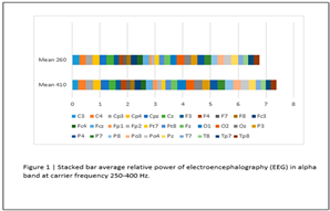
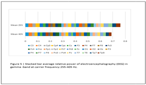
According to the tables obtained from the paired t-test, there are significant changes only in the gamma frequency band at the carrier frequency of 400 and 250 in the areas of Cp3/C3/F4/Fc3/Fz/O1/P8/Po3/Po4/Pz/Tp8.
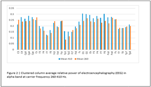
But based on the graphs and paired t-test obtained from the changes of the alpha frequency bands in the carrier frequencies of 250 and 400 Hz, we realized that there were also changes in the alpha frequency band, but it was not at a significant level.
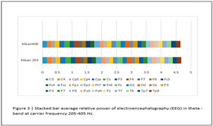
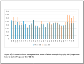
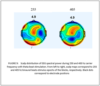
And in the theta frequency band at the carrier frequency of 250 and 405 Hz, no noticeable changes have been observed, but in the same frequency range, i.e. 255 and 405, based on the principle of theta-gamma coupling, there have been positive changes in the frequency band at the carrier frequency of 255 Hz.
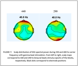
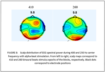
Based on the changes in the topographic map of the brain at different carrier frequencies in the same frequency bands shown, it can be inferred that the greatest effect that is caused by changing the carrier frequency on different brain areas is 1- the extent of the effect 2- the intensity of the effect.
Summary
The obtained results show that the changes caused by the gamma frequency band in the carrier frequency of 250 and 400 Hz are significant in a period of 3 minutes, and the decreasing trend of the changes caused by gamma to theta in our research shows that as the frequency band decreases The changes should be reduced within a certain period of 3 minutes.
In other words, we need more time than 3 minutes to create positive and significant changes in lower frequency bands.
The issue of gamma changes in the theta frequency band between 255 and 405 Hz shows that firstly, binaural beats stimulation can increase gamma waves based on the principle of theta-gamma coupling with theta waves.
Secondly, the issue of increasing the gamma frequency band in the carrier frequency of 255 compared to 405 shows that in order to be most effective in stimulating the binaural rhythm, specific frequency bands must be set in the specific carrier frequencies.
Another result obtained from the changes in the topography of the brain in different carrier frequencies shows that the greatest effect of the carrier frequency on the brain map is on 2 states: 1- the extent of the effect of the involved areas 2- the intensity of the effect of the involved areas.
Discussion
The results obtained from our research in a neurophysiological way showed the issue of energy transfer in different carrier frequencies raised by Licklider and showed that with the increase of the carrier frequency, there are positive changes in the relative power of the frequency bands.
Also, Licklider explained the issue of the frequency limitation of the carrier frequencies in the binaural beats in the theory of synchronizing the number of pulses with auditory neurons, from this theory it can be deduced that the number of pulses created by the carrier frequency is important. This is also shown in our research with the decreasing trend of the changes in frequency bands from gamma to theta.
In other words, it has been shown in our research that in a certain period of 3 minutes, as the frequency band decreases, the changes that occur also decrease, that is, the number of pulses that are sent in time is also important.
That's why we need more time than 3 minutes to create positive and significant changes in lower frequency bands.
Another important issue that has been identified in this research is the gamma changes in the theta frequency band between 255 and 405 Hz, which shows that, firstly, binaural rhythm stimulation, considering that it has the ability to stimulate the lower regions of the brain, can be based on the principle of theta gamma coupling. with theta waves, increase gamma waves as well.
Secondly, the matter of increasing the gamma frequency band at the carrier frequency of 255 compared to 405, contrary to the energy transfer equation between the frequency band and the carrier frequency that Licklider proposed, shows that the use of high frequency carrier frequencies in any frequency band is not important. Rather, in order to be most effective in stimulating the binaural beat, specific frequency bands should be set in the specific carrier frequencies.
Also, this finding shows that in addition to the limitation that Licklid raised in his article regarding frequencies above 500 Hz, that the number of pulses should increase to such an extent that the cortex cannot do the necessary processing, there are limitations between the carrier frequency and frequency bands. And regarding the theta frequency band, it shows that the carrier frequency of 400 Hz cannot have the same effect as the 250 Hz frequency in the theta-gamma coupling. And in the gamma and alpha frequency bands, the carrier frequency of 250 Hz cannot have the effect of the carrier frequency of 400 Hz. be
Therefore, for the best effect in binaural rhythm stimulation, there are limitations in the carrier frequencies and the carrier frequency should be adjusted with the frequency band.
The next important thing that happened in our study was the large changes in brain waves in response to different frequencies among the participants, which clarified important things about the effect of the carrier frequency in the binaural beat, which makes it only relevant to the subject. The transfer of energy in high frequencies should not be stopped and the basic brain state of people should also be measured.
In this case, we need to deal with it in detail in a new article.
References
- Baker, D. H. , Vilidaite, G., McClarnon, E., Valkova, E., Bruno, A., & Millman, R. E. (2020). Binaural summation of amplitude modulation involves weak interaural suppression. Scientific Reports 10(1), 3560.
- Becher, A. K. , Höhne, M., Axmacher, N., Chaieb, L., Elger, C. E., & Fell, J. (2015). Intracranial electroencephalography power and phase synchronization changes during monaural and binaural beat stimulation. European Journal of Neuroscience 41(2), 254–263. [PubMed]
- Carlson, A. B. , & Crilly, P. B. (2010). Communication Systems, McGraw-Hill: Higher Education. https://books.google.com/books?id=8qOUCgAAQBAJ.
- Chaieb, L. , Wilpert, E. C., Reber, T. P., & Fell, J. (2015). Auditory beat stimulation and its effects on cognition and mood states. Frontiers in psychiatry 6, 70.
- da Silva Junior, M. , de Freitas, R. C., dos Santos, W. P., da Silva, W. W. A., Rodrigues, M. C. A., & Conde, E. F. Q. (2019). Exploratory study of the effect of binaural beat stimulation on the EEG activity pattern in resting state using artificial neural networks. Cognitive systems research 54, 1-20.
- Frenzel Jr, L. (2018). Radio/wireless. Electronics Explained 159-194.
- Gao, X. , Cao, H., Ming, D., Qi, H., Wang, X., Wang, X., Chen, R., & Zhou, P. (2014). Analysis of EEG activity in response to binaural beats with different frequencies. International Journal of Psychophysiology, 94(3), 399–406. [PubMed]
- Ingendoh, R. M. , Posny, E. S., & Heine, A. (2023). Binaural beats to entrain the brain? A systematic review of the effects of binaural beat stimulation on brain oscillatory activity, and the implications for psychological research and intervention. Plos one, 18(5), e0286023. [Google Scholar]
- Ioannou, C. I. , Pereda, E., Lindsen, J. P., & Bhattacharya, J. (2015). Electrical brain responses to an auditory illusion and the impact of musical expertise. Plos one 10(6), e0129486.
- Jirakittayakorn, N. , & Wongsawat, Y. (2017). Brain responses to a 6-Hz binaural beat: effects on general theta rhythm and frontal midline theta activity. Frontiers in neuroscience 11, 365.
- Joris, P. X. , Van De Sande, B., & van der Heijden, M. (2005). Temporal damping in response to broadband noise. I. Inferior colliculus. Journal of neurophysiology 93(4), 1857–1870. [PubMed]
- Kim, H.-W. , Happe, J., & Lee, Y. S. (2023). Beta and gamma binaural beats enhance auditory sentence comprehension. Psychological research 87(7), 2218–2227.
- Licklider, J. C. R. , Webster, J., & Hedlun, J. (1950). On the frequency limits of binaural beats. The journal of the acoustical society of america 22(4), 468–473.
- Oster, G. (1973). Auditory beats in the brain. Scientific American 229(4), 94-103.
- Perez, H. D. O. , Dumas, G., & Lehmann, A. (2020). Binaural beats through the auditory pathway: from brainstem to connectivity patterns. Eneuro 7.
- Schwarz, D. W. , & Taylor, P. (2005). Human auditory steady state responses to binaural and monaural beats. Clinical Neurophysiology 116(3), 658–668. [PubMed]
- Shamsi, E. , Ahmadi-Pajouh, M. A., & Ala, T. S. (2021). Higuchi fractal dimension: An efficient approach to detection of brain entrainment to theta binaural beats. Biomedical signal processing and control 68, 102580.
- Solca, M. , Mottaz, A., & Guggisberg, A. G. (2016). Binaural beats increase interhemispheric alpha-band coherence between auditory cortices. Hearing research 332, 233-237.
- Ungan, P. , Yagcioglu, S., & Ayik, E. (2019). Event-related potentials to single-cycle binaural beats and diotic amplitude modulation of a tone. Experimental brain research, 237, 1931-1945. [Google Scholar]
- Vernon, D. , Peryer, G., Louch, J., & Shaw, M. (2014). Tracking EEG changes in response to alpha and beta binaural beats. International Journal of Psychophysiology 93(1), 134–139. [PubMed]
- Wernick, J. S. , & Starr, A. (1968). Binaural interaction in the superior olivary complex of the cat: an analysis of field potentials evoked by binaural-beat stimuli. Journal of neurophysiology 31(3), 428-441.
|
Disclaimer/Publisher’s Note: The statements, opinions and data contained in all publications are solely those of the individual author(s) and contributor(s) and not of MDPI and/or the editor(s). MDPI and/or the editor(s) disclaim responsibility for any injury to people or property resulting from any ideas, methods, instructions or products referred to in the content. |
© 2024 by the authors. Licensee MDPI, Basel, Switzerland. This article is an open access article distributed under the terms and conditions of the Creative Commons Attribution (CC BY) license (http://creativecommons.org/licenses/by/4.0/).


