Submitted:
15 October 2024
Posted:
15 October 2024
You are already at the latest version
Abstract
Keywords:
1. Introduction
2. Materials and Methods
2.1. Starting Materials
2.2. Synthesis of Cat-Cu Complex
2.3. Characterization of the Complex
- Instrumentation
- b. In vitro biological activity assay
3. Results and Discussions
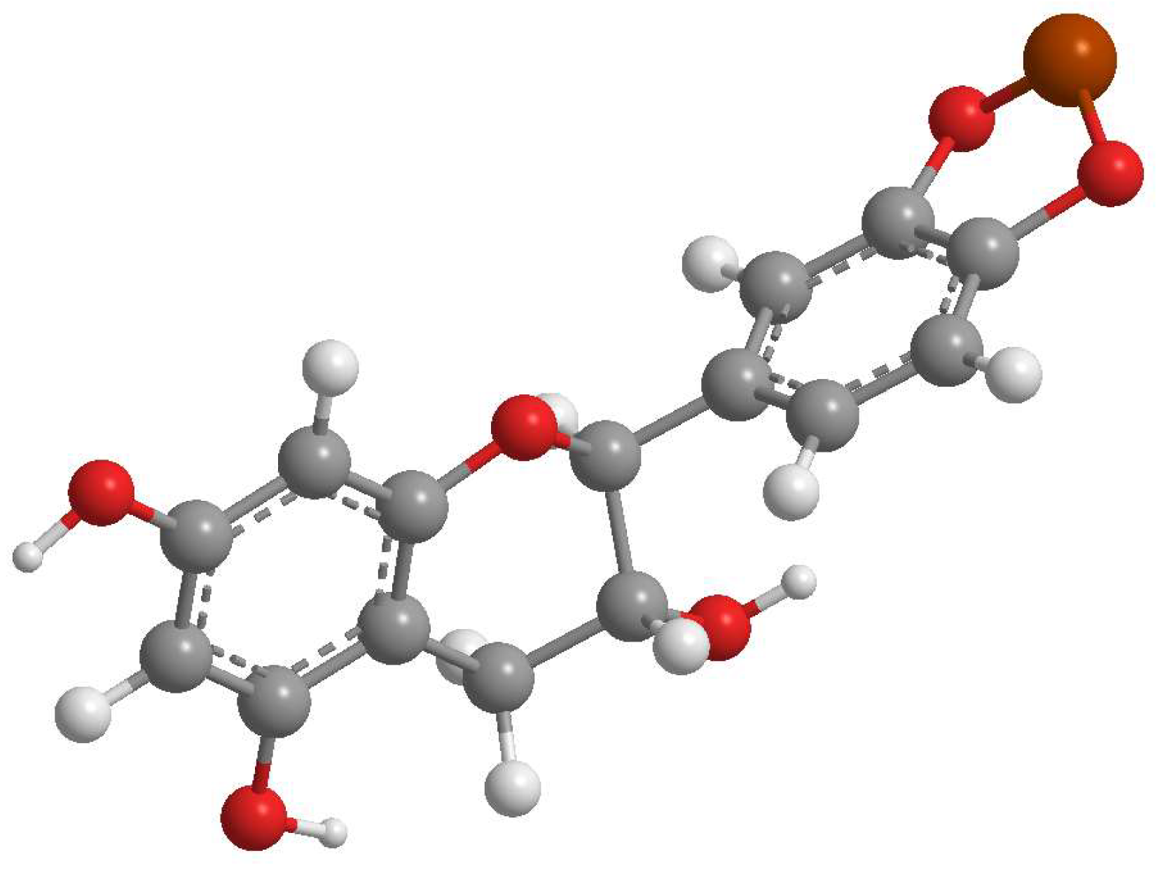
3.1. Synthesis and Complexation Yield

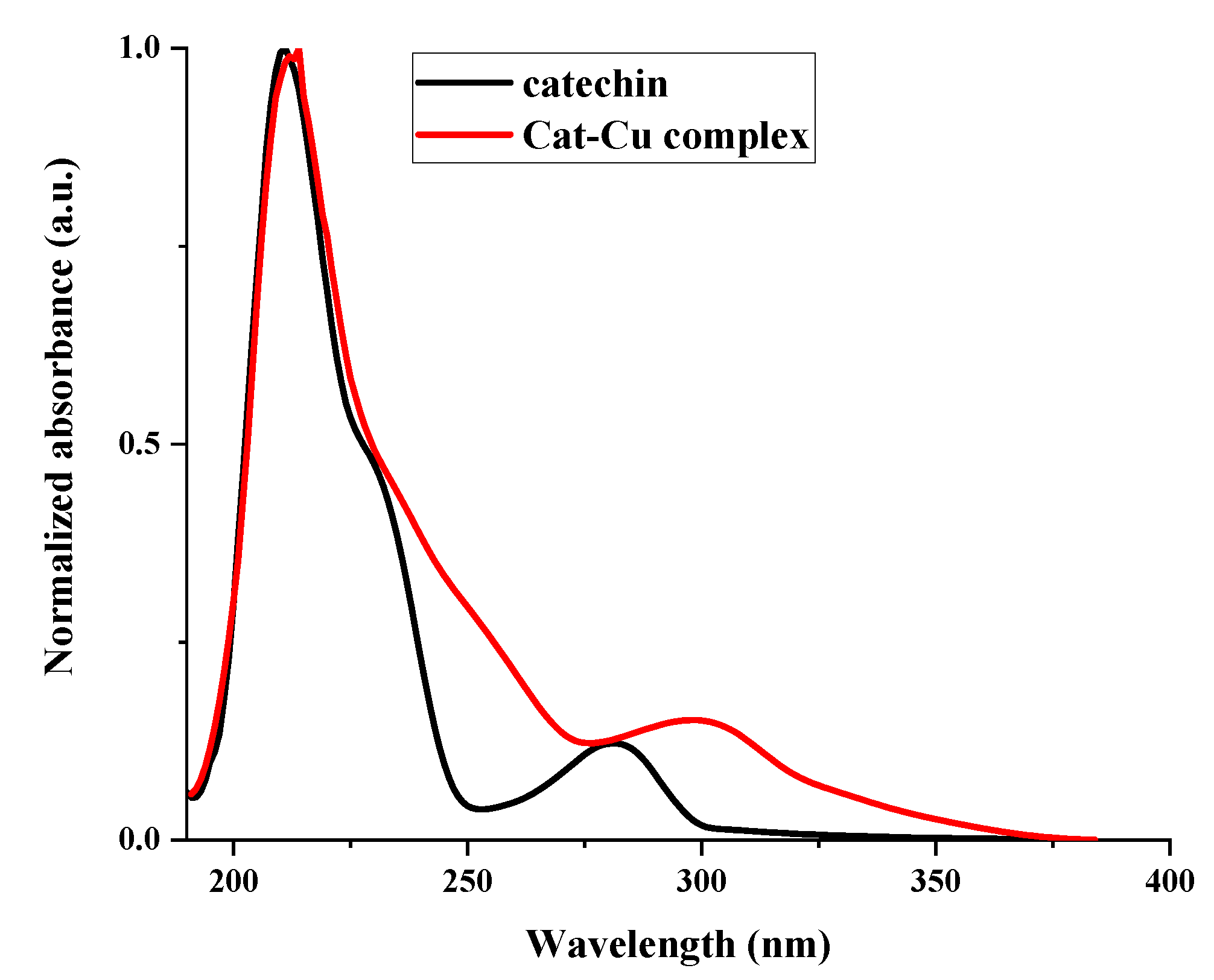
3.2. UV-VIS
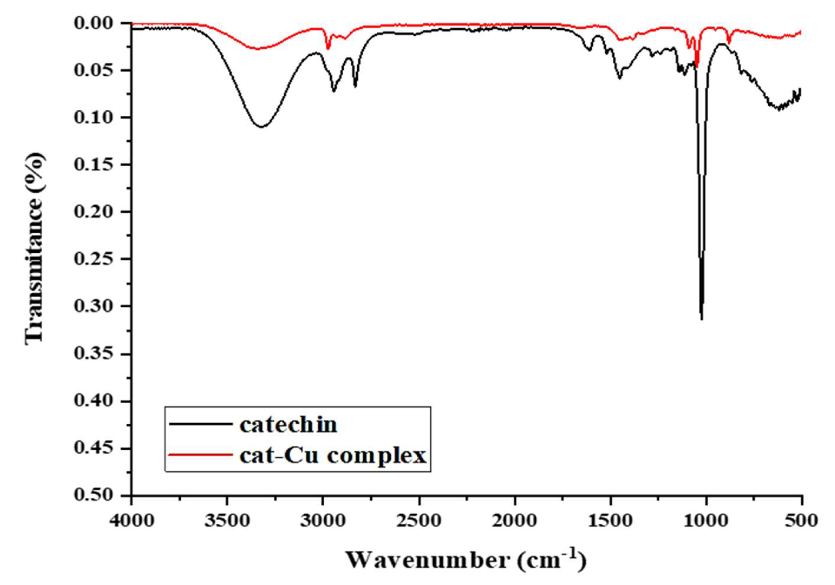
3.3. FT-IR
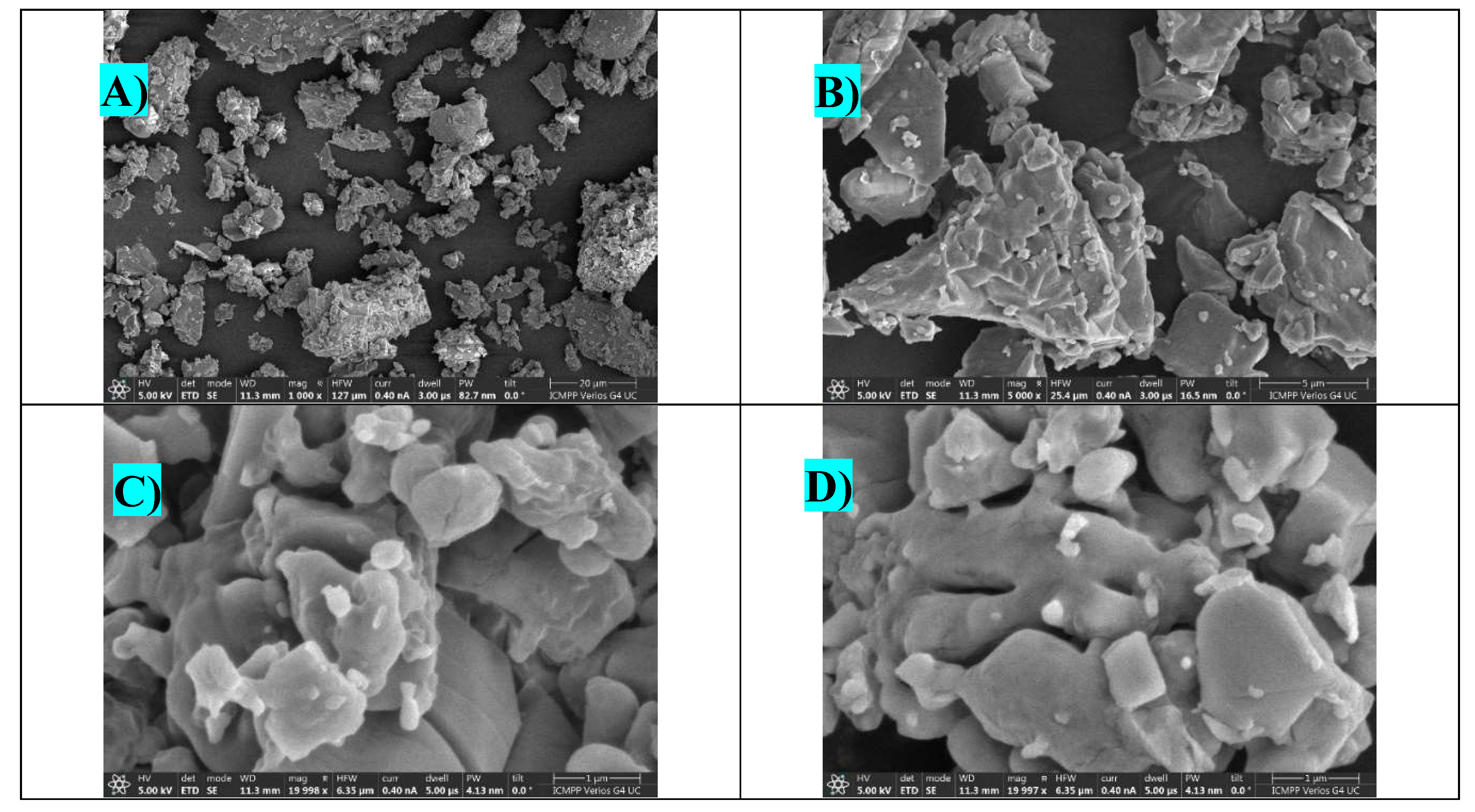
3.4. Morphological Analysis
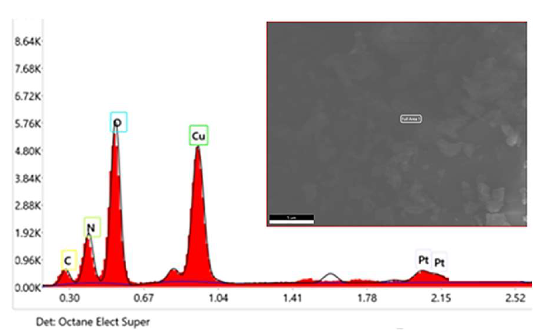
3.5. EDX
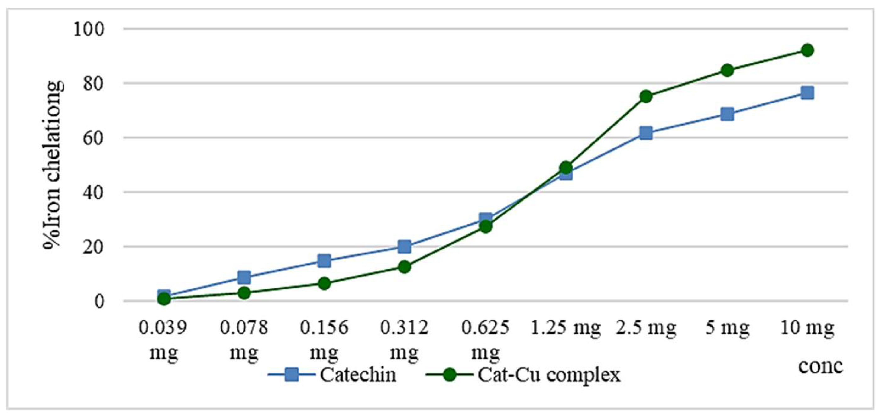
3.6. In Vitro Antioxidant Activity
3.7. Lipoxygenase Activity
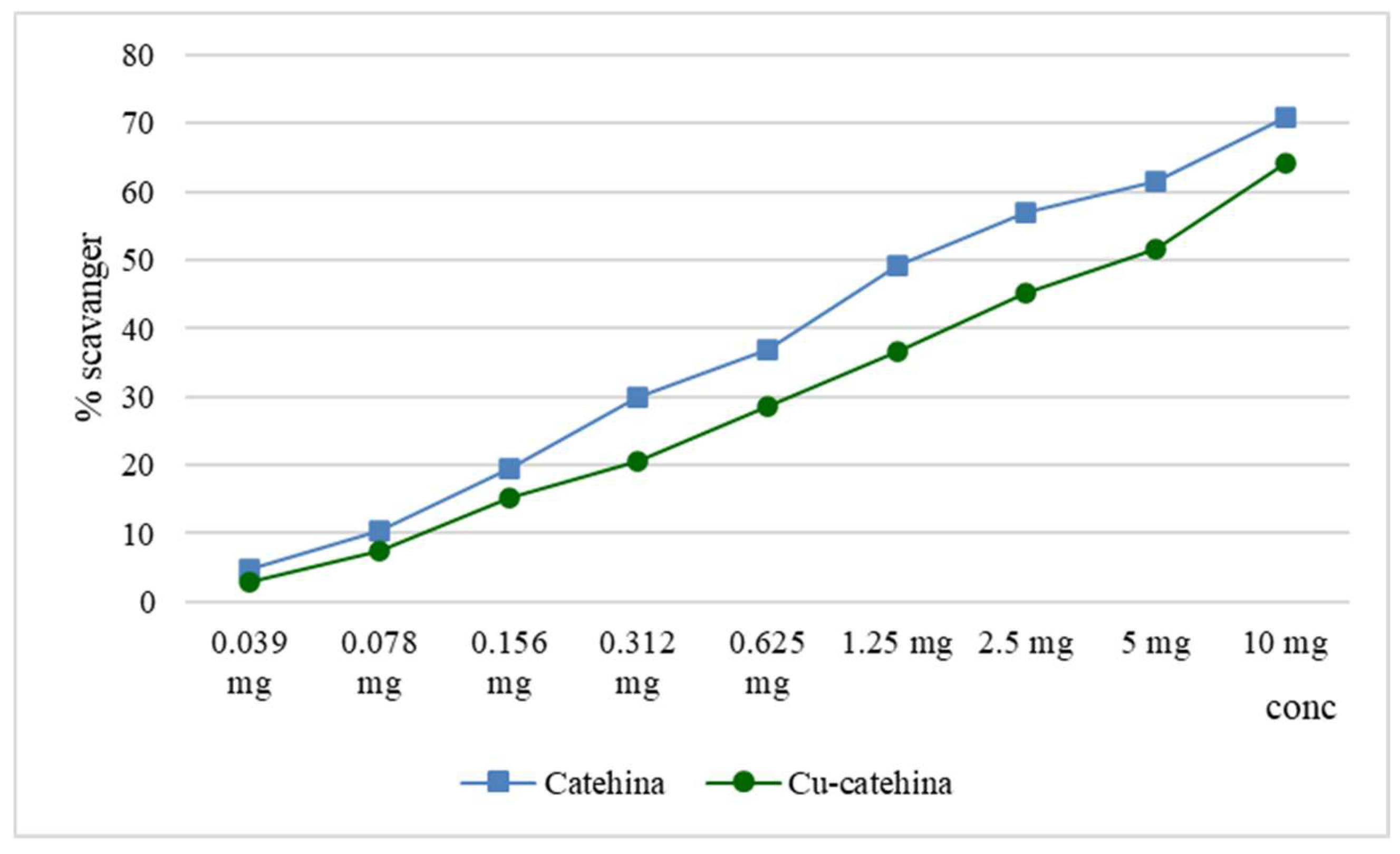
3.8. Determination of the Scavenger Capacity of the Hydroxyl Radical
3.9. In Vitro Antidiabetic Activity
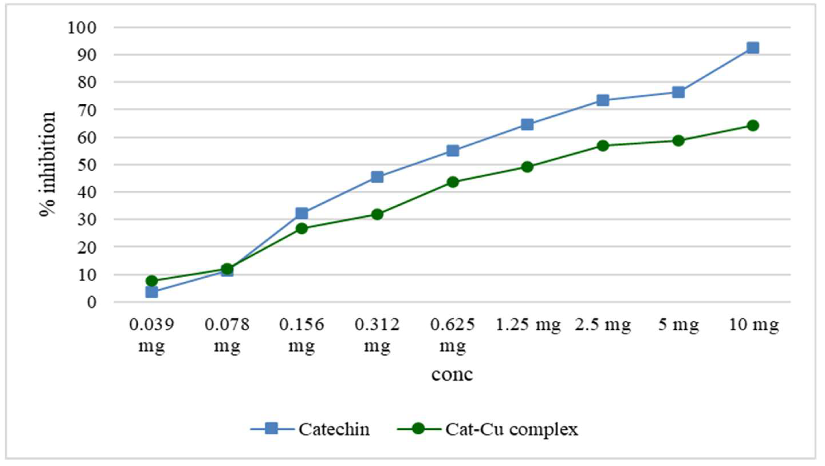
3.9.1. Inhibition of Alpha-Amylase
3.9.2. Inhibition of Alpha-Glucosidase
3.10. Antibacterial Activity
4. Conclusions
Author Contributions
Funding
Institutional Review Board Statement
Acknowledgments
Conflicts of Interest
References
- Humulescu, I.; Flutur, M.-M.; Cioanca, O.; Mircea, C.; Robu, S.; Marin-Batir, D.; Spac, A.; Corciova, A.; Hancianu, M. Comparative Chemical and Biological Activity of Selective Herbal Extracts. Farmacia 2021, 69, 861–866. [Google Scholar] [CrossRef]
- Tan, Z.; Deng, J.; Ye, Q.; Zhang, Z. The Antibacterial Activity of Natural-Derived Flavonoids. Curr Top Med Chem 2022, 22, 1009–1019. [Google Scholar] [CrossRef] [PubMed]
- Finaud, J.; Lac, G.; Filaire, E. Oxidative Stress : Relationship with Exercise and Training. Sports Med 2006, 36, 327–358. [Google Scholar] [CrossRef] [PubMed]
- Ferrali, M.; Signorini, C.; Caciotti, B.; Sugherini, L.; Ciccoli, L.; Giachetti, D.; Comporti, M. Protection against Oxidative Damage of Erythrocyte Membrane by the Flavonoid Quercetin and Its Relation to Iron Chelating Activity. FEBS Lett 1997, 416, 123–129. [Google Scholar] [CrossRef]
- Lungu, I.; Huzum, B.; Humulescu, I.A.; Cioancă, O.; Morariu, D.; Șerban, I.-L.; Hăncianu, M. Flavonoids as promising therapeutic and dietary agents. Med.-Surg. J. 2020, 124, 151–156. [Google Scholar]
- Panche, A.N.; Diwan, A.D.; Chandra, S.R. Flavonoids: An Overview. J Nutr Sci 2016, 5, e47. [Google Scholar] [CrossRef]
- Chen, C.-M.; Wu, C.-T.; Yang, T.-H.; Chang, Y.-A.; Sheu, M.-L.; Liu, S.H. Green Tea Catechin Prevents Hypoxia/Reperfusion-Evoked Oxidative Stress-Regulated Autophagy-Activated Apoptosis and Cell Death in Microglial Cells. J Agric Food Chem 2016, 64, 4078–4085. [Google Scholar] [CrossRef]
- Kostyuk, V.A.; Potapovich, A.I.; Vladykovskaya, E.N.; Korkina, L.G.; Afanas’ev, I.B. Influence of Metal Ions on Flavonoid Protection against Asbestos-Induced Cell Injury. Arch Biochem Biophys 2001, 385, 129–137. [Google Scholar] [CrossRef]
- Iancu C, Mircea C, Petrariu FL, Cioancă O, Stan C, Corciovă A, Murărașu A, Filip N, Hăncianu M, The evaluation of normo-glycemic and cyto-regenerative effects of Pelargonium species extracts. Farmacia 2020, 68, 135–141. [CrossRef]
- Tan, Z.; Deng, J.; Ye, Q.; Zhang, Z. The Antibacterial Activity of Natural-Derived Flavonoids. Curr Top Med Chem 2022, 22, 1009–1019. [Google Scholar] [CrossRef]
- Lungu, I.; Panainte, A.; Mircea, C.; Tuchiluș, C.; Ștefanache, A.; Szasz, F.; Grigorie, D.; Robu, S; Cioancă, O. Catechin-Zinc-Complex: Synthesis, Characterization and Biological Activity Assessment. Farmacia 2023, 71. [Google Scholar] [CrossRef]
- Kumamoto, M.; Sonda, T.; Nagayama, K.; Tabata, M. Effects of pH and Metal Ions on Antioxidative Activities of Catechins. Biosci Biotechnol Biochem 2001, 65, 126–132. [Google Scholar] [CrossRef] [PubMed]
- Bors, W.; Heller, W.; Michel, C.; Saran, M. Flavonoids as Antioxidants: Determination of Radical-Scavenging Efficiencies. Methods Enzymol 1990, 186, 343–355. [Google Scholar] [CrossRef] [PubMed]
- Ameena, S.; Rajesh, N.; Anjum, S.M.; Khadri, H.; Riazunnisa, K.; Mohammed, A.; Kari, Z.A. Antioxidant, Antibacterial, and Anti-Diabetic Activity of Green Synthesized Copper Nanoparticles of Cocculus Hirsutus (Menispermaceae). Appl Biochem Biotechnol 2022, 194, 4424–4438. [Google Scholar] [CrossRef]
- Souza, R.F.V.; De Giovani, W.F. Antioxidant Properties of Complexes of Flavonoids with Metal Ions. Redox Report 2004, 9, 97–104. [Google Scholar] [CrossRef] [PubMed]
- Sarker, U.; Oba, S. Nutraceuticals, Antioxidant Pigments, and Phytochemicals in the Leaves of Amaranthus spinosus and Amaranthus viridis Weedy Species. Sci Rep. 2019, 9, 20413. [Google Scholar] [CrossRef] [PubMed]
- Bukhari, S.B.; Memon, S.; Mahroof-Tahir, M.; Bhanger, M.I. Synthesis, Characterization and Antioxidant Activity Copper–Quercetin Complex. Spectrochim Acta A Mol Biomol Spectrosc 2009, 71, 1901–1906. [Google Scholar] [CrossRef]
- Sharma, N.; Phan, H.T.; Chikae, M.; Takamura, Y.; Azo-Oussou, A.F.; Vestergaard, M.C. Black Tea Polyphenol Theaflavin as Promising Antioxidant and Potential Copper Chelator. J Sci Food Agric 2020, 100, 3126–3135. [Google Scholar] [CrossRef]
- Szczepanik, J.; Podgórski, T.; Domaszewska, K. The Level of Zinc, Copper and Antioxidant Status in the Blood Serum of Women with Hashimoto’s Thyroiditis. Int J Environ Res Public Health 2021, 18, 7805. [Google Scholar] [CrossRef]
- Ali, H.; Houghton, P.J.; Soumyanath, A. Alpha-Amylase Inhibitory Activity of Some Malaysian Plants Used to Treat Diabetes; with Particular Reference to Phyllanthus Amarus. J Ethnopharmacol 2006, 107, 449–455. [Google Scholar] [CrossRef]
- Apostolidis, E.; Lee, C.M. In Vitro Potential of Ascophyllum Nodosum Phenolic Antioxidant-Mediated Alpha-Glucosidase and Alpha-Amylase Inhibition. J Food Sci 2010, 75, H97–102. [Google Scholar] [CrossRef]
- Choudhary, D.K.; Chaturvedi, N.; Singh, A.; Mishra, A. Characterization, Inhibitory Activity and Mechanism of Polyphenols from Faba Bean (Gallic-Acid and Catechin) on α-Glucosidase: Insights from Molecular Docking and Simulation Study. Prep Biochem Biotechnol 2020, 50, 123–132. [Google Scholar] [CrossRef] [PubMed]
- Rajeshkumar, S.; Menon, S.; Venkat Kumar, S.; Tambuwala, M.M.; Bakshi, H.A.; Mehta, M.; Satija, S.; Gupta, G.; Chellappan, D.K.; Thangavelu, L.; et al. Antibacterial and Antioxidant Potential of Biosynthesized Copper Nanoparticles Mediated through Cissus Arnotiana Plant Extract. J Photochem Photobiol B 2019, 197, 111531. [Google Scholar] [CrossRef] [PubMed]
- El-Fattah, A.A.; Azzam, M.; Elkashef, H.; Elhadydy, A. Antioxidant Properties of Milk: Effect of Milk Species, Milk Fractions and Heat Treatments. Int. J. Dairy Sci. 2019, 15, 1–9. [Google Scholar] [CrossRef]
- Hasanin, M.; Al Abboud, M.A.; Alawlaqi, M.M.; Abdelghany, T.M.; Hashem, A.H. Ecofriendly Synthesis of Biosynthesized Copper Nanoparticles with Starch-Based Nanocomposite: Antimicrobial, Antioxidant, and Anticancer Activities. Biol Trace Elem Res 2022, 200, 2099–2112. [Google Scholar] [CrossRef]
- Shahrajabian, M.H.; Sun, W.; Cheng, Q. Importance of Epigallocatechin and Its Health Benefits. Free Radicals and Antioxidants 2021, 10, 47–51. [Google Scholar] [CrossRef]
- Wang, J.; Wang, X.; He, Y.; Jia, L.; Yang, C.S.; Reiter, R.J.; Zhang, J. Antioxidant and Pro-Oxidant Activities of Melatonin in the Presence of Copper and Polyphenols In Vitro and In Vivo. Cells 2019, 8, 903. [Google Scholar] [CrossRef]
- Ferrali, M.; Signorini, C.; Caciotti, B.; Sugherini, L.; Ciccoli, L.; Giachetti, D.; Comporti, M. Protection against Oxidative Damage of Erythrocyte Membrane by the Flavonoid Quercetin and Its Relation to Iron Chelating Activity. FEBS Lett 1997, 416, 123–129. [Google Scholar] [CrossRef]
- Serpen, A.; Gökmen, V.; Fogliano, V. Total antioxidant capacities of raw and cooked meats. Meat Sci. 2012, 90, 60–65. [Google Scholar] [CrossRef]
- Jhong, C.-H.; Riyaphan, J.; Lin, S.-H.; Chia, Y.-C.; Weng, C.-F. Screening Alpha-Glucosidase and Alpha-Amylase Inhibitors from Natural Compounds by Molecular Docking in Silico. Biofactors 2015, 41, 242–251. [Google Scholar] [CrossRef]
- Kim, K.T.; Rioux, L.E.; Turgeon, S.L. Alpha-Amylase and Alpha-Glucosidase Inhibition Is Differentially Modulated by Fucoidan Obtained from Fucus Vesiculosus and Ascophyllum Nodosum. Phytochemistry 2014, 98, 27–33. [Google Scholar] [CrossRef]
- Etxeberria, U.; De La Garza, A.L.; Campin, J.; Martnez, J.A.; Milagro, F.I. Antidiabetic Effects of Natural Plant Extracts via Inhibition of Carbohydrate Hydrolysis Enzymes with Emphasis on Pancreatic Alpha Amylase. Expert Opin Ther Targets 2012, 16, 269–297. [Google Scholar] [CrossRef] [PubMed]
- Jiang, C.; Chen, Y.; Ye, X.; Wang, L.; Shao, J.; Jing, H.; Jiang, C.; Wang, H.; Ma, C. Three Flavanols Delay Starch Digestion by Inhibiting α-Amylase and Binding with Starch. Int J Biol Macromol 2021, 172, 503–514. [Google Scholar] [CrossRef] [PubMed]
- Ademiluyi, A.O.; Oboh, G. Phenolic-Rich Extracts from Selected Tropical Underutilized Legumes Inhibit α-Amylase, α-Glucosidase, and Angiotensin I Converting Enzyme in Vitro. J Basic Clin Physiol Pharmacol 2012, 23, 17–25. [Google Scholar] [CrossRef] [PubMed]
- Michalczyk, K.; Cymbaluk-Płoska, A. The Role of Zinc and Copper in Gynecological Malignancies. Nutrients 2020, 12, 3732. [Google Scholar] [CrossRef]
- Lisa EL, Carac G, Barbu V, Robu SI. The synergistic antioxidant effect and antimicrobial efficacity of propolis, myrrh and chlorhexidine as beneficial toothpaste components. Rev. Chim.-Buchar 2017, 68, 2060–2065. [CrossRef]
- Marin DB, Cioanca OA, Apostu MI, Tuchilus CG, Mircea C, Robu SI, Tutunaru D, Corciova AN, Hancianu M. The Comparative Study of Equisetum pratense, E. Sylvaticum, E. Telmateia: Accumulation of silicon, antioxidant and antimicrobial screening. Revista de Chimie 2019, 70, 2519–2523. [CrossRef]
- Maftei NM, Bogdan RE, Boev M, Marin DB, Ramos-Villarroel AY, Iancu AV. Innovative Fermented Soy Drink with the Sea Buckthorn Syrup and the Probiotics Co-Culture of Lactobacillus Paracasei ssp. Paracasei (L. Casei® 431) and Bifidobacterium Animalis ssp. Lactis (Bb-12®).
- Yu, J.; Huang, X.; Ren, F.; Cao, H.; Yuan, M.; Ye, T.; Xu, F. Application of antimicrobial properties of copper. Appl Organomet Chem 2024, 38, e7506. [Google Scholar] [CrossRef]
- Sadani, K. , Nag, P., Pisharody, L., Thian, X. Y., Bajaj, G., Natu, G.,... & Mukherji, S. Polyphenol stabilize copper nanoparticle formulations for rapid disinfection of bacteria and virus on diverse surfaces. Nanotechnology 2021, 33(3), 035701. [Google Scholar]
- Wang, Z. , & An, P. Characterization of copper complex nanoparticles synthesized by plant polyphenols. BioRxiv 2017, 134940. [Google Scholar]
- Salah, I.; Parkin, I. P.; Allan, E. Copper as an antimicrobial agent: Recent advances. RSC advances 2021, 11, 18179–18186. [Google Scholar] [CrossRef]
- Rajalakshmi, S. , Fathima, A., Rao, J. R., & Nair, B. U. Antibacterial activity of copper (II) complexes against Staphylococcus aureus. RSC advances 2014, 4, 32004–32012. [Google Scholar]
- Scattareggia Marchese, A. , Destro, E., Boselli, C., Barbero, F., Malandrino, M., Cardeti, G.,... & Lanni, L. Inhibitory Effect against Listeria monocytogenes of Carbon Nanoparticles Loaded with Copper as Precursors of Food Active Packaging. Foods 2022, 11. [Google Scholar]
| Tested substance | S. aureus ATCC 25923 | E. coli ATCC 25922 | P. aeruginosa ATCC 27853 | C. albicansATCC 90028 |
|---|---|---|---|---|
| Catechin | 20.0±0.00 | 0 | 16.0±0.00 | 0 |
| Copper sulphate | 14.0±0.00 | 0 | 0 | 0 |
| cat-Cu complex | 17.3±0.57 | 0 | 0 | 17.0±0.00 |
| blank (DMSO) | 0 | 0 | 0 | 0 |
| I. Ciprofloxacin (5 µg/disc) | 30.0±0.00 | 34.0±0.00 | 31.3±0.57 | Not tested |
| I.Fluconazol (25 µg/disc) | Not tested | Not tested | Not tested NT* | 30.0±0.00 |

Disclaimer/Publisher’s Note: The statements, opinions and data contained in all publications are solely those of the individual author(s) and contributor(s) and not of MDPI and/or the editor(s). MDPI and/or the editor(s) disclaim responsibility for any injury to people or property resulting from any ideas, methods, instructions or products referred to in the content. |
© 2024 by the authors. Licensee MDPI, Basel, Switzerland. This article is an open access article distributed under the terms and conditions of the Creative Commons Attribution (CC BY) license (http://creativecommons.org/licenses/by/4.0/).


