Preprint
Article

Phytochemical Characterization, Bioactive Properties, and Acute Toxicity of Wild Vitex agnus-castus L. methanolic extracts

Submitted:

16 October 2024

Posted:

17 October 2024

You are already at the latest version

Abstract
Vitex agnus-castus L.(VAC) belonging to the Verbenaceae family is a Mediterranean plant which is widely used in traditional Tunisian medicine. The aim of this study was to investigate the phytochemical composition, antioxidant, antiviral and cytotoxic potential of methanolic VAC fruits extracts. Quantitative analyses of the different constituents of extracts were evaluated by high-performance liquid chromatography with photodiode-array detection (HPLC-DAD). Spectrophotometric methods and chemical tests were used for phytochemical characterization and antioxidant activity. The cytotoxic study was evaluated using epidermoid carcinoma Hep-2 cells, while the antiviral assessment against clinical isolates of coxsackievirus B3 (CBV-3) was determined by using a HEp cell survival assay. VAC extracts presented several components and minerals The results of HPLC-DAD indicated the presence 7 of phenolic compounds with a remarkable abondance of chlorogenic and coumaric acids as polyphenols compounds. The data also showed that VAC extracts had an important antioxidant effect (IC50=0.061 mg/mL) with a reduced cytotoxicity (CC50= 213 µg/ml), and these effects are displayed in a dose-dependent manner. However, all the tested extracts are inactive against CBV-3. The results achieved confirm the important role of this plant as a source of therapeutic activities.
Keywords: 
Subject: 
Biology and Life Sciences  -   Biochemistry and Molecular Biology

1. Introduction

Medicinal plants have long been used in several traditional healing systems to treat a variety of diseases since ancient times because of their healing qualities [1]. Thousands of wild plant species worldwide exhibit medicinal properties and are still in use as part of alternative medicine. These days, a lot of individuals are interested in "green therapy," or using natural plant-based substances that have been shown to be less harmful and have no adverse effects than pharmaceutical medications [2]. They are also significant sources of numerous bioactive chemicals, which have a variety of uses in therapy, including analgesic and anti-inflammatory effects. Research has demonstrated that in addition to having antimicrobial activity against a variety of bacteria, plant extracts (and/or natural products derived from them) can also modify bacterial resistance mechanisms, boost the effectiveness of antibiotics taken concurrently, and, in certain situations, even reverse resistance mechanisms that have already been established. Since many synthetic pharmaceuticals have proven to have harmful side effects, there has been a noticeable tendency in recent years to investigate natural materials as intriguing alternatives. The main driver of interest in herbal remedies is the widespread belief that using these complementary therapies is safer, more affordable, and has no negative consequences [3]. Phytochemical and pharmacognostic analyses are, in fact, necessary to verify the efficacy and safety of using medicinal plants. As a result, many herbs are actually medicinal plants that are easily obtained and always in season, but few people are aware of their incredible qualities. Thus, several scientists have conducted numerous studies using different plant extracts to determine the, antibacterial, anti-inflammatory, antioxidant, and many other medicinal properties of these extracts. Among these beneficial plants, Vitex agnus-castus L. (VAC) was used by practitioners of phytotherapy to treat several ailments and is still the chief alternative for most people worldwide [4,5].
The majority of the plants in the largest genus of the Verbenaceae family, Vitex L., are employed in many traditional medical systems across the globe. The genus Vitex L., sometimes referred to as the chaste tree genus, is the largest in the Verbenaceae family and has over 230 species spread across the globe [2]. The majority of Vitex species are tiny trees or deciduous shrubs [6]. These species are widely dispersed throughout Southeast Asia and are primarily found in warm, temperate regions of Asia and Europe [7,8]. Many research studies have already examined Vitex species, namely Vitex agnus castus (VAC), the plant of interest in this study, and they are widely known as sources of beneficial medications in many geographic areas. VAC also known as the chasteberry and monk’s pepper is a small tree, the lilac-purple flowers of the plant bloom from June to early fall and the fruits ripen in the fall [7,8]
Traditionally, Vitex plants have long been used for several forms of treatment of menstruation disorders, fertility problems, menopausal symptoms, diarrhea, asthma, fever, cold, headache, migraine, gastrointestinal infections, and breast pain [9,10]. This genus possesses a wide range of biological features, including antibacterial activity, according to recent investigations [11]. Numerous investigations on the antibacterial qualities of Vitex L. have yielded data that demonstrate the antimicrobial activity of several parts of the plant, including the leaf, bark, root, stem, flower, fruit, and seed, against a variety of microbes. The Vitex species has yielded various previously identified chemicals, primarily alkaloids, flavonoids, and terpenoids, according to phytochemical study [11].
It was recently shown in a study that the methanolic extracts of VAC had vasorelaxant effects on the aortic rings of rabbits, indicating that Vitex agnus cactus contains bioactive chemicals that can regulate blood vessel function. Extracts or specific isolated compounds of genus Vitex have been previously shown to exert numerous functions and used for treating certain menstrual disorders, infertility, hyperprolactinemia, acne, corpus luteum insufficiency, menopause, cyclical mastalgia, inflammatory conditions, and cyclic breast pain, disrupted lactation, diarrhea, and flatulence [12,13,14,15]. Fruits and leaves of VAC are often used in Albanian traditional medicine to address a variety of issues related to female reproduction. There have been prior reports on the tracheorelaxant characteristics and underlying processes of VACE's phytochemical makeup [16]. However, to our best knowledge, the toxicity, minerals composition and the antiviral activity of methanolic extracts of the fruits of Vitex agnus cactus (VFME) described in the present work have not been reported earlier. The effect of VFME was observed at a relatively narrow range of concentrations.
One of the species that is available for use in traditional medicine is VAC, also referred to as "Kaf Maryem" locally in Tunisia. To the best of our knowledge, no particular phytochemical research has been conducted on the mineral composition, cytotoxicity, or antiviral activity of Tunisian VAC fruits (methanolic extracts). Validating the application of VAC in Tunisian traditional medicine was, thus, the goal of this investigation. Consequently, a phytochemical investigation was carried out for the first time, and VFME was assessed for cytotoxicity, mineral content, and antiviral activity.

2. Materials and Methods

2.1. Plant Material

2.1.1. Collection and Identification of the Plant

The study complies with relevant institutional, national, and international standards and regulations while using plant material. The fruits of Vitex agnus cactus was harvested at the edge of Nefza, of the province of Beja which corresponds to the following coordinates: latitude 36.9752, longitude 9.08095 36° 58′ 31″ North, 9° 4′ 51″ Est, altitude 23 m. The plant was collected from July to september 2022 after the full blooming stage. The complete taxonomic identification of the species was conducted by Prof. Fethia Harzallah Skhiri from the High Institute of Biotechnology in Monastir, Tunisia.. Fruits were rinsed, dried in a dry place, aired in the absence of light, and finally milled using a grinder.

2.1.2. Preparation of the Crude Plant Extract

Seeds of VAC (Vitex agnus-castus, L) (20 g) was soaked in 300 ml of methanol for 16 hours at room temperature under constant shaking and then filtered by passing through a filter paper. After that, the filtered liquid (filtrate) was concentrated in a rotary evaporator under pressure, dried out, and transferred to containers. the extract was kept in a refrigerator (4°C). Appropriate dilutions of the crude plant extract from the stock were freshly made on the day of the experiment. The dried crude extracts were rediscovered in the corresponding solvent to prepare different concentrations (1000, 500, 250, 125 µg/ml).

2.1.3. Phytochemical Analyses and Polyphenol Content

Total Phenolic Content (TPC). The TPC of VFME extracts was performed using Folin–Ciocalteu's procedure and gallic acid as the standard [17]. 0.25 ml of the sample was combined with 1.25 ml of Folin–Ciocalteu’s reagent (diluted ten-fold), and 1 ml of Na 2CO3 (75 mg/ml). After incubation at 40°C for 30min, the absorbance of the mixture was measured at 765 nm. All determinations were prepared in triplicate, and quantification was done on the basis of the standard curve of gallic acid. The TPC was expressed as mg gallic acid equivalents (GAE) per g of extract.
Total flavonoids. The amount of flavonoids was determined according to the method of Barros and collaborators [18]. Distilled water (1,250 μl), and sodium iodide Na2NO2 (75 μl) [19]. The extracts (250 μl), 5%) were shaken. Then,150 μl of AlCl3 (10%) was added and allowed to stand for 6 min be-fore adding 500 μl of NaOH (1 M) and 250 μl of distilled water. The mixture was left to ambient temperature for 15 min; absorbance was then recorded at 510 nm. The total flavonoid content was ex-pressed in milligrams of catechin equivalent (CE) per gram of samples. Analysis of each sample was carried out in triplicate.
Flavonoid content. The content of flavonols was determined by AlCl3 method as described by Miliauskas and Van Beek [20]. Briefly,500 μl of the plant extract was mixed with 500 μl aluminum trichloride (2%) and 1500 μl of acetate of sodium (5%). The mixture was shaken and allowed at room temperature in obscurity for 2 hr30 min. The absorption at 440 nm was then noted. Standard rutin samples were carried out from 0.05 g rutin. All determinations were performed in triplicate. The amount of flavonols in plant extracts was determined in milligrams of rutin equivalents (RUE) per g of extracts.

2.1.4. Determination of Tannin Contents

The assay of condensed tannins is carried out using the vanillin method [21,22] This method is based on the ability of vanillin to react with condensed tannin units in an acid medium to produce a colored complex (carbonium ion colored red) measured at 500 nm (23). 400μl of each extract (1 mg/mL) are added to750 μl of a 4% methanol solution of vanillin, then 375 µL of concentrated hydrochloric acid is added. After 20 min of reaction in the dark, the absorbance is read at 500 nm. The concentration of condensed tannins is calculated from the regression equation of the calibration range, established with the tannic acid reference standard (0-300μg/ml) and expressed in micrograms of tannic acid equivalents per gram of extract (mg EAT/g).

2.1.5. Determination of Minerals

Determination of Potassium and Calcium: To obtain free atoms and ions, the powder materials of both tested plant species were weighed and placed in numbered capsules. The samples were then heated in a muffle furnace for two hours at T1, or 220°C, and for six hours at T2, or 550°C, to ensure the breaking of chemical bonds. We refer to this stage as calcination. Each capsule then received a dose of strong hydrochloric acid. Five milliliters of N/10 hydrochloric acid were added to these samples after they had been heated on a hot plate until all the acid had evaporated. Filtration was the next action. Following the extraction of the solutions, 50 mL volumetric flasks were filled with distilled water to the mark of the dipstick.
Ultimately, flame photometry was used to determine the elements potassium, sodium, and calcium; as a result, stock solutions and calibration solutions for each element had to be made.

2.1.6. Determination of Nitrogen

The nitrogen was determined using the methodology described by Martin Prével et al. [24]. With the help of a catalyst and concentrated sulfuric acid, the vegetable powder was mineralized. Ammonia was produced from organic nitrogen, and the soda gathered in boric acid replaced the ammonia. Chloridric acid was used for titration. A catalyst, 40% sodium hydroxide, 2% boric acid, 0.1 mol/L chloridric acid, 15% sodium hydroxide, and concentrated sulfuric acid were the reagents utilized. 200 mg of the vegetable powder (moisture content: 0–10%) was added to a flask without letting it settle on the neck. The flask was then sealed and allowed to react for 30 minutes with 5 mL of concentrated sulfuric acid and 200 mg of catalyst. After giving it a gentle heat, it was brought to a boil for an hour, or until it became yellow. The tubes were placed one at a time into the Vapodest for possible distillation once the mineralization process was finished, and the pH was assessed.
Free Radical Scavenging Activity. The antioxidant activity of VAC methanolic extracts was performed by means of the stable DPPH on the basis of a modified method, reported by Kartal et al. Upon its reduction by an antiradical compound, 1.1-diphenyl-2-picrylhydrazil (DPPH) loose its absorption band at 517nm. Briefly 180µl of different concentrations of VAC methanolic extracts (0.009–0.312µg/ ml) was added to 1620 µl of DPPH, prepared daily. The absorbance was calculated at 517nm and corresponded to the ability of extract to reduce the DPPH to the yellow-colored diphenylpicrylhydrazine. The ascorbic acid was used as a reference. The antioxidant activity was expressed as IC50. A higher antiradical activity of EO corresponds to a lower IC50 value. This activity was measured using the following equation: % inhibition=[(A0−A1)]/A0×100 where A0 is the absorbance value of the DPPH blank sample and A1 is the absorbance value of the test solution. A1 was evaluated as the difference between the absorbance value of the test solution and the absorbance value of its blank.
Antioxidant Activity To trap ABTS+ cations, Stämpflian and his associates' spectrophotometric analysis method was employed [25]. Equivalent quantities of a potassium persulfate K2S2O8 solution were combined with a 39.2 mg stock solution of ABTS, maintained at ambient temperature and protected from light, to create the ABTS radical cation. This process was carried out 16 hours before the solution was used. The solution was then diluted with ethanol until the absorbance at 734 nm was between 0.7 and 0.8. For every analysis series, 25 µL of each extract was mixed with 975 µL of this newly prepared solution, and after 20 minutes, the reading at 734 nm was taken. Ascorbic acid served as a positive control. The antioxidant activity was calculated based on the solution’s discoloration and expressed as the percentage inhibition (PI) of absorbance at 734 nm, the wavelength at which the ABTS+• radical exhibits a distinctive absorption band. Inhibition (%) = [(Abs control − Abs extract)/Abs control] × 100. All assays were performed in triplicate.
HPLC-DAD analysis: A 324 nm detection set is the foundation of the chromatographic technique used for analysis. The diode array detector (DAD)-equipped Agilent 1100 Series HPLC equipment is utilized. Agilent 1200 is a computer-controlled chromatographic apparatus. The various analytes in the methanolic extract are separated using a reversed phase Kinetex Evo C18 column that is heated to 30 °C in an oven. A combination of water, acetonitrile, and formic acid makes up the mobile phase. Acetonitrile and 1% formic acid are combined to generate mobile phase A. Formic acid (1%), and water (99%) combine to form mobile phase B. The gradient program is conducted in the following way: 10% A, 90% B (0 min), 20% A, 80% B, (20 min), 25% A, 75% B (30 min), 35% A, 65% B, (40 min) and 10% A, 90% B10 (50 min). The elution rate of the mobile phase is 1 mL/minute. Before being injected, samples are filtered using a 0.20 µm filter. There is a 20 µL injection volume. By comparing retention durations and UV spectra of phenolic compounds with those of accessible, genuine standards, the peaks of these compounds can be identified [26,27]. Sigma-Aldrich is the source of all standards. Congruent retention periods and UV spectra are used to identify peaks by comparison with readily available genuine standards. By comparing the area of the peak of interest with that seen in a standard chromatogram that corresponds to a known concentration, one can estimate the quantification.

2.1.7. Cytotoxicity of Vitex Fruits Methanolic Extracts against HEp-2 Cells

The potential cytotoxicity of Vitex fruits methanolic extracts was carried out by the MTT (3-[4, 5-dimethylthiazol-2-yl]-2, 5-diphenyl tetrazolium bromide)-based viability assay and measured in laryngeal carcinoma cell line or on HEp-2 cells that should be the gold standard. In brief, cells were seeded at a density of 5×10³ per well and were incubated for 24h at 37°C into 96-well plates prior to the exposure of the samples. On the day of treatment, the culture medium was removed. Cells were then incubated with 100 μl of serially diluted methanolic plant extract;at a concentration ranging from 1 to 0.007 mg/ml. The test samples were prepared by dissolving the methanolic extract in DMSO followed by further dilution to reach the desired final concentration. Each concentration was tested in triplicate. After incubation 72 h of treatment with the methanolic extracts cells were treated with 50 µl of MTT solution (5 µg/ml in PBS). The MTT solution was then replaced with 100 µl of DMSO to dissolve the formed formazan crystals. The wells with only the medium and MTT were used as controls for each plate. To dissolve the dark blue formazan crystals formed by the tetrazolium formazan reaction, 100 µl of solubilization buffer (10% sodium dodecyl sulfate in 0.1% [vol/vol] HCl) was added to all the wells. The optical density was measured at 570 nm after overnight incubation at 37°C (28). The percentage of cell survival was expressed as (live cell number in the test group A0/ live cell number in the control group A1) × 100

2.1.8. Antiviral Activity Test of Methanolic Extracts

The viral strain used in this study consists of clinical isolates of CVB3, originally provided by the Laboratory of Communicable Diseases and Biologically Active Substances (LR99ES27). The antiviral activity assay is based on the inhibition of the characteristic cytopathic effect (CPE) induced by the virus on HEp-2 cells. The cells were cultured at a density of 5 × 10⁵ cells per well in 96-well round-bottom plates and incubated for 24 hours at 37°C in a humidified atmosphere containing 5% CO₂. Confluent HEp-2 cells were treated according to the cytotoxicity assay protocol. A series of two-fold serial dilutions of Vitex extract, starting from the CC50, were prepared. Then, 50 µL of a titrated CVB3 viral suspension, containing 10² TCID50/mL (50% tissue culture infectious dose/mL), was added to each well with the varying extract concentrations. Only concentrations with minimal or no cytotoxic effects on the cells (ranging from 0.062 mg/mL to 0.031 mg/mL, 0.015 mg/mL, and 0.007 mg/mL) were selected for antiviral activity testing. Each concentration was evaluated in triplicate under three different experimental conditions. Following a 72-hour incubation period at 37°C in a humidified atmosphere with 5% CO₂, the supernatant was collected, and cell viability in extract-treated groups was assessed using the MTT assay, as performed in the cytotoxicity test. Briefly, the extract-virus mixture was removed, and 50 µL of MTT solution (5 mg/mL) was added to each well. After 3 hours of incubation at 37°C in a 5% CO₂ atmosphere, the optical density (OD) was measured at 570 nm using a Multiskan FC plate reader. To dissolve the formazan crystals, 100 µL of DMSO was added to each well. The results are expressed as the percentage of inhibition of the viral CPE, compared to the negative control (without plant extract). The percentage of CPE inhibition was calculated relative to the viral control, and the 50% inhibitory concentration (IC50)—the concentration at which 50% viral inhibition occurs—was determined using linear regression analysis. Two types of controls were included for each condition: untreated cells (cells with medium only, serving as the viable cell control) and cells infected with the virus (virus control). Both controls were included in all experiments. The plates were routinely examined under an inverted microscope to monitor the viral CPE. The antiviral activity of the extract was assessed by determining its 50% inhibitory concentration (IC50), which represents the concentration required to achieve 50% viral inhibition. This approach involves identifying the extract concentration at which 50% of the cell monolayers exhibit inhibition of the viral CPE. The percentage of viral CPE inhibition for each extract is calculated using the following equation: % inhibition of viral CPE = (T - Vc / Cc - Vc) *100. Where: T is the OD of cells treated with an extract,Vc is the OD of cells inoculated with the virus alone,Cc is the OD of the non-inoculated cell control.

3. Results and Discussion

3.1. Phytochemical Screening

In this study, the total phenolic content of VFME was measured by using the Folin–Ciocalteu method, and the results (16.89±0.04 mg/g extract) are presentedTable 1. The flavonoid amount was also determined (Table 3) and the obtained value was 10.28 ±0.22 mg/g extract. For further characterization and for the first time, the flavonols and tannins contents of VFME were also measured, resulting in values of 04.37±0.01mg RE/g and 19.13±0.86 mg HE/g, respectively (Table 1).
GAE/g extract: milligrams of Gallic Acid Equivalents (GAE) per gram of extract; mean ± standard deviation, n = 3. b milligrams of catechin equivalents (CE) per milligram of extract (mg CE/g). Mg of rutin equivalents (RE) per gram of extract is expressed as c mg RE/g. mg of catechin equivalents (CE) per gram of extract is expressed as d mg CE/g. E.g., milligrams of hydroxytyrosol equivalents (HE) per milligram of extract (mg HE/g).
Because of their chemical structure, phenolic compounds are widely extracted from a variety of fruits, vegetables, and herbs, where they act as a barrier against reactive oxygen species-induced oxidative stress [29]. Additionally, polyphenols are recognized for their capacity to stop fatty acids from oxidizing, adding value to plants that are utilized as dietary ingredients [30]. In this study and as it has been cited above, the Folin–Ciocalteu’s method was used to measure the total phenolic content of VFME. The obtained concentration (16.89±0.04 GAE mg/g extract) was three times higher than that reported by other authors for the seeds methanolic extract of V. agnus cactus growing in Morocco (5:26 ± 0:44 GAE mg/g extract). The flavonoid amount in VFME (10.28 ±0.22mg QE/g extract) presented in our study was almost ten times higher than the amount reported for Moroccan V. agnus-cactus methanolic fruit extracts (1, 13 ± 0:20 mg QE/g extract) [31]. In the present study, the flavonols and tannins contents of VFME were also measured for the first time.
In addition to variable phenolic compounds, seeds of V. agnus-cactus (VAC) contain other important molecules. It’s has been reported that seeds of VAC are rich in polar lipids particularly phospholipids:such as lisophosphatidylcholine, phosphatidylinosit, phospatidylethanolamine and N-acylphosphatidylethanolamine, in neutral lipids, hydrocarbons, triglycerids, free fatty acids and sterines. The same study identified following free fatty acids: lauric, myristic, palmitic, stearic, linolic, linolenic, arachidic and begenic, unsaturated oleic and polyunsaturated linolic and linolenic acids indicates to its high biological activity and importance for usage in medicine [32]. A study conducted in turkey showed that the palmitic acid (16: 0), stearic acid (18 : 0), oleic acid (18 : 1), 18 : 2, and linolenic acid (18 : 3) fatty acids were commonly determined in seeds of VAC [33]. In fact, Unsaturated fatty acids which cannot be synthesized by human and animals are known as essential fatty acids. This group includes five fatty acids, unsaturated 18: 1, 16: 1, 18: 2, 18 : 3, and 20 : 4 [33]. VAC L. seeds having high nutritive value were recommended for processing as healthy food products [32]. In the current study, the phytochemical analysis of different extracts ofV. agnus-cactus showed the existence of variable phenolic compounds, which might act as efflux pump inhibitors and reduce antimicrobial resistance

3.2. Determination of Minerals

Our plant stands out for its exceptional mineral content, surpassing levels found in similar indigenous Vitex. Indeed, nitrogen, potassium and calcium, crucial macroelements, are present in significantly higher quantities (Table 2). Notably, iron content in our plant is an impressive amount. Regarding the mineral composition of VFME, the values of some microelements assessed in this study are shown in Table 2. It appears that in VFME, nitrogen, calcium and potassium were the major macroelements found, with values of 1640, 1620 and 1000 mg/100 g (dw), respectively. However, phosphorus and sodium presented the minority macroelements with values of 270 and 120mg/100 g (dw) respectively. Among the analyzed microelements, iron, boron and zinc were the most abundant (20.355, 6.1 and 2.752 mg/100 g fw, respectively) in VFME, while copper is detected in trace amounts (0.44 mg/100 g dw). Few data have been provided concerning the nutrition potential of Vitex agnus cactus (L.) fruits. The amount of minerals in our plant is higher than that observed in other local VAC but in decoction extracts since it’s for the first time that mineral content of methanolic extracts was assessed. In fact, Boujbiha and al presented that minerals of V. agnus-castus obtained from the Southeast of Tunisia (Gabes) were 1428.84 for K and 781.29 mg/100 g (dw), for Ca. Whereas the iron and zinc content were (5.89 and 0.469 mg/100 g fw, respectively. While manganese and copper were detected in trace amounts (1.9 and 0.44 mg/100 g (fw), respectively) [34,35]. A study conducted in 2012 showed the trace element levels of Vitex agnus-castus L. seeds in Turkey. They showed that their extract had higher amount of copper, zinc and iron (3.00 mg/kg for Cu, 7.00 mg/kg for Zn, 93.73 mg/kg for Fe), and less nitrogen compared to our extracts (4.42 mg/kg for Ni) [36]. Numerous variables, including soil, climate, composition, and types of solvents employed during the extraction process, might be utilized to explain this difference. As far as we are aware, no additional information has been offered regarding the nutritional value of VAC methanolic extracts.
It's also critical to note that the mineral levels found in our extracts fall within the range of suggested daily intake for humans. For minerals, namely, 900–1200 mg/day for Ca; 9–30 mg/day for Fe; 2000 to 10,000 mg/day for Na; 400–420 mg/day for Mg; 500–1250mg/day for the P; 10–20 mg/day for Zn; and 2000 mg/day for K suggesting potential contributions to fulfilling crucial dietary needs. Consequently, Vitex agnus-castus deserves protection in Tunisian flora and its domestication should be promoted. Additionally, it has been reported VAC are also rich in vitamin. A Turkish study showed that the seeds of Vitex agnus-castus contain vitamin levels: α-tocopherol, R-tocopherol and K were determined as 18.20, 9.70 and 27.79μg/g [36]. The human body depends on elements for effective functioning. These elements interact with biomolecules to play a crucial role in the biological system; deficiencies in these elements can result in major metabolic disorders, while excesses of them can be harmful [37]. Studies have been conducted on the levels of trace elements in many diseases, including chronic kidney, liver, and lung conditions, yielding significant findings. Essential components for the metabolism of enzymes are Fe, Cu, Mn, and Zn. Due to their immunomodulatory properties, they affect how susceptible a person is to be contracting certain viral infections and how they progress.

3.3. HPLC-DAD Analysis

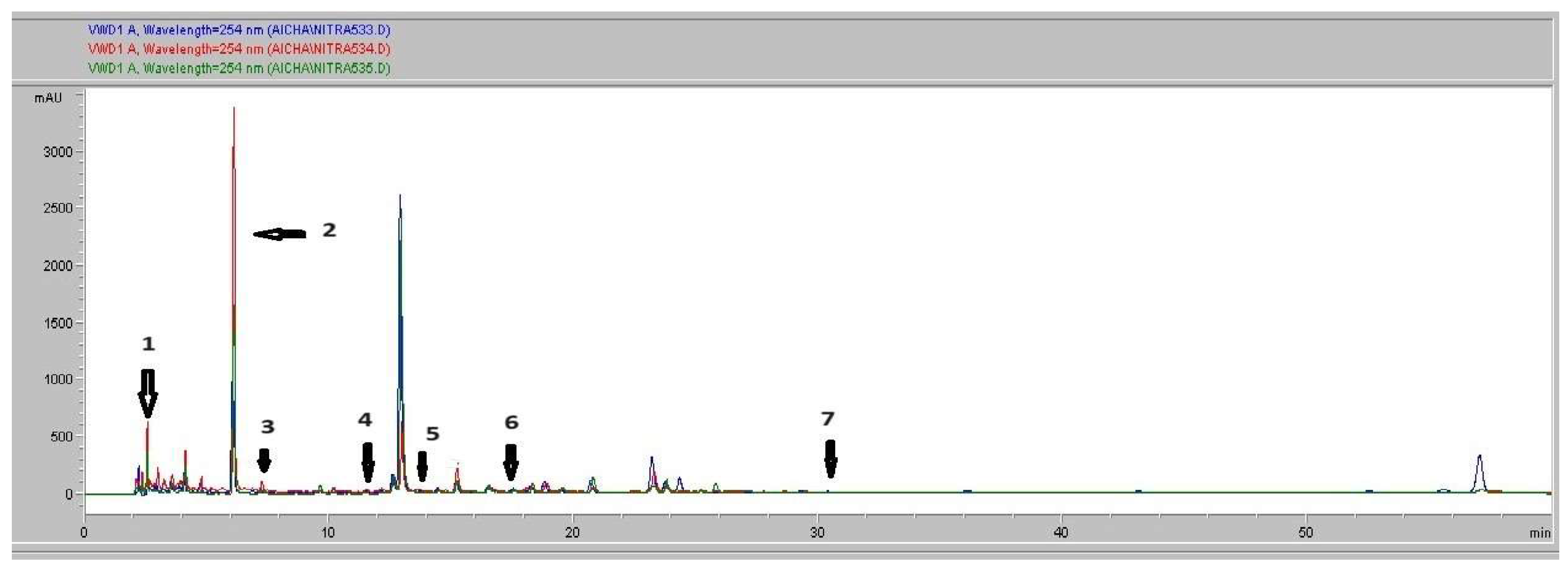
The active substances contained in methalonic Vitex agnus-castus fruits extract (VFME) were separated and evaluated using HPLC-DAD analysis system. The chromatographic separation of the methanolic extract is presented in Figure 1. Analysis of this chromatogram recorded at 254 nm noted the presence of phenolic compounds. A total of seven phenolic compounds were detected and identified on the basis of their retention times referring to the chromatogram of the standards (Figure 2). The compounds were labelled according to their order of elution. The phenolic compounds detected in MVFE included (Gallic acid: peak 1, RT = 2.63 min, Chlorogenic acid: peak 2, RT = 6.11 min, Caffeic acid peak 3, RT = 7.39, p-Coumaric acid: peak 4, RT = 11.46 min Trans Ferrulic Acid: peak 5, RT = 13.52min, Isoquercetin: peak 6, RT = 17.6 min and Quercetin: peak 7, RT = 31. (Table 3). Figure 2 shows the superposition of the two chromatograms: the chromatogram of the authentic standards bought from Sigma Aldrich and the chromatogram of the methanolic extract. Peak identification is dependent on the agreement between the analyte of interest's spectrum and the matching authentic standard, in addition to retention duration.
Table 3. Identification and quantification of seven phenolic compounds of methalonic Vitex agnus-castus fruits extract acquired at 254 nm.
Peak Compound
Identification
Similarity
Area
RT (min)
Retention Time
Content (µg/ ml) Quantity
1 Gallicacid 607 ,33 2.63 23
2 Chlorogenicacid 8819,53 6.11 709.89
3 Caffeicacid 247,66 7.39 19.093
4 p-Coumaricacid 633,6 11.46 99.33
5 Trans-ferulicacid 1435,63 13.52 85.842
6 Isoquercetin 1208,73 17.6 49.46
7 Quercetin 577,78 31.03 21.14
Our results revealed the richness of the methanol extract in chlorogenic acid p-coumaric acid, trans -ferulic acid and iso quercetin. Nevertheless, caffeic acid, quercetin and gallic acid appear in the extract with a smaller proportion. Similar work has been carried out on in vitro cultures of young shoots of Vitex agnus-castus L. to analyze the content of flavonoids and phenolic acids. Their results indicated that in vitro crop biomass is also rich in other compounds such as neochlorogenic acid, rutine and cinaroside [38]. Another study demonstrated the abundance of Vitex fruits in luteolin, 3,3′-dihydroxy-5,6,7,4′-tetramethoxyflavone, 3,7-dimethylquercetin, 3-O-methylkaempferol, 3-hidroxy5,6,7.4-tramethoxyflavon, 3-methylequercetine,3-methyllkaempférol,5,3′,5′-rihydroxymethoxyyflavanone, 5,7,3′,5′′-tetrahydroxyflavanon, apigenin, artemetine, vitexin, orientine, isovitexin, isoorientine, kaferempol, penduletine and eupatorine [39]. All these phytochemical compounds contained in the Vitex agnus-castus plant could be considered as a potential biotechnological source in the pharmaceutical and agri-food industries.
Our results are in agreement with a previous study of Berrani et al. [40]., conducted in Morroco which stated that Vitex extracts showed an important diversity and variability between plant with the chemical analysis by HPLC-MS. The authors demonstrated that the chlorogenic acid was the main phenolic compound and is present in all plant parts especially in seeds with higher concentration (122900 ± 4547:28μg/kg). Moreover, the same study reported that the 11 types of flavonoids were identified; the most abundant is luteolin whereas isoquercetin was the most present flavonoids in our samples. Importantly, other previous studies have also revealed numerous phenolic compounds in V. agnus castus extracts [6, 41,42]. Indeed, the rate of phenolic compounds found in our results is not exactly similar to those identified in these reposted works. This difference is certainly due the geographical origins of the plant. However, in our study, the variability between chemical compounds in different parts can be explained by the ability of each organ to synthesis secondary metabolites but also to metabolic regulation [43].
The study of Boujbiha et al showed a presence of a total of 41 headspace volatile compounds sampled by Solid-phase microextraction (SPME) were characterized in Tunisian fruits of V. agnus-castus by GC–MS (Decoction extracts). They showed that 99.7% of the total volatile compounds was detected and distributed in five chemical classes: monoterpene hydrocarbons (17.9%), sesquiterpene hydrocarbons (45.9%), oxygenated monoterpenes (34.5%), oxygenated sesquiterpenes (1.3%), and non-terpene derivatives (0.1%). Among the 41 detected compounds, germacrene D (11.8%), 1,8-cineole (30.3%), were the major ones. Nonetheless, the literature indicates that Turkish VAC volatile oils traditionally included the same main components [42]. Similarly, the volatile fraction of V. agnus-castus leaves collected in Iran was shown to have the same primary components [44]. It should be noted that although while the main chemicals are nearly identical, there are notable variations in their proportional concentrations. These variations may be caused by factors such as age, the extraction technique, or the length of the distillation phase.[45].

3.4. Antioxidant Activity

Antioxidants are extensively important substances that retain the capability to cover the body from damage caused by free revolutionary- convinced oxidative stress [46]. In the current study, the antioxidant activity for all. agnus castus extracts was tested in the hunt for new bioactive composites from natural cofers. The antioxidant capacity of VFME was evaluated by checking the IC50 using two different methods namely DPPH and ABTS. As presented in Table 4, the methanolic extract of V. agnus-castus showed important antioxidant activity. The highest antioxidant potential with a value of IC50 = 0.051 ± 1.19 mg/mL was obtained by DPPH assay, whereas a value of IC50 = 0.585 ± 0.59 mg/mL was obtained by ABTS. We concluded that the DPPH activity assay was the most sensitive one in terms of IC50 by comparing the two methods.
Antioxidants are essential for preventing oxidative stress, which is the root cause of many diseases, including cancer, diabetes, heart disease, atherosclerosis, neurological diseases, and aging [47]. The literature states that a variety of techniques have been used to assess the antioxidant potential of natural substances, such as volatile molecules, flavonoids, and polyphenols. As previously mentioned, the antioxidant ability of VFME was assessed in this instance by comparing the IC50 values obtained from two separate techniques, DPPH and ABTS. By comparing the acquired results with those reported in the scientific literature, it can be noticed that the methanolic seed extracts obtained a similar IC50 value (IC50=0.612 ± 0:007(48)) from a DPPH experiment. However, our extracts have a better antioxydant activity comparing to aqueous extracts of the fruits of V. agnus-castus cultivated in Tunisia and in Turkey [41]. Similarly, it has been demonstrated that V. agnus-castus leaf extracts obtained from Mo-rocco display a greater level of antioxidant activity (IC50 = 1.01 ± 0.019 mg/mL) and a highly significant positive correlation (p < 0.05) with their flavonoid and phenol content [48]. Numerous studies have clearly shown the substantial correlation between plant extracts' antioxidant activity and their total phenolic content [46, 49]. Our results are in good agreement with these studies. In sum; V. agnus castus aqueous, methanol extracts present the loftiest antioxidant exertion compared with reference antioxidant ascorbic acid, for DPPH and ABTS assays. The polyphenols content in different plant extracts acts as reducing agents and antioxidants by the hydrogen giving property of their hydroxyl groups [50]. Hence, we could conclude that these polyphenols are responsible for the observed antioxidant exertion in this study [28].

3.5. Antiviral Activity

Antiviral activity was estimated by the IC50 value, defined as the concentration of the substance that provides approximately 50% protection against the virus-induced cytopathic effect. For the antiviral tests, only non-cytotoxic concentrations were selected. HEp-2 cells infected with CVB3 were treated with varying non-cytotoxic concentrations of methanolic extracts from the fruits of Vitex agnus-castus L. Indeed, similar research has documented the antiviral effects of ethyl acetate extracts from the leaves and fruits of a Vitex species, specifically Vitex polygama. This study found that the ethyl acetate extract from the leaves exhibited superior antiviral activity against herpes simplex virus type 1 (HSV-1) compared to the extract from the fruits [51].
However, the methanolic extract showed no antiviral activity, despite the presence of numerous phytochemical compounds in Vitex fruits reported in the literature, such as flavonoids, terpenoids, polyphenols, coumarins, saponins, alkaloids, and iridoids, which are known for their antibacterial and antiviral properties [52]. The discrepancy in antiviral effects observed in different studies is likely attributed to variations in extraction solvents, the concentration of flavonoids and tannins in the extracts, and the specific mode of action of the compounds against the tested virus.

4. Conclusion

This study delves into the antioxidant, cytotoxic and antiviral methanolic extracts from Vitex agnus-castus fruits. By meticulously analyzing the chemical composition, with a particular emphasis on phenolic acids, flavonoids, and tannins, we aim to uncover the underlying bioactive compounds responsible for these properties. The extracts demonstrated significant antioxidant activity in vitro and exhibited moderate cytotoxicity, suggesting their potential as a valuable natural resource for the pharmaceutical products or industry. Vitex agnus-castus fruits could serve as a readily accessible source of antioxidants, offering promising applications in the development of novel anticancer and pharmaceutical products.

References

  1. Francis, A.; Bruno, D.G. Endobiogeny and Medicinal Plants: From Meaning in Physiology to Practice in Phytotherapy. Elsevier Masson, 2020, 400.
  2. Yi, F.; Xu, L.J.; Meng, H.; Dong, Y.M.; Liu, H.B.; Xiao, P.G. In Silico Approach to Reveal the Pharmacological Material Basis of Traditional Medicinal Plants. Menton. Med. 2018, 13, 33. [Google Scholar]
  3. Rani, A.; Sharma, A. The Genus Vitex: A Review. Pharmacogn. Rev. 2013, 7(14), 188–198. [Google Scholar] [CrossRef] [PubMed]
  4. Bertone-Johnson, E.R.; Hankinson, S.E.; Johnson, S.R.; Manson, J.E. Timing of Alcohol Use and the Incidence of Premenstrual Syndrome and Probable Premenstrual Dysphoric Disorder. J. WomensHealth (Larchmt) 2009, 18(12), 1945–1953. [Google Scholar] [CrossRef] [PubMed]
  5. Masure, C. Chaste Tree (Vitex agnus-castus L.): Interest and Use in Premenstrual Syndrome. Doctoral Thesis, Pharmaceutical Sciences, 2018, 127.
  6. Bruneton J. Toxic Plants: Dangerous Vegetation for Humans and Animals. 3rd revised and expanded edition. Éditions Tec & Doc Lavoisier &EMinter, Paris. 2005.
  7. Ulusoy, Ş.; İnal, E.; Küpeli Akkol, E.; Çiçek, M.; Kartal, M.; Sobarzo-Sánchez, E. Evaluation of the anti-obesity effect of Sambucus nigra L. (elderberry) and Vitex agnus-castus L. (chasteberry) extracts in high-fat diet-induced obese rats. Front. Pharmacol. 2024, 15, 1410854. [Google Scholar] [CrossRef]
  8. Chen, S.N.; Friesen, J.B.; Webster, D.; Nikolic, D.; Van Breemen, R.B.; Wang, Z.J.; Fong, H.H.; Farnsworth, N.R.; Pauli, G.F. Phytoconstituents from Vitex agnus-castus Fruits. Fitoterapia 2011, 82(4), 528–533. [Google Scholar] [CrossRef]
  9. Azofeifa, G.; Quesada, S.; Boudard, F.; Morena, M.; Cristol, J.P.; Perez, A.M.; Vaillant, F.; Michel, A. Antioxidant and anti-inflammatory in vitro activities of phenolic compounds from tropical highland blackberry (Rubus adenotrichos). J. Agric. Food Chem. 2013, 61, 5798–5804. [Google Scholar] [CrossRef]
  10. Lacampagne, S. Localization and Characterization of Tannins in Grape Skins: Impact of the Physicochemical Organization of Cell Walls on Tannic Composition, Fruit Quality, and Typicity of Bordeaux Grapes. Ph.D. Thesis, Sciences, Bordeaux 2, 2010, p. 294.
  11. Thaçi, S.; Krasniqi, B.; Dërmaku-Sopjani, M.; Rifati-Nixha, A.; Abazi, S.; Sopjani, M. Vasorelaxant Effects of the Vitex agnus-castus Extract. Evid. BasedComplement. Altern. Med. 2022,(1), 7708781. [CrossRef]
  12. Stöckigt, J.; Sheludk, Y.; Unger, M.; Gerasimenko, I.; Warzecha, H.; Stöckigt, D. High-Performance Liquid Chromatographic, Capillary Electrophoretic and Capillary Electrophoretic-Electrospray Ionisation Mass Spectrometric Analysis of Selected Alkaloid Groups. J. Chromatogr. A 2002, 967(1), 85–113. [Google Scholar] [CrossRef]
  13. Arokiyaraj, S.; Perinbam, K.; Vivek, P.; Udaya Prakash, N.K. Free Radical Scavenging and In Vitro Cytotoxic Activity of Agnuside from Vitex agnus-castus (Verbenaceae). J. Pharm. Res. 2012, 5(5), 2548–2552. [Google Scholar]
  14. Lee, H.J.; Wang, C.J.; Kuo, H.C.; Chou, F.P.; Jean, L.F.; Tseng, T.H. Induction of Apoptosis by Luteolin in Human Hepatoma HepG2 Cells Involving Mitochondria Translocation of Bax/Bak and Activation of JNK. Toxicol. Appl. Pharmacol. 2005, 203(2), 124–131. [Google Scholar] [CrossRef]
  15. Jia, Z.; Tang, M.; Wu, J. The determination of flavonoid contents in mulberry and their scavenging effects on superoxide radicals. Foods 1999, 64, 555–559. [Google Scholar] [CrossRef]
  16. Akkol, E.K.; Karpuz, B.; Türkcanoğlu, G.; Coşgunçelebi, F.G.; Taştan, H.; Aschner, M.; Khatkar, A.; Sobarzo-Sánchez, E. The Phytochemical Profile and Biological Activity of Malva neglecta Wallr. in Surgically Induced Endometriosis Model in Rats. Molecules 2022, 27, 7869. [Google Scholar] [CrossRef] [PubMed]
  17. Awah, F.M.; Uzoegwu, P.N.; Ifeonu, P.; Oyugi, J.O.; Rutherford, J.; Yao, X.; Fehrmann, F.; Fowke, K.R.; Eze, M.O. Free radical scavenging activity, phenolic contents, and cytotoxicity of selected Nigerian medicinal plants. Foods 2012, 131, 1279–1286. [Google Scholar] [CrossRef]
  18. Barros, H.R.; Ferreira, T.A.; Genovese, M.I. Antioxidant capacity and mineral content of pulp and peel from commercial cultivars of citrus from Brazil. Foods 2012, 134, 1892–1898. [Google Scholar] [CrossRef] [PubMed]
  19. Jia, Z.; Tang, M.; Wu, J. The determination of flavonoid contents in mulberry and their scavenging effects on superoxide radicals. Foods 1999, 64, 555–559. [Google Scholar] [CrossRef]
  20. Miliauskas, G.; Venskutonis, P.R.; Van Beek, T.A. Screening of radical scavenging activity of some medicinal and aromatic plant extracts. Foods 2004, 85, 231–237. [Google Scholar] [CrossRef]
  21. Schofield P,Mbugua DM, Pell A N. . Analyses of condensed tannins: a review Animal Food and Technology.2001, 91, 21-40. [CrossRef]
  22. Broadhurst, R.B.; Jones, W.T. Analysis of condensed tannins using acidified vanillin. J. Sci. Food Agric. 1978, 29, 788–794. [Google Scholar] [CrossRef]
  23. Heimler, D.; Vignolini, P.; Dini, M.G.; Romani, A. Antiradical activity and polyphenol composition of local Brassicaceae edible varieties. Foods 2006, 99, 464–469. [Google Scholar] [CrossRef]
  24. Savy, M.; Martin-Prével, Y.; Traissac, P.; Delpeuch, F. Measuring dietary diversity in rural Burkina Faso: comparison of a 1-day and a 3-day dietary recall. Public Health Nutr. 2007, 10, 71–78. [Google Scholar] [CrossRef]
  25. Stämpfli, R.; Brühwiler, P.; Mourad, S.; Verdejo, R.; Shaffer, M. Development and Characterisation of Carbon Nanotube-Reinforced Polyurethane Foams. EMPA Activities 2007, 26, 51.Landoulsi, A.; Roumy, V.; Riviere, C.; Sahpaz, S.; Benhamida, J.; Hennebelle, T. Chemical composition and pharmacological activities of Tunisian species of the genus Eryngium L. (Apiaceae). Open Sci. 2018, 1-23.
  26. Zhang W, Torabinejad M, Li Y. Evaluation of cytotoxicity of MTAD using the MTT-tetrazolium method. J Endod. 2003,29(10), 654-7. tfc. [CrossRef]
  27. He, Y.; Chipman, P.R.; Howitt, J.; et al. Interaction of Coxsackievirus B3 with the Full Length Coxsackievirus-Adenovirus Receptor. Nat. Struct. Biol. 2001, 8(10), 874-878.Kimmis BD, Downing C, Tyring S. Hand-foot-and-mouth disease caused by coxsackievirus A6 on the rise. Cutis. 2018, 102(5), 353-356.
  28. Berrani A, Marmouzi I, Kharbach M, et al. Anabasis aretioidesCoss. &Moq. phenolic compounds exhibit in vitro hypoglycemic, antioxidant and antipathogenic properties. J Basic Clin PhysiolPharmacol. 2018. 30(2), 251-257. [CrossRef]
  29. Kikalishvili, B.; Zurabashvili, D.; Sulakvelidze, T.; Malania, M.; Turabelidze, D. Georgian Med. News 2016, (256-257), 77-81.
  30. Ozkaya, F., Unak, P., Medine, E.I. et al. 18FDG conjugated magnetic nanoparticle probes: synthesis and in vitro investigations on MCF-7 breast cancer cells. J RadioanalNucl Chem 295, 1789–1796 (2013). [CrossRef]
  31. Lee HJ, Wang CJ, Kuo HC, Chou FP, Jean LF, Tseng TH. Induction apoptosis of luteolin in human hepatoma HepG2 cells involving mitochondria translocation of Bax/Bak and activation of JNK. Toxicol Appl Pharmacol. 2005, 203(2), 124-131. [CrossRef]
  32. Ohyama K, Akaike T, Imai M, Toyoda H, Hirobe C, Bessho T. Human gastric signet ring carcinoma (KATO-III) cell apoptosis induced by Vitex agnus-castus fruit extract through intracellular oxidative stress. Int J Biochem Cell Biol. 2005;37(7):1496-1510. [CrossRef]
  33. Boujbiha, M.A.; Chahdoura, H.; Adouni, K.; Ziani, B.E.C.; Snoussi, M.; Chakroun, Y.; Ciudad-Mulero, M.; Fernández-Ruiz, V.; Achour, L.; Selmi, B.; Morales, P.; Flamini, G.; Mosbah, H. Wild Vitex agnus-castus L.: Phytochemical Characterization, Acute Toxicity, and Bioactive Properties. Molecules2023, 28(13), 5096. [CrossRef]
  34. Ahmed, D.; Chaudhary, M.A.; Raza, A.; et al. Comparative study of antibacterial activity and mineral contents of various parts of Verbena officinalis Linn. Asian Journal of Chemistry 2012, 24(1), 68–72. [Google Scholar]
  35. Mesaik, M.A.; Azizuddin; Murad, S.; Khan, K.M.; Tareen, R.B.; Ahmed, A.; Atta-ur-Rahman; Choudhary, M.I. Isolation and immunomodulatory properties of a flavonoid, casticinfromVitex agnus-castus.Phytother Res.2009, 23(11), 1516–20. [CrossRef]
  36. Skrzypczak-Pietraszek, E.; Piska, K.; Pietraszek, J. Enhanced production of the pharmaceutically important polyphenolic compounds in Vitex agnus-castusL. shoot cultures by precursor feeding strategy. Eng. Life Sci. 2018, 18(5), 287–297. [Google Scholar] [CrossRef] [PubMed]
  37. Adamov, G.V.; Rendyuk, T.D.; Saybel, O.L.; Dargaeva, T.D.; Tsitsilin, A.N.; Bokov, D.O. Vitex agnus-castus: Botanical features and area, chemical composition of fruit, pharmacological properties, and medicinal uses. J. Appl. Pharm. Sci. 2022, 12(03), 034–044. [Google Scholar] [CrossRef]
  38. Berrani, A.; Marmouzi, I.; Bouyahya, A.; Kharbach, M.; El Hamdani, M.; El Jemli, M.; Lrhorfi, A.; Zouarhi, M.; Faouzi, M.E.A.; Bengueddour, R. Phenolic Compound Analysis and Pharmacological Screening of Vitex agnus-castus Functional Parts. BioMed Res. Int. 2021, 2021, 6695311. [Google Scholar] [CrossRef] [PubMed]
  39. Asdadi, A.; Hamdouch, A.; Oukacha, A.; Moutaj, R.; Gharby, S.; Harhar, H.; El Hadek, M.; Chebli, B.; Idrissi Hassani, L.M. Study on chemical analysis, antioxidant and in vitro antifungal activities of essential oil from wild Vitex agnus-castus L. seeds growing in the area of Argan Tree of Morocco against clinical strains of Candida responsible for nosocomial infections. J. Mycol. Med. 2015, 25(4), e118–e127. [Google Scholar] [CrossRef]
  40. Imai, M.; Yuan, B.; Kikuchi, H.; Saito, M.; Ohyama, K.; Hirobe, C.; Oshima, T.; Hosoya, T.; Morita, H.; Toyoda, H. Growth inhibition of a human colon carcinoma cell, COLO 201, by a natural product, Vitex agnus-castus fruits extract, in vivo and in vitro. Adv. Biol. Chem. 2012, 2, 20–28. [Google Scholar] [CrossRef]
  41. Dixon, R.A.; Dickinson, A.J. A century of studying plant secondary metabolism-From "what?" to "where, how, and why?". Plant Physiol. 2024, 195(1), 48–66. [Google Scholar] [CrossRef]
  42. Allahtavakoli, M.; Honari, N.; Pourabolli, I.; Arababadi, M.K.; Ghafarian, H.; Roohbakhsh, A.; Shamsizadeh, A. Vitex agnus-castus extract improves learning and memory and increases the transcription of estrogen receptor α in the hippocampus of ovariectomized rats. Base Clin. Neurosci. 2015, 6(3), 185. [Google Scholar]
  43. Powers, C.N.; Setzer, W.N. A molecular docking study of phytochemical estrogens mimicking plant-based dietary supplements. In Silico Pharmacol. 2015, 3(1), 1–63. [Google Scholar] [CrossRef]
  44. Basavalingappa, R.H.; Arumugam, R.; Lasrado, N.; Yalaka, B.; Massilamany, C.; Gangaplara, A.; Riethoven, J.J.; Xiang, S.H.; Steffen, D.; Reddy, J. Viral myocarditis involves the generation of autoreactive T cells with multiple antigen specificities that localize in lymphoid and non-lymphoid organs in the mouse model of CVB3 infection. Mol. Immunol. 2020, 124, 218–228. [Google Scholar] [CrossRef]
  45. Dejan, S.; Marina, S.; Jasmina, G.L.; Ana, D.; Ana, C.I.; Mihailo, R.; Dragoljub, G. Chemical composition and antimicrobial activity of Vitex agnus-castus L. fruits and leaves essential oils. Food Chem. 2011, 128(4), 1017–1022. [Google Scholar] [CrossRef]
  46. Chouikha, A.; Rezig, D.; Driss, N.; et al. Circulation and Molecular Epidemiology of Enteroviruses in Paralyzed, Immunodeficient, and Healthy Individuals in Tunisia, a Country with a Polio-Free Status for Decades. Viruses 2021, 13(3), 380. [Google Scholar] [CrossRef] [PubMed]
  47. Mone, K.; Lasrado, N.; Sur, M.; Reddy, J. Vaccines against Group B Coxsackieviruses and Their Importance. Vaccines 2023, 11(2), 274. [Google Scholar] [CrossRef] [PubMed]
  48. Centers for Disease Control and Prevention (CDC). Increased detections and severe neonatal disease associated with coxsackievirus B1 infection—United States, 2007. MMWR Morb. Mortal. Wkly. Rep. 2008, 57(20), 553-556.
  49. Khan, M.A.; Rahman, A.A.; Islam, S.; et al. A Comparative Study on the Antioxidant Activity of Methanolic Extracts from Different Parts of Morus alba L. (Moraceae). BMC Res. Notes 2013, 6, 24. [CrossRef]
  50. Gonçalves, J.L.; Leitão, S.G.; Monache, F.D.; Miranda, M.M.; Santos, M.G.; Romanos, M.T.; Wigg, M.D. In vitro antiviral effect of flavonoid-richextracts of Vitex polygama (Verbenaceae) against acyclovir-resistantherpes simplex virus type 1. Phytomedicine 2001, 8(6), 477–480. [Google Scholar] [CrossRef]
  51. Gonçalves JL, Leitão SG, Monache FD, Miranda MM, Santos MG, Romanos MT, Wigg MD. In vitro antiviral effect of flavonoid-rich extracts of Vitex polygama (Verbenaceae) against acyclovir-resistant herpes simplex virus type 1. Phytomedicine. 2001 Nov;8(6):477-80. [CrossRef] [PubMed]
  52. Sogame, M.; Naraki, Y.; Sasaki, T.; Seki, M.; Yokota, K.; Masada, S.; Hakamatsuka, T. Quality Assessment of Medicinal Products and Dietary Supplements Containing Vitex agnus-castus by HPLC Fingerprint and Quantitative Analyses. Chem. Pharm. Bull. 2019, 67(6), 527–533. [Google Scholar] [CrossRef]
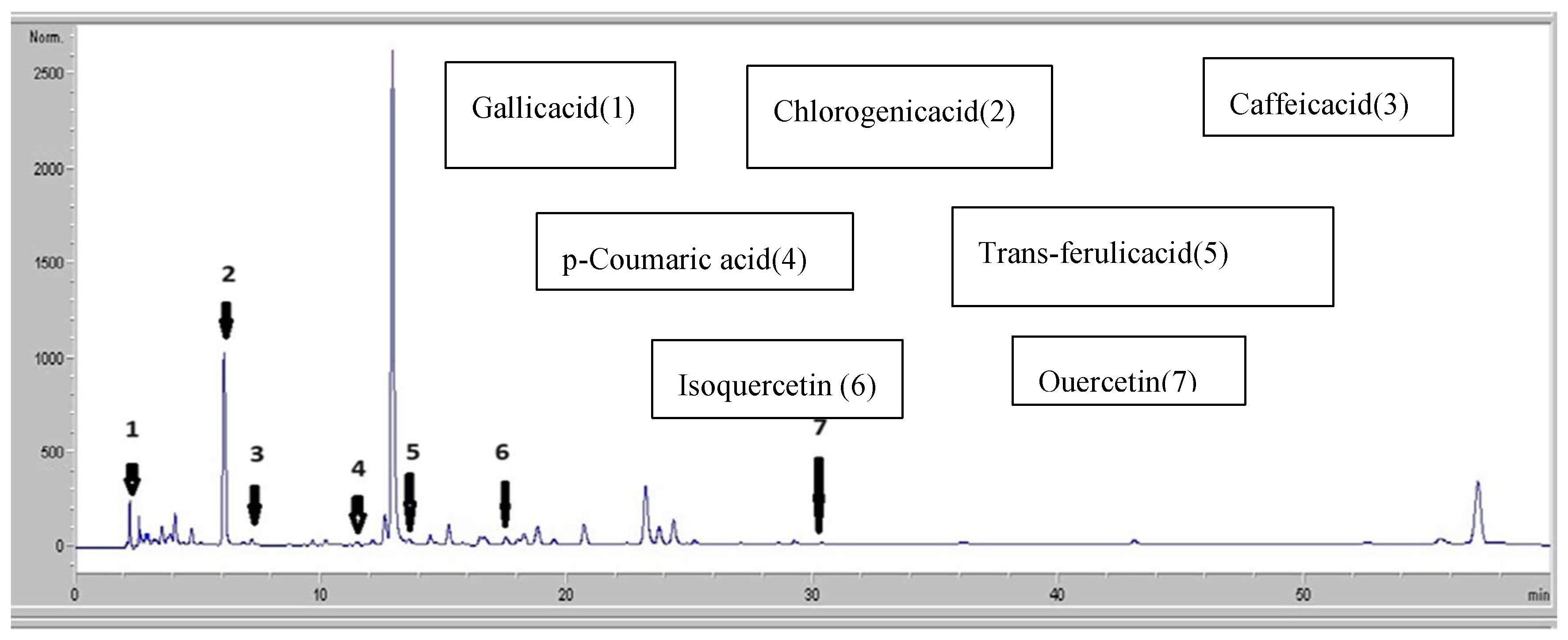
Figure 1. Chromatographic profile of Vitex agnus-castus L. acquired at 254 nm.
Preprints 121427 g001
Figure 2. Chromatographic profile of standards recorded at 254 nm.
Preprints 121427 g002
Table 1. Phytochemical compounds of Vitex agnus cactus fruits methanolic extract (VFME).
Phytochemical Compounds Values (mg/g VACME)
Total phenolic content (mg G AE/g Ext) 16.89±0.04
Total flavonoid content (mg ECat/g Ext) 10.28 ±0.22
Total flavonol content (mg ER/g Ext) 04.37±0.01
Total tannin content (mg EAT/g Ext) 19.13±0.86
Table 2. Minerals composition.
Content
(mg/100 g dw)
N 1640
P 270
K 1620
Ca 1000
Na 120
Mg 500
Fe 203.55
Cu 4.60
B 61.00
Zn 27.53
N: nitrogen; P: phosphorus; K: potassium; Ca: calcium; Na: sodium; Mg: magnesium: iron; Cu : copper; B : boron ; Zn: zinc.
Table 4. Antioxidant activity of V. agnus-castus methalonic extracts.
IC50 (mg/ml) Ascorbic Acid Methalonic extract
DPPH scavenging assay 0.018 ±1.27b 0.501±1.19b
ABTS scavenging assay 0.067±0.08b 0.585±0.59b
Disclaimer/Publisher’s Note: The statements, opinions and data contained in all publications are solely those of the individual author(s) and contributor(s) and not of MDPI and/or the editor(s). MDPI and/or the editor(s) disclaim responsibility for any injury to people or property resulting from any ideas, methods, instructions or products referred to in the content.
Copyright: This open access article is published under a Creative Commons CC BY 4.0 license, which permit the free download, distribution, and reuse, provided that the author and preprint are cited in any reuse.
Alerts
Prerpints.org logo

Preprints.org is a free preprint server supported by MDPI in Basel, Switzerland.

Subscribe

© 2025 MDPI (Basel, Switzerland) unless otherwise stated