Preprint
Review

Gold Nanoparticles: Multifunctional Properties, Synthesis and Future Prospects

This version is not peer-reviewed.

Submitted:

23 October 2024

Posted:

24 October 2024

You are already at the latest version

A peer-reviewed article of this preprint also exists.

Abstract

Gold nanoparticles (NPs) are among the most commonly employed metal NPs in biological applications, with their distinctive physicochemical features. Their extraordinary optical properties, stemming from the strong localized surface plasmon resonance (LSPR), contribute to the devel-opment of novel approaches in the areas of bioimaging, biosensing, and cancer research, especially for photothermal and photodynamic therapy. The ease of functionalization with various lig-ands provides a novel approach to the precise delivery of these molecules to targeted areas. Gold NPs ability to transfer heat and electricity positions them as valuable materials for advancing thermal management and electronic systems. Moreover, their inherent characteristics, such as in-ertness, give rise to the synthesis of novel antibacterial and antioxidant agents as they provide a biocompatible and low-toxic approach. Chemical and physical synthesis methods are utilized to produce gold NPs. The pursuit of more ecologically sustainable and economically viable large-scale technologies, such as environmentally benign biological processes referred to as green/biological synthesis, has garnered increasing interest among global researchers. Green synthesis methods are favorable among other synthesis techniques as they minimize the necessity for hazardous chemicals in the reduction process due to their simplicity, cost-effectiveness, energy efficiency, and biocompatibility. This article discusses the importance of gold NPs, their optical and conductivity properties, antibacterial, antioxidant, and anticancer properties, synthesis methods, contemporary uses, and biosafety, emphasizing the need to understand toxicology principles and green commercialization strategies.

Keywords: 
Subject: 
Chemistry and Materials Science  -   Nanotechnology

1. Introduction

In recent years, traditional biomedical techniques have been effectively supplanted by contemporary nanotechnology approaches, offering enhanced accuracy, sensitivity, efficiency, and rapid measurement. Many studies have been done on gold NPs for biological applications because they have many useful physical and chemical properties, such as being easy to make, biocompatible, non-toxic, having a high surface-to-volume ratio, and being able to change their size. The size and form of gold NPs affect their physical properties and color diversity, while bulk gold has different attributes than nanoscale particles [1].
The distinctive characteristics of gold NPs have driven researchers to develop them for applications in catalysis, biolabeling, nonlinear photonic systems, and drug delivery [2] (Table 1). Gold NPs optical characteristics, characterized by their strong LSPR, enable utilization in advancing bioimaging, biosensing studies along with commonly employed spectroscopic techniques such as Surface Enhanced Raman Scattering (SERS), which enables precise detection of analytes at extremely low, down to femtomolar and picomolar, levels [3]. It also extends to cancer therapy where researchers are using gold NPs’ photothermal characteristics to improve overall efficiency of the photothermal treatment, especially to facilitate localized heating processes [4]. Free-moving electrons on gold NPs’ outer membrane provide high electrical conductivity efficiency by facilitating a conductive pathway. This has recently led to the development of novel devices in the electronics field, such as conductive inks, where gold NPs are used as additives, coating and doping materials [5]. Further, gold NPs high thermal conductivity is exploited by researchers for production of thermal management systems, including heat pipes for electronics cooling, where rapid and even distribution of heat is a crucial requirement [6].
One of the most recognized research areas focusing on the extensive employment of gold NPs includes drug delivery. Gold NPs facile synthesis combined with their synergistic effects with various ligands, such as drugs, nucleic acids, chemotherapeutic agents, proteins, glycans, antibacterials and photosensitizers, paves the way for the creation of highly efficient delivery platforms [7]. From another aspect, where gold NPs are not integrated with other molecules, there is an increasing number of research highlighting the novel and nature-friendly antimicrobials, along with antioxidant and anticancer agents, synthesized using gold NPs [8,9,10]. This is mostly due to the recently developing green synthesis approaches, which takes advantage of the gold NPs inherent properties including low toxicity, high stability and biocompatibility.
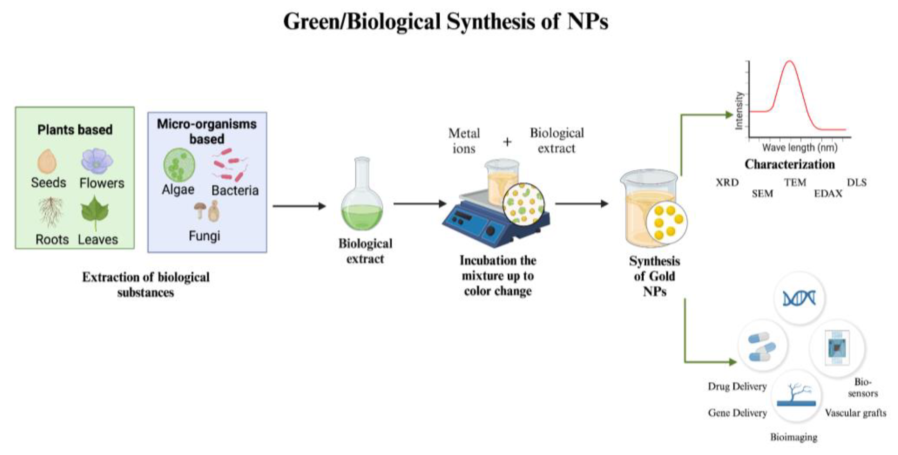
In the synthesis of gold NPs, two primary approaches are employed: top-down and bottom-up. Top-down approaches include the extraction of bulk material to form self-assembled nanoscale objects, whereas bottom-up approaches require the assembly of tiny atoms or reducing ions to form crucial nanostructures. One safe, effective, and energy-efficient way to manufacture NPs is through biological/green synthesis. This method uses a variety of biological resources, including eukaryotes and prokaryotes, to generate NPs in vivo. The bioreduction of metallic ions to NPs and the stability of these particles are significantly influenced by metabolites, which include proteins, fatty acids, carbohydrates, enzymes, and phenolic substances [11]. Therefore, numerous biological systems, including plants, bacteria, yeasts, and fungi, are actively investigating new pathways for the synthesis of safe nanoproducts for the manufacturing of gold NPs [12]. One of the greatest options for the large-scale production of gold NPs with well-defined size and morphology is the plant-based green synthesis technology because of its affordable cost [13]. However, chemical methods are frequently employed because of their simplicity; nevertheless, for reduction, harmful chemicals are required. While biological approaches use the same reducing and functionalizing agents, chemical procedures offer a wider range of functionalization options for creating NPs with different functionalizing agents. Chemical synthesis methods control the size of the particles, whereas biological processes yield big particles. While chemical synthesis needs a high temperature, room temperature synthesis is a potential biological technique. On the other hand, environmental advantages, high-purity particles free of hazardous chemicals, exact control over NP size and shape, and simple industrial uses like laser ablation or sputtering are all provided by the physical production of gold NPs. Its disadvantages over chemical synthesis include higher energy consumption, more expensive equipment, and lower yields [12,14].
Gold NPs have garnered significant interest in the realm of biomedicine and its applications. The substantial potential for future applications presented by the rapid growth of gold NPs technology can be attributed to their diversified characteristics and huge volume-specific surface areas in comparison to bulk gold. Due to these characteristics, gold NPs are now important for the creation of superior nanoelectronic chips as well as a range of biomedical and environmental applications [15,16]. They have also been employed in the food and beverage sectors [17]. Additionally, the Food and Drug Administration (FDA) approved gold NPs for a variety of biomedical uses, which led to an expansion of application areas, including medication carriers, cancer therapy, and other biological uses [18]. The extensive everyday use of gold NPs may elevate the likelihood of human exposure to these NPs. Numerous investigations into the toxicological effects of gold NPs from both academic and commercial sources have been disclosed [19]. Despite its potential in medicinal, environmental, and industrial applications, evidence regarding the acute and long-term health impacts of manufactured NPs remains scarce. These NPs can traverse the bloodstream without eliciting immunological rejection, yet their diminutive size and surface charges raise concerns. Gold NPs, produced in diverse forms, sizes, and charges, possess notable physicochemical features that raise new health concerns. Currently, there is insufficient knowledge regarding the health impacts of gold NPs, and no regulatory safety guidelines exist for their hazardous qualities [20].
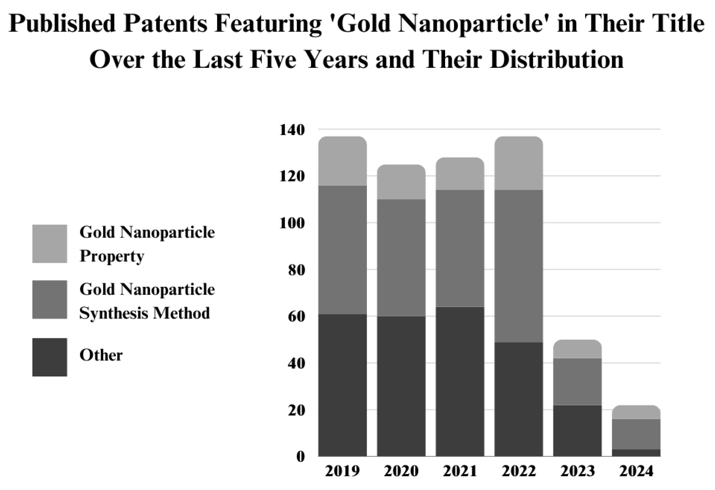
Gold NPs are among the most frequently discussed nanomaterials in nanotechnology. According to the Web of Science (WoS) database, approximately 10,000 papers titled "gold nanoparticles" are published annually (Figure 1). Although this is a significant number, the total publications on gold NPs are relatively low compared to silver NPs, as discussed in our previous paper [22,23,24]. Silver NPs are widely researched in the current literature, including combined and comparative studies involving gold NPs. One reason behind this research difference between these two NPs could be the challenges associated with green synthesis methods, particularly for gold NPs. Silver NPs have more established synthesis routes and demonstrate significant activity in certain areas. These challenges are well-documented in the literature, with a focus on missing points in optimizing the physicochemical properties of gold NPs during synthesis [25,26,27]. This may explain the pie-chart showing the distribution of papers that include "property" and "synthesis" keywords in their titles. It can be considered that the remaining articles ("others") mainly focus on investigating gold NP applications, including biological activities. However, based on this, current literature primarily explores synthesis methods and the physicochemical aspects of gold NPs, rather than their broad applications, as is common with silver NPs. Therefore, up-to-date reviews on gold NPs, especially those focusing on their synthesis processes and physical properties, are extremely crucial for the future of gold NP research.
This review paper presents an overview of the synthesis of gold NPs by the use of diverse synthesis methods classified as physical, chemical, and biological/green synthesis approaches. It examines their varied properties. This paper examines the various parameters influencing the synthesis of gold NPs to achieve optimum sizes and shapes, as well as the characterization methods employed to gain a deeper understanding of their characteristics. The significance of gold NPs, their improved optical and conductivity qualities, and their antibacterial, antioxidant, and anticancer capabilities are all covered in this article. With an emphasis on gold NP research, this article examines the biosafety of gold NPs, underscoring potential hazardous effects on cells, tissues, and organs, and stressing the necessity for a thorough understanding and adherence to toxicology principles. It draws attention to the increasing need for environmentally friendly industrial applications and makes noteworthy recommendations for developments in green commercialization strategies.

2. Properties of Gold Nanoparticles

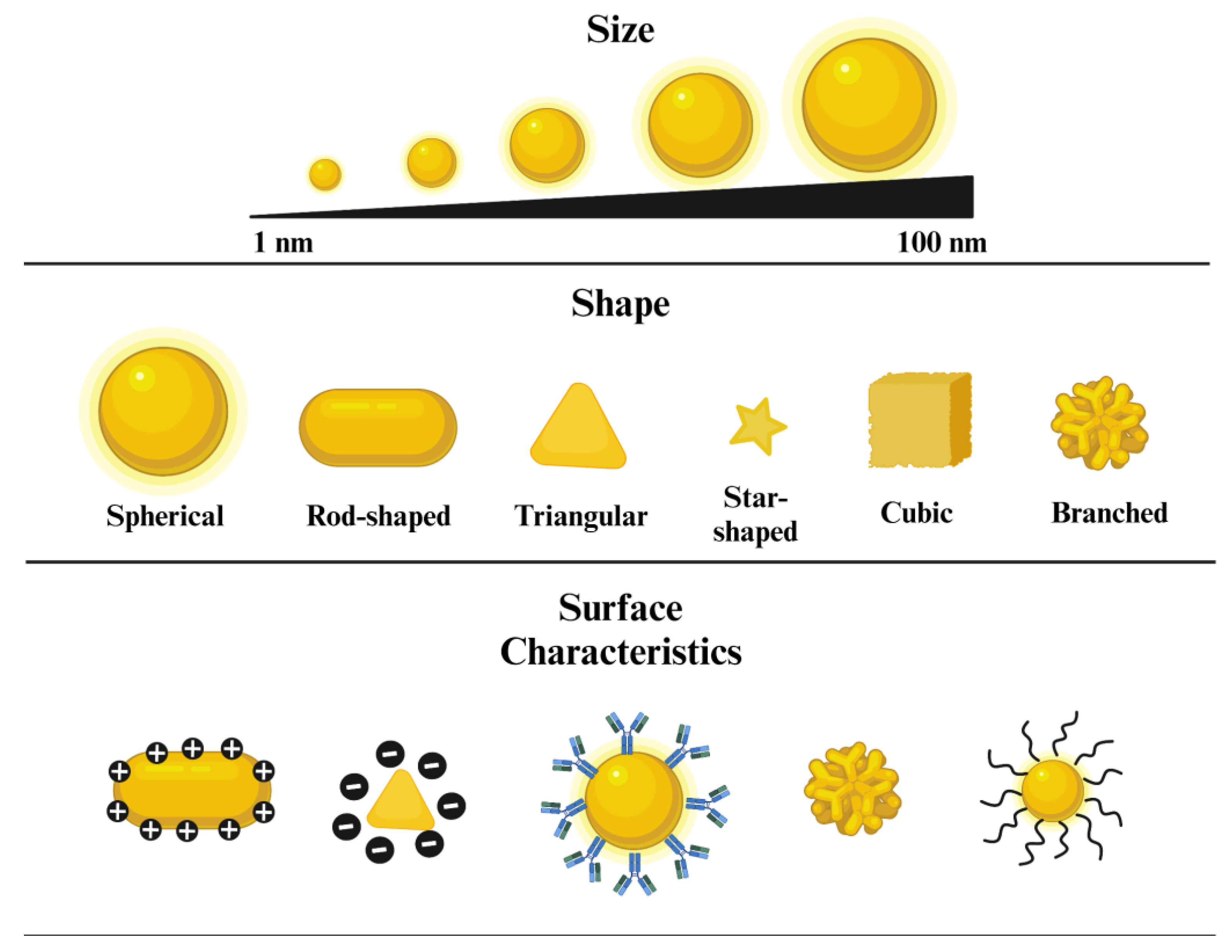
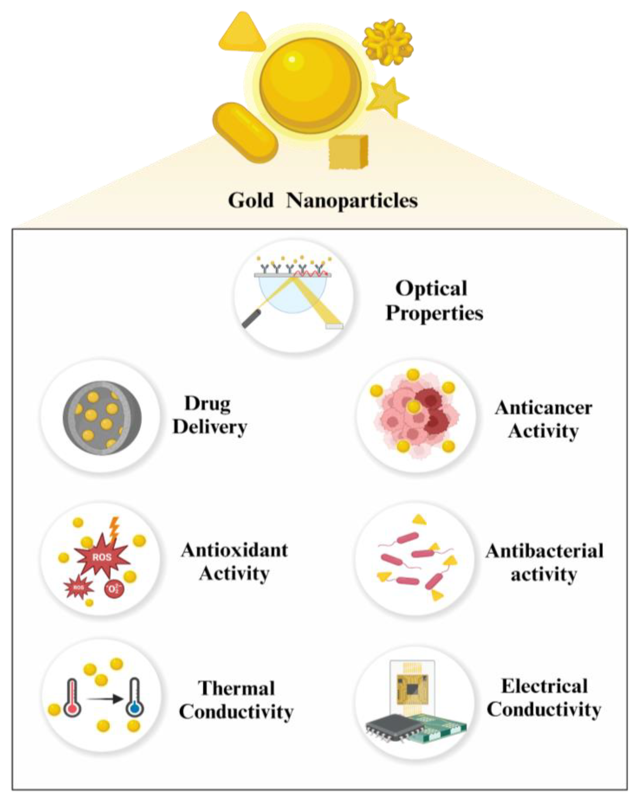
Metal NPs possess remarkable physical and chemical attributes that differ significantly from their bulkier counterparts. In this manner, they have been widely utilized in diverse fields of science, especially in the biomedical area. Among metal NPs, gold NPs were one of the most studied owing to their extraordinary characteristics. These include size and shape-dependent optical and electrical features, anticancer activity, facile surface functionalization with other molecules, and efficient delivery of drugs. In this section, we have pointed out the distinctive properties of gold NPs, from physical to biological (Figure 2), to underline their importance across multiple scientific disciplines (Figure 3).

2.1. Size

Gold NPs exhibit size-dependent physical, chemical, and biological characteristics that significantly determine their applicability in various fields [28]. It increases their interaction with their surroundings, by providing a high surface-area-to-volume ratio, further enhancing optical properties, catalytic activity, and biocompatibility [29]. For example, Shafiqa et al. investigated the dependence of surface plasmon resonance (SPR) concerning the size of gold nanospheres, ranging from 20 to 100 nm. Results revealed that absorption efficiency increases with NPs size, with a size of 70 nm demonstrating optimal efficiency compared to their counterparts [30]. Considering the catalytic behavior, Suchomel et al. explored the size-dependent activity of gold NPs, focusing on sizes varying from 6 to 22 nm. It has been shown that catalytic activity increased with decreasing size, which is primarily attributed to a higher surface-area-to-volume ratio [31].
Another aspect is that the size of the NPs is known to be a major determinant of their potential adverse effects. In this manner, Lee et al. investigated the size-dependent toxicity of gold NPs in human neural precursor cells and rat cerebral cortex. Utilizing two groups of gold NPs, 5 and 100 nm, they have demonstrated a correlation between size and the toxic effects. Findings revealed that smaller NPs (5 nm), exhibited higher toxicity compared to larger ones (100 nm), evidenced by a significant reduction in cell viability [32].

2.2. Shape

The shape of the NPs plays a key role in the definition of their resultant properties and further uses [33]. Gold NPs can be fabricated in various shapes such as spheres, cubes, triangles, rods, and stars [34]. Each of these shapes comes with distinct advantages and disadvantages. To exemplify, Jiji et al. investigated the catalytic performance of differently shaped gold NPs for the reduction of p-nitroaniline. Findings demonstrated that gold nanorods (NRs) were superior in terms of catalytic efficiency, compared to their spherical and dog bone counterparts [35]. From another perspective, Hameed et al. focused on the antibacterial activity of different-shaped gold NPs. Emphasizing the increased bactericidal efficiency at lower doses of NPs, they have shown that gold NPs interacted with foodborne bacterial pathogens, such as Escherichia coli (E. coli), Pseudomonas aeruginosa (P. aeruginosa), and Staphylococcus aureus (S. aureus), in a shape-dependent manner. In more detail, gold nanocubes were most bactericidal in comparison to nanospheres and nanostars, indicated by a zero percent survival rate [36]. Also, the shape of the gold NPs highly influences their optical traits. Hua et al. examined the nonlinear optical properties of gold NPs with varying shapes, including NRs, nanostars, and nanoshells. Utilizing Z-scan measurements and pump-probe spectroscopy, they have revealed that gold NRs exhibited the strongest saturable absorption while nanoshells showed the weakest response [37].
Moreover, shape is an important factor that affects the efficiency of gold NP-based delivery systems. One of the primary examples of this aspect includes the delivery of nucleic acids, such as small interfering RNAs (siRNAs). Accordingly, Morgan et al. demonstrated how various shaped gold NPs (nanoshells, nanocages, and NRs) influence the effectiveness of siRNA delivery. Their results highlighted that NRs were the most efficient in terms of the release of the attached siRNA strands, even though nanoshells could be loaded with three times more siRNA cargo [38]. In another study, Xie et al. evaluated the effect of NP shape concerning the cellular uptake into RAW264.7 cells. Focusing on stars, rods, and triangles, they have found that different-shaped gold NPs are prone to demonstrate cellular uptake through different pathways, with triangles being the most efficient among them [39]. These results not only highlighted the possible mechanisms of cellular uptake but also created a novel perspective for the further design of nanomaterials in drug delivery systems.
Besides, similar to their size, there are various studies in the current literature underlining the relationship between toxicity and the shape of the gold NPs. To name one, Sun et al. investigated the in vivo toxic effects and the biodistribution of sphere, rod, and cube-shaped gold NPs using a mice model. Their findings showed that the ideal shape of the NPs was the spheres since they have shown the highest biocompatibility. On the other hand, rod-shaped NPs were observed as the most toxic, evidenced by increased adverse effects at lower concentrations of exposure [40]. In an in vitro study, Wozniak et al. examined the size and shape-dependent cytotoxicity profiles of gold nanospheres, nanoflowers, NRs, nanostars, and nanoprisms on HEK293T and HeLa cell lines. It was observed that NRs and nanospheres were the most toxic since they have led to the greatest decrease in the number of cells, specifically at the concentrations of 32, 100, and 300 µM. However, nanostars, nanoprisms, and nanoflowers remained less lethal at the same concentrations and only caused toxic effects at the highest doses after 72 hours of exposure [41].

2.3. Surface Characteristics

2.3.1. Surface Charge

Surface charge is particularly important in determining the stability, biodistribution, and interaction of the NPs with their environment [42]. Typically, as a result of the negatively charged identity of the cell membrane, positively charged NPs interact more easily with cells compared to their negatively charged or neutral counterparts [43]. Taking this as an advantage, like most of the NPs, positively charged gold NPs are employed as efficient delivery agents in the biomedical area, considering their increased cellular uptake characteristics [18]. As an example, Noh et al. employed cationic gold NPs for the delivery of plasmid DNA (pVAXmIL-2) into C2C12 cells. Through the experiments, they examined gold NP-plasmid DNA complexes with various complexation rates and reported an enhancement in transfection efficiency, particularly at the ratio of 2400:1. To add more, a 4-fold increase in expression levels was also reported after intramuscular administration of the NP-DNA complex, compared to the DNA alone [44].
On the other hand, although anionic gold NPs have relatively low efficiency in terms of cellular uptake, they have also been widely investigated. In such a study, Lee et al. explored the influence of surface charge modifications of the gold NRs on skin penetration. Using transmission electron microscopy (TEM), they have measured the ability of differently charged NPs penetration to the skin. Results of the image analysis revealed that negatively charged gold NPs penetrated more rapidly than the positive ones, emphasizing the importance of surface chemistry for the development of suitable approaches in the near future [45].
To add more, negatively charged gold NPs have proven to be less toxic. In this manner, Bozic et al. assessed the toxic effects of gold NPs on Daphnia Magna, by focusing on different variables such as surface chemistry, charge, and the ligand type. Following both chronic and acute assays, results demonstrated that positively charged gold NPs were more toxic in comparison to negatively charged counterparts [46]. Similarly, Feng et al. focused on the adverse effects of positive and negatively charged gold NPs on Gram-negative Shewanella oneidensis MR-1 and Gram-positive Bacillus subtilis (B. subtilis). Findings revealed that cationic gold NPs, especially PAH-gold NPs, were more toxic to both types of bacteria [47]. Overall, these results are primarily attributed to the preferential binding of positively charged NPs towards negatively charged surfaces of the cell membranes.

2.3.2. Surface Functionalization

Manipulation of the surface characteristics of NPs enables their utilization in various areas since it provides a promising opportunity for novel uses [48]. In the case of gold NPs, various approaches such as covalent attachment (thiolation) [49], electrostatic interaction (citrate capping) [50], bioconjugation (with proteins, antibodies and DNA) [51] and coating with polymers (PEG, chitosan) [52] can be followed. These interactions, as a result of the strong affinity of gold NPs towards functional groups, facilitate the targeted delivery of these NPs into cells, as well as the biosensing and bioimaging applications [53].
In their research, Encabo-Berzosa et al. investigated the efficiency of PEG-functionalized polyethylenimine (PEI) gold NPs, as nonviral vectors, in comparison to commercially available lipoplexes. Bounding NPs to differently sized plasmids, from 4 to 40 kbp, their performance was evaluated on HeLa and Hek293t cells. Results indicated the potential applicability of gold NP complexes, as they have shown the same or increased levels of expression than the commercially available alternatives. It was also mentioned that pegylation of the gold NPs not only reduced the toxic effects arising from the PEI but also increased the dispersion of the NPs in the culture media [54]. In another study, Medley et al. synthesized aptamer-conjugated gold NPs to address the limitations associated with the detection of cancerous cells, including the high-priced instrumentation and time-consuming methods. Developing a novel colorimetric assay, they were able to differentiate between target and control cells, by observing the color change arising from the assembly of gold NPs on the cell surface. This novel approach emphasized the potential use of functionalized gold NPs in oncology, along with providing a highly sensitive and cost-effective method to the current literature [55]. Also, functionalization of the surface characteristics of gold NPs can be applied to mitigate the toxic effects, one of the major concerns associated with nanomaterials [56].

2.4. Optical Properties

In previous years gold NPs have been the main focus of numerous research in the diagnostics area, as a result of their extraordinary optical properties [57]. The most highlighted of these properties include LSPR, which is defined as the collective oscillating motion of conduction electrons near the NP surface when illuminated with light [58]. This phenomenon is primarily known for enabling gold NPs to absorb and scatter light in a broad range of the spectrum, consequently creating the basis for novel approaches in imaging, sensing, and labeling studies [59]. For example, functionalized gold NPs are being extensively used in bioimaging research, especially in oncology for the diagnosis of tumors, as they improve the contrast and resolution in the biological environment [60]. In the same manner, there is a lot of research in current literature focusing on the incorporation of gold NPs into sensing devices, with the aim of developing highly sensitive biosensors that demonstrate lower limits of detection [61].
The optical properties of gold NPs are highly influenced by their morphological characteristics, including their shape. To exemplify, differently shaped gold NPs have been shown to exhibit dissimilar LSPR peaks [62]. While nanospheres exhibit one unique LSPR band around 520 nm, NRs possess two split bands, transverse LSPR (t-LSPR) and longitudinal LSPR (l-LSPR), owing to their two-dimensional structure. Although t-LSPR peaks are located around 520 nm regardless of the dimension, l-LSPR peaks can be detected at higher wavelengths, from 650 to 1050 nm, depending on the NP aspect ratio [63]. On the other hand, branched or complex structures (including nanostars), with their asymmetric shapes, create multiple sharp resonance peaks which give rise to unique optical traits [64].
LSPR is also responsible for the distinctive colors of the gold NPs. Since surface absorption bands are known to be size and shape-dependent, a variation in these parameters causes a shift in the absorption peaks, thus, a change in the color is observed [65]. Generally, gold NPs show visible colors, from red to purple, when dispersed in a liquid medium and demonstrate absorption peaks at 500 to 550 nm. However, when the particle size increases, the color of the solution differs depending on the size of the NPs [66]. In line with that, Hao et al. utilized citrate-stabilized gold NPs for the visual detection of melamine, which is a non-protein nitrogen source, illegally integrated into milk products to increase the measured concentration of total protein [67]. Benefiting from the strong electrostatic interaction between melamine and the gold NPs, melamine levels down to 10 ppb were successfully detected upon a color change over a short period of time. It was also emphasized that the size of the gold NPs plays an important role in colorimetric detection, evidenced by an improvement in the sensitivity, from 15 to 5 ppm, when the size of the nanomaterials increased from 5 to 30 nm.
To summarize, researchers take advantage of the optical properties of gold NPs in various applications, primarily in rapid diagnostic tests, such as colorimetric and immunoassays [68].
Moreover, the LSPR characteristics of gold NPs extend to bioanalytical techniques such as SERS. SERS is a specialized form of Raman spectroscopy that is based on the principle of enhancing Raman scattering signals by using metal surfaces (or NPs) for the sensitive detection of molecular substances at lower concentrations, even down to 10-12 (picomolar) and 10-15 (femtomolar) levels [69]. The employment of gold NPs as SERS substrates has been widely emerged thanks to their strong LSPR. As an example, Xu et al. developed a novel sensor for the detection and analysis of pesticides, 2,4-dichlorophenoxyacetic acid (2,4-D), pymetrozine, and thiamethoxam, in food. Incorporating gold NPs within the mesoporous silica film in a densely packed form, researchers established a highly sensitive and stable SERS substrate containing multiple “hot spots”. These hot spots successfully amplified the Raman signals and allowed for the detection of trace amounts of pesticides, supported by a recorded limit of detection as low as 0.79 pg/mL for 2,4-D [70].
Additionally, gold NPs exhibit remarkable photothermal properties that allow for the development of novel approaches, especially in oncology [71]. One of these approaches includes photothermal therapy (PTT), where the energy of light absorbed by the NPs is converted into heat, in order to trigger cellular apoptosis [72]. As an example, Faid et al. showed the effectiveness of citrate-capped gold NPs in PTT as photothermal agents. Conducting experiments on breast cancer cell line (MCF-7), they have revealed the higher photothermal efficiency of gold NPs, in compared irradiation with 532 nm laser alone, supported by a significant 34% reduction in cell viability [73].
Another therapeutic approach where gold NPs are utilized is photodynamic therapy (PDT). PDT is based on the combined use of molecular oxygen and photosensitizers for the inhibition of cancer cell proliferation [74]. During the process, a photosensitizing agent is administered into the target tissue and illuminated with light. Upon exposure to specific wavelengths, the photosensitizing agent becomes activated and induces the generation of reactive oxygen species (ROS), eventually causing the death of cancerous cells [75]. In this context, gold NPs are commonly used to promote the efficiency of PDT. For example, Eshghi et al. synthesized Protoporphyrin IX (PpIX) conjugated gold NPs to overcome several drawbacks, such as the low solubility and aggregation associated with PpIX. In vitro tests on HeLa cells highlighted gold NPs as effective PDT agents, evidenced by a significant reduction in cell viability at all tested concentrations [76].
Recently, one of the most studied aspects of gold NPs has been their optical characteristics. The main reason behind this is the recent advances in biomedical research, in which the optical properties of gold NPs are frequently highlighted. Moreover, strong LSPR of the gold NPs plays an important role in this field by leading to improvements in diagnostic and therapeutic technologies. Hence, considering both previous and ongoing research regarding gold NPs, understanding and optimizing these attributes in a detailed manner would be crucial for advancing their applications across various fields.

2.5. Electrical Conductivity

Metals are recognized as remarkable electrical conductivity agents due to their unique atomic structure [77]. Presence of free moving electrons on their outer membrane leads to formation of a conduction pathway, where electrical current can pass through with minimal resistance [78]. This characteristic is also applied at nanoscale, in which metal NPs are widely employed as conductive additives in electronics, catalysis and sensing studies [79,80,81]. Among metal NPs, gold NPs stand out with their outstanding electrical conductivity [82]. Their high efficiency as doping and coating materials have led to significant advances in current research, by increasing the overall outcome of the electric conduction process [83,84]. As an example, Tomalih et al. utilized gold NPs, prepared by the laser ablation method, in order to enhance the electrical properties of Polyvinyl Pyrrolidone (PVP)/Polyvinyl Alcohol (PVA) blend. Confirming successful distribution of NPs within the composite, they have evaluated the effect of gold NPs, with varying ratios, on the electrical conductivity performance. Findings highlighted an improvement on the AC conductivity of gold NP-doped PVP/PVA blends, especially when the NP concentration is increased [85]. In a similar way, Baei et al. developed an alternative strategy to address the electrical property limitations of implanted heart grafts, which are used for regeneration of the infarcted heart. Through the experiments, they have demonstrated an enhancement following incorporation of gold NPs into chitosan hydrogels, compared to the samples containing chitosan alone. It was also emphasized that concentration of the NPs hold significance, highlighted by a 1.9-fold increase in electrical conductivity when different concentrations of gold NPs (36 and 72 mM) are used [86]. Further, it is important to mention gold NPs’ wide utilization in the industrial area. Their superior electrical conductivity compared to other metal NPs have positioned them as one of the primary preferences, when developing inks for electronics. Besides, gold NPs’ high stability, along with resistance to oxidation and corrosion, is considered advantageous for the fabrication of conductive inks [87]. However, despite its potential, high cost of gold NPs emerges as a limitation that hinders its applicability for mass production [88]. Lastly, gold NPs’ electrical conductivity can be influenced by their physical properties. Multiple studies in current literature have shown that, the electrical conduction efficiency of gold NPs is inversely proportional to their size [89], Hence, their synthesis methods and characterization must be tailored carefully, especially for the applications requiring electrical conductivity, along with the requirement of further research.

2.6. Thermal Conductivity

Apart from their remarkable electrical characteristics, gold NPs exhibit high thermal conductivity, arising from the efficient transfer of heat by the movement of both phonons and electrons [90]. Hence, they have been widely employed in various areas, including electronics, energy systems and biomedicine [91]. To be more specific, gold NP-incorporated electronic cooling, solar desalination and thermal management systems, in which the efficiency of heat transfer and dissipation is crucial, are being developed [92].
Similarly, gold NPs integration into composite nanofluids is considered an effective approach, where researchers take advantage of gold NPs’ stability and high surface area to enhance thermal conductivity [93].
Furthermore, thermal properties of gold NPs can be affected by their physical attributes. As an example, Essajai et al. investigated the influence of gold NP shapes on the thermal conductivity of nanofluids. Focusing on gold nanospheres and NRs, they have revealed that rod-shaped gold NPs were more effective in improving the nanofluid thermal conductivity in comparison to their spherical counterparts. These results were attributed to several factors, such as solid-liquid interface, enhanced mobility of liquid atoms in nanofluids, and surface effects [94]. Yet, contradicting results were achieved when the size of the NPs was considered. Shalkevich et al. evaluated the impact of size on the gold NPs’ ability to transfer heat. Synthesizing spherical NPs with sizes varying from 2 to 45 nm, they have measured the conductivity of nanofluids with concentrations of 0.00025-1 vol%. The results indicated a negligible enhancement in thermal conductivity, with only 40 nm sized gold NPs showing 1.4% improvement [94].
In brief, the superior thermal conductivity of gold NPs highlights them as promising materials for various uses. However, a full understanding of the factors that influence their efficiency is required to maximize their potential.

2.7. Delivery

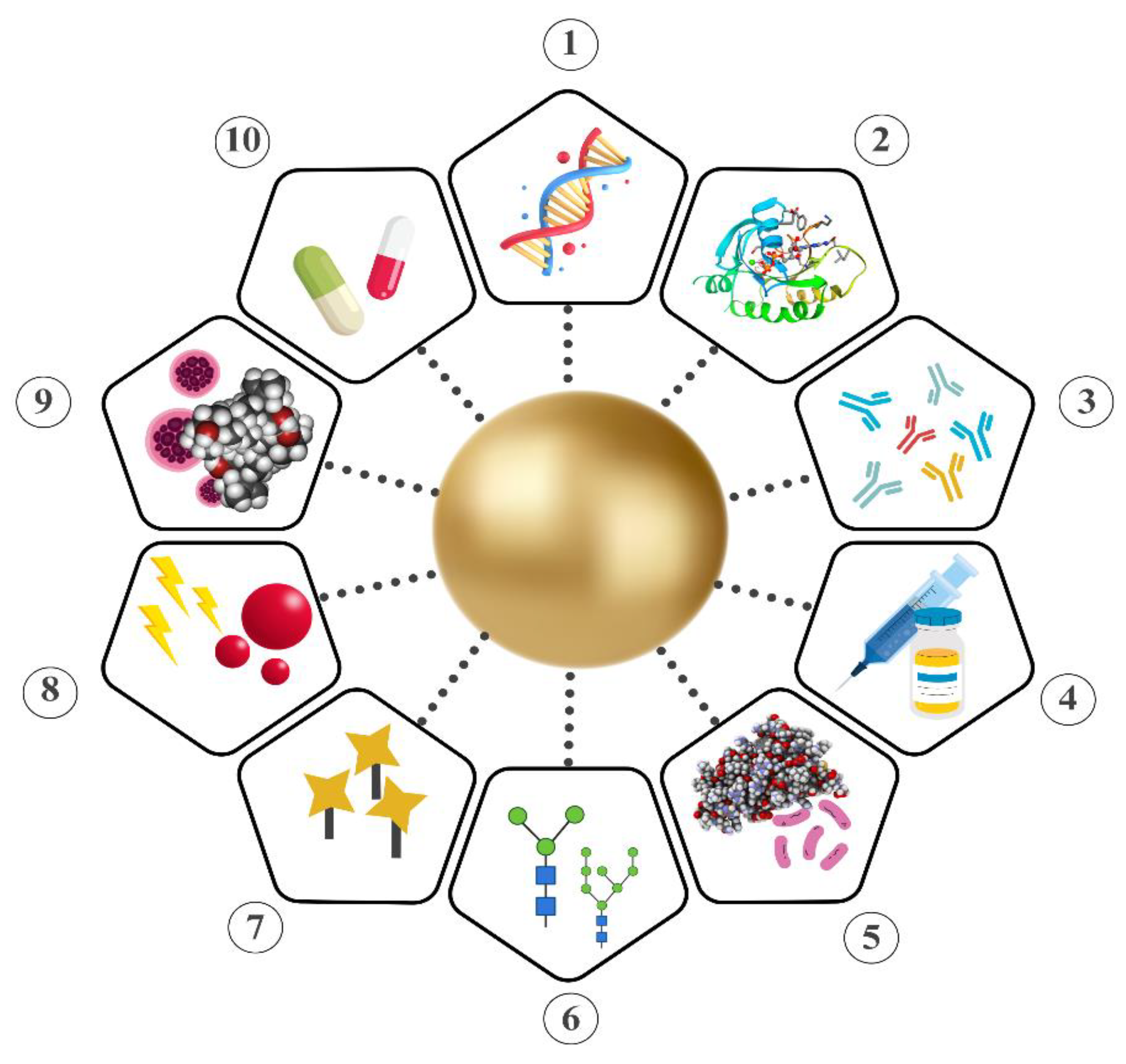
Gold NPs are considered promising carriers for therapeutics due to their facile synthesis, ease of functionalization, tunable physical properties, and low toxicity [95]. They are frequently used for the delivery of biomolecules, including proteins, nucleic acids, and chemotherapeutic agents, since they provide an effective approach for delivery systems [96] (Figure 4).
Gold NPs’ size can be tuned for specific applications to enhance overall efficiency and facilitate deeper penetration into target tissues. For instance, research has focused on the synthesis of smaller gold NPs to overcome the blood-brain barrier (BBB), which is regarded as one of the biggest obstacles when developing future therapies where the direct delivery of functional drugs to the brain is desired [97]. On the other hand, modifying gold NPs’ surface with various ligands or functional groups, such as therapeutic agents, amino acids, proteins, peptides, oligonucleotides, and DNA, not only enhances their interaction with surroundings but also enables a more precise targeting. Therefore, researchers are developing delivery systems with enhanced sensitivity and specificity [98].
Moreover, gold NPs are known as highly biocompatible and low-toxic materials, which is mostly attributed to their intrinsic properties, including chemical inertness, non-reactivity, and non- immunogenicity [99]. This makes them one of the safest materials for delivery systems, especially with their capability of not harming the healthy tissue while delivering drugs to the target area.

Protein Delivery

Proteins are one of the most abundantly found organic molecules within the cells. Consisting of one or multiple chains of amino acids, they serve as essential components for both cellular integrity and activity. Also, they play significant roles in building and repairing tissues, supporting immune function and producing the enzymes and hormones necessary for biological processes [100]. Gold NPs present an efficient platform for the delivery of proteins by mitigating certain drawbacks. Some of these drawbacks include poor solubility through cell membrane and instability against digestive enzymes [101]. Proteins can be conjugated to the gold NPs using various approaches, either with covalent or noncovalent bonding. Covalent bonding, primarily utilizing thiols, serve as a basis for the chemical attachment with molecular cargo. Amines are also used for covalent bonding, although they possess a lower bond strength relative to thiols [102]. On the other hand, non-covalent attachments include electrostatic and hydrophobic interactions, where the balance of these two is regarded as the key to achieve maximum binding affinity [103]. Non-covalent bonding is also highlighted by being reversible, which allows protein cargo to be easily released from the NPs under certain conditions [104].
Recently, gold NPs have been employed as efficient carriers for peptides, regarded as building blocks of proteins. Farhangi et al. synthesized laminin peptide decorated gold NPs, with spherical shape and having a size of 9.75 ± 2.40 nm. Their aim was to target and repair MS lesions through administration of laminin. In vivo experiments on mice models revealed significant accumulation of peptide-modified NPs in the lesion area, with gold content reaching up to 4.675 ± 0.56 μg/g of brain tissue in contrast to 1.073 ± 0.66 μg/g for the control group [105]. Similarly, Liu et al. focused on the delivery of peptide CopA3 along with ginsenoside CK, which has been known to exhibit antimicrobial effects (including antibacterial and anti-inflammatory) and anticancer properties. Synthesizing spherical gold NPs, ranging in diameter between 10 to 30 nm, researchers aimed to assess anti-inflammatory properties of bioconjugated NPs. Modified gold NP suppressed the expression of pro-inflammatory cytokines, IL-6, iNOs, COX-2, TNF-α and IL-1β, considerably reducing their levels by 37.0%, 34.7%, 51.9%, 24.7% and 33.6%, following pre-treatment. It was also stated that NPs significantly alleviated the ROS levels, with concentrations of 20 µg/mL and 40 µg/mL leading to inhibition of ROS production by 40.4% and 65.05%, respectively [106].

Nucleic Acid Delivery

Delivery of nucleic acids is considered crucial for the development of therapeutic approaches in the treatment of various diseases, including genetic disorders and cancers [107]. However certain limitations such as toxicity, rapid degradation, poor transfection efficiency and lack of targeting hinders their utilization on a broader scale [108]. Hence, gold NPs come forward as highly stable, less toxic and biologically inert inorganic nanomaterials, as they can significantly improve the overall efficiency of the delivery process [109]. Guo et al. developed siRNA-gold NP complexes by using the electrostatic interaction between the positively charged amino groups on the surface of the gold NPs and negatively charged siRNA. Using positively charged spherical gold NPs, ranging in size between 2 to 200 nm, the capacity of gold NPs in the delivery of nucleic acids were evaluated. It was stated that gold NPs were similarly efficient to those traditionally used vectors, thus they can be used in the creation of novel approaches for the delivery of genetic cargo [110]Similarly, Mout et al. utilized gold NPs in CRISPR/Cas9 system, which has been one of the main focuses of latest genetic research due to its potential to treat human genetic diseases. By co-assembling gold NPs with single guide RNA and Cas9 protein, efficiency of the vectors in delivering nucleic acids and proteins to the cytoplasm were assessed. Results revealed approximately 90% delivery efficiency in various cell types, along with up to 30% gene editing efficiency [111]. Another similar study delivered SARS-CoV-2 DNA vaccine through chitosan-gold nanostars [112]. The DNA vaccine was delivered with intranasal administration in mice and induced significant antibody levels without a significant reduction for weeks. The induced levels of antibodies increased the IgG and IgA levels in a noticeable amount.
Recently Li et al. synthesized gold NPs to deliver siRNA against Glucose transporter protein-1 (Glut1). Glut1 plays a critical role in the progression of cancer by enhancing glucose transport and is also known to be associated with aggressive tumor behavior when overexpressed. Their findings revealed successful inhibition of human lung cancer cell proliferation (A549) and in vivo xenograft tumor growth by the administration of gold NP complex, achieved through the knockdown of Glut1 that limits glucose uptake [113]. DNA conjugation is also used in anticancer activity, which was evaluated in research that used DNA aptamers to detect cancer cells and destroy it [114].
Based on these findings, it can be concluded that gold NPs possess a wide array of uses in the delivery of nucleic acids, including gene delivery, gene editing and cancer research. Hence, they can be regarded as efficient platforms for future approaches, owing to their facile synthesis, ease of functionalization and high biocompatibility.

Chemotherapeutic Agent Delivery

Gold NPs are considered highly effective carriers for the targeted delivery of chemotherapeutic agents, since they possess advantageous characteristics including low toxicity, inertness, high drug loading capacity and being able to easily be functionalized with a broad range of organic molecules [115]. Mechanism of the release of the chemotherapeutic agents from the gold NPs can depend on either internal or external stimuli, such as pH and light, respectively [7].
As an example, Khutale et al. synthesized spherical gold NPs, ranging from 20 to 25 nm, for the development of a pH-responsive system concerning the delivery of chemotherapeutic agent Doxorubicin (Dox). In this system, researchers utilized polyamidoamine (PAMAM) G4 dendrimer, which facilitated the attachment of Dox through amide bonds. In vitro studies revealed an approximate 50% of Dox release over 96 hours at pH 4.0 (in comparison to the pH 7.0), attributed to the cleavage of amide bond between Dox and the dendrimer in acidic conditions [116]. Similarly, Joshi et al. conjugated gold NPs (having an approximate size of 7 nm) with chloroquine, an antimalarial drug that recently came forward with its anticancer properties. Through in vitro experiments on MCF-7 cell lines, they have revealed a pH-dependent release of chloroquine, accompanied by enhancement of cytotoxicity compared to the unmodified NPs. In more detail, approximately 62% of the drug was released at pH 6.0 after 48 hours, which is reaching up to 81% at lower pH conditions in the same period [117].
On the other hand, Niikura et al. focused on the photoresponsive release of Dox from the cross-linked gold NP vesicles. Following incubation of Dox-encapsulated gold NPs with HeLa cells for 2 hours, diode laser, at 532 nm, was applied for 5 minutes to induce the Dox release. After laser irradiation, a significant increase in the ratio of dead HeLa cells were observed compared to the samples without laser activation, confirming the effectiveness of the drug delivery system [118].
Moreover, overall stability of the payload can be improved through functionalization with gold NPs. Kalimuthu et al. showed that pegylated gold NPs provide a more stable and less toxic environment for peptide-drug-conjugates (PDCs), materials that are being developed to deliver anticancer drugs but often hindered by their low stability and short half-live in biological fluids. Through in vitro experiments, researchers showed a considerable increase in the half-lives of gold NP-coated PCDs, extending from 10.6 to 15.4 minutes to 21.0 to 22.3 hours [119].

Glycan Delivery

In addition to proteins, nucleic acids and chemotherapeutic agents, glycans are another class of biomolecules that are employed by researchers for the delivery by gold NPs. Glycans are important carbohydrate segments. In nature, glycans can be found either free or bound to other structures including proteins, lipids, or peptides. They are essential for a number of cellular processes involved in both health and illness. By participating in cell adhesion and receptor activation, they defend the host from viral and microbial invasions. Protein folding, conformation, solubility, immunogenicity, antigenicity, and resistance to proteolysis are all strongly influenced by glycans. The ability of glycans to form the microbial community of the developing gastrointestinal tract and act as a source of prebiotics is one of their most well-studied roles [120].
In the last years, use of the glycans with NPs have been one of the emerging topics. Researchers particularly focused on developing vaccine adjuvants, especially in the area of oncology, by taking advantage of the glycans’ advantageous properties. To exemplify, Parry et al. synthesized gold NPs, with sizes ranging from 5 to 20 nm, decorated with Tn-antigen. Tn-antigen is a mucin-type O-glycan whose expression is correlated with cancer and various human disorders and regarded as promising candidates for the development of immunotherapies due to its strong interaction with tumors [121]. Immunological assays showed that these NPs generated long-lasting and strong immune responses, shown by higher levels of antibody titers (IgG), in compared to the samples delivered without NP conjugation [122]. Recently, Thomas-Moore et al. used gold NPs as carriers for glycans in PDT. Researchers modified 16 nm gold NPs with glycan-functionalized polyacrylamide probes, such as galactose, glucose, lactose and mannose. These glycan-modified gold NPs were then tested for their ability to inhibit the proliferation of breast cancer cell lines (SK-BR-3 and MDA-MB-231). Findings revealed that gold NPs selectively targeted cancerous cells through glycan-lectin interactions, with galactose-functionalized NPs showing the highest uptake [123].
From another perspective, glycans are also combined with gold NPs to treat infections caused by various pathogens. Since multivalent glycan-lectin interactions are leveraged by pathogens to bind and infect host cells, researchers can create surfaces that resemble pathogen structures by attaching glycans to gold NPs [124]. This mimicry creates a collective arrangement of glycans, similar to glycocalyx structure that covers the cell surface, and provides an enhancement of lectin binding as well as increased stability, solubility and cost-effectiveness [125]. In this manner Kulkarni et al. utilized glycan encapsulated spherical gold NPs, with an approximate diameter of 4 nm, for inhibition of Shiga toxins (Stx1 and Stx2) released from Shigella dysentriae and E. coli O157:H7. These toxins are known to be the main reason for various lethal disorders, such as hemolytic uremic syndrome that may cause severe damage to the human body. Conducting experiments on luc2P Vero cells, they have observed a significant improvement in ED50 value from 0.07 ng/mL to 25.6 ng/mL (for Stx1) and 0.6 ng/mL to 92 ng/mL (for Stx2). This indicated effective inhibition of Shiga toxins, requiring 366 and 153 times more Stx1 and Stx2 to inhibit protein synthesis, respectively, in the presence of glycan conjugated NPs [125].
Given the synergistic effect of gold NPs with proteins, nucleic acids, chemotherapeutic drugs and glycans, it is possible to develop novel drug delivery systems that would contribute significantly to the area of biomedicine. Hence, focusing on crucial parameters, such as optimization, delivery efficiency and biocompatibility, will be crucial for expanding the utility of gold NPs in future therapeutic strategies.

2.8. Anticancer Activity

Gold NPs’ extraordinary characteristics, such as inherent stability, resistance to oxidation, ease of surface functionalization, biocompatibility and strong optical traits, position them as ideal candidates for a broad array of biomedical uses [72]. In addition to their wide utilization in imaging, sensing and delivery systems, their employment is increasingly expanding especially in the design and development of novel anticancer agents [57]. In such a study Babaei et al. focused on the anticancer efficiency of spherical gold NPs, with an average diameter of 17 nm, on human glioblastoma U-87 and U-251 cell lines. Following 72 hours of exposure, it was stated that gold NPs triggered the induction of autophagy and significantly inhibited the proliferation of cancer cells [126]. Similarly, Safwat et al. synthesized spherical gold NPs, with sizes ranging from 9 to 17 nm, to enhance the anticancer effect of antimetabolite drug 5-Fluorouracil (5-FU) on colorectal cancer cells. Findings revealed a significant improvement on anticancer effect, with functionalized gold NPs reaching up to 2-fold increase in effectiveness compared to 5-FU alone [127].
From another point of view, green synthesis methods have become highly popular among nanomaterial research, which has been also applied to gold NPs for the development of eco-friendly and novel systems to mitigate toxicity [128]. Padalia et al. green synthesized gold NPs using aqueous leaf extract of Ziziphus nummularia. Spherical NPs with an average size of 11.65 nm showed dose-dependent in vitro toxic effects on T-47D, HeLa and human fibroblast normal cell lines. When concentration of gold NPs increased from 2 to 200 μg/mL, percent cell viability of T-47D, HeLa and human fibroblast cells decreased from 100 to 44%, 94 to 39% and 100 to 50%, respectively [129]. In another study Babu et al. employed marine seaweed Acanthophora spicifera for the green synthesis of gold NPs. Spherical shaped NPs, with a size less than 20 nm, exhibited cytotoxicity towards human colon adenocarcinoma (HT-29) cell line, with a half-maximal inhibitory concentration (IC50) of 21.86 µg/mL [130]. Considering both chemical and nature friendly approaches, Virmani et al. conducted a comparative study on the anticancer potential of gold NPs synthesized through different methods. Comparing chemically synthesized (spherical, hexagonal and oval) and green synthesized (spherical) NPs, from Ocimum tenuiflorum, they have revealed superior anticancer properties through biologically synthesis. Cell viability assays on HeLa cell line indicated that, treatment with biologically synthesized gold NPs at a concentration of 200 µg/mL were able to decrease cell viability down to 50%. On the contrary, chemically synthesized NPs showed 80% viability even at higher, 400 µg/mL, concentration [131].
Overall, it can be concluded that gold NPs hold promising potential in cancer treatment, especially considering the recent advances on widely utilized green synthesis approaches. Further improvement of these methods will not only enable less expensive and nature friendly production but also contribute to the development of novel anticancer agents utilizing nanomaterials such as gold NPs.

2.9. Antibacterial Activity

NPs have emerged as novel, cost-effective and highly stable tools in antibacterial research, showing significant efficiency even against antibiotic resistant strains of bacteria [132]. Among these, silver NPs have come forward as the most studied nanomaterials, as they exhibit high antibacterial activity towards a wide range of pathogens, with diverse mechanisms involving the constant release of silver ions [22]. Since gold NPs do not include such an effective mechanism, they are not regarded as powerful antibacterial agents as silver NPs. Still, multiple papers in current literature highlight gold NPs, usually combined with various molecules, as effective antibacterials. Lee et al. showed that gold NPs demonstrated antibacterial activity against E. coli, with a minimum inhibitory concentration (MIC) value of 16 μg/mL, without increasing the ROS levels. Gold NPs, with a diameter of 30 nm, inhibited cell growth by induction of membrane depolarization, overexpression of caspase-such as proteins and DNA fragmentation, which collectively resulted in an apoptosis-like cellular death pathway [133]. From another perspective, Piktel et al. assessed the effect of shape on the antibacterial activity of gold NPs. Following synthesis of rod, star, spherical-like and peanut-shaped NPs, antibacterial efficiency was tested on E. coli, S. aureus and P. aeruginosa. Findings revealed ROS-mediated potent bactericidal activity, followed by outer and inner membrane permeabilization, with non-spherical shaped gold NPs [134]. Additionally, Radhi et al. synthesized spherical gold NPs using a one-step pulsed laser ablation method, then evaluated their bactericidal efficiency against four different bacterial strains. Gold NPs with a concentration of 1250 µg/mL showed high activity against P. aeruginosa and S. aureus, while Acinetobacter Baumannii (A.baumannii) was strongly affected even at 1000 µg/mL. In addition, Streptococcus mutans showed sensitivity towards all tested concentrations [135].
Researchers are also trying to maximize gold NPs’ potential by developing unique approaches, through surface functionalization with various molecules, such as antibiotics, peptides, or employing unique green synthesis methods [136]. For example, Hagbani et al. synthesized cefotaxime loaded spherical gold NPs. In vitro tests highlighted potent antibacterial activity of these NPs against both Gram-positive and Gram-negative strains, with MIC values down to 0.68 µg/mL, 0.73 µg/mL, 0.87 µg/mL and 1.03 µg/mL for S. aureus, E. coli, P. aeruginosa and Klebsiella oxytoca, respectively [137]. Similarly, Jana et al. showed gold NPs conjugated with virstatin can be employed as alternatives to antibiotics, since multidrug resistance is becoming a major problem. Spherical NPs with an average diameter of 17 nm effectively inhibited the proliferation of Vibrio cholerae (V. cholerae), with an IC50 value of 0.1 nM [138].
On the other hand, there is a growing interest in green synthesis methods, as they provide nature-friendly and cost-effective alternatives to traditional production steps. Elias et al. used Melaleuca cajuputi leaf extract to synthesize gold NPs. Spherical gold NPs, with sizes ranging from 10 to 50 nm, caused bactericidal effects on Vibrio parahaemolyticus, showing MIC and minimum bactericidal concentration (MBC) of 0.0075 g/mL [139]. In addition, Kerdtoob et al. recently developed a novel approach, where gold NPs are synthesized from Gram-positive Streptomyces monashensis MSK03. Biosynthesized gold NPs, possessing an average particle size of 23.2 nm and spherical shape, were tested on drug-resistant A. baumannii and P. aeruginosa. Accordingly, antibacterial activity was observed with measured inhibition zones of 9.44 (±0.80) mm for A. baumannii and 11.20 (±0.67) mm for P. aeruginosa [140].

2.10. Antioxidant Activity

Oxidative stress is caused by the imbalance in the production of ROS. It results in a dangerous state where cellular damage occurs [141]. Hence, the requirement of antioxidant systems to alleviate harmful effects, such as the disturbance of cellular integrity, is significant. Metallic NPs (including gold) have attracted attention as promising antioxidants, for their strong catalytic activity in radical scavenging reactions [142]. This is mainly due to the ability of gold NPs’ to adsorb molecules and scavenge free radicals on their surface [143]. Researchers also highlighted a possible interaction between conduction electrons of the gold NPs and unpaired electrons from the free radical, improving the overall antioxidant efficiency [144]. Besides, the inherent properties of gold NPs contribute to their utilization in this research area, since they demonstrate chemical inertness, high resistance to oxidation and biocompatibility [145].
Recently it has been shown that gold NPs synthesized by green synthesis methods have significant antioxidant capabilities [128]. By using various quantities of NPs, researchers assessed the antioxidant capability of gold NPs derived from oak gum in terms of their ability to scavenge DPPH radicals. As a positive control, we employed butylated hydroxytoluene, and we contrasted the results of the scavenging activity with the potential of oak gum. For oak gum and NPs, the scavenging activity was 54.56% and 21.54%, respectively [146]. The antioxidant potential of gold NPs produced from Curcuma pseudomontana (C. pseudomontana) was also examined by researchers employing DPPH scavenging activity, H2O2, reducing power, nitric oxide, and the CUPRAC assay, with ascorbic acid serving as the standard material. Results demonstrated a considerable correlation between the quantity of NPs and their capacity to stifle the activity of free radicals. Gold NPs also have superior scavenging capabilities compared to ordinary materials [147]. It has been discovered that the aqueous flower extract of Achillea biebersteinii functions as a capping/reducing factor in gold NP biosynthesis, which is a quick, safe, and economical procedure by Mobaraki et al. Superoxide radical reduction abilities of the extract and Ab-goldNPs were excellent and comparable to typical antioxidant rutin [148]. Another recent study employing an aqueous extract of Allium sativum L. leaf showed that gold NPs had a similar antioxidant activity and had high potential for DPPH inhibition, with an IC50 value of 231 μg/mL [149].
These investigations have demonstrated the superior scavenging performance of gold NPs and highlighted their use as promising antioxidants in further research [150].
Table 1. Research highlighting the properties and applications of Gold NPs in the past five years (2019- 2024).
Highlighted Activity Synthesis Method Property Result Ref
Electrical conductivity Chemical reduction Size = 10 nm

Shape = Spherical
Gold NP-decorated porous carbon microspheres were developed as electrode materials for supercapacitors to enhance electrochemical performance. [151]
Electrical conductivity Electrodeposition Size = Ranging from 20 to 30 nm

Shape = -
Gold NPs were incorporated into amperometric sensors to enhance their sensitivity and electrochemical performance. [152]
Electrical conductivity Chemical reduction Size = Ranging from 1 to 6 nm.

Shape = Spherical
Incorporation of gold NPs into supercapacitor dielectric composites enhanced electrical conductivity and specific capacitance [153]
Electrical conductivity NPs were purchased commercially Size = -

Shape = -
Enhancement in electrical conductivity and electrochemical properties of gelatin methacrylate hydrogels were observed following incorporation of gold NPs. [154]
Electrical conductivity Turkevich method Size = Ranging from 9 to 46 nm

Shape = Spherical
Gold NPs (combined with copper nanowires) enhanced electrochemical conductivity in amperometric sensors, achieving up to 2.3-fold increase in performance. [155]
Electrical conductivity Laser ablation Size = Ranging from 19.43 nm and 32.76 nm.

Shape = -
Gold NPs increased the electrical conductivity of PVP/PVA matrix, with higher concentrations leading to greater AC values. [85]
Electrical conductivity Seeded growth Size = Ranging from 15 nm to 80 nm for spherical NPs.

Length of 40.4 nm and width of 12.0 nm for rod-shaped NPs.

Shape = Spherical and Rod-shaped.
Addition of gold NPs enhanced the fluid's conductivity, with smaller spherical NPs improving electrical properties more effectively than their counterparts.

[156]
Electrical conductivity Green synthesis from plant extract

Laser ablation
Size = Ranging from 3 nm to 24 nm for green synthesized NPs; 2 nm to 30 nm for those synthesized through laser ablation.

Shape = triangular, hexagonal, spherical and irregular
Incorporation of gold NPs enhanced the electrical properties of polymer blend.

Increased AC and DC conductivity, along with improved dielectric permittivity, was observed.
[157]
Electrical conductivity Electrodeposition Size = -

Shape = -
Increase in electron transfer was observed following modification of carbon electrodes with gold NPs.

[158]
Electrical conductivity Laser ablation Size = Average size of 77 ± 4 nm.

Shape = Spherical
Addition of Gold NPs into cement-based composites enhanced electrical conductivity, decreased electrical resistance and increased the piezoelectric response by up to 57 times [159]
Thermal conductivity Laser ablation Size = Average diameter of 6.3 nm

Shape = Crystalline structure
Incorporation of gold NPs improved thermal conductivity of the nanofluid.

Achieving 0.41 W/mK, 26% increase compared to the base fluid was observed.
[160]
Thermal conductivity Chemical reduction Size = Ranging from 20 to 40 nm.

Shape = Spherical
Incorporation of gold NPs into silica gel composites enhanced thermal conductivity by approximately 10–15%. [161]
Thermal conductivity NPs were purchased commercially Size = Having a diameter of 41 nm

Shape = Rod-shaped
Gold NPs improved the thermal properties of tissue-mimicking phantoms by increasing temperature response during photothermal therapy. [162]
Thermal conductivity Chemical reduction Size = -

Shape = -
Incorporation of gold NPs improved thermal conductivity of carbon nanotube fibers by 70%. [163]
Thermal conductivity - Size = Having approximate diameter of 4 nm

Shape -
Gold NPs enhanced heat transfer in the tree-structured polymer networks by increasing the number of thermal transfer channels [164]
Optical Properties Seeded growth Size = Length of 45 nm and 65 nm

Shape = Rod-shaped
Highly active SERS substrates were developed using hollow gold-silver NRs. [165]
Optical Properties Chemical reduction Size = Having a diameter of 13 nm

Shape = -
Optofluidic biosensor was developed using DNA-functionalized gold NPs for detection of mutated β-thalassemia sequence. [166]
Optical Properties NPs were purchased commercially. Size = Having a diameter of 50 nm.

Shape = Rod-shaped.
Gold NPs were developed to enhance photothermal performance in localized tumor treatment. [167]
Optical Properties NPs were purchased commercially Size = Average size of 95.74 nm

Shape = Spherical
Colorimetric biosensor utilizing gold NPs was developed for the enzyme-free detection of Klebsiella pneumoniae (K. pneumoniae). [168]
Optical Properties Turkevich method Size = Having an approximate diameter around 40 nm

Shape = Spherical
Electrochemical sensor utilizing gold NPs was developed for the detection of catechol. [169]
Optical Properties Turkevich method Size = 14 nm

Shape = Spherical
Gold NP-based lateral flow immunoassay was developed for the detection of tuberculosis antigens CFP-10 and ESAT-6. [170]
Optical Properties NPs were purchased commercially Size = 40 nm

Shape =
Gold NP-based electrochemical immunosensors were developed for the detection of HER-1 and HER-2 biomarkers in breast cancer. [171]
Optical Properties Chemical reduction Size = Average size of 40 nm

Shape =
Gold NP integrated plasmonic biosensors were developed for the early detection of Familial Mediterranean Fever. [172]
Optical Properties Chemical reduction Size = Average size of 53.88 ± 1.81 nm

Shape = Spherical
Gadolinium-functionalized gold NPs were developed for dual-modal imaging and photothermal therapy in tumors. [173]
Optical Properties Chemical reduction Size = Average diameter of 16 ± 1 nm

Shape = Spherical
Gold NPs were developed for the rapid detection of microRNAs in milk samples to assess milk quality and cattle health. [174]
Delivery Chemical reduction Size= Ranging from 5 nm to 20 nm

Shape = Spherical
Targeted drug delivery system against SARS-CoV-2 was developed. [175]
Delivery Seeded-growth Size = Average hydrodynamic size of 98.6 ± 0.6 nm

Shape = Urchin-like
Nasal drug delivery system utilizing gold nanourchins as a carrier for targeted brain delivery was developed. [176]
Delivery Chemical reduction Size = 2 nm.

Shape = Spherical
Delivery system using ultra-small gold NPs to cross the BBB was developed. [177]
Delivery Chemical reduction Size = Ranging from 13 nm to 18 nm

Shape = Spherical
Non-viral gene delivery system utilizing gold NPs as carriers for hepatocellular carcinoma treatment was developed. [178]
Delivery Turkevich method Size = Average diameter of 12 nm

Shape = Spherical
Delivery system using gold NPs for the controlled release of dacarbazine was developed.

[179]
Delivery Chemical reduction Size = Average diameter of 35 nm

Shape = Spherical
Resveratrol-gold NP delivery system to inhibit cataract formation was developed. [180]
Delivery Brust-Schiffrin method Size = 4 nm.

Shape = -
Delivery system using gold NPs, functionalized with cRGD peptides, for the delivery of anticancer drug DM1 was developed. [181]
Delivery Gold NPs, with a concentration of 3000 ppm, were purchased commercially Size = Having a diameter of 10 nm

Shape = -
Gold NP-photosensitizer conjugates were developed to enhance the efficiency of PDT in targeting lung cancer stem cells. [182]
Delivery Chemical reduction Size = Average diameter of 24 nm

Shape = -
Gold NP-conjugated MRI contrast agents were developed to enhance the specificity and sensitivity of MRI imaging. [183]
Delivery Chemical reduction Size = Average diameter of 13 nm

Shape = -
Gold NPs were developed for the effective delivery of miR-206 to reduce cell viability and induce apoptosis in breast cancer cells. [184]
Anticancer activity Chemical reduction Size = Average size of 90.6 (± 9.6) nm

Shape = Rod-shaped
Anticancer agents using gold NPs decorated with bovine serum albumin were developed. [185]
Anticancer activity Green synthesis using licorice root extract

Size = 2.647 nm to 16.25 nm size range


Shape = Spherical
At high concentrations, the gold Np mediated by licorice root demonstrated superior antiproliferative action against MCF-7. [186]
Anticancer activity Green synthesis using marine microbe Vibrio alginolyticus (V. alginolyticus)

Size = 100 - 150 nm

Shape =
With a maximum cell death inhibition of 25 mg/mL, the biosynthesized gold NPs showed a dose-dependent inhibitory effect on colon cancer cell growth. [187]
Anticancer activity Green synthesis using Fusarium solani Size = 40-45 nm

Shape = Needle and spindle like shape
On MCF-7 and HeLa cells, these gold NPs had strong cytotoxic effects. [188]
Anticancer activity Green synthesis using Trachyspermum ammi Size = Average 16.63 nm

Shape = Spherical and spheroidal
HepG2 cancer cell lines were shown to respond favorably to these NPs as anticancer agents.

The synthesized NPs' ability to suppress biofilm formation against pathogens, Listeria monocytogenes and Serratia marcescens (S. marcescens), at SUB-MICs.

[189]
Anticancer activity Green synthesis using Mangifera indica Size = 20 nm

Shape = round, triangle, and irregular shape

Modest antibacterial, cytotoxic, and dose-dependent antioxidant activities were demonstrated by gold NPs. [190]
Anticancer activity Green synthesis using Vicoa indica leaf extract Size = Average size of 13 nm

Shape = Spherical
Anticancer activity against lung cancer cell line (A549), with a IC50 value of 73.56 µg/mL, was observed . [191]
Anticancer activity Green synthesis using Gelidium pusillum Size = Average diameter of 12 ± 4.2 nm


Shape = Spherical
Gold NPs demonstrated anticancer activity against cancerous cells ( MDA-MB-23), supported by an IC50 value of 43.09 ± 1.6 µg/mL. [192]
Anticancer activity Green synthesis using Schizophyllum commune Size = Average size of 90 nm

Shape = Spherical
Gold NPs demonstrated dose-dependent anticancer activity against A549 lung cancer cells.

Increasing the doses, from 15 μg/mL to 25 μg/mL, led to decreased cell viability.
[193]
Anticancer activity Green synthesis using Cyclopia genistoides leaf extract Size = Average size of 37 nm

Shape = Spherical and pentagonal
Dose-dependent anticancer activity was observed against PC-3, Caco-2, and MCF-7 cells.

PC-3 cell death increased by 2.5-fold compared to MCF-7 cells at a concentration of 100 µg/mL of gold NPs.
[194]
Antimicrobial activity Green synthesis using Presley leaf, Petroselinum crispum (P. crispum), extract Size = Ranging from 20 to 80 nm

Shape = Multi-shaped and spherical
Gold NPs(A) (2.5 mL extract used) demonstrated antibacterial inhibition against two Gram-negative pathogenic bacteria and demonstrated the highest anticancer efficiency against human colon cancer cells (HCT116). [195]
Antimicrobial activity Green synthesis using Mentha longifolia (M. longifolia) leaves extracts Size = 3.45 ± 2 nm

Shape = Round oval
The NPs markedly enhance antibacterial, antioxidant, antinociceptive, analgesic, and sedative actions [196]
Antimicrobial activity Green synthesis using Citrus macroptera (C. macroptera) Size = 20 nm

Shape = Pseudo-spherical
The gold NPs that were manufactured demonstrate antibiofilm action against P. aeruginosa biofilm. Additionally, they primarily show cytotoxic effects on HepG2. [197]
Antimicrobial activity Green synthesis using Cynodon dactylon L. Pers (C. dactylon) Size = 21- 33 nm

Shape = Spherical and irregular

Gold NPs exhibited significant antibacterial efficacy against pathogenic bacteria such as Enterobacter cloacae, Staphylococcus haemolyticus, Staphylococcus petrasii subsp. pragensis, and Bacillus cereus, with inhibition zones of between 12 and 13 mm. [198]
Antimicrobial activity Green synthesis using Scutellaria baicalensis Size = 20-40 nm

Shape = Spherical
Gold NPs had strong cytotoxic, antibacterial, and antioxidant properties. They were not hazardous to RAW 264.7 or A549 cells, according to in vitro cytotoxicity data. [199]
Antimicrobial activity Green synthesis using Jatropha integerrima (J. integerrima) Size = 38.8 nm

Shape = Spherical
Maximum and minimum antibacterial activity against B. subtilis and E. coli is demonstrated by the gold NPs. B. subtilis, S. aureus, E. coli, and K. pneumoniae were shown to have MICs of 5.0, 10, 2.5, and 2.5 lg/mL, respectively, when gold NPs were used.

[200]
Antimicrobial activity Green synthesis using Platycodon grandiflorum Size = 15 nm

Shape = Spherical
The P. grandiflorum gold NPs that were produced demonstrated effective antibacterial action against B. subtilis (11 mm) and E. coli (16 mm). [201]
Antimicrobial activity Green synthesis using Arthrospira platensis extract Size = Average size of 10.98 nm

Shape = Rod-shaped
Antibacterial activity against Streptococcus pneumoniae was observed with a MIC value of 12 μg/mL. [202]
Antimicrobial activity Green synthesis using Lysinibacillus odysseyi PBCW2 Size = Average size of 31.6 ± 9.7 nm

Shape = Spherical
Antibacterial activity was observed against both Gram-positive and Gram-negative strains (S. aureus, E. coli, V. cholerae Shigella dysenteriae, Aeromonas hydrophila and Salmonella typhi)

MIC and MBC values were found between 25 to 40 μg/mL and 60–85μg/mL, respectively.
[203]
Antimicrobial activity Seeded-growth Size = 82.57 nm

Shape = Rod shaped
Gold NPs demonstrated antibacterial and antifungal activity against E. coli, S. aureus, and Candida albicans (C. albicans), at concentrations ranging from 0.25 ng/mL to 0.125 ng/mL. [204]
Antioxidant activity Green synthesis using Oak gum extract Size = Average 10-15 nm

Shape = crystalline structure
It was found that the material demonstrated remarkable antioxidant properties through DPPH radical scavenging experiments. [146]
Antioxidant activity Green synthesis using C. pseudomontana isolated curcumin Size = Average 20 nm

Shape = Spherical
Effective antibacterial, anti-inflammatory, and antioxidant properties were exhibited by the gold NPs. [147]
Antioxidant activity Green synthesis using Achillea bieber- steinii flower extract

Size = Average 8 nm

Shape = Spherical
It was discovered that the Ab-gold NPs were efficient against the DPPH radicals. In addition, they showed better DPPH scavenging action than the plant extract did. [148]
Antioxidant activity Green synthesis using Paracoccus haeundaensis BC74171T Size = Average size of 20.93 ± 3.46

Shape = Spherical
Antioxidant activity was observed, with a DPPH radical scavenging percentage ranging from 13.04 ± 3.14% at 10 μg/ml to 73.04 ± 3.01% at 320 μg/ml. [205]
Antioxidant activity Green synthesis using Vitex negundo (V. negundo) leaf extract Size = Ranging from 20 to 70 nm

Shape = Spherical
DPPH radical scavenging activity reached 84.64% at a concentration of 120 µg/mL, along with an IC50 value of 62.18 µg.

Nitric oxide assay indicated 69.79% scavenging activity with IC50 value of 70.45 µg, for the same tested concentrations
[206]
Antioxidant activity Green synthesis using Hubertia ambavilla plant extract Size = Average size of 50 nm

Shape = Flower-shaped
DPPH radicals were neutralized with an IC50 value of 16.5 μg/mL.

Dose-dependent reduction in UV-A induced MMP-1 production in normal human dermal fibroblast cells was observed, achieving IC50 of 9.25 μg/mL.
[207]
Antioxidant activity Green synthesis using Glaucium flavum leaf extract Size = Average size of 32 nm

Shape = Spherical
DPPH assay revealed dose-dependent antioxidant effect of gold NPs.

At concentrations of 125 μg/mL, 500 μg/mL, and 1000 μg/mL, the NPs achieved reductions in DPPH radicals of 23%, 37%, and 44%, respectively.
[208]
Antioxidant activity Green synthesis using Capsicum annum fruit extract Size = Ranging from 20 to 30 nm

Shape = Spherical
DPPH assay showed 86% efficiency of NPs, at a concentration of 100 µg/mL, in comparison to Vitamin C that displayed 69.3% efficiency for the same tested concentrations. [209]
Antioxidant activity Green synthesis using Nostoc calcicola Size = Ranging from 20 to 140 nm

Shape = Triangular, spherical and cuboidal
DPPH radicals were effectively neutralized, with an IC50 value of 55.97 μg/mL. [210]
Antioxidant activity Green synthesis using curcumin isolated from C. pseudomontana Size = Average diameter of 20 nm

Shape = Spherical
DPPH, hydrogen peroxide, nitric oxide, reducing power and CUPRAC assays showed dose-dependent antioxidant activity.

At highest concentration of 25 μg/mL, NPs exhibited inhibition rates of 85.2%, 83.2%, 84.5%, 87.9% and 85.6%, respectively.
[147]

3. Synthesis of Gold Nanoparticles

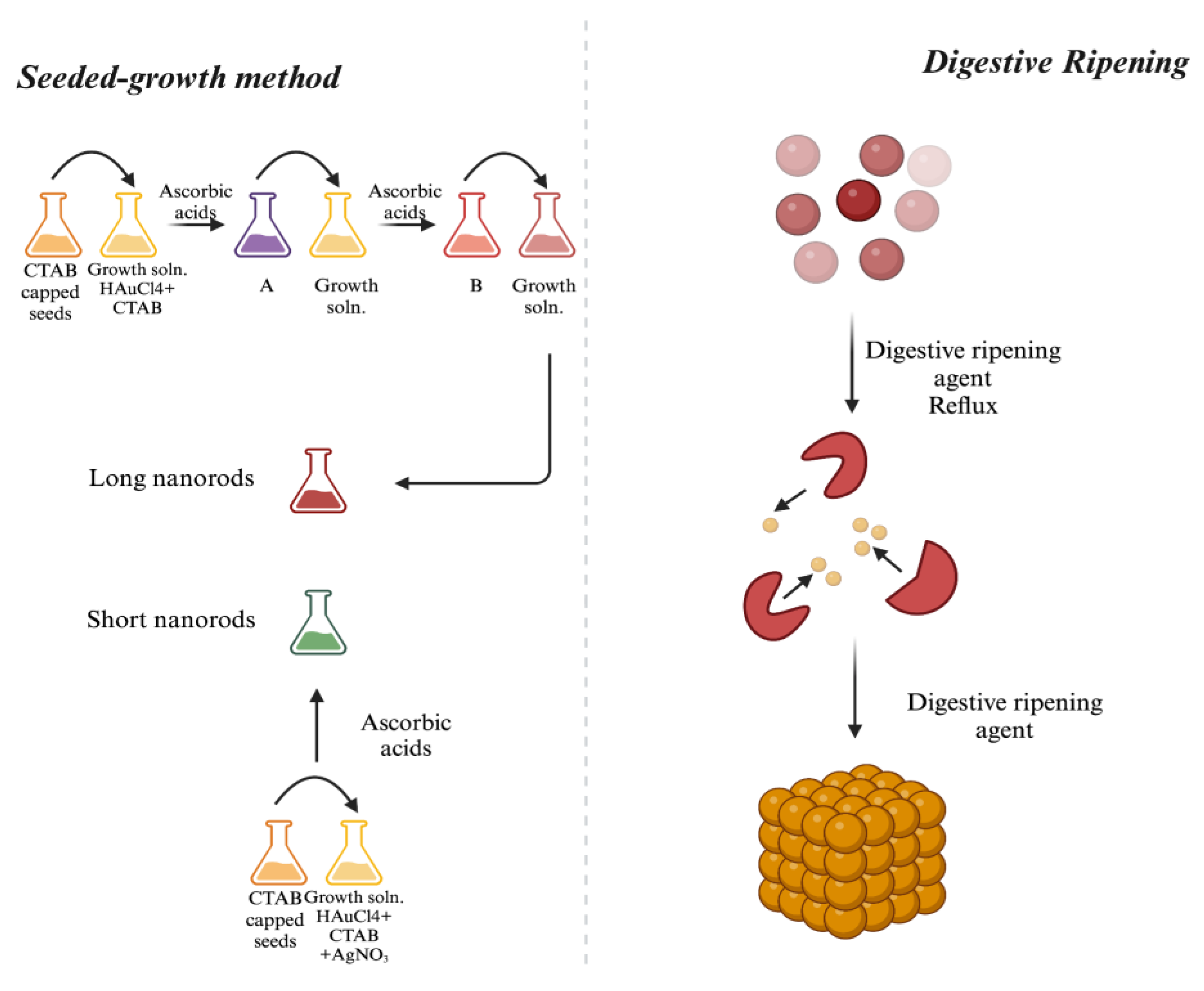
There are two types of approaches used in the synthesis of gold NPs: top-down and bottom-up schemes. Top-down approaches entail the fragmentation of complex material into NPs, whereas bottom-up approaches begin at the atomic scale. Figure 5 depicts the fundamental procedures included in each method. Laser ablation [211], ion sputtering [212],UV and IR irradiation [211], and aerosol technology are examples of synthesis methods that use the top-down approach. On the other hand, the bottom-up approach involves the reduction of Au3+ to Au0.
Thus, based on the top-down and bottom-up mechanisms, the production of gold NPs encompasses a range of techniques, such as chemical, physical, and green/biological procedures, which will be further below.

3.1. Physical Synthesis

The γ-irradiation approach has been demonstrated to be the most effective for synthesizing gold NPs with adjustable dimensions and high purity. The γ-irradiation technique is employed to produce gold NPs ranging from 5 to 40 nm in size. This approach employed a natural polysaccharide alginate solution as a stabilizer [213].
Gold NPs are produced by a photochemical process employing chloroauric acid (HAuCl4) and an aqueous glycine solution subjected to UV irradiation. The initiator comprises amino acid-capped gold NPs that are subsequently functionalized with glycine [214].
Using cetyltrimethyl ammonium bromide (CTAB) as a binding agent and citric acid as a reducing agent, the microwave irradiation method was used to produce gold NPs [215]. Gold NPs are manufactured using either the heating approach or the photochemical reduction method. Citrate, tartrate, and malate ligands were employed for lowering HAuCl4 [216]. A photochemical methodology has been documented for the synthesis of gold-polyethylene glycol core-shell NPs, measuring 10 - 50 nm in aqueous solution, utilizing redox and polymerization processes. This approach involved the reduction of gold salt through radical production, utilizing polyethylene glycol diacrylate via a UV process facilitated by a photo-initiator, 2-Hydroxy-2-methyl-1-phenyl-1-propane [217].
Laser ablation synthesis in solution is a straightforward process for the fabrication of NPs from various solvents. The irradiation of various metals submerged in a solution by a laser beam generates a plasma that produces NPs. The top-down methodology for metal reduction to NPs, employing laser ablation techniques, provides a stable solution devoid of stabilizing agents or chemicals [218]. Laser ablation in an aqueous biocompatible media can be used to produce gold NPs, which significantly alters the surface chemistry as well as the physical and biological characteristics [219].
Because of their distinct surface chemistry, laser-synthesized NPs can react with a range of innovative biocompatible materials [220]. For example, they may have O− functionalization at high pH due to the presence of some higher oxidation states, which enables hydrogen bonding interactions. Without any group ligand-specific binding, gold NPs can be non-covalently conjugated to a wide range of substances, enhancing their potential as innovative biocompatible materials for unexplored or underdeveloped biological applications [221].
In order to prevent contamination by certain undesirable compounds that can obstruct biological research, gold NPs have been generated and functionalized utilizing the capabilities and benefits of fs laser ablation by Correard et al. Their findings demonstrated that stable and monodisperse gold NPs may be produced using laser ablation in solutions containing dextran, polyethylene glycol, and water [222]. The formation of gold NPs by pulsed laser ablation of bulk, high-purity gold in deionized water was examined in a similar study. The scientists irradiated the gold surface with a Q-switched Nd:YAG laser, then looked at how the thickness of the liquid layer and the post-ablative modification processes affected the size, aggregation, and ablation effectiveness of the gold NPs. The average particle size was found to be decreased from 15.12 nm to 9.5 nm by laser-induced NP modification, and the size distribution was narrowed with 532 nm pulses. This allowed for the control of NP redistribution and average size in ablated colloid solutions. Without the need of any additional chemical reagent, Wender et al. produced stable gold NPs by simply ablating an Au foil that was positioned inside or outside of four ionic liquids (ILs) using a laser. Following laser ablation, irregularly shaped spherical gold NPs with a diameter ranging from 5 to 20 nm were created. The produced NPs' size and form were found to be correlated with the location of NP nucleation and growth, which might be either within or at the IL surface. In fact, the stabilization of the gold NPs created by laser ablation outside of the ILs was significantly influenced by the surface ion orientation and chemical makeup of the IL/air interface [223].
Vacuum sputtering is a method employed in the fabrication of thin films or coatings, such as gold NPs. This method relies on the application of a potential difference between the two electrodes within the vacuum chamber, creating an electric field. An inert gas enters the chamber and undergoes ionization. A metal target (cathode) is bombarded with argon plasma. As a result, atomic clusters are extracted from the target location and deposited onto a surface or into a liquid solution [224]. ILs make it possible to prepare the metal NP system, which lowers the need for successive stabilizers. In order to prepare solutions via cathode sputtering, low vapor pressure is required. With the groundbreaking study of the Kuwabata group in 2006, the relatively new field of research on the creation of NPs by the sputtering method onto ILs was launched. Sputtering 1-ethyl-3-methylimidazolium tetrafluoroborate (EMI.BF4) directly onto its surface produced gold NPs gold NPs with a diameter of 5.5 nm and a deviation of 0.9 nm [225]. By using plasma sputtering in a solution medium in an open system at atmospheric pressure, Hu et al. were able to create gold clusters (average particle size about 1.5 nm) and NPs (average particle size approximately 3.5 nm) with spherical morphology. The solvent medium and temperature have an impact on the particle sizes. They showed that gold NPs of varying sizes might find use in a variety of industries [226].
A straightforward mechanical technique called ball milling uses attrition to create NPs. It moves kinetic energy from the reduction material to the grinding medium. Consolidation and compaction, an industrial-scale procedure where NPs are "put back together," yield enhanced characteristics [22].

3.2. Chemical Synthesis

3.2.1. Turkevich Method

The Turkevich method, initially documented in 1951, is a well acknowledged approach for producing spherical gold NPs in the size range of 10-30 nm, therefore establishing it as the pioneering chemical system for gold NPs synthesis. This technique entails the reduction of gold precursors by the use of reducing agents such as trisodium citrate, ascorbic acid, amino acid, and polymers. Subject the gold precursor to heat until it reaches boiling point, then add a 1% solution of trisodium citrate while stirring vigorously. Formation of gold NPs is indicated by the color transition from pale yellow to wine red.
The Turkevich method is a systematic and consistent technique used to generate spherical particles within the size range of 10-30 nm. However, as particles exceed 30 nm in size, they lose their spherical shape and exhibit limited yield [227].

3.2.2. Electrochemical Method

Using tetra alkyl ammonium salts as stabilizers in a nonaqueous solution, Reetz et al. 's 1994 work showed that NPs may be synthesized electrochemically, enabling the size-selective synthesis of transition metal particles [228,229]. Through the use of tetra alkyl ammonium salts as stabilizers of metal clusters in a nonaqueous solution, their research demonstrated that size-selective nano scales of transition metal particles may be configured electrochemically. The superiority of the electrochemical technique over alternative NP manufacturing methods has been confirmed, mostly attributed to its modest equipment requirements, low cost, reduced processing temperature, good quality, and facile yield control [230,231]. Using a basic two-electrode cell, the gold NPs were synthesized electrochemically by oxidizing the anode and reducing the cathode [232]. The electrochemical synthesis method can be used to produce gold NPs on the surface of multi-walled carbon nanotubes supported by glassy carbon electrodes [233].

3.2.3. The Brust-Schiffrin Method

Brust-Schiffrin synthesis (BSS), initially presented in 1994, is a technique for synthesizing stable thiol functionalized metal NPs, specifically designed for the preparation of gold NPs in organic solutions [234]. The procedure entails the immersion of a water-based gold precursor into an organic solvent, namely tetraoctylammonium bromide (TOAB), followed by its reduction with sodium borohydride (NaBH4) in the presence of alkanethiol. The transition in color from orange to brown signifies the synthesis of gold NPs [235].
The Brust approach is a straightforward technique for producing thermally and air-stable gold NPs with precise size and dispersion. However, its drawbacks include producing less evenly distributed gold NPs and requiring the use of immiscible organic solvents [236].

3.2.4. Seeded-Growth Method

Using the seeded growth technique, gold NPs can be produced in a range of geometries including rods, cubes, and tubes. This technique produces gold NPs of diminutive dimensions, measuring between 5 and 40 nm in diameter. It is straightforward, efficient, and inexpensive. This procedure entails the reduction of the precursor using a potent reducing agent (NaBH4) to generate seed particles. These particles are subsequently introduced into a metal solution that contains weak reducing agents (ascorbic acid or sodium citrate) to facilitate the formation of OH- groups and structure-directed agents (Figure 6). Variations in the concentration of metal seed, reducing agents, and structure-directed agents can yield diverse nanostructures [235].
Effective synthesis of rod-shaped gold NPs by seed-mediated growth requires precise control of parameters such as HAuCl4 concentration, temperature, and seed number. Greater concentrations of HAuCl4 led to the formation of larger seed rods with reduced aspect ratios, whereas elevated temperatures resulted in the formation of rods with lower aspect ratios [237].

3.2.5. Digestive Ripening

An efficient approach to produce uniform gold NPs in the presence of an abundance of ligands (digestive ripening agents) is known as digestive ripening. The fundamental procedure involves subjecting a colloidal solution to elevated temperatures (about 138oC) for 2 minutes, followed by additional heating at 110oC for 5 hours, utilizing alkanethiol. Thermal conditions are the primary determinant of the size distribution of gold colloids [238]. Furthermore, there are alternative techniques that provide the synthesis of gold NPs by the utilization of ultrasonic vibrations (Figure 6) [239,240].
A simple and useful chemical strategy for producing monodispersed NPs is the digestive ripening method. One other advantage of this approach is the substantial production of NPs [241]. An underlying drawback of the digestive ripening technique is the challenge of regulating the morphology of NPs when subjected to extremely high temperatures [242].

3.3. Green/Biological Synthesis

Although chemical and physical approaches are straightforward for rapidly producing gold NPs, their applicability in biology is limited by the requirement for toxic and expensive reducing agents or applications (Table 1) [243]. Hence, there is an increasing need to create environmentally sustainable and economical methods for the synthesis of NPs that do not involve the use of hazardous substances. NPs synthesis using green methods has gained significant interest as an environmentally benign and sustainable approach in recent years. Green techniques involve the synthesis of NPs using microbes, enzymes, and plant compounds or plant extracts (Figure 7) [244].

3.3.1. Microorganism-Based Gold NPs Synthesis

Microorganisms, such as bacteria and fungi, are frequently employed in the manufacture of gold NPs. These microbes engage with organic metals and can produce gold NPs through both extracellular and intracellular mechanisms. Microorganisms are cultivated in settings with gold precursors, subsequently extracted and purified by various procedures. The dimensions and morphology of gold NPs can be regulated by microbial growth factors [245].
When gold NPs interact with bases that include phosphorus or sulfur, they cause free radicals and break respiratory chains, which ultimately results in cell death. To further contribute to cell death, they might further decrease ATPase function and inhibit tRNA attachment to ribosomal subunits [246]. Gold NPs generate surplus electrons, exterminating Leishmania and creating ROS (O2 and OH). These radicals obliterate the pathogen's DNA and other cellular constituents [247]. These gold NPs may also impede transmembrane H+ efflux, representing an additional possible mechanism [248]. Due to their diminutive size and approximately 250 times diminished antibacterial efficacy compared to bacterial cells, they are more prone to adhere to cell walls and impede processes that often lead to cell death [249]. Herdt et al. argued that interaction with a gold surface can damage DNA [250].
The precipitation of gold NPs in bacterial cells upon exposure to Au+3 ions was observed by Beveridge and Murray. Organic phosphate compounds contribute to the formation of octahedral gold, potentially serving as agents for biota-gold complexation. Both Fe+3 reducing bacteria and Shewanella algae are capable of reducing Au+3 ions in anaerobic conditions [251]. S. algae and hydrogen gas fully decompose Au ions, resulting in the formation of gold NPs of 10-20 nm in size [252]. V. alginolyticus marine bacteria was employed by Shunmugam et al. to synthesize gold NPs. The highest suppression of colon cancer cell proliferation was seen at 25 mg/mL when the biosynthesized gold NPs were applied. The environmentally benign and economically viable green synthesis of gold NPs has anti-inflammatory and anti-cancer properties against colon cancer [187]. Unlike the other studies, gradient centrifugation was employed by Qui et al. to eliminate the hazardous components of gold NPs from S. aureus. Muscle cell viability was observed to benefit from the pure gold NPs, which also provided protection against damage caused by cardiotoxins. They showed promise in facilitating myocardial infarction healing by reducing infarct area and enhancing heart function when constructed onto an elastic scaffold to form a cardiac patch [253].
The incorporation of fungi in the synthesis of NPs is a relatively new development and shows potential for huge-scale NP manufacturing. Indeed, fungus release substantial quantities of the enzymes relevant to NP formation and are more easily cultivated in both laboratory and industrial settings. Fusarium oxysporum (F. oxysporum), Verticillium sp., Thermomonospora sp., and Rhodococcus sp. have been documented as fungal and actinomycete species capable of synthesizing NPs either intra- or extracellularly [254,255].
Because of the fungi's favorable growth characteristics, gold NPs may be produced on a huge scale in the industrial setting. Furthermore, fungi demonstrate high gold NP monodispersity. Yeast species including Pichia jadinii and Yerrowiali polytica, as well as fungal species including F. oxysporum and Verticillium sp., have a good ability to manufacture gold NPs [256].
The fungal manufacturing of gold NPs can occur both extracellularly and intracellularly. The internal mechanism can be elucidated by the reduction of sugars, proteins such as ATPase, glyceraldehyde-3-phosphate dehydrogenase, and 3-glucan-binding proteins implicated in the energy metabolism of fungal cells [257]. Au+3 permeate the cell membrane and are reduced by cytosolic redox mediators. It remains ambiguous whether the diffusion of Au+3 ions transpire across the membrane via active bioaccumulation or passive biosorption [258]. The research on fungal ultrathin slices revealed the concentration of gold NPs in the vacuoles of cells [259]. The extracellular synthesis of gold NPs transpires through the adsorption of AuCl4 ions onto cell wall enzymes via electrostatic interactions with positively charged groups [260]. NADPH-dependent oxidoreductases, located either on the cell surface or within the cytoplasm, are essential enzymes in the biosynthesis of gold NPs, similar to their role in the synthesis of other NPs, such as silver NPs [261,262].
Algae, a distinctive source of chemicals such as fucoidan, neutral glucan, and alginic acid, has considerable medicinal importance owing to their antibacterial, anticoagulant, and antifouling properties. The production process can occur via both extracellular and intracellular pathways, rendering algae a unique subject for investigating gold NP characteristics [263]. Blue-green algae, such as Spirulina subsalsa, which is a member of the Spirulinaceae family, have been used for manufacturing gold NPs. The green algae Rhizoclonium hieroglyphicum and Rhizoclonium riparium are members of the Cladophoraceae family. Chakraborty et al. [264] and Nayak et al. [265] have reported finding diatoms Nitzschia obtusa and Navicula minimum. Turbinaria conoides algae extract was used to produce gold NPs in a green way. Color change and spectral data, such as a large surface plasmon resonance band at 520–525 nm, confirmed the synthesis of gold NPs. TEM and crystalline structure with a size range of 6–10 nm was used to corroborate the structure of the produced Au [266]. In a similar study, Tetraselmis suecica, a marine microalga, was used as a reducing agent during the synthesis of gold NPs. The formation was verified by Ultraviolet–Visible (UV-vis) spectroscopy, which showed a peak at 530 nm. A Bragg reflection was detected using FTIR and X-ray diffraction (XRD) spectroscopy. The generation of well-dispersed gold NPs was confirmed by TEM and laser light scattering, with 79 nm being the most common particle size [267]. Optical, physical, chemical, and antibacterial characteristics of self-assembled gold NPs were revealed by their production employing extract from Chlorella vulgaris by Annamalai et al. From 2 to 10 nm in diameter, spherical self-assembled cores were seen in the gold NPs. They tested positive for S. aureus and C. albicans through biological screening, indicating that they may have use in green chemistry and as a strong medication [268]. Chakraborty et al. propose that secreted algal enzymes contribute to the biosynthesis of gold NPs, with NADPH-dependent reductase playing a pivotal role. This enzyme functions as a NADH electron carrier, effectively reducing Au ions to gold NPs via an enzymatically mediated electron transfer process within the inner mitochondrial membrane matrix [269]. An efficient, quick, and one-pot approach was used to achieve the synthesis of gold NPs utilizing Cystoseira baccata extract, a brown macroalgae that is present in the Atlantic, Baltic, and Mediterranean Seas. Zeta potential measurement, TEM, High-resolution TEM, Scanning Electron Microscopy (SEM), and UV-vis spectroscopy were used to analyze the biosynthesized gold NPs. To stop coalescence and aggregation, the extract was applied as a shielding substance [270]. Tetraselmis kochinensis, a green alga with a size range of 5–35 nm, has been used in the environmentally benign biosynthesis of gold NPs Senapati et al. Because the cytoplasmic membrane and cell wall include enzymes that reduce metal ions, these gold NPs have a significant impact on the cell wall. They may find use in catalysis, biological applications, and medication administration [271].
In addition to complete organisms, biological products such as nucleic acids, amino acids, lipids, proteins (enzymes), and viroid capsules were utilized in the manufacture of gold NPs. These compounds contain carbonyl and hydroxyl functional groups, which facilitate the reduction of Au+3 ions to Au0, subsequently stabilized by stabilizing agents. Although numerous studies have documented the production of microbial gold NPs, the precise process remains inadequately defined and is now under investigation. According to the assumed process of microbial synthesis, the nucleation and development of gold NPs are caused by the enzymes released by microorganisms reducing Au+3 ions to Au0 [272]. The organic phosphate compounds may facilitate the formation of a complex with gold salts, leading to the in vitro accumulation of gold NPs in B. subtilis [273].

3.3.2. Plant, Fruit, and Waste Extracts-Based Gold NPs Synthesis

When compared to microbial synthesis, plant-mediated inorganic NP synthesis has a number of benefits, such as the ability to create enormous quantities of NPs on an industrial scale and the avoidance of the labor-intensive maintenance of cell cultures [274]. Revaluing plant secondary metabolites based on polyphenols as effective reducing agents for metallic precursors is the mechanism-guided synthesis procedure. The hydroxyl groups present in plant-derived polyphenols were discovered to play a successful role in the reduction of gold ions by promoting the oxidation reaction and the particular synthesis of quinine forms [275].
Numerous research studies have documented the synthesis of gold NPs from plant leaf extracts. Leaf extracts of C. pseudomontana [147], Lawsonia inermis [276], and Scutellaria barbata [277] have been used to produce gold NPs. Antonio Zuorro's study involved the synthesis of NPs from both the leaves and the juice extract of the plant. This study employed kiwi fruit juice to synthesize and stabilize gold NPs [278]. The juice of C. macroptera was successfully used to synthesize gold NPs. The NPs produced from C. macroptera juice extract exhibit a surface plasmon resonance band at 544 nm [197]. Gold NPs were produced by combining an appropriate quantity of Punica Granatum juice under mild reaction conditions in a separate study [279].
Furthermore, Jasminum auriculatum leaf extract, which functions as a stabilizing and reducing agent, is used in a trouble-free, ecologically friendly method for the biogenic manufacture of gold NPs as reported by Balasubramanian et al. The biogenic gold NPs production was validated by the surface plasmon resonance peak in the UV-visible absorption spectra at 547 nm. The biogenic gold NPs have an average size of 8–37 nm and are spherical, according to TEM and SEM examination. The biogenic gold NPs are a flexible choice for heterogeneous catalysis, as demonstrated by their catalytic reduction activity on p-nitrophenol. The biogenic gold NPs' cytotoxicity demonstrated that the NPs significantly inhibited the proliferation of the human cervical cancer cell line at a dose-dependent level, with an IC50 value of 104 μg/mL [280]. In a different study, Al-Radadi attempted to generate innovative, environmentally friendly, and reasonably priced gold NPs using licorice root extract. On MCF-7 and HePG-2 (liver) cell lines, the cytotoxicity of green produced gold NPs was also evaluated using an MTT technique. When tested against bacterial and fungal strains, as well as cell lines, gold NPs showed little antibacterial, antifungal, and reflected anticancer activities [186]. Ullah et al. also evaluated the in vitro antibacterial efficacy of gold NPs generated from the aqueous extract of Tamarindus indica, and they examined the in vivo sedative and analgesic properties of the crude extract. The samples exhibited significant antibacterial efficacy against K. pneumoniae, B. subtilis, and Staphylococcus epidermidis, demonstrating a 10-12 mm inhibition zone. Gold NPs exhibited stability under elevated temperature, varying pH levels, and in a 1 mM saline solution. They demonstrated sedative and analgesic properties, underscoring the significance of phytochemical-mediated gold NP production [281]. The study by Ruf et al. examined the analogous biological activities of green produced gold NPs, favoring M. longifolia leaf extracts for their manufacture. The dimensions and morphology of the NPs were validated using sophisticated techniques, revealing polydisperse and spherical configurations of gold NPs measuring 13.45±2 nm. In comparison to MLE, the NPs markedly enhance antibacterial, antioxidant, antinociceptive, analgesic, and sedative actions [196]. El-Borady et al. utilized Presley leaf (P. crispum) extract for the first time to produce gold NPs, which were then employed as antioxidant, anticancer, antibacterial, and photocatalytic agents. The research employed four extract volumes to synthesize gold NPs, yielding four distinct sizes and morphologies. The NPs were evaluated using many approaches, with gold NPs(D) (20 mL plant extract) exhibiting the highest antioxidant capacity. The NPs exhibited antibacterial action against two Gram-negative bacteria, but not against Gram-positive bacteria. The photocatalytic efficacy for the degradation of methylene blue dye was attained rapidly [195]. Using seed extract from Trachyspermum ammi, the researchers created gold NPs for the study. They then investigated the NPs' effectiveness against drug-resistant biofilms of S. marcescens and L. monocytogenes, as well as their potential to inhibit HepG2 cancer cell lines. Against these bacteria, the NPs demonstrated strong biofilm inhibitory effect, blocking important components such as exopolysaccharide, motility, and CSH. Additionally, they reduced the amount of intracellular GSH in HepG2 cancer cells, which made them more susceptible to the production of ROS and led to apoptosis [189].
Human cervical, breast, and normal cell lines were used as test subjects for Mangifera indica gold NPs. With respect to concentration, the cell viability varied, ranging from 98-67 to 98-60%. Gold NPs' potential for anticancer activity is highlighted in this work [190]. J. integerrima Jacq. flower extract was used to create gold NPs; the Fast Fourier Transform Infrared (FT-IR) revealed phenolic chemicals, and the UV-vis spectrum revealed a high peak at 547 nm. Their crystalline nature was confirmed by XRD, SAED, TEM, and (dynamic light scattering) DLS. gold NPs had MICs of 5.0, 10, 2.5, and 2.5 lg/mL, respectively, against B. subtilis and E. coli. Their crystalline nature was proven by their spherical form and 38.8 nm size [200]. Similar to this, a straightforward and environmentally friendly method was used to create gold NPs that contained an aqueous extract of C. dactylon. When C. dactylon-loaded gold NPs were tested at varying concentrations (0.625–100 μg/mL) against the MCF-7 cell line, the IC50 was determined to be 31.34 μg/mL using the MTT assay [198].
The field of study is showing promise for the advancement of environmentally friendly nanomaterials, which could lead to the creation of sustainable solutions for pressing health and environmental issues. To fully realize this field's potential and tackle difficult problems in a variety of industries, more research and development are needed.

3.4. Variables Influencing NPs Synthesis

The manufacture, characterization, and applications of NPs are affected by several factors, including substrate selection and catalytic activity during synthesis. The dynamic characteristics of produced NPs change under various environmental conditions and influences. Critical parameters influencing NPs formation encompass solution pH, temperature, extract concentration, raw material concentration, particle size, and synthesis processes. This section delineates essential aspects affecting NPs biogenesis, emphasizing the necessity of accounting for these factors in the manufacture, characterization, and application of NPs [282].

pH

The shape and size of NPs are significantly influenced by the pH levels of the substrate and the surrounding media. Medium to larger-sized NPs is usually produced in an acidic pH environment, and changes in the substratum's pH cause the NPs' size and form to change [283]. Polyakova et al. used citrus lemon to investigate how pH affected the synthesis of gold NPs. The findings demonstrated that whereas basic pH (7–9) slows down NPs growth, acidic pH (2.5–5) tends to promote NPs growth by quickening the aggregation step. It was discovered by the researchers that the creation of 8-nm-sized gold NPs can occur at a pH of 5 [284].

Temperature

Temperature substantially affects the formation kinetics of gold NPs and the mechanics of particle growth. Fluctuations in temperature factors influence the ultimate aspect ratio, length, and diameter of gold NRs. Reduced temperatures, between 21 and 35°C, led to the formation of shorter and thinner gold NRs due to the energy necessary for particle growth [285]. The physical approach necessitates temperatures over 350°C, whereas chemical procedures demand temperatures below 350°C. The synthesis of green technology generally necessitates temperatures below 100°C or at ambient temperature, as the temperature of the reaction medium affects the characteristics of the NPs [286].

Pressure

Pressure is crucial for NP production. The pressure exerted on the reaction medium influences the morphology and dimensions of the produced NPs. The rate of metal ion reduction utilizing biological agents is significantly accelerated at ambient pressure settings [287].

Time

Researchers investigated the impact of reaction time on gold NPs manufactured from palm leaves by extracting samples from the reaction media at intervals ranging from 5 to 480 minutes. The UV spectroscopy results indicated an increase in absorption wavelength from 5 minutes to 60 minutes. After 60 minutes, the wavelength stabilizes, indicating the complete reduction of gold ions [288]. Temporal differences may arise from numerous factors, including particle aggregation owing to prolonged storage, alterations in particle size with time, and inherent shelf life, all of which influence their potential [289].

Concentration of Source Extract/Biomass and Salt

The efficacy of NPs is frequently determined by the concentration of plant extract and salt solution. Several studies have observed changed form and increased production of NPs. This is why it's frequently necessary to determine the right concentrations [290]. Variations in salt concentrations affecting NPs production have also been observed. The synthesis of NPs was enhanced by elevating the salt content, achieving optimal yield at 0.7 mM concentrations [291].

3.5. Characterization of Gold NPs

Dimensions, surface arrangement, and morphology determine the physical characteristics of gold NPs. Key to the work is characterization, which verifies the production of gold NPs with a color change from pale yellow to ruby red. Further characterization methods are employed to have a deeper understanding of their characteristics (Figure 8) [235].

3.5.1. Ultraviolet–Visible Spectroscopy (UV-vis)

Gold NPs size, concentration, and aggregation level may all be estimated using UV-vis spectroscopy, which is a very helpful method. UV–vis spectroscopy examination is a prevalent method for assessing the production of metal NPs by investigating their distinctive optical properties, which are contingent upon their size and morphology [292]. In the UV-vis spectrum, the gold NPs that were made show a clear single SPR band at 527 nm. In addition, most laboratories have UV-vis spectrometers; the analysis doesn't change the material, and spectrum registration takes little time. The extinction spectra of gold NP can be evaluated by Mie theory, contingent upon the modification of the metal's dielectric constant to account for NP dimensions and physicochemical conditions, as evidenced by direct measurements on individual ensembles of NPs [293].
To offer a straightforward and rapid approach for assessing the size and concentration of gold NPs, Haiss et al. contrasted theoretical findings with experimental data. Gold NPs measuring between 5 and 100 nm were produced and analyzed using TEM and UV-vis. The dimensions and concentration of gold NPs can be directly ascertained from UV-vis spectra utilizing eqs 10-13 for the computation of d and eq 14 for the determination of N, together with the relevant fitting parameters [294]. The assessment of gold NP aggregation can be accomplished using UV-vis spectroscopic techniques. Indeed, when gold NPs combine, a shoulder appears at a wavelength of roughly 600 nm, which is near the distinctive SPR band. Overall, without sample pretreatment, this spectroscopic approach offers an effortless and quick analysis method for quality control right after their synthesis. For an accurate characterization of particle size, UV-VIS spectroscopy in conjunction with other analytical techniques is therefore essential [295].

3.5.2. X-ray Diffractometer (XRD)

The crystalline structure of gold NPs was determined by XRD research [296]. Using equipment that operated at a voltage of 20 mA and was effective at 40 kV, the sample preparation process entailed reducing the gold NP solution to be drop-coated on a glass surface using Cu Kα radiations [297]. The crystalline characteristics of gold NPs synthesized by K. pneumoniae were examined using XRD diffraction patterns, and the average size of these NPs was determined employing the Debye-Scherrer equation [298]. Four unique 2θ peaks at 38.1, 44.3, 64.1, and 77.7 are observed in gold NPs, which closely resemble the typical Bragg reflections of a face centered cubic lattice. Intense diffraction peak at 38.1 suggests that Au0 has a preferential growth orientation in the 111 directions [299].

3.5.3. Transmission Electron Microscope (TEM) and Scanning Electron Microscope (SEM)

While the optical microscope has a resolution limit of microns, electron microscopes such as TEM can picture NPs on the nanoscale scale. TEM pictures are used to identify the size and surface morphology of NPs, making them appropriate for the structural and chemical characterization of nanomaterials at the nanoscale. TEMs can be magnified up to 50 million times to enhance the visibility of features [300]. On the other hand, the sizes and forms of gold NPs in the dry condition can be established using TEM imaging. It is possible to see that gold NPs have a distinct structural shape and accurate size measurements [301].
Furthermore, this approach requires sample preparation, which may lead to artifacts like gold NP aggregation [295]. Surface alterations are undetectable by normal TEM techniques; specialized equipment, such as cryo-TEM, is necessary. Surface alterations are undetectable by ordinary TEM techniques; specialized equipment, such as cryo-TEM [302], glycerol spraying/low-angle rotating metal shadowing TEM [303], and others, is necessary.
SEM is a method that employs direct viewing to analyze NP morphology through electron microscopy. It provides benefits for morphological and dimensional analysis, although it also presents disadvantages, including insufficient data regarding size distribution and the actual population mean. The apparatus comprises an electron cannon, condenser lenses, and a vacuum system. SEM generates three primary types of images: external X-ray maps, backscattered electron images, and secondary electron images [304]. Preparing the sample for analysis with SEM involves coating copper grids with tiny layers of carbon. To produce these films, a small amount of material was dropped onto the grip, and the remaining solution was removed using blotting paper. The film was then further dried under a mercury lamp for a minimum of five minutes [305]. The average diameter of the gold NPs, which were discovered to be in a variety of geometries, including rectangle, square, cubic, and triangular, was 60 nm [298]. Notwithstanding these benefits, this technique is labor-intensive and expensive, frequently necessitating supplementary information regarding the size distribution.

3.5.4. Energy-Dispersive X-ray Spectroscopy (EDAX)

Energy Dispersive X-ray Spectroscopy (EDAX) is a radiological method employed to determine the fundamental composition of substances. Integrated with SEM/TEM, the microscope's imaging capability facilitates specimen identification. Digital X-ray (EDX) data exhibits spectrum peaks, including the distinctive absorption band peak of metallic gold nanocrystallites in gold NPs, detected at around 2.2 keV [306].

3.5.5. Dynamic Light Scattering (DLS)

Despite its long-standing history and popularity, people often arbitrarily select the weighting and mean of DLS, a prevalent method for assessing NP size, to conform to other techniques and expectations [307]. In contrast to other more complex methods, DLS is comparatively inexpensive and simple to use, making it the main instrumentation option for assessing the size and size distribution of NP suspensions [308]. More than 50% of drug products incorporating nanomaterials submitted to the US Food and Drug Administration's Center for Drug Evaluation and Research in recent years used DLS to characterize size. Even though DLS measurements are widely used in the characterization of NPs, they nevertheless present a number of difficulties because of dubious data interpretation and processing [309].
Gold NPs exhibit exceptional light scattering properties at or near their surface plasmon resonance wavelength. Jans et al. preference was for DLS as a highly practical and effective instrument for gold NP bioconjugation and biomolecular binding investigations. The conjugation of protein A with gold NPs under various experimental settings, together with the quality and stability of the resulting conjugates, was extensively monitored and evaluated using DLS [310]. Liu et al. employ DLS to enhance the understanding of potential interactions between gold NP materials and biomolecules both in vivo and in vitro, specifically focusing on gold NRs. In gold NRs with particular aspect ratios, the size distribution diagram displays two peaks: one at 70–80 nm and the other with a hydrodynamic diameter of 5-7 nm. Rotational diffusion produces the small peak, but when proteins adhere to the NRs, the peak associated with rotational diffusion significantly changes. They proved that DLS is a useful instrument for characterizing NRs. It provides information on gold NR–protein interactions that is supplementary to molecular spectroscopy methods [311].

3.5.6. Fourier Transform Infrared Spectroscopy (FT-IR)

For the identification of the functional group present between a reducing agent and a gold precursor in suspensions of NPs, FT-IR is a highly sensitive method. It validates the functionalization of gold NPs by illustrating the interactions between them and the reactive agent [312].
The utilization of contemporary computing tools enables the quantitative analysis of NPs to be accomplished in a few seconds.

3.5.7. X-ray Photoelectron Spectroscopy (XPS)

X-ray photoelectron spectroscopy (XPS) is an effective technique for analyzing surface properties (less than 10 nm) and is extensively utilized across various disciplines in science and engineering [313]. It is a quantitative method renowned for its superior capacity to ascertain chemical states, extensive applicability, and non-destructive characteristics. Nonetheless, it possesses deficiencies including inadequate spatial resolution (15 mm), moderate absolute sensitivity (0.01 - 1.00%), incapacity to identify hydrogen or helium, and protracted analytical durations. The deficiencies are progressively being rectified as the applications of the method expand. XPS applications encompass the analysis of surface functionality in organic and plastic coatings, the assessment of oxidation states in catalysts and nanomaterials, and the provision of both quantitative and qualitative data. These applications are compatible with diverse sample types, including gaseous, liquid, or solid, and are non-destructive [314]. XPS offers valuable insights into the coordination chemistry of ligands on NP surfaces, particularly in comparison to uncoordinated ligands [315].

3.5.8. Thermogravimetric Analysis (TGA)

Nanomaterials' physicochemical characterization needs more analytical methods to figure out their shape and make-up, since their surface make-up can change when they come into contact with biological fluids or environmental factors. Precisely defining the surfaces and functional coatings on NPs is essential for achieving complete and uniform coverage during modification [316]. TGA is a dependable analytical method for evaluating the purity of nanomaterials by the observation of mass changes during heating. It generates a decomposition curve, indicating the oxidation temperature and remaining mass. The residual mass may result from inorganic leftovers, metal catalysts associated with synthesis, or contaminants. Nonetheless, TGA is both deleterious and costly. It can also evaluate organic residues, surface melting characteristics, and oxidation resistance [317].

4. Toxicity

Though their broad application depends on biosafety evaluation, gold NPs are interesting biomedical instruments. The need to assess their effects on health and learn more about their toxicity and biocompatibility is developing [318]. At first, the cytotoxic effects of novel nanomaterials are assessed since they may change the way cell’s function. Nevertheless, implicit biocompatibility is not always ensured by the absence of cytotoxicity. The basis for biocompatibility is how a material interacts with its biological surroundings; it guarantees that treated biomaterials won't produce any harmful or defensive reactions. This idea is essential to determining biocompatibility [319].
Biocompatibility is often defined as the ability of a certain substance or device to function well with living tissue or organisms. When a nanomaterial interacts with its host without causing harmful effects such oxidative stress, DNA damage, mutagenesis, or apoptosis, biocompatibility is generally achieved. Prior to in vivo testing, cytotoxicity—the detrimental effect on a particular cell line—is usually evaluated using in vitro assays. Nevertheless, regardless of the methodology, existing studies on the toxicity of gold NPs are incongruous [320].
Toxicity mechanisms of NPs are classified into two groups, including oxidative and non-oxidative mechanisms (Figure 9). One of the toxicity mechanisms of NPs is thought to be the production of ROS, which can inhibit antioxidants and cause oxidative stress, potentially resulting in inflammation and damage to molecules and cell membranes [321]. Additional oxidative stress can harm DNA, which triggers cell death processes in the cell [322]. Ozcicek et al.'s study demonstrated a correlation between elevated ROS generation and gold NP concentrations ranging from 1 μg/mL to 100 μg/mL [56].
In assessing the toxicity of gold NPs, one must consider various potential pathways that may be involved, including but not limited to genotoxicity, ROS generation, mitochondrial damage, cell death pathways, hazardous material leakage, the interaction of endocrine disruption with molecules, or alterations in cell morphology. Although oxidative stress is a well-known mechanism of NP toxicity, it is important to understand that there are a number of non-oxidative mechanisms by which gold NPs can cause toxicity. Due to its ability to disrupt multiple metabolic pathways and have an impact on amino acid synthesis, mitochondrial toxicity is the primary cause of gold NP cytotoxicity [323].
Numerous journals have published reviews of NPs' toxicity, primarily focusing on NP-cell/organism interactions within specific contexts (Table 2). Because high doses are employed in in vitro tests, which are not feasible for in vivo investigations due to the progressive clearance of NPs by renal and fecal excretion, in vitro and in vivo studies cannot be directly compared. Therefore, minimal dosages should be employed in in vitro tests for the assessment of nanotoxicity [324,325]. Furthermore, unlike with traditional chemicals or medications, the dosage–response connection cannot be ascertained by specifying the NP dose as a concentration. Other measurements, such as surface area or the quantity of NPs, are probably more relevant [326].
NPs show promise in biological applications; however, pharmacokinetics, biodistribution, and toxicity measurements require further focus, particularly when human subjects are involved. Nanomaterials ought to be both large enough to load particular components and small enough to get through biological barriers [328].
Humans are exposed to metal NPs through the bioapplication of nanomaterials; hence, a comprehensive evaluation of the health concerns associated with designed gold NPs is required. For preclinical studies of nanosafety and toxicity risk, this calls for the creation of new standardization and certification assays that assess physicochemical properties, sterility, pyrogenicity, biodistribution, ADME, pharmacokinetics, and in vivo and in vitro toxicity [329]. The ongoing efforts to standardize gold NP risk assessment methodologies still require refinement. While Standards Developing Organizations (SDOs) are working to produce key standards for nanomaterials, none have yet gained widespread acceptance. Nanomaterials are thought of similarly to conventional chemicals [99].
Requirements for compounds used in food, medicine, cosmetics, and devices have been established by US and European authorities. In the US, the FDA sets these criteria, although South Korea, China, and Japan have their own requirements for characterizing nanomaterials. South American countries' industries and regulatory environments heavily rely on The Brazilian Health Regulatory Agency (ANVISA) laws. The types of nanomaterials and their intended uses must be taken into consideration when regulating their characterization, standards, and evaluation techniques [330].
When considering bioapplication, a systemic toxicity assessment of gold NPs is typically carried out using animal models, including mice or rats (Table 2) [331]. It is essential that gold NPs enter the body through the subcutaneous, intravenous, and oral routes. These particles have the ability to change or undergo metabolism in response to biological components. Numerous factors influence the nanotoxic effect of gold NPs; hence, a detailed investigation is necessary [322].
Different-shaped gold NPs (spherical, rod-shaped, triangle-shaped, star-shaped, octahedron-shaped, plate-shaped, and prism-shaped) have been synthesized and their cytotoxicity evaluated. Steckiewicz et al. examined the cytotoxic effects of rod-shaped, star-shaped, and spherical gold NPs on human fetal osteoblast hFOB 1.19 and pancreatic duct cell hTERT-HPNE cell lines. Gold NP rods had the highest toxicity to human cells, whereas gold NP spheres demonstrated the lowest toxicity [332]. Wang et al. indicated that gold NP NRs exhibited significantly greater toxicity than gold NP hexapods. This emphasized that the cytotoxicity of gold NPs is contingent upon their form [333]. The arrangement of surface atoms in gold NPs may have altered due to their varying geometries, particularly when comparing spherical shapes to star or rod configurations. A greater number of atoms at angles and edges may lead to enhanced interactions with biomolecules, resulting in toxicity in rods and stars, which is generally not seen in spherical NPs [334].
The immunological organs of broiler chickens were studied, and it was discovered that adding 15 ppm gold NPs to drinking water resulted in DNA fragmentation, elevated IL-6, histopathological alterations, oxidative damage, and a significant drop in antibody titer against avian influenza and newcastle disease [335].
The suitability of gold NPs for surface modifications is crucial not only for controlling their toxicity but also for enhancing their effectiveness in imaging, diagnostic, and anticancer applications. Localized and specific agglomerations of gold NPs are key to the efficient detection or elimination of targeted structures. However, this also poses a major drawback in treatment areas, such as anticancer therapies and drug delivery, where agglomeration can present significant risks to human health [336]. To prevent off-target effects during gold NP treatment, their surface characteristics must be precisely controlled during the synthesis process. The surface charge of gold NPs significantly affects their biocompatibility, influencing uptake levels and potentially leading to non-specific binding [337]. The surface chemistry of gold NPs, modified with conjugated molecules like PEG or chitosan, can also significantly influence their biodistribution [338].

5. Future Trends

Since they are nontoxic and biocompatible, gold NPs have become increasingly important in many areas of nanotechnology. Green synthesis pathways should prove beneficial in places where traditional gold NPs are already having an impact. Because of their strong antibacterial action, plant-based gold NPs are anticipated to be useful in the fight against antimicrobial resistance. A new generation of broad-spectrum antimicrobial medications might be developed as a result of gold NPs; by 2021, the global gold NPs market is expected to increase at a compound annual growth rate of 18.84 percent. Gold NPs have a practical and affordable market through green/biological synthesis, especially for commercial uses such as point-of-care testing, large-scale diagnostics, vaccine research, and home use [350]. Human health has advanced significantly as a result of recent approvals and trials, including the use of gold NPs in nanomedicine. However, the international community needs to create particular protocols for the preclinical development and characterization of these products because of their distinct physicochemical and optical features and possible bioapplications.
One of the most prominent applications of gold NPs is in anticancer research. Not only are they efficient anticancer agents, but they can also be utilized in cancer imaging and drug delivery, enabling multiple approaches to anticancer research [28]. The need for advanced methodologies and novel agents in anticancer research is well-established. Gold NPs are among the most suitable metal NPs for surface modification. Given their application in anticancer research and their ability to be modified with antibodies, patient-specific anticancer drug delivery and treatments are expected to become a major trend in the near future [351]. Given the importance of alternative treatments in anticancer research and their low toxicity combined with green synthesis methods [352], gold NPs are expected to become one of the leading nanomaterials in future anticancer research.
Another unique characteristic of gold NPs is their capability in colorimetric assays for molecular detection and diagnostic applications. Various types of molecules, such as pharmaceutical compounds, heavy metals and enzymes can be detected with gold NP-based colorimetric sensors. Moreover, various agents found in foods, including chemicals, microorganisms, and antibiotics, can also be detected using colorimetric sensors [353]. Most importantly, the color change is detectable with the naked eye, up to a certain threshold, offering a significant advantage in sensing experiments. These wide-ranging possibilities for gold NP-based sensing applications are all due to their unique optical properties. Thanks to their significant SPR, many PTT-based applications show great promise in diagnostics.
Green synthesis, surface modification, and the excellent optical properties of gold NPs are also being applied to another emerging field: wastewater treatment and detection in environmental applications. Various gold NP-based sensors can be used for dye absorption, heavy metal detection, and the immobilization of chemical toxins [354]. However, the lack of research on gold NP toxicity and the absence of optimized large-scale production using green methods still hinder the development of these promising trends, especially in environmental applications [355].
On the other hand, the distribution of patents registered in the last 5 years shows similarity in terms of the involvement of properties and synthesis when compared to the published articles on gold NPs (Figure 10). From 2019 to 2022, there is consistent patent registration, suggesting the involvement of gold NPs in various fields. The fact that properties and synthesis methods in these patents comprise nearly 50% of total patent registrations further highlights the development of gold NPs in these areas. The distribution of patent registrations is consistent with the distribution of published articles in recent years (Figure 1). One noticeable trend is the drop in 2023, with a similar potential for 2024. The same decline is also observed in the number of published papers, which may indicate a shift in research focus or a saturation where current challenges hinder research progress. At this point, the importance of synthesis methods should be highlighted, especially with their constant involvement (nearly 30%) in both patent registrations and published articles. Despite the visible drop, gold NP synthesis methods remain a major research area, indicating their importance in the future of gold NPs.

6. Conclusions

Gold NPs, a category of metal NPs, possess considerable promise in biological domains owing to their wide range of forms and dimensions, spanning from 1 to 100 nm. Known for their facile synthesis, surface modification, biocompatibility, non-toxicity, high surface-to-volume ratio, and size tunability, gold NPs find extensive use in diverse biological applications. Because of their strong localized surface plasmon resonance, gold NPs have revolutionized biosensing, bioimaging, cancer research, and photothermal and photodynamic therapy. They are useful in medicinal applications because of their simple functionalization with a variety of ligands, which enables targeted administration. They are also perfect for creating antioxidants and antibacterials due to their electrical and heat management capabilities.
Since physical and chemical synthesis requires the use of hazardous materials, green synthesis—which uses plants and microbes—is garnering interest in the scientific community. Also, effective characterization approaches are therefore necessary since factors impacting NP quality and quantity are critical to their applications in the environmental, electrical, medicinal, and drug delivery sectors.
In this article, we provided a detailed overview of the physical and chemical properties, production, characterization, and toxicity of gold NPs. This review covers the methods for characterizing NPs as well as the factors that affect their production. More in-depth toxicity studies, distinct surface functionalization methods, quantitative phytochemical reduction kinetics, and deeper mechanistic insights have all been provided by recent research on gold NPs, which has improved our understanding and may offer a more complete picture of their molecular and functional aspects. Future studies in green nanotechnology will offer a thorough comprehension of these elements as well as cutting-edge technology for effective uses in the pharmaceutical and biological sectors. For the purpose of creating green commercialization strategies for the safe and sustainable use of gold NPs in a variety of industries, it is essential to comprehend the biological and environmental effects of these particles.
Given these factors, it is crucial to evaluate and alleviate any potential adverse effects in order to ensure safer outcomes. All of these characteristics point to the significant role that gold NPs play in nanotechnology, where their special set of qualities not only encourages novel applications but also emphasizes the necessity of more research.

Author Contributions

Conceptualization, S.K.; writing—original draft preparation, H.D., E.A., F.E. writing—review and editing, S.K. M.B., visualization, H.D., E.A., F.E. All authors have read and agreed to the published version of the manuscript.

Funding

This research received no external funding.

Institutional Review Board Statement

Not applicable

Informed Consent Statement

Not applicable.

Data Availability Statement

Not applicable

Conflicts of Interest

The authors declare no conflicts of interest.

References

  1. Elahi, N.; Kamali, M.; Baghersad, M.H. Recent Biomedical Applications of Gold Nanoparticles: A Review. Talanta 2018, 184, 537–556. [Google Scholar] [CrossRef] [PubMed]
  2. Pal, R.; Panigrahi, S.; Bhattacharyya, D.; Chakraborti, A.S. Characterization of Citrate Capped Gold Nanoparticle-Quercetin Complex: Experimental and Quantum Chemical Approach. Journal of Molecular Structure 2013, 1046, 153–163. [Google Scholar] [CrossRef]
  3. Si, P.; Razmi, N.; Nur, O.; Solanki, S.; Pandey, C.M.; Gupta, R.K.; Malhotra, B.D.; Willander, M.; De La Zerda, A. Gold Nanomaterials for Optical Biosensing and Bioimaging. Nanoscale Adv. 2021, 3, 2679–2698. [Google Scholar] [CrossRef] [PubMed]
  4. Riley, R.S.; Day, E.S. Gold Nanoparticle-mediated Photothermal Therapy: Applications and Opportunities for Multimodal Cancer Treatment. WIREs Nanomed Nanobiotechnol 2017, 9, e1449. [Google Scholar] [CrossRef]
  5. Naghdi, S.; Rhee, K.Y.; Hui, D.; Park, S.J. A Review of Conductive Metal Nanomaterials as Conductive, Transparent, and Flexible Coatings, Thin Films, and Conductive Fillers: Different Deposition Methods and Applications. Coatings 2018, 8, 278. [Google Scholar] [CrossRef]
  6. Tsai, C.Y.; Chien, H.T.; Ding, P.P.; Chan, B.; Luh, T.Y.; Chen, P.H. Effect of Structural Character of Gold Nanoparticles in Nanofluid on Heat Pipe Thermal Performance. Materials Letters 2004, 58, 1461–1465. [Google Scholar] [CrossRef]
  7. Ghosh, P.; Han, G.; De, M.; Kim, C.; Rotello, V. Gold Nanoparticles in Delivery Applications☆. Advanced Drug Delivery Reviews 2008, 60, 1307–1315. [Google Scholar] [CrossRef]
  8. Venkatesh, G.; Serdaroğlu, G.; Üstün, E.; Haripriya, D.; Vennila, P.; Siva, V.; Haseena, S.; Sowmiya, V.; Pradhiksha, A. Green Synthesis, Characterization, Anti-Cancer and Antimicrobial Activity of AuNPs Extracted from Euphorbia Antiquorum Stem and Flower: Experimental and Theoretical Calculations. Journal of Drug Delivery Science and Technology 2024, 95, 105583. [Google Scholar] [CrossRef]
  9. Liu, H.; Zhang, M.; Meng, F.; Wubuli, A.; Li, S.; Xiao, S.; Gu, L.; Li, J. HAuCl4-Mediated Green Synthesis of Highly Stable Au NPs from Natural Active Polysaccharides: Synthetic Mechanism and Antioxidant Property. International Journal of Biological Macromolecules 2024, 265, 130824. [Google Scholar] [CrossRef]
  10. Rashidipour, M.; Soroush, S.; Ashrafi, B.; Sepahvand, A.; Rasoulian, B.; Sohrabi, S.S.; Babaeenezhad, E. Green Synthesis of Gold Nanoparticles (AuNPs) Using Aqueous Extract and Essential Oils from Satureja Khuzestanica Jamzad: Evaluation of Their Antibacterial and Antifungal Activities. Biologia 2023, 79, 333–342. [Google Scholar] [CrossRef]
  11. Thakur, R.; Yadav, S. Smart Multifaceted Potential Microbial Inoculant Isolated from Rhizospheric Soils of Bergenia Ciliata and Possible Role in Developing Green Biosynthesized Nanoparticles. Biocatalysis and Agricultural Biotechnology 2024, 57, 103087. [Google Scholar] [CrossRef]
  12. Hammami, I.; Alabdallah, N.M.; Jomaa, A.A.; Kamoun, M. Gold Nanoparticles: Synthesis Properties and Applications. Journal of King Saud University - Science 2021, 33, 101560. [Google Scholar] [CrossRef]
  13. Kajani, A.A.; Bordbar, A.-K.; Zarkesh Esfahani, S.H.; Razmjou, A. Gold Nanoparticles as Potent Anticancer Agent: Green Synthesis, Characterization, and in Vitro Study. RSC Adv. 2016, 6, 63973–63983. [Google Scholar] [CrossRef]
  14. Ahmed, S.; Annu; Ikram, S. ; Yudha S., S. Biosynthesis of Gold Nanoparticles: A Green Approach. Journal of Photochemistry and Photobiology B: Biology 2016, 161, 141–153. [Google Scholar] [CrossRef]
  15. Zhang Toxicologic Effects of Gold Nanoparticles in Vivo by Different Administration Routes. IJN 2010, 771. [CrossRef]
  16. Pissuwan, D.; Niidome, T.; Cortie, M.B. The Forthcoming Applications of Gold Nanoparticles in Drug and Gene Delivery Systems. Journal of Controlled Release 2011, 149, 65–71. [Google Scholar] [CrossRef]
  17. Fröhlich, E.; Roblegg, E. Models for Oral Uptake of Nanoparticles in Consumer Products. Toxicology 2012, 291, 10–17. [Google Scholar] [CrossRef]
  18. Patra, C.R.; Bhattacharya, R.; Mukhopadhyay, D.; Mukherjee, P. Fabrication of Gold Nanoparticles for Targeted Therapy in Pancreatic Cancer. Advanced Drug Delivery Reviews 2010, 62, 346–361. [Google Scholar] [CrossRef]
  19. Jo, M.-R.; Bae, S.-H.; Go, M.-R.; Kim, H.-J.; Hwang, Y.-G.; Choi, S.-J. Toxicity and Biokinetics of Colloidal Gold Nanoparticles. Nanomaterials 2015, 5, 835–850. [Google Scholar] [CrossRef]
  20. Sani, A.; Cao, C.; Cui, D. Toxicity of Gold Nanoparticles (AuNPs): A Review. Biochemistry and Biophysics Reports 2021, 26, 100991. [Google Scholar] [CrossRef]
  21. Document Search - Web of Science Core Collection Available online:.
  22. https://www.webofscience.com/wos/woscc/basic-search (accessed on 10 October 2024).
  23. Duman, H.; Eker, F.; Akdaşçi, E.; Witkowska, A.M.; Bechelany, M.; Karav, S. Silver Nanoparticles: A Comprehensive Review of Synthesis Methods and Chemical and Physical Properties. Nanomaterials 2024, 14, 1527. [Google Scholar] [CrossRef] [PubMed]
  24. Eker, F.; Duman, H.; Akdaşçi, E.; Bolat, E.; Sarıtaş, S.; Karav, S.; Witkowska, A.M. A Comprehensive Review of Nanoparticles: From Classification to Application and Toxicity. Molecules 2024, 29, 3482. [Google Scholar] [CrossRef] [PubMed]
  25. Eker, F.; Duman, H.; Akdaşçi, E.; Witkowska, A.M.; Bechelany, M.; Karav, S. Silver Nanoparticles in Therapeutics and Beyond: A Review of Mechanism Insights and Applications. Nanomaterials 2024, 14, 1618. [Google Scholar] [CrossRef]
  26. Xu, F.; Li, Y.; Zhao, X.; Liu, G.; Pang, B.; Liao, N.; Li, H.; Shi, J. Diversity of Fungus-Mediated Synthesis of Gold Nanoparticles: Properties, Mechanisms, Challenges, and Solving Methods. Critical Reviews in Biotechnology 2024, 44, 924–940. [Google Scholar] [CrossRef]
  27. Kalimuthu, K.; Cha, B.S.; Kim, S.; Park, K.S. Eco-Friendly Synthesis and Biomedical Applications of Gold Nanoparticles: A Review. Microchemical Journal 2020, 152, 104296. [Google Scholar] [CrossRef]
  28. Patil, S.; Chandrasekaran, R. Biogenic Nanoparticles: A Comprehensive Perspective in Synthesis, Characterization, Application and Its Challenges. Journal of Genetic Engineering and Biotechnology 2020, 18, 67. [Google Scholar] [CrossRef]
  29. Ghobashy, M.M.; Alkhursani, Sh.A.; Alqahtani, H.A.; El-damhougy, T.K.; Madani, M. Gold Nanoparticles in Microelectronics Advancements and Biomedical Applications. Materials Science and Engineering: B 2024, 301, 117191. [Google Scholar] [CrossRef]
  30. Bansal, S.A.; Kumar, V.; Karimi, J.; Singh, A.P.; Kumar, S. Role of Gold Nanoparticles in Advanced Biomedical Applications. Nanoscale Adv. 2020, 2, 3764–3787. [Google Scholar] [CrossRef]
  31. Shafiqa, A.R.; Abdul Aziz, A.; Mehrdel, B. Nanoparticle Optical Properties: Size Dependence of a Single Gold Spherical Nanoparticle. J. Phys.: Conf. Ser. 2018, 1083, 012040. [Google Scholar] [CrossRef]
  32. Suchomel, P.; Kvitek, L.; Prucek, R.; Panacek, A.; Halder, A.; Vajda, S.; Zboril, R. Simple Size-Controlled Synthesis of Au Nanoparticles and Their Size-Dependent Catalytic Activity. Sci Rep 2018, 8, 4589. [Google Scholar] [CrossRef]
  33. Lee, U.; Yoo, C.-J.; Kim, Y.-J.; Yoo, Y.-M. Cytotoxicity of Gold Nanoparticles in Human Neural Precursor Cells and Rat Cerebral Cortex. Journal of Bioscience and Bioengineering 2016, 121, 341–344. [Google Scholar] [CrossRef] [PubMed]
  34. Sun, Y.; Xia, Y. Shape-Controlled Synthesis of Gold and Silver Nanoparticles. Science 2002, 298, 2176–2179. [Google Scholar] [CrossRef] [PubMed]
  35. Khan, A.; Rashid, R.; Murtaza, G.; Zahra, A. Gold Nanoparticles: Synthesis and Applications in Drug Delivery. Trop. J. Pharm Res 2014, 13, 1169. [Google Scholar] [CrossRef]
  36. Jiji, S.G.; Gopchandran, K.G. Shape Dependent Catalytic Activity of Unsupported Gold Nanostructures for the Fast Reduction of 4-Nitroaniline. Colloid and Interface Science Communications 2019, 29, 9–16. [Google Scholar] [CrossRef]
  37. Hameed, S.; Wang, Y.; Zhao, L.; Xie, L.; Ying, Y. Shape-Dependent Significant Physical Mutilation and Antibacterial Mechanisms of Gold Nanoparticles against Foodborne Bacterial Pathogens (Escherichia Coli, Pseudomonas Aeruginosa and Staphylococcus Aureus) at Lower Concentrations. Materials Science and Engineering: C 2020, 108, 110338. [Google Scholar] [CrossRef]
  38. Hua, Y.; Chandra, K.; Dam, D.H.M.; Wiederrecht, G.P.; Odom, T.W. Shape-Dependent Nonlinear Optical Properties of Anisotropic Gold Nanoparticles. J. Phys. Chem. Lett. 2015, 6, 4904–4908. [Google Scholar] [CrossRef]
  39. Morgan, E.; Wupperfeld, D.; Morales, D.; Reich, N. Shape Matters: Gold Nanoparticle Shape Impacts the Biological Activity of siRNA Delivery. Bioconjugate Chem. 2019, 30, 853–860. [Google Scholar] [CrossRef]
  40. Xie, X.; Liao, J.; Shao, X.; Li, Q.; Lin, Y. The Effect of Shape on Cellular Uptake of Gold Nanoparticles in the Forms of Stars, Rods, and Triangles. Sci Rep 2017, 7, 3827. [Google Scholar] [CrossRef]
  41. Sun, Y.-N.; Wang, C.-D.; Zhang, X.-M.; Ren, L.; Tian, X.-H. Shape Dependence of Gold Nanoparticles on In Vivo Acute Toxicological Effects and Biodistribution. j nanosci nanotechnol 2011, 11, 1210–1216. [Google Scholar] [CrossRef]
  42. Woźniak, A.; Malankowska, A.; Nowaczyk, G.; Grześkowiak, B.F.; Tuśnio, K.; Słomski, R.; Zaleska-Medynska, A.; Jurga, S. Size and Shape-Dependent Cytotoxicity Profile of Gold Nanoparticles for Biomedical Applications. J Mater Sci: Mater Med 2017, 28, 92. [Google Scholar] [CrossRef]
  43. Xiao, K.; Li, Y.; Luo, J.; Lee, J.S.; Xiao, W.; Gonik, A.M.; Agarwal, R.G.; Lam, K.S. The Effect of Surface Charge on in Vivo Biodistribution of PEG-Oligocholic Acid Based Micellar Nanoparticles. Biomaterials 2011, 32, 3435–3446. [Google Scholar] [CrossRef] [PubMed]
  44. Yang, K.; Zhang, S.; He, J.; Nie, Z. Polymers and Inorganic Nanoparticles: A Winning Combination towards Assembled Nanostructures for Cancer Imaging and Therapy. Nano Today 2021, 36, 101046. [Google Scholar] [CrossRef]
  45. Noh, S.M.; Kim, W.-K.; Kim, S.J.; Kim, J.M.; Baek, K.-H.; Oh, Y.-K. Enhanced Cellular Delivery and Transfection Efficiency of Plasmid DNA Using Positively Charged Biocompatible Colloidal Gold Nanoparticles. Biochimica et Biophysica Acta (BBA) - General Subjects 2007, 1770, 747–752. [Google Scholar] [CrossRef] [PubMed]
  46. Lee, O.; Jeong, S.H.; Shin, W.U.; Lee, G.; Oh, C.; Son, S.W. Influence of Surface Charge of Gold Nanorods on Skin Penetration. Skin Research and Technology 2013, 19. [Google Scholar] [CrossRef] [PubMed]
  47. Bozich, J.S.; Lohse, S.E.; Torelli, M.D.; Murphy, C.J.; Hamers, R.J.; Klaper, R.D. Surface Chemistry, Charge and Ligand Type Impact the Toxicity of Gold Nanoparticles to Daphnia Magna. Environ. Sci.: Nano 2014, 1, 260–270. [Google Scholar] [CrossRef]
  48. Feng, Z.V.; Gunsolus, I.L.; Qiu, T.A.; Hurley, K.R.; Nyberg, L.H.; Frew, H.; Johnson, K.P.; Vartanian, A.M.; Jacob, L.M.; Lohse, S.E.; et al. Impacts of Gold Nanoparticle Charge and Ligand Type on Surface Binding and Toxicity to Gram-Negative and Gram-Positive Bacteria. Chem. Sci. 2015, 6, 5186–5196. [Google Scholar] [CrossRef]
  49. Ahmad, F.; Salem-Bekhit, M.M.; Khan, F.; Alshehri, S.; Khan, A.; Ghoneim, M.M.; Wu, H.-F.; Taha, E.I.; Elbagory, I. Unique Properties of Surface-Functionalized Nanoparticles for Bio-Application: Functionalization Mechanisms and Importance in Application. Nanomaterials 2022, 12, 1333. [Google Scholar] [CrossRef]
  50. Pandey, P.; Singh, S.P.; Arya, S.K.; Gupta, V.; Datta, M.; Singh, S.; Malhotra, B.D. Application of Thiolated Gold Nanoparticles for the Enhancement of Glucose Oxidase Activity. Langmuir 2007, 23, 3333–3337. [Google Scholar] [CrossRef]
  51. Zhang, S.; Moustafa, Y.; Huo, Q. Different Interaction Modes of Biomolecules with Citrate-Capped Gold Nanoparticles. ACS Appl. Mater. Interfaces 2014, 6, 21184–21192. [Google Scholar] [CrossRef]
  52. Zhang, L.; Mazouzi, Y.; Salmain, M.; Liedberg, B.; Boujday, S. Antibody-Gold Nanoparticle Bioconjugates for Biosensors: Synthesis, Characterization and Selected Applications. Biosensors and Bioelectronics 2020, 165, 112370. [Google Scholar] [CrossRef]
  53. Muddineti, O.S.; Ghosh, B.; Biswas, S. Current Trends in Using Polymer Coated Gold Nanoparticles for Cancer Therapy. International Journal of Pharmaceutics 2015, 484, 252–267. [Google Scholar] [CrossRef] [PubMed]
  54. Tiwari, P.M.; Vig, K.; Dennis, V.A.; Singh, S.R. Functionalized Gold Nanoparticles and Their Biomedical Applications. Nanomaterials 2011, 1, 31–63. [Google Scholar] [CrossRef] [PubMed]
  55. Encabo-Berzosa, M.M.; Sancho-Albero, M.; Sebastian, V.; Irusta, S.; Arruebo, M.; Santamaria, J.; Martín Duque, P. Polymer Functionalized Gold Nanoparticles as Nonviral Gene Delivery Reagents. J Gene Med 2017, 19, e2964. [Google Scholar] [CrossRef] [PubMed]
  56. Medley, C.D.; Smith, J.E.; Tang, Z.; Wu, Y.; Bamrungsap, S.; Tan, W. Gold Nanoparticle-Based Colorimetric Assay for the Direct Detection of Cancerous Cells. Anal. Chem. 2008, 80, 1067–1072. [Google Scholar] [CrossRef]
  57. Ozcicek, I.; Aysit, N.; Cakici, C.; Aydeger, A. The Effects of Surface Functionality and Size of Gold Nanoparticles on Neuronal Toxicity, Apoptosis, ROS Production and Cellular/Suborgan Biodistribution. Materials Science and Engineering: C 2021, 128, 112308. [Google Scholar] [CrossRef]
  58. Huang, X.; Jain, P.K.; El-Sayed, I.H.; El-Sayed, M.A. Gold Nanoparticles: Interesting Optical Properties and Recent Applications in Cancer Diagnostics and Therapy. Nanomedicine 2007, 2, 681–693. [Google Scholar] [CrossRef]
  59. He, M.-Q.; Yu, Y.-L.; Wang, J.-H. Biomolecule-Tailored Assembly and Morphology of Gold Nanoparticles for LSPR Applications. Nano Today 2020, 35, 101005. [Google Scholar] [CrossRef]
  60. Cordeiro, M.; Ferreira Carlos, F.; Pedrosa, P.; Lopez, A.; Baptista, P. Gold Nanoparticles for Diagnostics: Advances towards Points of Care. Diagnostics 2016, 6, 43. [Google Scholar] [CrossRef]
  61. Luo, D.; Wang, X.; Burda, C.; Basilion, J.P. Recent Development of Gold Nanoparticles as Contrast Agents for Cancer Diagnosis. Cancers 2021, 13, 1825. [Google Scholar] [CrossRef]
  62. Hua, Z.; Yu, T.; Liu, D.; Xianyu, Y. Recent Advances in Gold Nanoparticles-Based Biosensors for Food Safety Detection. Biosensors and Bioelectronics 2021, 179, 113076. [Google Scholar] [CrossRef]
  63. Depciuch, J.; Stec, M.; Kandler, M.; Baran, J.; Parlinska-Wojtan, M. From Spherical to Bone-Shaped Gold Nanoparticles—Time Factor in the Formation of Au NPs, Their Optical and Photothermal Properties. Photodiagnosis and Photodynamic Therapy 2020, 30, 101670. [Google Scholar] [CrossRef] [PubMed]
  64. Shi, W.; Casas, J.; Venkataramasubramani, M.; Tang, L. Synthesis and Characterization of Gold Nanoparticles with Plasmon Absorbance Wavelength Tunable from Visible to Near Infrared Region. ISRN Nanomaterials 2012, 2012, 1–9. [Google Scholar] [CrossRef]
  65. Nehl, C.L.; Hafner, J.H. Shape-Dependent Plasmon Resonances of Gold Nanoparticles. J. Mater. Chem. 2008, 18, 2415. [Google Scholar] [CrossRef]
  66. Njoki, P.N.; Lim, I.-I.S.; Mott, D.; Park, H.-Y.; Khan, B.; Mishra, S.; Sujakumar, R.; Luo, J.; Zhong, C.-J. Size Correlation of Optical and Spectroscopic Properties for Gold Nanoparticles. J. Phys. Chem. C 2007, 111, 14664–14669. [Google Scholar] [CrossRef]
  67. Alex, S.; Tiwari, A. Functionalized Gold Nanoparticles: Synthesis, Properties and Applications—A Review. j nanosci nanotechnol 2015, 15, 1869–1894. [Google Scholar] [CrossRef]
  68. Zeng, S.; Cai, M.; Liang, H.; Hao, J. Size-Dependent Colorimetric Visual Detection of Melamine in Milk at 10 Ppb Level by Citrate-Stabilized Au Nanoparticles. Anal. Methods 2012, 4, 2499. [Google Scholar] [CrossRef]
  69. Wilson, R. The Use of Gold Nanoparticles in Diagnostics and Detection. Chem. Soc. Rev. 2008, 37, 2028. [Google Scholar] [CrossRef]
  70. Porter, M.D.; Lipert, R.J.; Siperko, L.M.; Wang, G.; Narayanan, R. SERS as a Bioassay Platform: Fundamentals, Design, and Applications. Chem. Soc. Rev. 2008, 37, 1001. [Google Scholar] [CrossRef]
  71. Xu, Y.; Kutsanedzie, F.Y.H.; Hassan, M.; Zhu, J.; Ahmad, W.; Li, H.; Chen, Q. Mesoporous Silica Supported Orderly-Spaced Gold Nanoparticles SERS-Based Sensor for Pesticides Detection in Food. Food Chemistry 2020, 315, 126300. [Google Scholar] [CrossRef]
  72. Huang, X.; Jain, P.K.; El-Sayed, I.H.; El-Sayed, M.A. Plasmonic Photothermal Therapy (PPTT) Using Gold Nanoparticles. Lasers Med Sci 2008, 23, 217–228. [Google Scholar] [CrossRef]
  73. Vines, J.B.; Yoon, J.-H.; Ryu, N.-E.; Lim, D.-J.; Park, H. Gold Nanoparticles for Photothermal Cancer Therapy. Front. Chem. 2019, 7, 167. [Google Scholar] [CrossRef] [PubMed]
  74. Faid, A.H.; Shouman, S.A.; Badr, Y.A.; Sharaky, M. Enhanced Photothermal Heating and Combination Therapy of Gold Nanoparticles on a Breast Cell Model. BMC Chemistry 2022, 16, 66. [Google Scholar] [CrossRef] [PubMed]
  75. Huang, H.; Liu, R.; Yang, J.; Dai, J.; Fan, S.; Pi, J.; Wei, Y.; Guo, X. Gold Nanoparticles: Construction for Drug Delivery and Application in Cancer Immunotherapy. Pharmaceutics 2023, 15, 1868. [Google Scholar] [CrossRef] [PubMed]
  76. Liu, X.-Y.; Wang, J.-Q.; Ashby, C.R.; Zeng, L.; Fan, Y.-F.; Chen, Z.-S. Gold Nanoparticles: Synthesis, Physiochemical Properties and Therapeutic Applications in Cancer. Drug Discovery Today 2021, 26, 1284–1292. [Google Scholar] [CrossRef]
  77. Eshghi, H.; Sazgarnia, A.; Rahimizadeh, M.; Attaran, N.; Bakavoli, M.; Soudmand, S. Protoporphyrin IX–Gold Nanoparticle Conjugates as an Efficient Photosensitizer in Cervical Cancer Therapy. Photodiagnosis and Photodynamic Therapy 2013, 10, 304–312. [Google Scholar] [CrossRef]
  78. Madhu, B.J. Electrical Characterization of Shielding Materials. In Advanced Materials for Electromagnetic Shielding; Jaroszewski, M., Thomas, S., Rane, A.V., Eds.; Wiley, 2018; pp. 89–108 ISBN 978-1-119-12861-8.
  79. Li, C.; Zhang, L.; Chen, J.; Li, X.; Sun, J.; Zhu, J.; Wang, X.; Fu, Y. Recent Development and Applications of Electrical Conductive MOFs. Nanoscale 2021, 13, 485–509. [Google Scholar] [CrossRef]
  80. Chen, D.; Qiao, X.; Qiu, X.; Chen, J. Synthesis and Electrical Properties of Uniform Silver Nanoparticles for Electronic Applications. J Mater Sci 2009, 44, 1076–1081. [Google Scholar] [CrossRef]
  81. Roshan, H.; Mosahebfard, A.; Sheikhi, M.H. Effect of Gold Nanoparticles Incorporation on Electrical Conductivity and Methane Gas Sensing Characteristics of Lead Sulfide Colloidal Nanocrystals. IEEE Sensors J. 2018, 18, 1940–1945. [Google Scholar] [CrossRef]
  82. Yaduvanshi, P.; Mishra, A.; Kumar, S.; Dhar, R. Enhancement in the Thermodynamic, Electrical and Optical Properties of Hexabutoxytriphenylene Due to Copper Nanoparticles. Journal of Molecular Liquids 2015, 208, 160–164. [Google Scholar] [CrossRef]
  83. Fan, J.; Cheng, Y.; Sun, M. Functionalized Gold Nanoparticles: Synthesis, Properties and Biomedical Applications. The Chemical Record 2020, 20, 1474–1504. [Google Scholar] [CrossRef]
  84. Tommalieh, M.J.; Ibrahium, H.A.; Awwad, N.S.; Menazea, A.A. Gold Nanoparticles Doped Polyvinyl Alcohol/Chitosan Blend via Laser Ablation for Electrical Conductivity Enhancement. Journal of Molecular Structure 2020, 1221, 128814. [Google Scholar] [CrossRef]
  85. Qian, T.; Yu, C.; Wu, S.; Shen, J. Gold Nanoparticles Coated Polystyrene/Reduced Graphite Oxide Microspheres with Improved Dispersibility and Electrical Conductivity for Dopamine Detection. Colloids and Surfaces B: Biointerfaces 2013, 112, 310–314. [Google Scholar] [CrossRef] [PubMed]
  86. Tommalieh, M.J.; Awwad, N.S.; Ibrahium, H.A.; Menazea, A.A. Characterization and Electrical Enhancement of PVP/PVA Matrix Doped by Gold Nanoparticles Prepared by Laser Ablation. Radiation Physics and Chemistry 2021, 179, 109195. [Google Scholar] [CrossRef]
  87. Baei, P.; Jalili-Firoozinezhad, S.; Rajabi-Zeleti, S.; Tafazzoli-Shadpour, M.; Baharvand, H.; Aghdami, N. Electrically Conductive Gold Nanoparticle-Chitosan Thermosensitive Hydrogels for Cardiac Tissue Engineering. Materials Science and Engineering: C 2016, 63, 131–141. [Google Scholar] [CrossRef]
  88. Tan, H.W.; An, J.; Chua, C.K.; Tran, T. Metallic Nanoparticle Inks for 3D Printing of Electronics. Adv Elect Materials 2019, 5, 1800831. [Google Scholar] [CrossRef]
  89. R. , V.K.R.; K., V.A.; P. S., K.; Singh, S.P. Conductive Silver Inks and Their Applications in Printed and Flexible Electronics. RSC Adv. 2015, 5, 77760–77790. [Google Scholar] [CrossRef]
  90. Abdelhalim, M.A.K.; Mady, M.M.; Ghannam, M.M. Rheological and Dielectric Properties of Different Gold Nanoparticle Sizes. Lipids Health Dis 2011, 10, 208. [Google Scholar] [CrossRef]
  91. Alkurdi, A.; Lombard, J.; Detcheverry, F.; Merabia, S. Enhanced Heat Transfer with Metal-Dielectric Core-Shell Nanoparticles. Phys. Rev. Applied 2020, 13, 034036. [Google Scholar] [CrossRef]
  92. Torrisi, L.; Torrisi, A. Gold Nanoparticles for Physics and Bio-Medicine Applications. Radiation Effects and Defects in Solids 2020, 175, 68–83. [Google Scholar] [CrossRef]
  93. Garwe, F.; Bauerschäfer, U.; Csaki, A.; Steinbrück, A.; Ritter, K.; Bochmann, A.; Bergmann, J.; Weise, A.; Akimov, D.; Maubach, G.; et al. Optically Controlled Thermal Management on the Nanometer Length Scale. Nanotechnology 2008, 19, 055207. [Google Scholar] [CrossRef]
  94. Jana, S.; Salehi-Khojin, A.; Zhong, W.-H. Enhancement of Fluid Thermal Conductivity by the Addition of Single and Hybrid Nano-Additives. Thermochimica Acta 2007, 462, 45–55. [Google Scholar] [CrossRef]
  95. Shalkevich, N.; Escher, W.; Bürgi, T.; Michel, B.; Si-Ahmed, L.; Poulikakos, D. On the Thermal Conductivity of Gold Nanoparticle Colloids. Langmuir 2010, 26, 663–670. [Google Scholar] [CrossRef] [PubMed]
  96. Dheyab, M.A.; Aziz, A.A.; Moradi Khaniabadi, P.; Jameel, M.S.; Oladzadabbasabadi, N.; Mohammed, S.A.; Abdullah, R.S.; Mehrdel, B. Monodisperse Gold Nanoparticles: A Review on Synthesis and Their Application in Modern Medicine. IJMS 2022, 23, 7400. [Google Scholar] [CrossRef] [PubMed]
  97. Graczyk, A.; Pawlowska, R.; Jedrzejczyk, D.; Chworos, A. Gold Nanoparticles in Conjunction with Nucleic Acids as a Modern Molecular System for Cellular Delivery. Molecules 2020, 25, 204. [Google Scholar] [CrossRef] [PubMed]
  98. Sokolova, V.; Mekky, G.; Van Der Meer, S.B.; Seeds, M.C.; Atala, A.J.; Epple, M. Transport of Ultrasmall Gold Nanoparticles (2 Nm) across the Blood–Brain Barrier in a Six-Cell Brain Spheroid Model. Sci Rep 2020, 10, 18033. [Google Scholar] [CrossRef]
  99. Amina, S.J.; Guo, B. A Review on the Synthesis and Functionalization of Gold Nanoparticles as a Drug Delivery Vehicle. IJN 2020, Volume 15, 9823–9857. [Google Scholar] [CrossRef]
  100. Kus-Liśkiewicz, M.; Fickers, P.; Ben Tahar, I. Biocompatibility and Cytotoxicity of Gold Nanoparticles: Recent Advances in Methodologies and Regulations. IJMS 2021, 22, 10952. [Google Scholar] [CrossRef]
  101. Yu, Y.-M.; Fukagawa, N.K. Protein and Amino Acids. In Present Knowledge in Nutrition; Elsevier, 2020; pp. 15–35 ISBN 978-0-323-66162-1.
  102. Rana, S.; Bajaj, A.; Mout, R.; Rotello, V.M. Monolayer Coated Gold Nanoparticles for Delivery Applications. Advanced Drug Delivery Reviews 2012, 64, 200–216. [Google Scholar] [CrossRef]
  103. Vigderman, L.; Zubarev, E.R. Therapeutic Platforms Based on Gold Nanoparticles and Their Covalent Conjugates with Drug Molecules. Advanced Drug Delivery Reviews 2013, 65, 663–676. [Google Scholar] [CrossRef]
  104. Khandelia, R.; Jaiswal, A.; Ghosh, S.S.; Chattopadhyay, A. Polymer Coated Gold Nanoparticle–Protein Agglomerates as Nanocarriers for Hydrophobic Drug Delivery. J. Mater. Chem. B 2014, 2, 6472–6477. [Google Scholar] [CrossRef]
  105. Xu, L.; Dong, S.; Hao, J.; Cui, J.; Hoffmann, H. Surfactant-Modified Ultrafine Gold Nanoparticles with Magnetic Responsiveness for Reversible Convergence and Release of Biomacromolecules. Langmuir 2017, 33, 3047–3055. [Google Scholar] [CrossRef] [PubMed]
  106. Farhangi, S.; Karimi, E.; Khajeh, K.; Hosseinkhani, S.; Javan, M. Peptide Mediated Targeted Delivery of Gold Nanoparticles into the Demyelination Site Ameliorates Myelin Impairment and Gliosis. Nanomedicine: Nanotechnology, Biology and Medicine 2023, 47, 102609. [Google Scholar] [CrossRef] [PubMed]
  107. Liu, Y.; Perumalsamy, H.; Kang, C.H.; Kim, S.H.; Hwang, J.-S.; Koh, S.-C.; Yi, T.-H.; Kim, Y.-J. Intracellular Synthesis of Gold Nanoparticles by Gluconacetobacter Liquefaciens for Delivery of Peptide CopA3 and Ginsenoside and Anti-Inflammatory Effect on Lipopolysaccharide-Activated Macrophages. Artificial Cells, Nanomedicine, and Biotechnology 2020, 48, 777–788. [Google Scholar] [CrossRef] [PubMed]
  108. Oliveira, C.; Ribeiro, A.J.; Veiga, F.; Silveira, I. Recent Advances in Nucleic Acid-Based Delivery: From Bench to Clinical Trials in Genetic Diseases. j biomed nanotechnol 2016, 12, 841–862. [Google Scholar] [CrossRef]
  109. Abo Dena, A.S.; El-Sherbiny, I.M. Biological Macromolecules for Nucleic Acid Delivery. In Biological Macromolecules; Elsevier, 2022; pp. 479–490 ISBN 978-0-323-85759-8.
  110. Luther, D.C.; Huang, R.; Jeon, T.; Zhang, X.; Lee, Y.-W.; Nagaraj, H.; Rotello, V.M. Delivery of Drugs, Proteins, and Nucleic Acids Using Inorganic Nanoparticles. Advanced Drug Delivery Reviews 2020, 156, 188–213. [Google Scholar] [CrossRef]
  111. Guo, J.; Armstrong, M.J.; O’Driscoll, C.M.; Holmes, J.D.; Rahme, K. Positively Charged, Surfactant-Free Gold Nanoparticles for Nucleic Acid Delivery. RSC Adv. 2015, 5, 17862–17871. [Google Scholar] [CrossRef]
  112. Mout, R.; Ray, M.; Yesilbag Tonga, G.; Lee, Y.-W.; Tay, T.; Sasaki, K.; Rotello, V.M. Direct Cytosolic Delivery of CRISPR/Cas9-Ribonucleoprotein for Efficient Gene Editing. ACS Nano 2017, 11, 2452–2458. [Google Scholar] [CrossRef]
  113. Kumar, U.S.; Afjei, R.; Ferrara, K.; Massoud, T.F.; Paulmurugan, R. Gold-Nanostar-Chitosan-Mediated Delivery of SARS-CoV-2 DNA Vaccine for Respiratory Mucosal Immunization: Development and Proof-of-Principle. ACS Nano 2021, 15, 17582–17601. [Google Scholar] [CrossRef]
  114. Li, J.; Yu, J.; Fang, Q.; Du, Y.; Zhang, X. Gold Nanoparticle Delivery of Glut1 SiRNA Facilitates Glucose Starvation Therapy in Lung Cancer. ChemBioChem 2024, 25, e202400239. [Google Scholar] [CrossRef]
  115. Zhang, S.; Chen, C.; Xue, C.; Chang, D.; Xu, H.; Salena, B.J.; Li, Y.; Wu, Z. Ribbon of DNA Lattice on Gold Nanoparticles for Selective Drug Delivery to Cancer Cells. Angew Chem Int Ed 2020, 59, 14584–14592. [Google Scholar] [CrossRef]
  116. Connor, D.M.; Broome, A.-M. Gold Nanoparticles for the Delivery of Cancer Therapeutics. In Advances in Cancer Research; Elsevier, 2018; Vol. 139, pp. 163–184 ISBN 978-0-12-814169-4.
  117. Khutale, G.V.; Casey, A. Synthesis and Characterization of a Multifunctional Gold-Doxorubicin Nanoparticle System for pH Triggered Intracellular Anticancer Drug Release. European Journal of Pharmaceutics and Biopharmaceutics 2017, 119, 372–380. [Google Scholar] [CrossRef] [PubMed]
  118. Joshi, P.; Chakraborti, S.; Ramirez-Vick, J.E.; Ansari, Z.A.; Shanker, V.; Chakrabarti, P.; Singh, S.P. The Anticancer Activity of Chloroquine-Gold Nanoparticles against MCF-7 Breast Cancer Cells. Colloids and Surfaces B: Biointerfaces 2012, 95, 195–200. [Google Scholar] [CrossRef] [PubMed]
  119. Niikura, K.; Iyo, N.; Matsuo, Y.; Mitomo, H.; Ijiro, K. Sub-100 Nm Gold Nanoparticle Vesicles as a Drug Delivery Carrier Enabling Rapid Drug Release upon Light Irradiation. ACS Appl. Mater. Interfaces 2013, 5, 3900–3907. [Google Scholar] [CrossRef]
  120. Kalimuthu, K.; Lubin, B.-C.; Bazylevich, A.; Gellerman, G.; Shpilberg, O.; Luboshits, G.; Firer, M.A. Gold Nanoparticles Stabilize Peptide-Drug-Conjugates for Sustained Targeted Drug Delivery to Cancer Cells. J Nanobiotechnol 2018, 16, 34. [Google Scholar] [CrossRef]
  121. Duman, H.; Kaplan, M.; Arslan, A.; Sahutoglu, A.S.; Kayili, H.M.; Frese, S.A.; Karav, S. Potential Applications of Endo-β-N-Acetylglucosaminidases From Bifidobacterium Longum Subspecies Infantis in Designing Value-Added, Next-Generation Infant Formulas. Front. Nutr. 2021, 8, 646275. [Google Scholar] [CrossRef]
  122. Ju, T.; Otto, V.I.; Cummings, R.D. The Tn Antigen—Structural Simplicity and Biological Complexity. Angew Chem Int Ed 2011, 50, 1770–1791. [Google Scholar] [CrossRef]
  123. Parry, A.L.; Clemson, N.A.; Ellis, J.; Bernhard, S.S.R.; Davis, B.G.; Cameron, N.R. ‘Multicopy Multivalent’ Glycopolymer-Stabilized Gold Nanoparticles as Potential Synthetic Cancer Vaccines. J. Am. Chem. Soc. 2013, 135, 9362–9365. [Google Scholar] [CrossRef]
  124. Thomas-Moore, B.A.; Dedola, S.; Russell, D.A.; Field, R.A.; Marín, M.J. Targeted Photodynamic Therapy for Breast Cancer: The Potential of Glyconanoparticles. Nanoscale Adv. 2023, 5, 6501–6513. [Google Scholar] [CrossRef]
  125. Budhadev, D.; Poole, E.; Nehlmeier, I.; Liu, Y.; Hooper, J.; Kalverda, E.; Akshath, U.S.; Hondow, N.; Turnbull, W.B.; Pöhlmann, S.; et al. Glycan-Gold Nanoparticles as Multifunctional Probes for Multivalent Lectin–Carbohydrate Binding: Implications for Blocking Virus Infection and Nanoparticle Assembly. J. Am. Chem. Soc. 2020, 142, 18022–18034. [Google Scholar] [CrossRef]
  126. Kulkarni, A.A.; Fuller, C.; Korman, H.; Weiss, A.A.; Iyer, S.S. Glycan Encapsulated Gold Nanoparticles Selectively Inhibit Shiga Toxins 1 and 2. Bioconjugate Chem. 2010, 21, 1486–1493. [Google Scholar] [CrossRef]
  127. Babaei, A.; Mousavi, S.M.; Ghasemi, M.; Pirbonyeh, N.; Soleimani, M.; Moattari, A. Gold Nanoparticles Show Potential in Vitro Antiviral and Anticancer Activity. Life Sciences 2021, 284, 119652. [Google Scholar] [CrossRef] [PubMed]
  128. Safwat, M.A.; Soliman, G.M.; Sayed, D.; Attia, M.A. Gold Nanoparticles Enhance 5-Fluorouracil Anticancer Efficacy against Colorectal Cancer Cells. International Journal of Pharmaceutics 2016, 513, 648–658. [Google Scholar] [CrossRef]
  129. Santhosh, P.B.; Genova, J.; Chamati, H. Green Synthesis of Gold Nanoparticles: An Eco-Friendly Approach. Chemistry 2022, 4, 345–369. [Google Scholar] [CrossRef]
  130. Padalia, H.; Chanda, S. Antioxidant and Anticancer Activities of Gold Nanoparticles Synthesized Using Aqueous Leaf Extract of Ziziphus Nummularia. BioNanoSci. 2021, 11, 281–294. [Google Scholar] [CrossRef]
  131. Babu, B.; Palanisamy, S.; Vinosha, M.; Anjali, R.; Kumar, P.; Pandi, B.; Tabarsa, M.; You, S.; Prabhu, N.M. Bioengineered Gold Nanoparticles from Marine Seaweed Acanthophora Spicifera for Pharmaceutical Uses: Antioxidant, Antibacterial, and Anticancer Activities. Bioprocess Biosyst Eng 2020, 43, 2231–2242. [Google Scholar] [CrossRef]
  132. Virmani, I.; Sasi, C.; Priyadarshini, E.; Kumar, R.; Sharma, S.K.; Singh, G.P.; Pachwarya, R.B.; Paulraj, R.; Barabadi, H.; Saravanan, M.; et al. Comparative Anticancer Potential of Biologically and Chemically Synthesized Gold Nanoparticles. J Clust Sci 2020, 31, 867–876. [Google Scholar] [CrossRef]
  133. Slavin, Y.N.; Asnis, J.; Häfeli, U.O.; Bach, H. Metal Nanoparticles: Understanding the Mechanisms behind Antibacterial Activity. J Nanobiotechnol 2017, 15, 65. [Google Scholar] [CrossRef]
  134. Lee, H.; Lee, D.G. Gold Nanoparticles Induce a Reactive Oxygen Species-Independent Apoptotic Pathway in Escherichia Coli. Colloids and Surfaces B: Biointerfaces 2018, 167, 1–7. [Google Scholar] [CrossRef]
  135. Piktel, E.; Suprewicz, Ł.; Depciuch, J.; Chmielewska, S.; Skłodowski, K.; Daniluk, T.; Król, G.; Kołat-Brodecka, P.; Bijak, P.; Pajor-Świerzy, A.; et al. Varied-Shaped Gold Nanoparticles with Nanogram Killing Efficiency as Potential Antimicrobial Surface Coatings for the Medical Devices. Sci Rep 2021, 11, 12546. [Google Scholar] [CrossRef]
  136. Radhi, A.B.; Khashan, K.S.; Sulaiman, G.M. Antibacterial Activity of Gold Nanoparticles Produced by One-Step Pulsed Laser Ablation in Liquid. Plasmonics 2024, 19, 1173–1185. [Google Scholar] [CrossRef]
  137. Okkeh, M.; Bloise, N.; Restivo, E.; De Vita, L.; Pallavicini, P.; Visai, L. Gold Nanoparticles: Can They Be the Next Magic Bullet for Multidrug-Resistant Bacteria? Nanomaterials 2021, 11, 312. [Google Scholar] [CrossRef] [PubMed]
  138. Al Hagbani, T.; Rizvi, S.; Hussain, T.; Mehmood, K.; Rafi, Z.; Moin, A.; Abu Lila, A.; Alshammari, F.; Khafagy, E.-S.; Rahamathulla, M.; et al. Cefotaxime Mediated Synthesis of Gold Nanoparticles: Characterization and Antibacterial Activity. Polymers 2022, 14, 771. [Google Scholar] [CrossRef] [PubMed]
  139. Jana, S.K.; Gucchait, A.; Paul, S.; Saha, T.; Acharya, S.; Hoque, K.M.; Misra, A.K.; Chatterjee, B.K.; Chatterjee, T.; Chakrabarti, P. Virstatin-Conjugated Gold Nanoparticle with Enhanced Antimicrobial Activity against the Vibrio Cholerae El Tor Biotype. ACS Appl. Bio Mater. 2021, 4, 3089–3100. [Google Scholar] [CrossRef] [PubMed]
  140. Elias, N.A.; Hassan, M.S.A.; Yusoff, N.A.H.; Harun, N.A.; Rahmah, S.; Sheikh, H.I.; Sung, Y.Y.; Abdullah, F.; Ishak, A.N.; Yee Ng, J.J.; et al. Antibacterial Properties of Synthesized Melaleuca Cajuputi-Leaf Gold Nanoparticles. Materials Letters 2024, 366, 136565. [Google Scholar] [CrossRef]
  141. Kerdtoob, S.; Chanthasena, P.; Rosyidah, A.; Limphirat, W.; Penkhrue, W.; Ganta, P.; Srisakvarangkool, W.; Yasawong, M.; Nantapong, N. Streptomyces Monashensis MSK03-Mediated Synthesis of Gold Nanoparticles: Characterization and Antibacterial Activity. RSC Adv. 2024, 14, 4778–4787. [Google Scholar] [CrossRef]
  142. Aruoma, O.I. Free Radicals, Oxidative Stress, and Antioxidants in Human Health and Disease. J Americ Oil Chem Soc 1998, 75, 199–212. [Google Scholar] [CrossRef]
  143. Ge, X.; Cao, Z.; Chu, L. The Antioxidant Effect of the Metal and Metal-Oxide Nanoparticles. Antioxidants 2022, 11, 791. [Google Scholar] [CrossRef]
  144. Razzaq, H.; Saira, F.; Yaqub, A.; Qureshi, R.; Mumtaz, M.; Saleemi, S. Interaction of Gold Nanoparticles with Free Radicals and Their Role in Enhancing the Scavenging Activity of Ascorbic Acid. Journal of Photochemistry and Photobiology B: Biology 2016, 161, 266–272. [Google Scholar] [CrossRef]
  145. Zhang, Z.; Berg, A.; Levanon, H.; Fessenden, R.W.; Meisel, D. On the Interactions of Free Radicals with Gold Nanoparticles. J. Am. Chem. Soc. 2003, 125, 7959–7963. [Google Scholar] [CrossRef]
  146. Sharpe, E.; Andreescu, D.; Andreescu, S. Artificial Nanoparticle Antioxidants. In ACS Symposium Series; Andreescu, S., Hepel, M., Eds.; American Chemical Society: Washington, DC, 2011; Vol. 1083, pp. 235–253; ISBN 978-0-8412-2683-8. [Google Scholar]
  147. Lu, L.; Zhao, Q.; Wang, Z.; Ju, F. Oak Gum Mediated Sustainable Synthesis of Gold Nanoparticles (Au NPs): Evaluation of Its Antioxidant and Anti-Colon Cancer Effects. Journal of Experimental Nanoscience 2022, 17, 377–388. [Google Scholar] [CrossRef]
  148. Muniyappan, N.; Pandeeswaran, M.; Amalraj, A. Green Synthesis of Gold Nanoparticles Using Curcuma Pseudomontana Isolated Curcumin: Its Characterization, Antimicrobial, Antioxidant and Anti- Inflammatory Activities. Environmental Chemistry and Ecotoxicology 2021, 3, 117–124. [Google Scholar] [CrossRef]
  149. Mobaraki, F.; Momeni, M.; Taghavizadeh Yazdi, M.E.; Meshkat, Z.; Silanian Toosi, M.; Hosseini, S.M. Plant-Derived Synthesis and Characterization of Gold Nanoparticles: Investigation of Its Antioxidant and Anticancer Activity against Human Testicular Embryonic Carcinoma Stem Cells. Process Biochemistry 2021, 111, 167–177. [Google Scholar] [CrossRef]
  150. Liu, Q.; Wu, F.; Chen, Y.; Alrashood, S.T.; Alharbi, S.A. Anti-Human Colon Cancer Properties of a Novel Chemotherapeutic Supplement Formulated by Gold Nanoparticles Containing Allium Sativum L. Leaf Aqueous Extract and Investigation of Its Cytotoxicity and Antioxidant Activities. Arabian Journal of Chemistry 2021, 14, 103039. [Google Scholar] [CrossRef]
  151. Baek, K.; Patra, J.K. Novel Green Synthesis of Gold Nanoparticles Using Citrullus Lanatus Rind and Investigation of Proteasome Inhibitory Activity, Antibacterial, and Antioxidant Potential. IJN 2015, 7253. [Google Scholar] [CrossRef] [PubMed]
  152. Ma, H.; Chen, Z.; Gao, X.; Liu, W.; Zhu, H. 3D Hierarchically Gold-Nanoparticle-Decorated Porous Carbon for High-Performance Supercapacitors. Sci Rep 2019, 9, 17065. [Google Scholar] [CrossRef]
  153. Farina, R.; Scalese, S.; Corso, D.; Capuano, G.E.; Screpis, G.A.; Coniglio, M.A.; Condorelli, G.G.; Libertino, S. Chronoamperometric Ammonium Ion Detection in Water via Conductive Polymers and Gold Nanoparticles. Molecules 2024, 29, 3028. [Google Scholar] [CrossRef]
  154. Chan, K.-Y.; Yang, D.; Demir, B.; Mouritz, A.P.; Lin, H.; Jia, B.; Lau, K.-T. Boosting the Electrical and Mechanical Properties of Structural Dielectric Capacitor Composites via Gold Nanoparticle Doping. Composites Part B: Engineering 2019, 178, 107480. [Google Scholar] [CrossRef]
  155. Barabadi, Z.; Bahmani, A.; Jalalimonfared, M.; Ashrafizadeh, M.; Rashtbar, M.; Sharifi, E.; Tian, H. Design and Characterization of Electroactive Gelatin Methacrylate Hydrogel Incorporated with Gold Nanoparticles Empowered with Parahydroxybenzaldehyde and Curcumin for Advanced Tissue Engineering Applications. J Mater Sci: Mater Med 2024, 35, 45. [Google Scholar] [CrossRef]
  156. Kusnin, N.; Yusof, N.A.; Mutalib, N.A.A.; Mohammad, F.; Abdullah, J.; Sabri, S.; Mustafa, S.; Mohamad Saman, A.F.; Mohd Faudzi, F.N.; Soleiman, A.A. Enhanced Electrochemical Conductivity of Surface-Coated Gold Nanoparticles/Copper Nanowires onto Screen-Printed Gold Electrode. Coatings 2022, 12, 622. [Google Scholar] [CrossRef]
  157. García-Garabal, S.; Domínguez-Pérez, M.; Cabeza, O.; Arosa, Y.; Varela, L.M.; Fernández-López, C.; Pérez-Juste, J.; Pastoriza-Santos, I. Effect of Gold Nanoparticles on Transport Properties of the Protic Ionic Liquid Propylammonium Nitrate. J. Chem. Eng. Data 2021, 66, 3028–3037. [Google Scholar] [CrossRef]
  158. Yassin, A.Y. Synthesized Polymeric Nanocomposites with Enhanced Optical and Electrical Properties Based on Gold Nanoparticles for Optoelectronic Applications. J Mater Sci: Mater Electron 2023, 34, 46. [Google Scholar] [CrossRef]
  159. Rhouati, A.; Zourob, M. Development of a Multiplexed Electrochemical Aptasensor for the Detection of Cyanotoxins. Biosensors 2024, 14, 268. [Google Scholar] [CrossRef] [PubMed]
  160. Triana-Camacho, D.A.; Mendoza Reales, O.A.; Quintero-Orozco, J.H. Low Concentrations of Gold Nanoparticles as Electric Charge Carriers in Piezoelectric Cement-Based Materials. Materials 2024, 17, 615. [Google Scholar] [CrossRef] [PubMed]
  161. Mbambo, M.C.; Madito, M.J.; Khamliche, T.; Mtshali, C.B.; Khumalo, Z.M.; Madiba, I.G.; Mothudi, B.M.; Maaza, M. Thermal Conductivity Enhancement in Gold Decorated Graphene Nanosheets in Ethylene Glycol Based Nanofluid. Sci Rep 2020, 10, 14730. [Google Scholar] [CrossRef]
  162. Shandurkov, D.; Danchova, N.; Spassov, T.; Petrov, V.; Tsekov, R.; Gutzov, S. Silica Gels Doped with Gold Nanoparticles: Preparation, Structure and Optical Properties. Gels 2023, 9, 663. [Google Scholar] [CrossRef]
  163. Asadi, S.; Korganbayev, S.; Xu, W.; Mapanao, A.K.; Voliani, V.; Lehto, V.-P.; Saccomandi, P. Experimental Evaluation of Radiation Response and Thermal Properties of NPs-Loaded Tissues-Mimicking Phantoms. Nanomaterials 2022, 12, 945. [Google Scholar] [CrossRef]
  164. Qiu, L.; Zou, H.; Wang, X.; Feng, Y.; Zhang, X.; Zhao, J.; Zhang, X.; Li, Q. Enhancing the Interfacial Interaction of Carbon Nanotubes Fibers by Au Nanoparticles with Improved Performance of the Electrical and Thermal Conductivity. Carbon 2019, 141, 497–505. [Google Scholar] [CrossRef]
  165. Wei, X.; Hernandez, R. Heat Transfer Enhancement in Tree-Structured Polymer Linked Gold Nanoparticle Networks. J. Phys. Chem. Lett. 2023, 14, 9834–9841. [Google Scholar] [CrossRef]
  166. Michałowska, A.; Kudelski, A. Hollow Gold–Silver Nanorods—A New, Very Efficient Nanomaterial for Surface-Enhanced Raman Scattering (SERS) Measurements. Molecules 2024, 29, 4540. [Google Scholar] [CrossRef]
  167. Barshilia, D.; Komaram, A.C.; Chau, L.-K.; Chang, G.-E. Waveguide-Enhanced Nanoplasmonic Biosensor for Ultrasensitive and Rapid DNA Detection. Micromachines 2024, 15, 1169. [Google Scholar] [CrossRef]
  168. Pedrosa, T.D.L.; De Oliveira, G.M.F.; Pereira, A.C.M.V.; Crispim, M.J.B.D.S.; Da Silva, L.A.; Da Silva, M.S.; De Souza, I.A.; Melo, A.M.M.D.A.; Gomes, A.S.L.; De Araujo, R.E. Tailoring Plasmonic Nanoheaters Size for Enhanced Theranostic Agent Performance. Bioengineering 2024, 11, 934. [Google Scholar] [CrossRef] [PubMed]
  169. Shahbazi, E.; Mollasalehi, H.; Minai-Tehrani, D. A Gold Nanoparticle Conjugated Single-Legged DNA Walker Driven by Catalytic Hairpin Assembly Biosensor to Detect a Prokaryotic Pathogen. Sci Rep 2024, 14, 22980. [Google Scholar] [CrossRef] [PubMed]
  170. Salvo-Comino, C.; Rassas, I.; Minot, S.; Bessueille, F.; Arab, M.; Chevallier, V.; Rodriguez-Mendez, M.L.; Errachid, A.; Jaffrezic-Renault, N. Voltammetric Sensor Based on Molecularly Imprinted Chitosan-Carbon Nanotubes Decorated with Gold Nanoparticles Nanocomposite Deposited on Boron-Doped Diamond Electrodes for Catechol Detection. Materials 2020, 13, 688. [Google Scholar] [CrossRef] [PubMed]
  171. Seele, P.P.; Dyan, B.; Skepu, A.; Maserumule, C.; Sibuyi, N.R.S. Development of Gold-Nanoparticle-Based Lateral Flow Immunoassays for Rapid Detection of TB ESAT-6 and CFP-10. Biosensors 2023, 13, 354. [Google Scholar] [CrossRef]
  172. Wignarajah, S.; Chianella, I.; Tothill, I.E. Development of Electrochemical Immunosensors for HER-1 and HER-2 Analysis in Serum for Breast Cancer Patients. Biosensors 2023, 13, 355. [Google Scholar] [CrossRef]
  173. Karaca Acari, I.; Kurul, F.; Avci, M.B.; Yasar, S.D.; Topkaya, S.N.; Açarı, C.; Ünsal, E.; Makay, B.; Köytepe, S.; Ateş, B.; et al. A Plasmonic Biosensor Pre-Diagnostic Tool for Familial Mediterranean Fever. Nat Commun 2024, 15, 8515. [Google Scholar] [CrossRef]
  174. Yang, C.; Mei, T.; Fu, Q.; Zhang, Y.; Liu, Y.; Cui, R.; Li, G.; Wang, Y.; Huang, J.; Jia, J.; et al. Silk Fibroin-Induced Gadolinium-Functionalized Gold Nanoparticles for MR/CT Dual-Modal Imaging-Guided Photothermal Therapy. JFB 2022, 13, 87. [Google Scholar] [CrossRef]
  175. Lopez-Benitez, K.; Alcazar-Gonzalez, P.; El Qassim, L.A.; Fernandez-Argüelles, M.T.; Vicente, F.; Royo, L.J.; Menendez-Miranda, M. Development of a Gold Nanoparticle-Based Sensor for Authentication of Organic Milk Based on Differential Levels of miRNA. Nanomaterials 2024, 14, 1364. [Google Scholar] [CrossRef]
  176. Park, J.; Han, H.; Ahn, J.K. Development of Targeted Drug Delivery System for the Treatment of SARS-CoV-2 Using Aptamer-Conjugated Gold Nanoparticles. Pharmaceutics 2024, 16, 1288. [Google Scholar] [CrossRef]
  177. Maaz, A.; Blagbrough, I.S.; De Bank, P.A. Gold Nanoparticles: Tunable Characteristics and Potential for Nasal Drug Delivery. Pharmaceutics 2024, 16, 669. [Google Scholar] [CrossRef]
  178. Kostka, K.; Sokolova, V.; El-Taibany, A.; Kruse, B.; Porada, D.; Wolff, N.; Prymak, O.; Seeds, M.C.; Epple, M.; Atala, A.J. The Application of Ultrasmall Gold Nanoparticles (2 Nm) Functionalized with Doxorubicin in Three-Dimensional Normal and Glioblastoma Organoid Models of the Blood–Brain Barrier. Molecules 2024, 29, 2469. [Google Scholar] [CrossRef] [PubMed]
  179. Zenze, M.; Singh, M. Receptor Targeting Using Copolymer-Modified Gold Nanoparticles for pCMV-Luc Gene Delivery to Liver Cancer Cells In Vitro. IJMS 2024, 25, 5016. [Google Scholar] [CrossRef] [PubMed]
  180. Quintana-Contardo, S.; Donoso-González, O.; Lang, E.; Guerrero, A.R.; Noyong, M.; Simon, U.; Kogan, M.J.; Yutronic, N.; Sierpe, R. Optimizing Dacarbazine Therapy: Design of a Laser-Triggered Delivery System Based on β-Cyclodextrin and Plasmonic Gold Nanoparticles. Pharmaceutics 2023, 15, 458. [Google Scholar] [CrossRef] [PubMed]
  181. Chen, Q.; Gu, P.; Liu, X.; Hu, S.; Zheng, H.; Liu, T.; Li, C. Gold Nanoparticles Encapsulated Resveratrol as an Anti-Aging Agent to Delay Cataract Development. Pharmaceuticals 2022, 16, 26. [Google Scholar] [CrossRef]
  182. Perrins, R.D.; McCarthy, L.-A.; Robinson, A.; Spry, K.L.; Cognet, V.; Ferreira, A.; Porter, J.; Garcίa, C.E.; Rodriguez, M.Á.; Lopez, D.; et al. Targeting Ultrasmall Gold Nanoparticles with cRGD Peptide Increases the Uptake and Efficacy of Cytotoxic Payload. Nanomaterials 2022, 12, 4013. [Google Scholar] [CrossRef]
  183. Crous, A.; Abrahamse, H. Effective Gold Nanoparticle-Antibody-Mediated Drug Delivery for Photodynamic Therapy of Lung Cancer Stem Cells. IJMS 2020, 21, 3742. [Google Scholar] [CrossRef]
  184. Queiroz, S.M.; Veriato, T.S.; Raniero, L.; Castilho, M.L. Gold Nanoparticles Conjugated with Epidermal Growth Factor and Gadolinium for Precision Delivery of Contrast Agents in Magnetic Resonance Imaging. Radiol Phys Technol 2023. [Google Scholar] [CrossRef]
  185. Chaudhari, R.; Nasra, S.; Meghani, N.; Kumar, A. MiR-206 Conjugated Gold Nanoparticle Based Targeted Therapy in Breast Cancer Cells. Sci Rep 2022, 12, 4713. [Google Scholar] [CrossRef]
  186. Mahmoud, N.N.; Salman, T.M.; Al-Dabash, S.; Abdullah, M.; Abu-Dahab, R. The Impact of Gold Nanoparticles Conjugated with Albumin on Prostate and Breast Cancer Cell Lines: Insights into Cytotoxicity, Cellular Uptake, Migration, and Adhesion Potential. J Nanopart Res 2024, 26, 101. [Google Scholar] [CrossRef]
  187. Al-Radadi, N.S. Facile One-Step Green Synthesis of Gold Nanoparticles (AuNp) Using Licorice Root Extract: Antimicrobial and Anticancer Study against HepG2 Cell Line. Arabian Journal of Chemistry 2021, 14, 102956. [Google Scholar] [CrossRef]
  188. Shunmugam, R.; Renukadevi Balusamy, S.; Kumar, V.; Menon, S.; Lakshmi, T.; Perumalsamy, H. Biosynthesis of Gold Nanoparticles Using Marine Microbe (Vibrio Alginolyticus) and Its Anticancer and Antioxidant Analysis. Journal of King Saud University - Science 2021, 33, 101260. [Google Scholar] [CrossRef]
  189. Clarance, P.; Luvankar, B.; Sales, J.; Khusro, A.; Agastian, P.; Tack, J.-C.; Al Khulaifi, M.M.; AL-Shwaiman, H.A.; Elgorban, A.M.; Syed, A.; et al. Green Synthesis and Characterization of Gold Nanoparticles Using Endophytic Fungi Fusarium Solani and Its In-Vitro Anticancer and Biomedical Applications. Saudi Journal of Biological Sciences 2020, 27, 706–712. [Google Scholar] [CrossRef] [PubMed]
  190. Perveen, K.; Husain, F.M.; Qais, F.A.; Khan, A.; Razak, S.; Afsar, T.; Alam, P.; Almajwal, A.M.; Abulmeaty, M.M.A. Microwave-Assisted Rapid Green Synthesis of Gold Nanoparticles Using Seed Extract of Trachyspermum Ammi: ROS Mediated Biofilm Inhibition and Anticancer Activity. Biomolecules 2021, 11, 197. [Google Scholar] [CrossRef] [PubMed]
  191. Donga, S.; Bhadu, G.R.; Chanda, S. Antimicrobial, Antioxidant and Anticancer Activities of Gold Nanoparticles Green Synthesized Using Mangifera Indica Seed Aqueous Extract. Artificial Cells, Nanomedicine, and Biotechnology 2020, 48, 1315–1325. [Google Scholar] [CrossRef]
  192. Deivanathan, S.K.; Prakash, J.T.J. Synthesis of Environmentally Benign of Gold Nanoparticles from Vicoa Indica Leaf Extracts and Their Physiochemical Characterization, Antimicrobial, Antioxidant and Anticancer Activity against A549 Cell Lines. Res Chem Intermed 2023, 49, 4955–4971. [Google Scholar] [CrossRef]
  193. Jeyarani, S.; Vinita, N.M.; Puja, P.; Senthamilselvi, S.; Devan, U.; Velangani, A.J.; Biruntha, M.; Pugazhendhi, A.; Kumar, P. Biomimetic Gold Nanoparticles for Its Cytotoxicity and Biocompatibility Evidenced by Fluorescence-Based Assays in Cancer (MDA-MB-231) and Non-Cancerous (HEK-293) Cells. Journal of Photochemistry and Photobiology B: Biology 2020, 202, 111715. [Google Scholar] [CrossRef]
  194. Alqurashi, Y.E.; Almalki, S.G.; Ibrahim, I.M.; Mohammed, A.O.; Abd El Hady, A.E.; Kamal, M.; Fatima, F.; Iqbal, D. Biological Synthesis, Characterization, and Therapeutic Potential of S. Commune-Mediated Gold Nanoparticles. Biomolecules 2023, 13, 1785. [Google Scholar] [CrossRef]
  195. Sharma, J.R.; Sibuyi, N.R.S.; Fadaka, A.O.; Meyer, S.; Madiehe, A.M.; Katti, K.; Meyer, M. Anticancer and Drug-Sensitizing Activities of Gold Nanoparticles Synthesized from Cyclopia Genistoides (Honeybush) Extracts. Applied Sciences 2023, 13, 3973. [Google Scholar] [CrossRef]
  196. El-Borady, O.M.; Ayat, M.S.; Shabrawy, M.A.; Millet, P. Green Synthesis of Gold Nanoparticles Using Parsley Leaves Extract and Their Applications as an Alternative Catalytic, Antioxidant, Anticancer, and Antibacterial Agents. Advanced Powder Technology 2020, 31, 4390–4400. [Google Scholar] [CrossRef]
  197. Rauf, A.; Ahmad, T.; Khan, A.; Maryam; Uddin, G. ; Ahmad, B.; Mabkhot, Y.N.; Bawazeer, S.; Riaz, N.; Malikovna, B.K.; et al. Green Synthesis and Biomedicinal Applications of Silver and Gold Nanoparticles Functionalized with Methanolic Extract of Mentha Longifolia. Artificial Cells, Nanomedicine, and Biotechnology 2021, 49, 194–203. [Google Scholar] [CrossRef]
  198. Majumdar, M.; Biswas, S.C.; Choudhury, R.; Upadhyay, P.; Adhikary, A.; Roy, D.N.; Misra, T.K. Synthesis of Gold Nanoparticles Using Citrus Macroptera Fruit Extract: Anti-Biofilm and Anticancer Activity. ChemistrySelect 2019, 4, 5714–5723. [Google Scholar] [CrossRef]
  199. Vinayagam, R.; Santhoshkumar, M.; Lee, K.E.; David, E.; Kang, S.G. Bioengineered Gold Nanoparticles Using Cynodon Dactylon Extract and Its Cytotoxicity and Antibacterial Activities. Bioprocess Biosyst Eng 2021, 44, 1253–1262. [Google Scholar] [CrossRef] [PubMed]
  200. Chen, L.; Huo, Y.; Han, Y.X.; Li, J.F.; Ali, H.; Batjikh, I.; Hurh, J.; Pu, J.Y.; Yang, D.C. Biosynthesis of Gold and Silver Nanoparticles from Scutellaria Baicalensis Roots and in Vitro Applications. Appl. Phys. A 2020, 126, 424. [Google Scholar] [CrossRef]
  201. Suriyakala, G.; Sathiyaraj, S.; Babujanarthanam, R.; Alarjani, K.M.; Hussein, D.S.; Rasheed, R.A.; Kanimozhi, K. Green Synthesis of Gold Nanoparticles Using Jatropha Integerrima Jacq. Flower Extract and Their Antibacterial Activity. Journal of King Saud University - Science 2022, 34, 101830. [Google Scholar] [CrossRef]
  202. Anbu, P.; Gopinath, S.C.; Jayanthi, S. Synthesis of Gold Nanoparticles Using Platycodon Grandiflorum Extract and Its Antipathogenic Activity under Optimal Conditions. Nanomaterials and Nanotechnology 2020, 10, 184798042096169. [Google Scholar] [CrossRef]
  203. Azmy, L.; Al-Olayan, E.; Abdelhamid, M.A.A.; Zayed, A.; Gheda, S.F.; Youssif, K.A.; Abou-Zied, H.A.; Abdelmohsen, U.R.; Ibraheem, I.B.M.; Pack, S.P.; et al. Antimicrobial Activity of Arthrospira Platensis-Mediated Gold Nanoparticles against Streptococcus Pneumoniae: A Metabolomic and Docking Study. IJMS 2024, 25, 10090. [Google Scholar] [CrossRef]
  204. Cherian, T.; Maity, D.; Rajendra Kumar, R.T.; Balasubramani, G.; Ragavendran, C.; Yalla, S.; Mohanraju, R.; Peijnenburg, W.J.G.M. Green Chemistry Based Gold Nanoparticles Synthesis Using the Marine Bacterium Lysinibacillus Odysseyi PBCW2 and Their Multitudinous Activities. Nanomaterials 2022, 12, 2940. [Google Scholar] [CrossRef]
  205. Soliman, W.E.; Elsewedy, H.S.; Younis, N.S.; Shinu, P.; Elsawy, L.E.; Ramadan, H.A. Evaluating Antimicrobial Activity and Wound Healing Effect of Rod-Shaped Nanoparticles. Polymers 2022, 14, 2637. [Google Scholar] [CrossRef]
  206. Patil, M.P.; Kang, M.; Niyonizigiye, I.; Singh, A.; Kim, J.-O.; Seo, Y.B.; Kim, G.-D. Extracellular Synthesis of Gold Nanoparticles Using the Marine Bacterium Paracoccus Haeundaensis BC74171T and Evaluation of Their Antioxidant Activity and Antiproliferative Effect on Normal and Cancer Cell Lines. Colloids and Surfaces B: Biointerfaces 2019, 183, 110455. [Google Scholar] [CrossRef]
  207. Veena, S.; Devasena, T.; Sathak, S.S.M.; Yasasve, M.; Vishal, L.A. Green Synthesis of Gold Nanoparticles from Vitex Negundo Leaf Extract: Characterization and In Vitro Evaluation of Antioxidant–Antibacterial Activity. J Clust Sci 2019, 30, 1591–1597. [Google Scholar] [CrossRef]
  208. Ben Haddada, M.; Gerometta, E.; Chawech, R.; Sorres, J.; Bialecki, A.; Pesnel, S.; Spadavecchia, J.; Morel, A.-L. Assessment of Antioxidant and Dermoprotective Activities of Gold Nanoparticles as Safe Cosmetic Ingredient. Colloids and Surfaces B: Biointerfaces 2020, 189, 110855. [Google Scholar] [CrossRef] [PubMed]
  209. Dehghani, F.; Mosleh-Shirazi, S.; Shafiee, M.; Kasaee, S.R.; Amani, A.M. Antiviral and Antioxidant Properties of Green Synthesized Gold Nanoparticles Using Glaucium Flavum Leaf Extract. Appl Nanosci 2023, 13, 4395–4405. [Google Scholar] [CrossRef] [PubMed]
  210. Patil, T.P.; Vibhute, A.A.; Patil, S.L.; Dongale, T.D.; Tiwari, A.P. Green Synthesis of Gold Nanoparticles via Capsicum Annum Fruit Extract: Characterization, Antiangiogenic, Antioxidant and Anti-Inflammatory Activities. Applied Surface Science Advances 2023, 13, 100372. [Google Scholar] [CrossRef]
  211. Mandhata, C.P.; Bishoyi, A.K.; Sahoo, C.R.; Swain, S.; Bej, S.; Jali, B.R.; Meher, R.K.; Dubey, D.; Padhy, R.N. Investigation of in Vitro Antimicrobial, Antioxidant and Antiproliferative Activities of Nostoc Calcicola Biosynthesized Gold Nanoparticles. Bioprocess Biosyst Eng 2023, 46, 1341–1350. [Google Scholar] [CrossRef]
  212. Tangeysh, B.; Moore Tibbetts, K.; Odhner, J.H.; Wayland, B.B.; Levis, R.J. Gold Nanoparticle Synthesis Using Spatially and Temporally Shaped Femtosecond Laser Pulses: Post-Irradiation Auto-Reduction of Aqueous [AuCl 4 ] . J. Phys. Chem. C 2013, 117, 18719–18727. [Google Scholar] [CrossRef]
  213. Birtcher, R.C.; Kirk, M.A.; Furuya, K.; Lumpkin, G.R.; Ruault, M.-O. In Situ Transmission Electron Microscopy Investigation of Radiation Effects. J. Mater. Res. 2005, 20, 1654–1683. [Google Scholar] [CrossRef]
  214. Rezende, T.S.; Andrade, G.R.S.; Barreto, L.S.; Costa, N.B.; Gimenez, I.F.; Almeida, L.E. Facile Preparation of Catalytically Active Gold Nanoparticles on a Thiolated Chitosan. Materials Letters 2010, 64, 882–884. [Google Scholar] [CrossRef]
  215. Ganeshkumar, M.; Sastry, T.P.; Sathish Kumar, M.; Dinesh, M.G.; Kannappan, S.; Suguna, L. Sun Light Mediated Synthesis of Gold Nanoparticles as Carrier for 6-Mercaptopurine: Preparation, Characterization and Toxicity Studies in Zebrafish Embryo Model. Materials Research Bulletin 2012, 47, 2113–2119. [Google Scholar] [CrossRef]
  216. Murawala, P.; Tirmale, A.; Shiras, A.; Prasad, B.L.V. In Situ Synthesized BSA Capped Gold Nanoparticles: Effective Carrier of Anticancer Drug Methotrexate to MCF-7 Breast Cancer Cells. Materials Science and Engineering: C 2014, 34, 158–167. [Google Scholar] [CrossRef]
  217. Guo, W.; Pi, Y.; Song, H.; Tang, W.; Sun, J. Layer-by-Layer Assembled Gold Nanoparticles Modified Anode and Its Application in Microbial Fuel Cells. Colloids and Surfaces A: Physicochemical and Engineering Aspects 2012, 415, 105–111. [Google Scholar] [CrossRef]
  218. Chen, K.-S.; Hung, T.-S.; Wu, H.-M.; Wu, J.-Y.; Lin, M.-T.; Feng, C.-K. Preparation of Thermosensitive Gold Nanoparticles by Plasma Pretreatment and UV Grafted Polymerization. Thin Solid Films 2010, 518, 7557–7562. [Google Scholar] [CrossRef]
  219. Amendola, V.; Meneghetti, M. Laser Ablation Synthesis in Solution and Size Manipulation of Noble Metal Nanoparticles. Phys. Chem. Chem. Phys. 2009, 11, 3805. [Google Scholar] [CrossRef] [PubMed]
  220. Kabashin, A.V.; Delaporte, Ph.; Pereira, A.; Grojo, D.; Torres, R.; Sarnet, Th.; Sentis, M. Nanofabrication with Pulsed Lasers. Nanoscale Res Lett 2010, 5, 454–463. [Google Scholar] [CrossRef] [PubMed]
  221. Sylvestre, J.-P.; Kabashin, A.V.; Sacher, E.; Meunier, M.; Luong, J.H.T. Stabilization and Size Control of Gold Nanoparticles during Laser Ablation in Aqueous Cyclodextrins. J. Am. Chem. Soc. 2004, 126, 7176–7177. [Google Scholar] [CrossRef] [PubMed]
  222. Petersen, S.; Barcikowski, S. In Situ Bioconjugation: Single Step Approach to Tailored Nanoparticle-Bioconjugates by Ultrashort Pulsed Laser Ablation. Adv Funct Materials 2009, 19, 1167–1172. [Google Scholar] [CrossRef]
  223. Braguer, D.; Correard, F.; Maximova, K.; Villard, C.; Roy, M.; Al-Kattan, A.; Sentis, M.; Gingras, M.; Kabashin, A.; Esteve, M.-A. Gold Nanoparticles Prepared by Laser Ablation in Aqueous Biocompatible Solutions: Assessment of Safety and Biological Identity for Nanomedicine Applications. IJN 2014, 5415. [Google Scholar] [CrossRef]
  224. Wender, H.; Andreazza, M.L.; Correia, R.R.B.; Teixeira, S.R.; Dupont, J. Synthesis of Gold Nanoparticles by Laser Ablation of an Au Foil inside and Outside Ionic Liquids. Nanoscale 2011, 3, 1240. [Google Scholar] [CrossRef]
  225. Siegel, J.; Kvítek, O.; Ulbrich, P.; Kolská, Z.; Slepička, P.; Švorčík, V. Progressive Approach for Metal Nanoparticle Synthesis. Materials Letters 2012, 89, 47–50. [Google Scholar] [CrossRef]
  226. Torimoto, T.; Okazaki, K.; Kiyama, T.; Hirahara, K.; Tanaka, N.; Kuwabata, S. Sputter Deposition onto Ionic Liquids: Simple and Clean Synthesis of Highly Dispersed Ultrafine Metal Nanoparticles. Applied Physics Letters 2006, 89, 243117. [Google Scholar] [CrossRef]
  227. Hu, X.L.; Takai, O.; Saito, N. Synthesis of Gold Nanoparticles by Solution Plasma Sputtering in Various Solvents. J. Phys.: Conf. Ser. 2013, 417, 012030. [Google Scholar] [CrossRef]
  228. Dong, J.; Carpinone, P.L.; Pyrgiotakis, G.; Demokritou, P.; Moudgil, B.M. Synthesis of Precision Gold Nanoparticles Using Turkevich Method. KONA 2020, 37, 224–232. [Google Scholar] [CrossRef] [PubMed]
  229. Reetz, M.T.; Helbig, W.; Quaiser, S.A.; Stimming, U.; Breuer, N.; Vogel, R. Visualization of Surfactants on Nanostructured Palladium Clusters by a Combination of STM and High-Resolution TEM. Science 1995, 267, 367–369. [Google Scholar] [CrossRef] [PubMed]
  230. Helbig, W. Size-Selective Synthesis of Nanostructured Transition Metal Clusters. J. Am. Chem. Soc. 1994, 116, 7401–7402. [Google Scholar] [CrossRef]
  231. Kuge, K.; Arisawa, M.; Aoki, N.; Hasegawa, A. Preparation of Gelatin Layer Film with Gold Clusters in Using Photographic Film. Jpn. J. Appl. Phys. 2000, 39, 6550. [Google Scholar] [CrossRef]
  232. Kamat, P.V.; Flumiani, M.; Hartland, G.V. Picosecond Dynamics of Silver Nanoclusters. Photoejection of Electrons and Fragmentation. J. Phys. Chem. B 1998, 102, 3123–3128. [Google Scholar] [CrossRef]
  233. Huang, C.-J.; Chiu, P.-H.; Wang, Y.-H.; Chen, K.-L.; Linn, J.-J.; Yang, C.-F. Electrochemically Controlling the Size of Gold Nanoparticles. J. Electrochem. Soc. 2006, 153, D193. [Google Scholar] [CrossRef]
  234. Song, Y.Z.; Li, X.; Song, Y.; Cheng, Z.P.; Zhong, H.; Xu, J.M.; Lu, J.S.; Wei, C.G.; Zhu, A.F.; Wu, F.Y.; et al. Electrochemical Synthesis of Gold Nanoparticles on the Surface of Multi-Walled Carbon Nanotubes with Glassy Carbon Electrode and Their Application. Russ. J. Phys. Chem. 2013, 87, 74–79. [Google Scholar] [CrossRef]
  235. Teimouri, M.; Khosravi-Nejad, F.; Attar, F.; Saboury, A.A.; Kostova, I.; Benelli, G.; Falahati, M. Gold Nanoparticles Fabrication by Plant Extracts: Synthesis, Characterization, Degradation of 4-Nitrophenol from Industrial Wastewater, and Insecticidal Activity – A Review. Journal of Cleaner Production 2018, 184, 740–753. [Google Scholar] [CrossRef]
  236. Patil, T.; Gambhir, R.; Vibhute, A.; Tiwari, A.P. Gold Nanoparticles: Synthesis Methods, Functionalization and Biological Applications. J Clust Sci 2023, 34, 705–725. [Google Scholar] [CrossRef]
  237. Herizchi, R.; Abbasi, E.; Milani, M.; Akbarzadeh, A. Current Methods for Synthesis of Gold Nanoparticles. Artificial Cells, Nanomedicine, and Biotechnology 2016, 44, 596–602. [Google Scholar] [CrossRef]
  238. Ward, C.J.; Tronndorf, R.; Eustes, A.S.; Auad, M.L.; Davis, E.W. Seed-Mediated Growth of Gold Nanorods: Limits of Length to Diameter Ratio Control. Journal of Nanomaterials 2014, 2014, 765618. [Google Scholar] [CrossRef]
  239. Sahu, P.; Prasad, B.L.V. Time and Temperature Effects on the Digestive Ripening of Gold Nanoparticles: Is There a Crossover from Digestive Ripening to Ostwald Ripening? Langmuir 2014, 30, 10143–10150. [Google Scholar] [CrossRef] [PubMed]
  240. Lee, J.-H.; Choi, S.U.S.; Jang, S.P.; Lee, S.Y. Production of Aqueous Spherical Gold Nanoparticles Using Conventional Ultrasonic Bath. Nanoscale Res Lett 2012, 7, 420. [Google Scholar] [CrossRef] [PubMed]
  241. Kundu, S.; Peng, L.; Liang, H. A New Route to Obtain High-Yield Multiple-Shaped Gold Nanoparticles in Aqueous Solution Using Microwave Irradiation. Inorg. Chem. 2008, 47, 6344–6352. [Google Scholar] [CrossRef]
  242. Sidhaye, D.S.; Prasad, B.L.V. Many Manifestations of Digestive Ripening: Monodispersity, Superlattices and Nanomachining. New J. Chem. 2011, 35, 755–763. [Google Scholar] [CrossRef]
  243. Shimpi, J.R.; Sidhaye, D.S.; Prasad, B.L.V. Digestive Ripening: A Fine Chemical Machining Process on the Nanoscale. Langmuir 2017, 33, 9491–9507. [Google Scholar] [CrossRef]
  244. Noruzi, M.; Zare, D.; Khoshnevisan, K.; Davoodi, D. Rapid Green Synthesis of Gold Nanoparticles Using Rosa Hybrida Petal Extract at Room Temperature. Spectrochimica Acta Part A: Molecular and Biomolecular Spectroscopy 2011, 79, 1461–1465. [Google Scholar] [CrossRef]
  245. Mohanpuria, P.; Rana, N.K.; Yadav, S.K. Biosynthesis of Nanoparticles: Technological Concepts and Future Applications. J Nanopart Res 2008, 10, 507–517. [Google Scholar] [CrossRef]
  246. Santra, T.S.; Tseng, F.-G. (Kevin); Barik, T.K. Biosynthesis of Silver and Gold Nanoparticles for Potential Biomedical Applications—A Brief Review. Journal of Nanopharmaceutics and Drug Delivery 2014, 2, 249–265. [Google Scholar] [CrossRef]
  247. Cui, Y.; Zhao, Y.; Tian, Y.; Zhang, W.; Lü, X.; Jiang, X. The Molecular Mechanism of Action of Bactericidal Gold Nanoparticles on Escherichia Coli. Biomaterials 2012, 33, 2327–2333. [Google Scholar] [CrossRef]
  248. Ahmad, T.; Irfan, M.; Bustam, M.A.; Bhattacharjee, S. Effect of Reaction Time on Green Synthesis of Gold Nanoparticles by Using Aqueous Extract of Elaise Guineensis (Oil Palm Leaves). Procedia Engineering 2016, 148, 467–472. [Google Scholar] [CrossRef]
  249. Ahmad, T.; Wani, I.A.; Lone, I.H.; Ganguly, A.; Manzoor, N.; Ahmad, A.; Ahmed, J.; Al-Shihri, A.S. Antifungal Activity of Gold Nanoparticles Prepared by Solvothermal Method. Materials Research Bulletin 2013, 48, 12–20. [Google Scholar] [CrossRef]
  250. R. , G.; D.V.L., S. Characterization and Antimicrobial Activity of Gold and Silver Nanoparticles Synthesized Using Saponin Isolated from Trianthema Decandra L. Industrial Crops and Products 2013, 51, 107–115. [Google Scholar] [CrossRef]
  251. Herdt, A.R.; Drawz, S.M.; Kang, Y.; Taton, T.A. DNA Dissociation and Degradation at Gold Nanoparticle Surfaces. Colloids and Surfaces B: Biointerfaces 2006, 51, 130–139. [Google Scholar] [CrossRef] [PubMed]
  252. Beveridge, T.J.; Murray, R.G. Sites of Metal Deposition in the Cell Wall of Bacillus Subtilis. J Bacteriol 1980, 141, 876–887. [Google Scholar] [CrossRef]
  253. Nomura, T.; Saitoh, N. Microbial Preparation of Gold Nanoparticles by Anaerobic Bacterium.
  254. Qiu, R.; Xiong, W.; Hua, W.; He, Y.; Sun, X.; Xing, M.; Wang, L. A Biosynthesized Gold Nanoparticle from Staphylococcus Aureus – as a Functional Factor in Muscle Tissue Engineering. Applied Materials Today 2021, 22, 100905. [Google Scholar] [CrossRef]
  255. Ahmad, A.; Mukherjee, P.; Senapati, S.; Mandal, D.; Khan, M.I.; Kumar, R.; Sastry, M. Extracellular Biosynthesis of Silver Nanoparticles Using the Fungus Fusarium Oxysporum. Colloids and Surfaces B: Biointerfaces 2003, 28, 313–318. [Google Scholar] [CrossRef]
  256. Mandal, D.; Bolander, M.E.; Mukhopadhyay, D.; Sarkar, G.; Mukherjee, P. The Use of Microorganisms for the Formation of Metal Nanoparticles and Their Application. Appl Microbiol Biotechnol 2006, 69, 485–492. [Google Scholar] [CrossRef]
  257. Shedbalkar, U.; Singh, R.; Wadhwani, S.; Gaidhani, S.; Chopade, B.A. Microbial Synthesis of Gold Nanoparticles: Current Status and Future Prospects. Advances in Colloid and Interface Science 2014, 209, 40–48. [Google Scholar] [CrossRef]
  258. Zhang, X.; He, X.; Wang, K.; Yang, X. Different Active Biomolecules Involved in Biosynthesis of Gold Nanoparticles by Three Fungus Species. j biomed nanotechnol 2011, 7, 245–254. [Google Scholar] [CrossRef]
  259. Das, S.K.; Liang, J.; Schmidt, M.; Laffir, F.; Marsili, E. Biomineralization Mechanism of Gold by Zygomycete Fungi Rhizopous Oryzae. ACS Nano 2012, 6, 6165–6173. [Google Scholar] [CrossRef] [PubMed]
  260. Menon, S.; S. , R.; S., V.K. A Review on Biogenic Synthesis of Gold Nanoparticles, Characterization, and Its Applications. Resource-Efficient Technologies 2017, 3, 516–527. [Google Scholar] [CrossRef]
  261. Durán, N.; Marcato, P.D.; Durán, M.; Yadav, A.; Gade, A.; Rai, M. Mechanistic Aspects in the Biogenic Synthesis of Extracellular Metal Nanoparticles by Peptides, Bacteria, Fungi, and Plants. Appl Microbiol Biotechnol 2011, 90, 1609–1624. [Google Scholar] [CrossRef] [PubMed]
  262. Senapati, S.; Ahmad, A.; Khan, M.I.; Sastry, M.; Kumar, R. Extracellular Biosynthesis of Bimetallic Au–Ag Alloy Nanoparticles. Small 2005, 1, 517–520. [Google Scholar] [CrossRef]
  263. Mikhailova, E.O. Silver Nanoparticles: Mechanism of Action and Probable Bio-Application. JFB 2020, 11, 84. [Google Scholar] [CrossRef]
  264. Ahluwalia, S.S.; Goyal, D. Microbial and Plant Derived Biomass for Removal of Heavy Metals from Wastewater. Bioresource Technology 2007, 98, 2243–2257. [Google Scholar] [CrossRef]
  265. Chakraborty, N.; Banerjee, A.; Lahiri, S.; Panda, A.; Ghosh, A.N.; Pal, R. Biorecovery of Gold Using Cyanobacteria and an Eukaryotic Alga with Special Reference to Nanogold Formation – a Novel Phenomenon. J Appl Phycol 2009, 21, 145–152. [Google Scholar] [CrossRef]
  266. Nayak, D.; Nag, M.; Banerjee, S.; Pal, R.; Laskar, S.; Lahiri, S. Preconcentration of 198Au in a Green Alga, Rhizoclonium. J Radioanal Nucl Chem 2006, 268, 337–340. [Google Scholar] [CrossRef]
  267. Rajeshkumar, S.; Malarkodi, C.; Gnanajobitha, G.; Paulkumar, K.; Vanaja, M.; Kannan, C.; Annadurai, G. Seaweed-Mediated Synthesis of Gold Nanoparticles Using Turbinaria Conoides and Its Characterization. J Nanostruct Chem 2013, 3, 44. [Google Scholar] [CrossRef]
  268. Shakibaie, M.; Forootanfar, H.; Mollazadeh-Moghaddam, K.; Bagherzadeh, Z.; Nafissi-Varcheh, N.; Shahverdi, A.R.; Faramarzi, M.A. Green Synthesis of Gold Nanoparticles by the Marine Microalga Tetraselmis Suecica. Biotech and App Biochem 2010, 57, 71–75. [Google Scholar] [CrossRef]
  269. Annamalai, J.; Nallamuthu, T. Characterization of Biosynthesized Gold Nanoparticles from Aqueous Extract of Chlorella Vulgaris and Their Anti-Pathogenic Properties. Appl Nanosci 2015, 5, 603–607. [Google Scholar] [CrossRef]
  270. Shankar, P.D.; Shobana, S.; Karuppusamy, I.; Pugazhendhi, A.; Ramkumar, V.S.; Arvindnarayan, S.; Kumar, G. A Review on the Biosynthesis of Metallic Nanoparticles (Gold and Silver) Using Bio-Components of Microalgae: Formation Mechanism and Applications. Enzyme and Microbial Technology 2016, 95, 28–44. [Google Scholar] [CrossRef] [PubMed]
  271. González-Ballesteros, N.; Prado-López, S.; Rodríguez-González, J.B.; Lastra, M.; Rodríguez-Argüelles, M.C. Green Synthesis of Gold Nanoparticles Using Brown Algae Cystoseira Baccata: Its Activity in Colon Cancer Cells. Colloids and Surfaces B: Biointerfaces 2017, 153, 190–198. [Google Scholar] [CrossRef]
  272. Senapati, S.; Syed, A.; Moeez, S.; Kumar, A.; Ahmad, A. Intracellular Synthesis of Gold Nanoparticles Using Alga Tetraselmis Kochinensis. Materials Letters 2012, 79, 116–118. [Google Scholar] [CrossRef]
  273. Nadeem, M.; Abbasi, B.H.; Younas, M.; Ahmad, W.; Khan, T. A Review of the Green Syntheses and Anti-Microbial Applications of Gold Nanoparticles. Green Chemistry Letters and Reviews 2017, 10, 216–227. [Google Scholar] [CrossRef]
  274. Reddy, A.S.; Chen, C.-Y.; Chen, C.-C.; Jean, J.-S.; Chen, H.-R.; Tseng, M.-J.; Fan, C.-W.; Wang, J.-C. Biological Synthesis of Gold and Silver Nanoparticles Mediated by the Bacteria Bacillus Subtilis. j nanosci nanotechnol 2010, 10, 6567–6574. [Google Scholar] [CrossRef]
  275. Kumar, V.; Yadav, S.K. Plant-mediated Synthesis of Silver and Gold Nanoparticles and Their Applications. J of Chemical Tech & Biotech 2009, 84, 151–157. [Google Scholar] [CrossRef]
  276. Wang, W.; Chen, Q.; Jiang, C.; Yang, D.; Liu, X.; Xu, S. One-Step Synthesis of Biocompatible Gold Nanoparticles Using Gallic Acid in the Presence of Poly-(N-Vinyl-2-Pyrrolidone). Colloids and Surfaces A: Physicochemical and Engineering Aspects 2007, 301, 73–79. [Google Scholar] [CrossRef]
  277. Kumari, P.; Meena, A. Green Synthesis of Gold Nanoparticles from Lawsoniainermis and Its Catalytic Activities Following the Langmuir-Hinshelwood Mechanism. Colloids and Surfaces A: Physicochemical and Engineering Aspects 2020, 606, 125447. [Google Scholar] [CrossRef]
  278. Wang, L.; Xu, J.; Yan, Y.; Liu, H.; Karunakaran, T.; Li, F. Green Synthesis of Gold Nanoparticles from Scutellaria Barbata and Its Anticancer Activity in Pancreatic Cancer Cell (PANC-1). Artificial Cells, Nanomedicine, and Biotechnology 2019, 47, 1617–1627. [Google Scholar] [CrossRef]
  279. Zuorro Antonio; Iannone Annalaura; Lavecchia Roberto; Natali Stefano Green Synthesis of Gold Nanoparticles Using Kiwifruit Juice. Chemical Engineering Transactions 2020, 81, 1393–1398. [CrossRef]
  280. Gubitosa, J.; Rizzi, V.; Lopedota, A.; Fini, P.; Laurenzana, A.; Fibbi, G.; Fanelli, F.; Petrella, A.; Laquintana, V.; Denora, N.; et al. One Pot Environmental Friendly Synthesis of Gold Nanoparticles Using Punica Granatum Juice: A Novel Antioxidant Agent for Future Dermatological and Cosmetic Applications. Journal of Colloid and Interface Science 2018, 521, 50–61. [Google Scholar] [CrossRef] [PubMed]
  281. Balasubramanian, S.; Kala, S.M.J.; Pushparaj, T.L. Biogenic Synthesis of Gold Nanoparticles Using Jasminum Auriculatum Leaf Extract and Their Catalytic, Antimicrobial and Anticancer Activities. Journal of Drug Delivery Science and Technology 2020, 57, 101620. [Google Scholar] [CrossRef]
  282. Ullah, U.; Rauf, A.; El-Sharkawy, E.; Khan, F.A.; Khan, A.; Bukhari, S.M.; Bawazeer, S.; Mabkhot, Y.N.; Malikovna, B.K.; Kazhybayeva, G.; et al. Green Synthesis, in Vivo and in Vitro Pharmacological Studies of Tamarindus Indica Based Gold Nanoparticles. Bioprocess Biosyst Eng 2021, 44, 1185–1192. [Google Scholar] [CrossRef]
  283. Fatima, Z.; Saleem, R.; Khan, R.R.M.; Liaqat, M.; Pervaiz, M.; Saeed, Z.; Muhammad, G.; Amin, M.; Rasheed, S. Green Synthesis, Properties, and Biomedical Potential of Gold Nanoparticles: A Comprehensive Review. Biocatalysis and Agricultural Biotechnology 2024, 59, 103271. [Google Scholar] [CrossRef]
  284. Binupriya, A.R.; Sathishkumar, M.; Vijayaraghavan, K.; Yun, S.-I. Bioreduction of Trivalent Aurum to Nano-Crystalline Gold Particles by Active and Inactive Cells and Cell-Free Extract of Aspergillus Oryzae Var. Viridis. Journal of Hazardous Materials 2010, 177, 539–545. [Google Scholar] [CrossRef]
  285. Polyakova, N.Yu.; Polyakov, A.Yu.; Sukhorukova, I.V.; Shtansky, D.V.; Grigorieva, A.V. The Defining Role of pH in the Green Synthesis of Plasmonic Gold Nanoparticles Using Citrus Limon Extract. Gold Bull 2017, 50, 131–136. [Google Scholar] [CrossRef]
  286. Sheen Mers, S.V.; Umadevi, S.; Ganesh, V. Controlled Growth of Gold Nanostars: Effect of Spike Length on SERS Signal Enhancement. ChemPhysChem 2017, 18, 1358–1369. [Google Scholar] [CrossRef]
  287. Rai, A.; Singh, A.; Ahmad, A.; Sastry, M. Role of Halide Ions and Temperature on the Morphology of Biologically Synthesized Gold Nanotriangles. Langmuir 2006, 22, 736–741. [Google Scholar] [CrossRef]
  288. Patra, J.K.; Baek, K.-H. Green Nanobiotechnology: Factors Affecting Synthesis and Characterization Techniques. Journal of Nanomaterials 2014, 2014, 417305. [Google Scholar] [CrossRef]
  289. Ahmad, A.; Syed, F.; Imran, M.; Khan, A.U.; Tahir, K.; Khan, Z.U.H.; Yuan, Q. Phytosynthesis and Antileishmanial Activity of Gold Nanoparticles by M Aytenus Royleanus: PHYTOSYNTHESIS AND ANTILEISHMANIAL ACTIVITY OF GOLD NANOPARTICLES. Journal of Food Biochemistry 2016, 40, 420–427. [Google Scholar] [CrossRef]
  290. Baer, D.R. Surface Characterization of Nanoparticles: Critical Needs and Significant Challenges. JSA 2011, 17, 163–169. [Google Scholar] [CrossRef] [PubMed]
  291. Vijayaraghavan, K.; Ashokkumar, T. Plant-Mediated Biosynthesis of Metallic Nanoparticles: A Review of Literature, Factors Affecting Synthesis, Characterization Techniques and Applications. Journal of Environmental Chemical Engineering 2017, 5, 4866–4883. [Google Scholar] [CrossRef]
  292. Bhaskaran, S.; Sharma, N.; Tiwari, P.; Singh, S.R.; Sahi, S.V. Fabrication of Innocuous Gold Nanoparticles Using Plant Cells in Culture. Sci Rep 2019, 9, 12040. [Google Scholar] [CrossRef]
  293. Paulkumar, K.; Gnanajobitha, G.; Vanaja, M.; Pavunraj, M.; Annadurai, G. Green Synthesis of Silver Nanoparticle and Silver Based Chitosan Bionanocomposite Using Stem Extract of Saccharum Officinarum and Assessment of Its Antibacterial Activity. Adv. Nat. Sci: Nanosci. Nanotechnol. 2017, 8, 035019. [Google Scholar] [CrossRef]
  294. Amendola, V.; Meneghetti, M. Size Evaluation of Gold Nanoparticles by UV−vis Spectroscopy. J. Phys. Chem. C 2009, 113, 4277–4285. [Google Scholar] [CrossRef]
  295. Haiss, W.; Thanh, N.T.K.; Aveyard, J.; Fernig, D.G. Determination of Size and Concentration of Gold Nanoparticles from UV−Vis Spectra. Anal. Chem. 2007, 79, 4215–4221. [Google Scholar] [CrossRef]
  296. Sýkora, D.; Kašička, V.; Mikšík, I.; Řezanka, P.; Záruba, K.; Matějka, P.; Král, V. Application of Gold Nanoparticles in Separation Sciences. J of Separation Science 2010, 33, 372–387. [Google Scholar] [CrossRef]
  297. Bennur, T.; Khan, Z.; Kshirsagar, R.; Javdekar, V.; Zinjarde, S. Biogenic Gold Nanoparticles from the Actinomycete Gordonia Amarae: Application in Rapid Sensing of Copper Ions. Sensors and Actuators B: Chemical 2016, 233, 684–690. [Google Scholar] [CrossRef]
  298. Husseiny, M.I.; El-Aziz, M.A.; Badr, Y.; Mahmoud, M.A. Biosynthesis of Gold Nanoparticles Using Pseudomonas Aeruginosa. Spectrochimica Acta Part A: Molecular and Biomolecular Spectroscopy 2007, 67, 1003–1006. [Google Scholar] [CrossRef]
  299. Rajeshkumar, S. Anticancer Activity of Eco-Friendly Gold Nanoparticles against Lung and Liver Cancer Cells. Journal of Genetic Engineering and Biotechnology 2016, 14, 195–202. [Google Scholar] [CrossRef] [PubMed]
  300. Krishnamurthy, S.; Esterle, A.; Sharma, N.C.; Sahi, S.V. Yucca-Derived Synthesis of Gold Nanomaterial and Their Catalytic Potential. Nanoscale Res Lett 2014, 9, 627. [Google Scholar] [CrossRef] [PubMed]
  301. Smith, D.J. Characterization of Nanomaterials Using Transmission Electron Microscopy. In Nanocharacterisation; Kirkland, A.I., Haigh, S.J., Eds.; The Royal Society of Chemistry, 2015; pp. 1–29 ISBN 978-1-84973-805-7.
  302. Piella, J.; Bastús, N.G.; Puntes, V. Size-Dependent Protein–Nanoparticle Interactions in Citrate-Stabilized Gold Nanoparticles: The Emergence of the Protein Corona. Bioconjugate Chem. 2017, 28, 88–97. [Google Scholar] [CrossRef] [PubMed]
  303. Goldberg-Oppenheimer, P.; Regev, O. Exploring a Nanotube Dispersion Mechanism with Gold-Labeled Proteins via Cryo-TEM Imaging. Small 2007, 3, 1894–1899. [Google Scholar] [CrossRef]
  304. Liu, S.; Lämmerhofer, M. Functionalized Gold Nanoparticles for Sample Preparation: A Review. Electrophoresis 2019, 40, 2438–2461. [Google Scholar] [CrossRef]
  305. Naganthran, A.; Verasoundarapandian, G.; Khalid, F.E.; Masarudin, M.J.; Zulkharnain, A.; Nawawi, N.M.; Karim, M.; Che Abdullah, C.A.; Ahmad, S.A. Synthesis, Characterization and Biomedical Application of Silver Nanoparticles. Materials 2022, 15, 427. [Google Scholar] [CrossRef]
  306. Ankamwar, B. Biosynthesis of Gold Nanoparticles (Green-gold) Using Leaf Extract of Terminalia Catappa. Journal of Chemistry 2010, 7, 1334–1339. [Google Scholar] [CrossRef]
  307. Almatroudi, A. Silver Nanoparticles: Synthesis, Characterisation and Biomedical Applications. Open Life Sciences 2020, 15, 819–839. [Google Scholar] [CrossRef]
  308. Farkas, N.; Kramar, J.A. Dynamic Light Scattering Distributions by Any Means. J Nanopart Res 2021, 23, 120. [Google Scholar] [CrossRef]
  309. Zheng, T.; Bott, S.; Huo, Q. Techniques for Accurate Sizing of Gold Nanoparticles Using Dynamic Light Scattering with Particular Application to Chemical and Biological Sensing Based on Aggregate Formation. ACS Appl. Mater. Interfaces 2016, 8, 21585–21594. [Google Scholar] [CrossRef]
  310. D’Mello, S.R.; Cruz, C.N.; Chen, M.-L.; Kapoor, M.; Lee, S.L.; Tyner, K.M. The Evolving Landscape of Drug Products Containing Nanomaterials in the United States. Nature Nanotech 2017, 12, 523–529. [Google Scholar] [CrossRef] [PubMed]
  311. Jans, H.; Liu, X.; Austin, L.; Maes, G.; Huo, Q. Dynamic Light Scattering as a Powerful Tool for Gold Nanoparticle Bioconjugation and Biomolecular Binding Studies. Anal. Chem. 2009, 81, 9425–9432. [Google Scholar] [CrossRef] [PubMed]
  312. Liu, H.; Pierre-Pierre, N.; Huo, Q. Dynamic Light Scattering for Gold Nanorod Size Characterization and Study of Nanorod–Protein Interactions. Gold Bull 2012, 45, 187–195. [Google Scholar] [CrossRef]
  313. Ngo, V.K.T.; Nguyen, D.G.; Huynh, T.P.; Lam, Q.V. A Low Cost Technique for Synthesis of Gold Nanoparticles Using Microwave Heating and Its Application in Signal Amplification for Detecting Escherichia Coli O157:H7 Bacteria. Adv. Nat. Sci: Nanosci. Nanotechnol. 2016, 7, 035016. [Google Scholar] [CrossRef]
  314. Krishna, D.N.G.; Philip, J. Review on Surface-Characterization Applications of X-Ray Photoelectron Spectroscopy (XPS): Recent Developments and Challenges. Applied Surface Science Advances 2022, 12, 100332. [Google Scholar] [CrossRef]
  315. Mahoney, J.; Monroe, C.; Swartley, A.M.; Ucak-Astarlioglu, M.G.; Zoto, C.A. Surface Analysis Using X-Ray Photoelectron Spectroscopy. Spectroscopy Letters 2020, 53, 726–736. [Google Scholar] [CrossRef]
  316. Ielo, I.; Rando, G.; Giacobello, F.; Sfameni, S.; Castellano, A.; Galletta, M.; Drommi, D.; Rosace, G.; Plutino, M.R. Synthesis, Chemical–Physical Characterization, and Biomedical Applications of Functional Gold Nanoparticles: A Review. Molecules 2021, 26, 5823. [Google Scholar] [CrossRef]
  317. Mansfield, E.; Tyner, K.M.; Poling, C.M.; Blacklock, J.L. Determination of Nanoparticle Surface Coatings and Nanoparticle Purity Using Microscale Thermogravimetric Analysis. Anal. Chem. 2014, 86, 1478–1484. [Google Scholar] [CrossRef]
  318. Pang, L.S.K.; Saxby, J.D.; Chatfield, S.P. Thermogravimetric Analysis of Carbon Nanotubes and Nanoparticles. J. Phys. Chem. 1993, 97, 6941–6942. [Google Scholar] [CrossRef]
  319. Carnovale, C.; Bryant, G.; Shukla, R.; Bansal, V. Identifying Trends in Gold Nanoparticle Toxicity and Uptake: Size, Shape, Capping Ligand, and Biological Corona. ACS Omega 2019, 4, 242–256. [Google Scholar] [CrossRef]
  320. Kohane, D.S.; Langer, R. Biocompatibility and Drug Delivery Systems. Chem. Sci. 2010, 1, 441–446. [Google Scholar] [CrossRef]
  321. Jia, H.Y.; Liu, Y.; Zhang, X.J.; Han, L.; Du, L.B.; Tian, Q.; Xu, Y.C. Potential Oxidative Stress of Gold Nanoparticles by Induced-NO Releasing in Serum. J. Am. Chem. Soc. 2009, 131, 40–41. [Google Scholar] [CrossRef] [PubMed]
  322. Khalili Fard, J.; Jafari, S.; Eghbal, M.A. A Review of Molecular Mechanisms Involved in Toxicity of Nanoparticles. Adv Pharm Bull 2015, 5, 447–454. [Google Scholar] [CrossRef] [PubMed]
  323. Jia, Y.-P.; Ma, B.-Y.; Wei, X.-W.; Qian, Z.-Y. The in Vitro and in Vivo Toxicity of Gold Nanoparticles. Chinese Chemical Letters 2017, 28, 691–702. [Google Scholar] [CrossRef]
  324. Ji, J.; Sun, J.; Zhang, Y.; Sun, X. Cell-Based Metabolomics Approach for Anticipating and Investigating Cytotoxicity of Gold Nanorods. Foods 2022, 11, 3569. [Google Scholar] [CrossRef]
  325. Yang, K.; Wan, J.; Zhang, S.; Zhang, Y.; Lee, S.-T.; Liu, Z. In Vivo Pharmacokinetics, Long-Term Biodistribution, and Toxicology of PEGylated Graphene in Mice. ACS Nano 2011, 5, 516–522. [Google Scholar] [CrossRef]
  326. Hu, X.; Li, D.; Gao, Y.; Mu, L.; Zhou, Q. Knowledge Gaps between Nanotoxicological Research and Nanomaterial Safety. Environment International 2016, 94, 8–23. [Google Scholar] [CrossRef]
  327. Dusinska, M.; Dusinska, M.; Fjellsb⊘, L.; Magdolenova, Z.; Rinna, A.; Runden Pran, E.; Bartonova, A.; Heimstad, E.; Harju, M.; Tran, L.; et al. Testing Strategies for The Safety of Nanoparticles Used in Medical Applications. Nanomedicine 2009, 4, 605–607. [Google Scholar] [CrossRef]
  328. Niżnik, Ł.; Noga, M.; Kobylarz, D.; Frydrych, A.; Krośniak, A.; Kapka-Skrzypczak, L.; Jurowski, K. Gold Nanoparticles (AuNPs)—Toxicity, Safety and Green Synthesis: A Critical Review. IJMS 2024, 25, 4057. [Google Scholar] [CrossRef]
  329. De Jong, W.H.; Hagens, W.I.; Krystek, P.; Burger, M.C.; Sips, A.J.A.M.; Geertsma, R.E. Particle Size-Dependent Organ Distribution of Gold Nanoparticles after Intravenous Administration. Biomaterials 2008, 29, 1912–1919. [Google Scholar] [CrossRef]
  330. Hall, J.B.; Dobrovolskaia, M.A.; Patri, A.K.; McNeil, S.E. Characterization of Nanoparticles for Therapeutics. Nanomedicine 2007, 2, 789–803. [Google Scholar] [CrossRef] [PubMed]
  331. Wacker, M.G.; Proykova, A.; Santos, G.M.L. Dealing with Nanosafety around the Globe—Regulation vs. Innovation. International Journal of Pharmaceutics 2016, 509, 95–106. [Google Scholar] [CrossRef] [PubMed]
  332. Kumar, V.; Sharma, N.; Maitra, S.S. In Vitro and in Vivo Toxicity Assessment of Nanoparticles. Int Nano Lett 2017, 7, 243–256. [Google Scholar] [CrossRef]
  333. Steckiewicz, K.P.; Barcinska, E.; Malankowska, A.; Zauszkiewicz–Pawlak, A.; Nowaczyk, G.; Zaleska-Medynska, A.; Inkielewicz-Stepniak, I. Impact of Gold Nanoparticles Shape on Their Cytotoxicity against Human Osteoblast and Osteosarcoma in in Vitro Model. Evaluation of the Safety of Use and Anti-Cancer Potential. J Mater Sci: Mater Med 2019, 30, 22. [Google Scholar] [CrossRef] [PubMed]
  334. Wang, S.; Lu, W.; Tovmachenko, O.; Rai, U.S.; Yu, H.; Ray, P.C. Challenge in Understanding Size and Shape Dependent Toxicity of Gold Nanomaterials in Human Skin Keratinocytes. Chemical Physics Letters 2008, 463, 145–149. [Google Scholar] [CrossRef]
  335. Olveira, S.; Forster, S.P.; Seeger, S. Nanocatalysis: Academic Discipline and Industrial Realities. Journal of Nanotechnology 2014, 2014, 1–19. [Google Scholar] [CrossRef]
  336. Hassanen, E.I.; Morsy, E.A.; Hussien, A.M.; Ibrahim, M.A.; Farroh, K.Y. The Effect of Different Concentrations of Gold Nanoparticles on Growth Performance, Toxicopathological and Immunological Parameters of Broiler Chickens. Bioscience Reports 2020, 40, BSR20194296. [Google Scholar] [CrossRef]
  337. Anik, M.I.; Mahmud, N.; Al Masud, A.; Hasan, M. Gold Nanoparticles (GNPs) in Biomedical and Clinical Applications: A Review. Nano Select 2022, 3, 792–828. [Google Scholar] [CrossRef]
  338. Damasco, J.A.; Ravi, S.; Perez, J.D.; Hagaman, D.E.; Melancon, M.P. Understanding Nanoparticle Toxicity to Direct a Safe-by-Design Approach in Cancer Nanomedicine. Nanomaterials 2020, 10, 2186. [Google Scholar] [CrossRef]
  339. Li, X.; Wang, B.; Zhou, S.; Chen, W.; Chen, H.; Liang, S.; Zheng, L.; Yu, H.; Chu, R.; Wang, M.; et al. Surface Chemistry Governs the Sub-Organ Transfer, Clearance and Toxicity of Functional Gold Nanoparticles in the Liver and Kidney. J Nanobiotechnol 2020, 18, 45. [Google Scholar] [CrossRef]
  340. Bailly, A.-L.; Correard, F.; Popov, A.; Tselikov, G.; Chaspoul, F.; Appay, R.; Al-Kattan, A.; Kabashin, A.V.; Braguer, D.; Esteve, M.-A. In Vivo Evaluation of Safety, Biodistribution and Pharmacokinetics of Laser-Synthesized Gold Nanoparticles. Sci Rep 2019, 9, 12890. [Google Scholar] [CrossRef] [PubMed]
  341. Yahyaei, B.; Nouri, M.; Bakherad, S.; Hassani, M.; Pourali, P. Effects of Biologically Produced Gold Nanoparticles: Toxicity Assessment in Different Rat Organs after Intraperitoneal Injection. AMB Expr 2019, 9, 38. [Google Scholar] [CrossRef] [PubMed]
  342. Megarajan, S.; Ameen, F.; Singaravelu, D.; Islam, M.A.; Veerappan, A. Synthesis of N-Myristoyltaurine Stabilized Gold and Silver Nanoparticles: Assessment of Their Catalytic Activity, Antimicrobial Effectiveness and Toxicity in Zebrafish. Environmental Research 2022, 212, 113159. [Google Scholar] [CrossRef] [PubMed]
  343. Laakso, S.; Mäki, M. Assessment of a Semi-automated Protocol for Multiplex Analysis of Sepsis-causing Bacteria with Spiked Whole Blood Samples. MicrobiologyOpen 2013, 2, 284–292. [Google Scholar] [CrossRef]
  344. Enea, M.; Pereira, E.; Peixoto De Almeida, M.; Araújo, A.M.; Bastos, M.D.L.; Carmo, H. Gold Nanoparticles Induce Oxidative Stress and Apoptosis in Human Kidney Cells. Nanomaterials 2020, 10, 995. [Google Scholar] [CrossRef]
  345. Zhao, P.; Chen, X.; Wang, Q.; Zou, H.; Xie, Y.; Liu, H.; Zhou, Y.; Liu, P.; Dai, H. Differential Toxicity Mechanism of Gold Nanoparticles in HK-2 Renal Proximal Tubular Cells and 786-0 Carcinoma Cells. Nanomedicine (Lond.) 2020, 15, 1079–1096. [Google Scholar] [CrossRef]
  346. Nirmala, J.G.; Akila, S.; Nadar, M.S.A.M.; Narendhirakannan, R.T.; Chatterjee, S. Biosynthesized Vitis Vinifera Seed Gold Nanoparticles Induce Apoptotic Cell Death in A431 Skin Cancer Cells. RSC Adv. 2016, 6, 82205–82218. [Google Scholar] [CrossRef]
  347. Majoumouo, M.S.; Sharma, J.R.; Sibuyi, N.R.S.; Tincho, M.B.; Boyom, F.F.; Meyer, M. Synthesis of Biogenic Gold Nanoparticles from Terminalia Mantaly Extracts and the Evaluation of Their In Vitro Cytotoxic Effects in Cancer Cells. Molecules 2020, 25, 4469. [Google Scholar] [CrossRef]
  348. Priya M R, K.; Iyer, P.R. Antiproliferative Effects on Tumor Cells of the Synthesized Gold Nanoparticles against Hep2 Liver Cancer Cell Line. Egypt Liver Journal 2020, 10, 15. [Google Scholar] [CrossRef]
  349. Sunayana, N.; Uzma, M.; Dhanwini, R.P.; Govindappa, M.; Prakash, H.S.; Vinay Raghavendra, B. Green Synthesis of Gold Nanoparticles from Vitex Negundo Leaf Extract to Inhibit Lipopolysaccharide-Induced Inflammation Through In Vitro and In Vivo. J Clust Sci 2020, 31, 463–477. [Google Scholar] [CrossRef]
  350. Enea, M.; Pereira, E.; Costa, J.; Soares, M.E.; Dias Da Silva, D.; Bastos, M.D.L.; Carmo, H.F. Cellular Uptake and Toxicity of Gold Nanoparticles on Two Distinct Hepatic Cell Models. Toxicology in Vitro 2021, 70, 105046. [Google Scholar] [CrossRef] [PubMed]
  351. Khan, T.; Ullah, N.; Khan, M.A.; Mashwani, Z.-R.; Nadhman, A. Plant-Based Gold Nanoparticles; a Comprehensive Review of the Decade-Long Research on Synthesis, Mechanistic Aspects and Diverse Applications. Advances in Colloid and Interface Science 2019, 272, 102017. [Google Scholar] [CrossRef] [PubMed]
  352. Kumar, A.; Das, N.; Rayavarapu, R.G. Role of Tunable Gold Nanostructures in Cancer Nanotheranostics: Implications on Synthesis, Toxicity, Clinical Applications and Their Associated Opportunities and Challenges. JNT 2023, 4, 1–34. [Google Scholar] [CrossRef]
  353. Andleeb, A.; Andleeb, A.; Asghar, S.; Zaman, G.; Tariq, M.; Mehmood, A.; Nadeem, M.; Hano, C.; Lorenzo, J.M.; Abbasi, B.H. A Systematic Review of Biosynthesized Metallic Nanoparticles as a Promising Anti-Cancer-Strategy. Cancers 2021, 13, 2818. [Google Scholar] [CrossRef]
  354. Liu, D.-M.; Dong, C. Gold Nanoparticles as Colorimetric Probes in Food Analysis: Progress and Challenges. Food Chemistry 2023, 429, 136887. [Google Scholar] [CrossRef]
  355. Sarfraz, N.; Khan, I. Plasmonic Gold Nanoparticles (AuNPs): Properties, Synthesis and Their Advanced Energy, Environmental and Biomedical Applications. Chemistry An Asian Journal 2021, 16, 720–742. [Google Scholar] [CrossRef]
  356. Heinemann, M.G.; Rosa, C.H.; Rosa, G.R.; Dias, D. Biogenic Synthesis of Gold and Silver Nanoparticles Used in Environmental Applications: A Review. Trends in Environmental Analytical Chemistry 2021, 30, e00129. [Google Scholar] [CrossRef]
  357. Google Patents. Available online: https://patents.google.com/ (accessed on 10 October 2024).
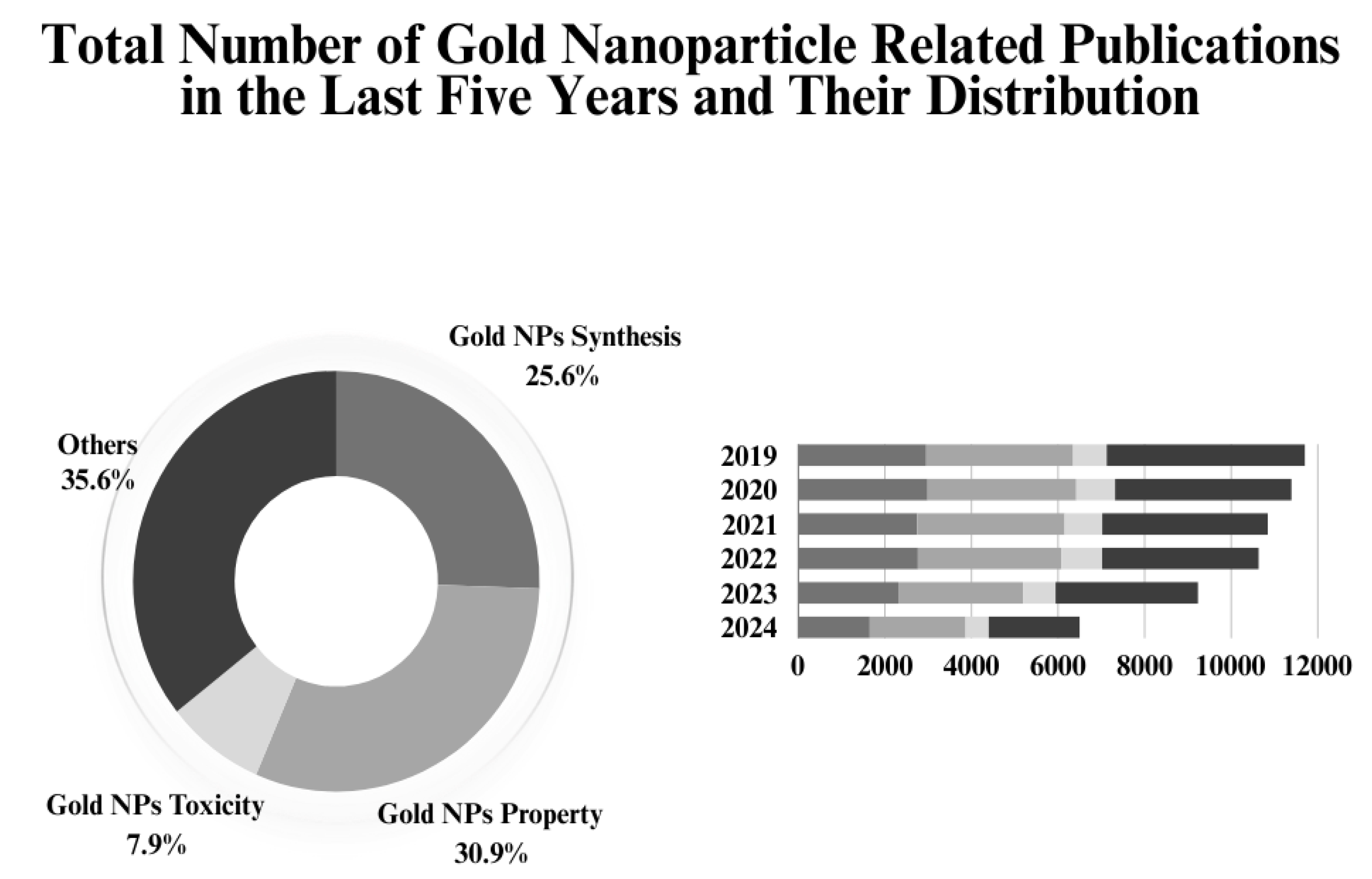
Figure 1. Comparison of total number of gold NPs related publications in the last five years [21].
Preprints 122112 g001
Figure 2. Gold NPs’ size ranges between 1 to 100 nm. They can have various shapes, including spherical, rod, triangular, star, cubic or branched, depending on the synthesis method and further use. Their surface characteristics can be manipulated using various approaches, such as the pegylation to enhance overall stability and biocompatibility of the NPs [28].
Preprints 122112 g002
Figure 3. Highlighted properties of gold NPs [12].
Preprints 122112 g003
Figure 4. Functionalized gold NPs can be utilized for the delivery of wide range of molecules, including nucleic acids (1), proteins (2), antibodies (3), vaccine adjuvants (4), antibacterials (5), glycans (6), imaging probes (7), photosensitizers (8), chemotherapeutic agents (9) and drugs (10) [7].
Preprints 122112 g004
Figure 5. Top-down and bottom-up methodologies for the production of NPs [98].
Preprints 122112 g005
Figure 6. Steps in the manufacture of gold NPs. The seeded-growth approach (A); The digestive ripening technique (B) [98].
Preprints 122112 g006
Figure 7. Graphical representation of green/biological synthesis of gold NPs [244].
Preprints 122112 g007
Figure 8. Characterization of Gold NPs [235].
Preprints 122112 g008
Figure 9. Graphical representation of gold NPs toxicity process both oxidative and non-oxidative [327].
Preprints 122112 g009
Figure 10. Graph representing the number of patents containing keywords that are related to properties and synthesis methods in comparison to the total number of patents published from 2019 to 2024 [356].
Preprints 122112 g010
Table 2. In vivo and In vitro Toxicity Studies of Gold NPs (2019-2024).
Type of Study Organism Particle Effects Ref
In vivo Mice Laser-ablated dextran-coated gold NPs The absence of acute and chronic toxicities and healthy animal behavior supported the safety of gold NPs, which were mostly stored in the liver and spleen and did not produce hepatic or renal toxicity. [339]
In vivo Broiler chicken Gold
NPs
The therapy significantly damaged the blood's oxidative capacity, altered histology, elevated the expression of the IL-6 and Nrf2 genes, fragmented DNA, and reduced the antibody titer against avian influenza and Newcastle disease. [335]
In vivo Rat Gold NPs Different doses of gold NPs had distinct hazardous effects on different organs. Non-toxic doses, on the other hand, had no effect on the testis and only mildly affected the liver and kidney. [340]
In vivo Zebrafish animal model N-myristoyltaurine stabilized gold NPs According to a toxicity study conducted on an animal model of zebrafish, gold NPs are safe. [341]
In vivo Rat Gold NPs Despite being non-toxic, the study discovered that greater doses of gold NPs, such 2 mg/kg, were harmful to every organ examined. [342]
In vitro Human kidney-2 (HK-2) cell and proximal tubular cells Gold NPs with different shapes (spheres and stars), capping (citrate and MUA), and diameters (13 nm and 60 nm) For HK-2 cells, the 13 nm nanospheres were the most hazardous, damaging the mitochondria and lysosomes and increasing the generation of ROS. Severe MUA-capped gold NPs led to apoptosis. Larger 60 nm gold NPs considerably decreased cellular viability but were less hazardous. [343]
In vitro HK-2 and 786-0 cells
Gold NPs Gold NPs with a diameter of 5 or 200 nm have the ability to trigger autophagy in HK-2 cells to shield them from harm and apoptosis in 786-0 cells to kill tumor cells. [344]
In vitro Keratinocyte cell line (HaCaT) and human epidermoid skin cancer cell line (A431) Vitis vinifera (V. vinifera) gold NPs V. vinifera seed gold NPs were non-toxic to normal HaCaT cells, but they suppressed the growth of A431 skin cancer cells by cytotoxicity and death. [345]
In vitro Cancer (Caco-2, MCF-7 and HepG2) and non-cancer (KMST-6) cell lines Terminalia mantaly (TM) extract gold NPs Using the MTT assay, the study investigated the cytotoxic effects of TM-gold NPs on cancer and non-cancer cell lines and discovered that certain extracts were more hazardous than others. [346]
In vitro Hep2 liver cancer cell line and Vero cell line Gold NPs The NPs exhibited remarkable non-toxic effects on normal VERO cell line and anticancer activities in treated Hep2 liver cancer cell line. T [347]
In vitro HeLa cell lines V. negundo extract gold NPs At greater dosages, gold NPs are hazardous to HeLa cells. [348]
In vitro Human HepaRG cells or primary rat hepatocytes (PRH) Gold NPs with different size (~ 15 nm and 60 nm), shape (nanospheres and nanostars) and capping [citrate- or 11-mercaptoundecanoic acid (MUA)], In serum-free media, the 15 nm MUA-capped nanospheres exhibited considerable toxicity to PRH and HepaRG cells, indicating that their restricted application in diagnostics should be disregarded. [349]
Disclaimer/Publisher’s Note: The statements, opinions and data contained in all publications are solely those of the individual author(s) and contributor(s) and not of MDPI and/or the editor(s). MDPI and/or the editor(s) disclaim responsibility for any injury to people or property resulting from any ideas, methods, instructions or products referred to in the content.
Copyright: This open access article is published under a Creative Commons CC BY 4.0 license, which permit the free download, distribution, and reuse, provided that the author and preprint are cited in any reuse.
Alerts
Prerpints.org logo

Preprints.org is a free preprint server supported by MDPI in Basel, Switzerland.

Subscribe

© 2025 MDPI (Basel, Switzerland) unless otherwise stated