Preprint
Article

Phytochemical Analysis and Anti-Diarrheal Activity of Stem Bark Decoctions of Pentadesma butyracea (Sabine) (Clusiaceae)

Altmetrics

Downloads

71

Views

31

Comments

0

Submitted:

22 October 2024

Posted:

24 October 2024

You are already at the latest version

Alerts
Abstract
Pentadesma butyracea is a medicinal plant of which stem bark decoctions are used in traditional medicine for the treatment of diarrheal in Gabon. The aims of the present work were to perform a phytochemical analysis of P. butyracea stem bark decoctions harvested in rain and dry seasons and to evaluate their biological proprieties in order to improve their pharmaceutical use. In a first approach, spectrophotometric analyses were used to quantify phenolics compounds, following by liquid chromatography coupled to mass spectrometry analysis that allowed the identification of flavanone-flavone dimers as the main metabolites of this stem bark decoction. Pharmacological analyses showed the absence of in vitro and in vivo toxicity, thus allowing the safety in use of this decoction. The antioxidant activity of the samples was also demonstrated. Of particular interest, the decoction of P. butyracea stem barks exhibited in vitro antidiarrheal activities thanks to the effects on contractions of ileum fragments isolated from rats. In addition, in vivo activities on castor oil-induced diarrhea in rats were also demonstrated. These results encourage the deepening of a bio-guided research on P. butyracea stem bark decoctions in order to propose standard traditional medical treatments in Gabon.
Keywords: 
Subject: Public Health and Healthcare  -   Public Health and Health Services

1. Introduction

Ethnopharmacology has gained a considerable reputation most notably in African and Asian countries. Ethnopharmacological research has become increasingly of interest for the development of bioactive phytochemicals as novel and effective preventive and therapeutic strategies for various diseases. However, to bridge the gap between local uses of medicinal plant extracts to a medication delivered in a pharmaceutical prescribing, ethnopharmacology has to meet the standards of pharmacological research practices which include pharmacological and clinical studies of traditional medicines, and the identification and accurate quantification of metabolites relevant for a specific biological activity. Functional intestinal disorders are chronic digestive symptoms indicating a dysfunction in the gastrointestinal tract without any evidence of an organic disease. The prevalence of functional intestinal disorders in Gabon was previously investigated in health care facilities [1]. This study revealed a frequency of 12.55 %, mainly concerning women (63%) and an average age of 46 years. Abdominal pain and constipation were the most recorded clinical signs and the most prevailing associated diseases were gastroesophageal reflux and haemorrhoids [1].
An ethnopharmacological and ethnobotanical study was carried out in Gabon by the Cultural and Technical Cooperation Agency (CTCA). These surveys were conducted in four major Gabonese towns (Libreville, Lambaréné, Franceville and Oyem) and medicinal plants used to treat various health symptoms have been listed. In this list, we have selected four plants used for the treatment of diarrhea symptoms, constipation or abdominal pain because these pathologies appear in people suffering from irritable bowel syndrome (IBS) (Table 1). Among them, stem bark decoction of Pentadesma butyracea known for its antidiarrheal activities in traditional medicine in Gabon was selected in the present study for phytochemical and pharmacological investigations.
P. butyracea is a large tree of dense forests belonging to Clusiaceae family. It is present in forests from Sierra Leone, Gabon to Cameroon [2,3,4,5]. In these countries, a kind of butter is traditionally prepared from its seeds and as a consequence, P. butyracea is known as « tallow tree » or « butter tree », « Krinda » in Côte d’Ivoire, « Abotoasebie » in Ghana, « Kpangnan » or « Sesseido » in Benin and « Agnuhé » in Gabon [5]. This butter is used in traditional medicine as a massage oil for skin and hair care and in the manufacture of soap for its softening, lubricating and healing qualities [4,6]. It is also reported as able to delay skin ageing [7]. In Ghana, the root decoction is used to fight intestinal worms [8]. In Gabon, stem bark maceration is also used for treatment against skin parasites [9]. Extracts isolated from P. butyracea organs (stem barks, leaves, seeds and roots) are also commonly used as traditional medical treatments of several diseases including breast pains and genitourinary system disorders [10,11]. With regards to previous phytochemical studies carried out on P. butyracea, various terpenes have been identified in essential oils [4,12], as well as xanthones and triterpenes in a methanol extract [13,14]. P. butyracea seeds also accumulate alkaloids, saponines, tannins and phenolic metabolites [15].
The aims of the present work are to perform phytochemical and pharmacological analyses of P. butyracea stem bark decoctions, one of the plant extracts used in traditional medicine in Gabon for the treatment of diarrhea symptoms. Stem barks were harvested in the rainy and dry seasons on the same tree and on a young tree, to investigate whether their extractable contents depend on seasons and age. Indeed, barks are indifferently collected all year round in Gabon for the preparation of decoctions from different trees. Phytochemical analysis of this extract was achieved by spectrophotometric quantification of phenolic compounds and then by identification of metabolites by ultrahigh performance liquid chromatography coupled to a quadrupole-Orbitrap mass spectrometer (UHPLC-ESI-MS/MS). Then, the safety, antioxidant activity and antidiarrheal potential in vitro and in vivo of a P. butyracea stem bark decoction have been investigated.

2. Results

2.1. Phytochemical Analyses of Decoctions of Pentadesma butyracea Stem Barks

2.1.1. Total Phenolic Contents

The yields of P. butyracea decoctions obtained from stem barks collected during the dry season (DPBD), the rainy season (DPBR) and on a young tree during the dry season (DPBY) were respectively 6.24%, 8.43% and 4.11% of the crude materials. The contents in phenolic compounds of these samples were estimated by spectrophotometry by measuring the reduction of the Folin–Ciocalteu reagent with gallic acid as standard. The total flavonoid contents of the decoctions of P. butyracea stem barks were then investigated using the aluminium chloride method and quercetin as reference. The data on phenolic content were determined from a calibration curve (Y = 0.016X + 0.06309, R2 = 0.9916) of gallic acid expressed in gallic acid equivalents per milligram (GAE/mg) of dry extract (Table 2). The phenolic contents of decoctions obtained from barks harvested during the dry season, i.e., DPBD and DPBY, were determined to be higher than the one obtained from barks collected during the rainy season (DPBR) (129 ± 10.3 µg GAE/mg and 62.1 ± 1.4 µg GAE/mg, respectively versus 44.1 ± 2.9 µg GAE/mg). The phenolic content of DPBD is three times higher than that of DPBR, although the harvesting was performed from the same tree. The results of flavonoid contents were obtained from the calibration curve (Y = 0.01458X - 0.04833, R2 = 0.9517) of quercetin expressed as quercetin equivalents per milligram (QE/mg) of dry extract (Table 2). The decoctions prepared from the same tree in both seasons (DPBD and DPBR) are the highest (150.3 ± 30.3 µg QE/mg and 83.0 ± 9.3 µg QE/mg, respectively). These results suggested that the accumulation of phenolic contents in P. butyracea stem barks depends on the harvesting season and on age of the tree.

2.1.2. Mass Spectrometry Identification of Biflavonoids in the Decoction of Pentadesma butyracea Stem Barks

Phytochemical analysis of P. butyracea stem bark decoction was performed by mass spectrometry on the DPBR sample. The metabolites were analyzed by ultra-high performance liquid chromatography coupled with a quadrupole-Orbitrap mass spectrometer (UHPLC-ESI-MS/MS) in negative and positive ionic modes. In addition, DPBR sample was submitted to a methanolysis and trimethylsilylation to convert free or O-linked monosaccharides, acids and phenolic compounds into their methylester or methylglycoside trimethylsilyl derivatives which were then identified by gas chromatography coupled to electron ionization mass spectrometry (GC-EI-MS) (Table S1).
Figure 1 shows the total ion current chromatogram for UHPLC-ESI-MS in the negative mode and the peak numbering assigned to main metabolites annotated by LC-ESI-MS/MS analysis and reported in Table 3. The metabolites annotation was based on the accurate mass measurements in both negative and positive ionization modes and their MS/MS fragmentation patterns in negative mode by comparison with literature and databases (Massbank, PubChem, HMDB). In negative ion mode, metabolites were detected either as deprotonated molecule [M−H] ̶ or as formate adduct ([M+HCOO] ̶ ). The elution peak between 0.5 and 1 min mainly contains small metabolites and sugars (compounds 1a to 1n in Table 3). For instance, [M−H] ̶ was assigned to 4-(4-deoxy-β-D-gluc-4-enuronosyl)-galacturonate resulting from the degradation of cell wall pectins by pectate lyase. In addition, LC peaks 2 and 3 were assigned to citrate and methyl citrate, respectively.
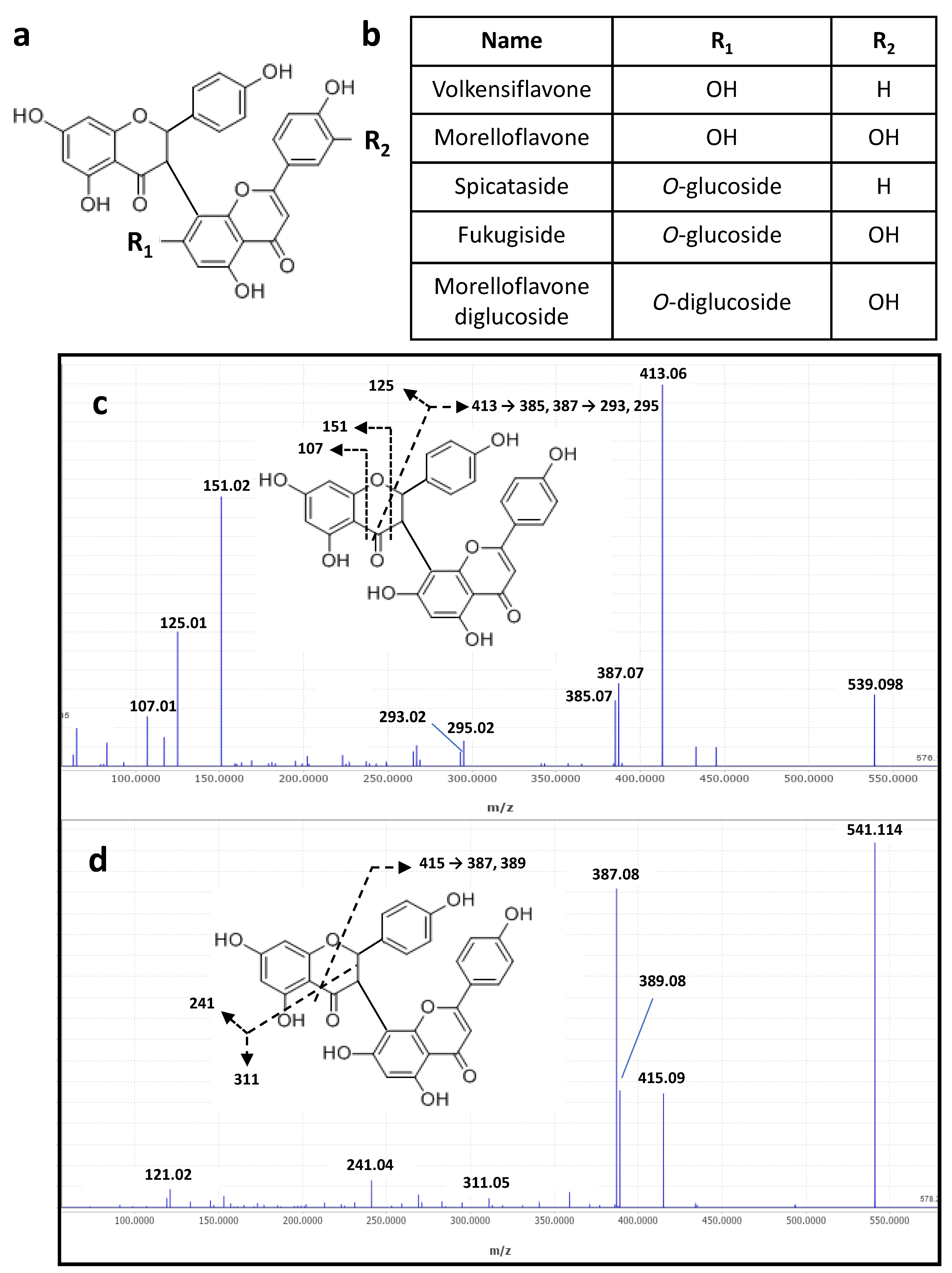
Main metabolites (n° 15-18, Figure 1 and Table 3) of the DPBR sample were determined as being flavanone-flavone dimers, namely volkensiflavone and morelloflavone, and their respective mono and diglucosides (Figure 2a,b). All together, these biflavonoids represented about 75 % of metabolites detected in the MS negative mode (Figure 2). Their negative MS/MS fragmentation patterns were consistent with published data on flavanone-flavone dimers [16] and on flavonoids [17]. For instance, MS/MS fragmentation of the [M-H]- of volkensiflavone at m/z 539.098 (C30H21O10) resulted in the loss of 126 Da, yielding an intense fragment ion at m/z 413.06 (Figure 2c). This fragment arose from the cleavage of the pyranose ring of the flavanone moiety of the biflavonoid. Others diagnostics ions mainly resulted from the secondary fragmentation of this major ion. For morelloflavone ([M-H] - at m/z 555.094) and morelloflavone mono and diglucoside ([M-H] - at m/z 717.146 and m/z 879.199), a shift of 16 Da was observed for main fragment ions. This is due to the presence of a luteolin instead of an apigenin flavone motif in these flavanone-flavone dimers. With regards to the mono and diglycosides derivatives, the sugar composition determined by GC-EI-MS analysis indicated that glucose is the main hexose identified in the DPBR sample (Table S1). We thus postulated that main glycoside derivatives were mono and diglucosides, namely spicataside and fukugiside, respectively (Table 3 and Figure 2b). Same conclusions were drawn on the basis of the investigation of the MS/MS fragmentation patterns of [M+H]+ ions of biflavonoids in the MS positive mode (Table 3 and Figure 2d).
In addition to biflavonoids, less abundant metabolites were eluted between 3.79 and 6.13 min (Figure 1). Among them, the presence of vitexine-2-O-rhamnoside was deduced from the MS/MS spectrum of the [M−H] at m/z 577.156 (C27H30O14) in accordance with the literature data [18] and the identification of rhamnose as the main deoxyhexose in the decoction (Table 3). Other metabolites were identified as deoxyhexosyl hexose disaccharide linked to phenolic or benzoic derivatives (metabolites 5, 10, 11 and 14). These benzoic acids were also detected in the GC-EI-MS analysis (Table S1). The deoxyhexosyl hexose disaccharide motif was proposed as being a rhamnosylglucoside sequence, also called rutinoside, on the basis of the sugar composition of the DPBR sample (Table S1). and in agreement with the fragmentation patterns of homologous metabolites reported in the literature [19]. Among these rhamnosylglucoside-containing metabolites, we identified antiarol rutinoside that was previously reported in plant extracts [20].
It is also worth noting that the detection by UHPLC-ESI-MS/MS of [M−H] at m/z 371.098, 387.093 and 417.103 that were assigned to benzoyl derivatives of polyhydroxy nonanedioic acid (Table 3). This diacid, also detected in the LC elution peak as a free diacid, could correspond to pentahydroxy 2, 3, 4, 6, 7 nonanedioic acid on the basis of its ESI-MS/MS fragmentation pattern (Figure S1b) and the EI-MS of its trimethylsilyl dimethylester derivative (Figure S1a). Its structure was also confirmed from the ESI-MS/MS fragmentation pattern of its 2-benzoyl derivative (metabolite 8, Table 3) as depicted in Figure S1c.

2.1.3. Evaluation of Biflavonoid Contents in the DPB Samples

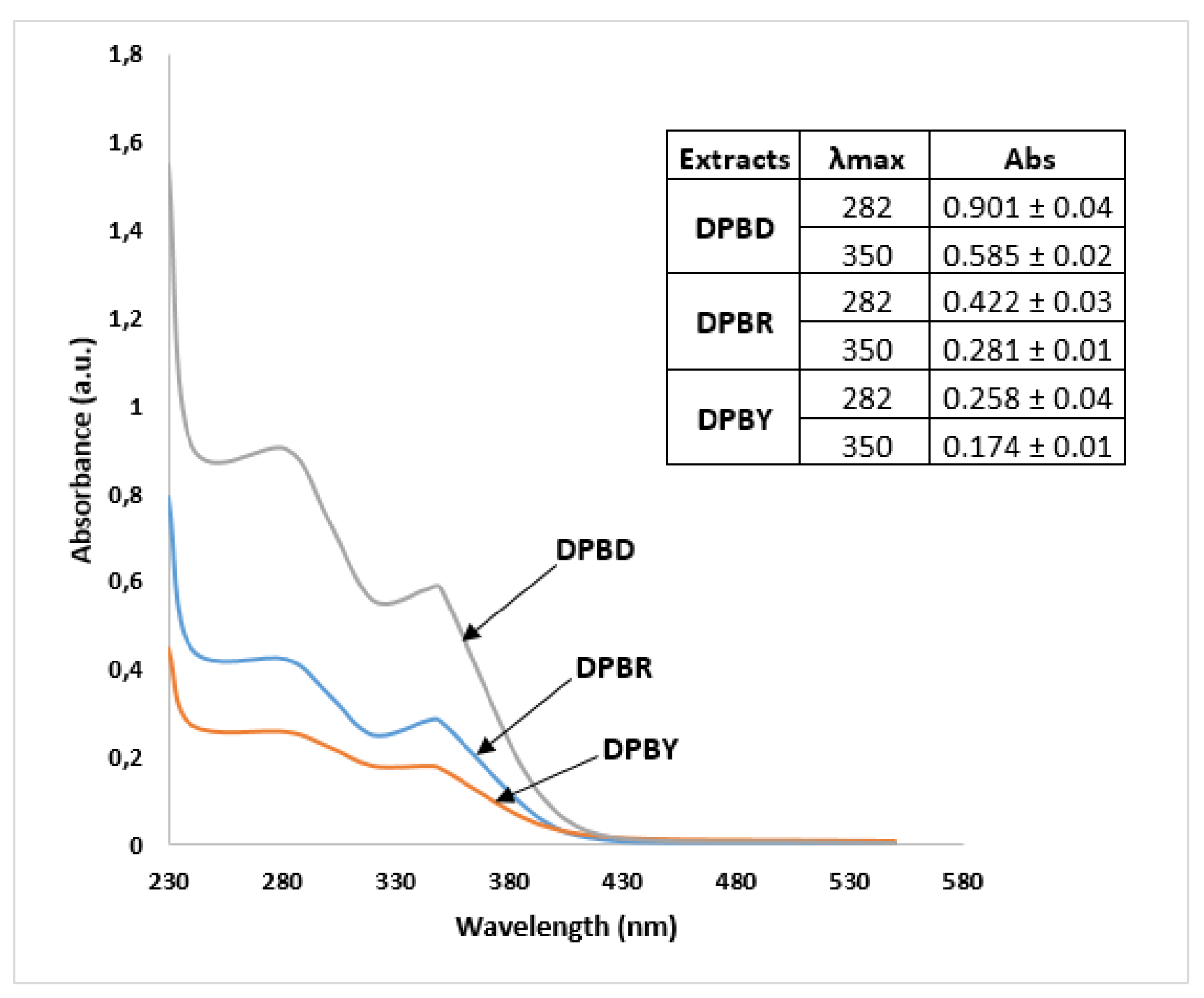
Flavanone-flavone dimers, such as volkensiflavone and morelloflavone, exhibit specific UV absorbances at λmax = 282 and 350 nm due to flavone and flavanone motifs [16]. Similar UV profiles and λmax were observed in the UV spectra of decoctions of P. butyracea stem barks indicating that mainly bioflavonoids (Figure 3), which represent about 75 % of phenolic metabolites of the extract (Table 3), contribute to UV absorbances at these wavelengths, although we cannot rule out the contribution of minor phenolic compounds to these UV profiles. As a consequence, quantification of biflavonoids at λmax = 282 and 350 nm was performed to investigate the biflavonoid contents in decoctions of P. butyracea stem barks from young or adult P. butyracea trees or between stem barks collected during either the dry or the rainy season (Figure 3). In accordance with data on phenolic contents determined by quantification through a spectrophotometric assay (Table 2), UV profiles confirmed that the decoction of P. butyracea stem barks collected during the dry season (DPBD) contains higher amounts of biflavonoids than samples collected during the rainy season (DPBR) or young trees (DPBY).

2.2. Pharmacological Analyses of Decoction of P. butyracea Stem Barks

2.2.1. Antioxidant Activity Assay

The DPPH radical scavenging activities of decoctions of stem barks from P. butyracea are presented in Table 4. Extracts from decoctions prepared from barks harvested during the dry season (DPBD and DPBY) presented a higher antioxidant activity than the decoction obtained from barks collected during the rainy season (DPBR) with IC50 of 8.1 ± 0.6; 11.0 ± 2.0 and 23.5 ± 2.1 µg/mL, respectively. All data were compared with the IC50 value of standard ascorbic acid (6.2 ± 1.2 µg/mL).

2.2.2. Toxicity Assays

To examine a putative cytotoxic activity of DPBR, we first used two human cell lines, HEK-293 and hCMEC/D3, obtained from embryonic kidney and adult cerebral vessels, respectively. We found that incubation of both cell lines with graded concentrations of DPBR had no impact on cell viability, even at high concentration (100 µg/L) and during a 48-h incubation period (Figure S2).
DPBR sample was also evaluated for acute oral toxicity in Wistar rats. Over a 14-day period, DPBR sample did not exhibit any toxicity or behavioural changes in animals that received at a single dose of 2,000 mg/kg of body weight. Moreover, we did not observe any weight loss over two weeks of observation (Figure S3). We thus concluded that DBPR does not induce any oral acute toxicity even at a high dose.

2.2.3. Activity of DPBR Sample on Smooth Muscle

The effect of different doses of DPBR was assessed in vitro on the contractile activity of rat ileal smooth muscle to study their impact on spontaneous ileal contractions. As reported in Table 5, DPBR was found to induce a dose-dependent decrease in contractile activity. Loperamide was used in the study as a positive control. DPBR decoction tested at 4 mg/mL totally relaxed the smooth muscle (100% of relaxation). This showed that DPBR exhibits a spasmolytic activity in vitro resulting from muscle-relaxing properties of metabolites of this plant extract. In view of these results, we then investigated the in vivo antidiarrheal activity on Wistars rats to corroborate the ethnobotanical information reported on stem barks of P. butyracea in Gabon.

2.2.4. In Vivo Antidiarrheal Activity of DPBR

The antidiarrheal activity of DPBR at doses of 250, 500 and 1,000 mg/kg of body weight was then evaluated in vivo in rats in the castor oil-induced diarrhea model and compared to loperamide at 5 mg/kg used as a positive control. The DPBR sample showed 100% protection against diarrhea at the concentration of 500 mg/kg of body weight (Table 6). In addition, the moisture content dropped down from 75 to 39% at the same dose and the inhibition of diarrhea was 100%. These data suggested that this decoction induces antisecretory mechanisms of water and electrolyte, and that the dose of 500 mg/kg can be taken as the reference dose for antidiarrheal treatment by this decoction. At the higher dose of 1,000 mg/kg of DPBR sample, the percentage of inhibition of defecation largely decreased that is likely due to an inhibitory response of the decoction.

3. Discussion

Plant compounds are widely used in traditional medicine for their healing power [21]. With regards to P. butyracea, previously reported phytochemical studies allowed the identification in seeds and leaves of various terpenes in essential oils [4,12], as well as xanthones and triterpenes in a methanol extract [13,14]. P. butyracea seeds also accumulate alkaloids, tannins, phenolics and flavonoids [15]. The presence of mucilage, coumarins, gallic tannins, flavones, sterols and saponins was also reported in P. butyracea leave decoctions [4]. The present study focuses on the phytochemical content, the safety and the antioxidant and in vitro and in vivo antidiarrheic activities of the stem bark decoctions of P. butyracea which are used in traditional Gabonese medicine as an antidiarrheal agent.
Various analytical techniques are usually performed for the identification of phytochemicals. Thin layer chromatography, HPLC, gas chromatography, mass spectrometry and NMR are usually carried out depending upon the nature of the compounds [22]. In our study, the spectrophotometric analysis of the stem bark decoctions of P. butyracea first showed that polyphenols are major metabolites in these samples, although their amounts depend on the season of harvesting and the age of the tree. Analyses of DPBR by GC-EI-MS allowed the identification of the constitutive monomers (monosaccharides, diacids and phenolic compounds). Then, UHPLC-ESI-MS/MS analysis revealed that the flavanone-flavone biflavonoids represent about 75% of the metabolites present in this decoction. It is to note that biflavonoids have been previously reported in different species of the Clusiacea family, such as in Pentadesma grandifolia [14]. Rhamnosylglucoside-containing metabolites were also identified in DPBR by ESI-MS/MS, as well as benzoyl derivatives of polyhydroxy nonanedioic acid that have never been reported in the literature to date.
Phenolic compounds, such as flavonoids or their derivatives, are widely used in human health. However, it remains crucial to evaluate their toxicological risk. Putative toxicity of the DPBR sample was first investigated in vitro on cell cultures. Our results showed that, even à high concentrations and for a 48-h period treatment, DPBR did not significantly affect cell survival of two different cell lines (Fig. S2). Moreover, feeding of rats with up to 2,000 mg/kg of body weight did not reveal any acute toxicity of DPBR on Wistar rats (Fig. S3). The oral administration of decoction of P. butyracea was thus considered as safe for Wistar rats considering the OECD toxicity guidelines 423. As a consequence, absence of toxicity of stem bark decoctions of P. butyracea could justify their use in local traditional medicine. This result is consistent with previous acute toxicity studies on the hydroalcoholic extract of P. butyracea seeds which showed no mortality of mice at the dose of 2,000 mg/kg [23], as well as for a leaf extract tested on rats at the dose of 3,000 mg/kg [11].
Antioxidant metabolites are important for human health because of their ability to neutralize free radicals. Various studies on P. butyracea leaves [11], seeds [15] and fruits [24] have reported the antioxidant activities of these extracts due to their high phenolic molecule contents. This is corroborated by our results which show that the polyphenol richest extract DPBD has the highest antioxidant activity with value of IC50 of 8.1 ± 0.6. This is also in accordance with its content in biflavonoids estimated by UV spectrophotometry by comparison to DPBY and DPBR (Figure 3). The antioxidant activities of biflavonoids have been previously reported in phytochemical analyses of Allanblackia floribunda or Garcinia madruno [16,25,26]. It can be assumed that these compounds perform their radical scavenging activity through the antioxidant activity of their phenolic motifs [27]. Moreover, the presence of numerous aromatic rings and hydroxyl groups are known to be essential for the ability of molecules to scavenge free radicals [28].
It has been reported that the oxidative stress could have direct or indirect effects on gastrointestinal tract responses and could thus be responsible for several troubles including the gastrointestinal troubles like spasm and diarrhea [29,30]. In vitro evaluation of the contractile activity showed that DPBR exhibits relaxing effects on smooth muscle of rat in a dose-dependent manner with 32 ± 5; 68 ± 1.7 and 100% at concentrations of 1; 2 and 4 mg/mL, respectively, against 55 ±.4 % for loperamide, a standard antagonist drug known to reduce propulsive motor activity in the jejunum [31]. The myorelaxant activity of P. butyracea bark decoction on smooth muscle has not yet been studied so far. However, several studies have previously reported on the effects of medicinal plant extracts on the smooth muscle activity. As illustration, the aqueous extract of Spondias mombin barks tested at 794 µg/mL showed an inhibition of about 95 % of the contraction of rabbit duodenum fragments [32]. Another study showed that the decoction of Sapium ellipticum inhibits ileal contractions with an EC50 of 33.29 µg/mL [33]. Momordica balsamina aqueous extract was also demonstrated to relax rabbit smooth muscle at concentrations ranging from 0.4 to 1.6 mg/mL [34]. The aqueous extract of leaves of Morinda morindoides with an EC50 of 360 μg/mL [35], the hydroalcoholic extract of Curcuma longa rhizomes (at 120 μg/mL) [36] and finally biflavonoids isolated from Allanblackia floribunda have been also shown to exhibit vasorelaxing activities [24]. Based on this information, the activity observed with the decoction of P. butyracea stem barks led us to investigate its in vivo antidiarrheal activity induced by castor oil in rats.
Castor oil is able to produce diarrhea symptoms in the same way that physiopathological processes do. Indeed, castor oil releases ricinoleic acid which causes local irritation and inhibition of the intestinal mucosa, resulting in the release of prostaglandins that induce gastrointestinal motility, secretion of water and electrolytes [37,38]. Diarrhea occurs when there is a disturbance in the motility of the smooth intestinal muscles that leads to a water imbalance in the gastrointestinal tract [39]. In our study, the antidiarrheal effect of DPBR at doses of 250 mg and 500 mg/kg body weight significantly delayed the diarrheal onset and decreased the frequency of defecation and weight of feces in a dose dependent manner. Protection was about 60% and 100% respectively, against 40% for loperamide tested at 5 mg/kg. The percentage of inhibition of defecation was 84.6% and 100% at the same doses against 46.2% for loperamide. Loperamide is the standard antidiarrheal agent that antagonizes the action of castor oil due to its anti-motility and anti-secretion properties [40]. In the castor oil-induced diarrhea model, the agents that inhibit the number and weight of fecal matter are considered to have antidiarrheal activity [41,42].
In our in vivo antidiarrheal assay, the percentage of inhibition of defecation and moisture largely decreased at a high concentration of DPBR sample (1,000 mg/mL) by comparison of lower doses. This is likely due to a phenomenon of hormesis which is characterized by an inverted U-shaped dose-response relationship with a stimulating response to small doses but an inhibitory response at high doses [43]. This phenomenon of hormesis was reported in previous studies on the antidiarrheal activity of α-terpineol and of a complex polysaccharide extracted from Anacardium occidentale [44,45].

4. Materials and Methods

4.1. Equipment

A balance Adventurer OHAUS was used for weighing and grinding of barks was performed using an industrial grinder Reisch:AEG typ: AM 80 NX2. An CHRIST Alpha 1-2 LDplus freeze dryer was used to dry the extracts. Spectrophotometric analysis was conducted using spectrophotometer UV-VIS (Drawell). Ultra-high performance liquid chromatography coupled to mass spectrometry (UHPLC-ESI-MS/MS) analyses were performed using an UHPLC system (Vanquish, Thermo Scientific, San Jose, CA, USA) coupled to a quadrupole-Orbitrap mass spectrometer (Exploris 120, Thermo scientific) equipped with an electrospray ionization source. Gas chromatography coupled to an electron impact mass spectrometer (GC-EI-MS) was performed on an Agilent 8860 GC instrument coupled to a 5977-mass selective detector (MSD) quadrupole MS instrument (Agilent Technologies, Palo Alto, CA, USA). The RIKADENKY organ isolation device was used to assess the effect of plant extracts on smooth muscle, a dissection kit was used to isolate the organs.

4.2. Plant Material

Pentadesma butyracea (Sabine) stem barks were collected in 2020 in Libreville (Gabon). They were authenticated at the National Herbarium of Gabon (NHG) where a sample is conserved as reference. In the laboratory, these materials were kept in a glass bell. Two harvests were made from the same tree, one in the rainy season (R), the other in the dry season (D) and another on a young tree in the dry season (Y).

4.3. Chemicals

2,2-di(4-tert-octylphenyl)-1-picrylhydrazyl free radical (DPPH), Folin-Ciocalteu reagent, disodium hydrogen phosphate (Na2HPO4), monobasic potassium phosphate (KH2PO4), dimethyl sulfoxide (DMSO), quercetin, gallic acid and ascorbic acid were purchased from Sigma Chemical Co. (St. Louis, MO, USA). Organic solvents, acids and other chemicals such as ethanol, methanol, hydrochloric acid, aluminium chloride, sodium carbonate, sodium chloride and potassium chloride were purchased from Merck (Darmstadt, Germany). All reagents and chemicals were of analytical grade and the organic solvents were of HPLC grade. All substances were stored in glass containers at room temperature.

4.4. Animal and Cell Lines Model

Adult Wistar rats were from the animal house of the Institute of Pharmacopoeia and Traditional Medicine (IPHAMETRA), Libreville, Gabon. These animals were fed with industrial pellets containing 29% protein and had access to drinking water. All tests were carried out according to protocols already approved by the Department of Pharmacology and Toxicology of IPHAMETRA (agreement N° 001; MESRSTT/IPHAMETRA) and met international standards for animal studies [46]. Human cells were embryonic kidney HEK-293 cells (ATCC®, CRL-1573™) and the human cerebral endothelial cell line (hCMEC/D3; kindly provided by Dr. Pierre-Olivier Couraud, Institute COCHIN, Paris, France).

4.5. Preparation of P. butyracea Stem Barks Decoctions

The stem barks were dried at the Department of Traditional Medicine of IPHAMETRA, Libreville, Gabon, for two weeks and then reduced to a fine powder using a grinder. Five hundred grams of the ground material were placed in a volume of 2 L of distilled water, brought to the boil at 100°C and stirred for 1 h. The aqueous solute was filtered, frozen, freeze-dried and named according to the collect period and oldness of the tree as such: DPBR (rain season), DPBD (dry season) and DPBY (young tree). The extract yields were calculated using the ratio of the mass of the decoction extract to the ground material. For bioassays, decoctions were then solubilized in 1% DMSO in water.

4.6. Determination of Total Phenolic Contents

The total phenol contents (TPC) of the decoctions of P. butyracea stem barks were determined by measuring the reduction of the Folin–Ciocalteu reagent into a blue solution by complexation of phenolic compounds of the samples [47]. Briefly, 1.5 mL of a Folin–Ciocalteu solution (10% in distilled water) was mixed with 500 µL of bark decoctions (1 mg/mL in distilled water) and then allowed to stand for 10 min. Afterward, 2 mL of 7.5% (w/v) sodium carbonate solution was added in each tube and kept at 37°C for 1 h in the dark. Then, the absorbance of respective solutions was determined at λ = 760 nm on a UV-VIS spectrophotometer against the reaction mixture (water + 10% Folin solution + 7.5% sodium carbonate) as a blank. Concentrations of gallic acid from 10 to 60 µg/mL were used to draw a standard calibration plot. TPC of decoctions were estimated as microgram of equivalent gallic acid (GAE) per milligram of extract (µg gallic acid/mg). The following formula was applied to calculate the total concentration of phenolic content: TPC = P × V/m where P is the gallic acid concentration in mg/mL, V is volume (mL) of the sample used in the extraction and m is the weight of pure dried sample used (mg). All tests were carried out in triplicates.

4.7. Determination of Total Flavonoid Contents

The total flavonoid contents (TFC) of the decoctions of P. butyracea stem barks were determined using the aluminium chloride (AlCl3) method [48]. Briefly, 1 mL of decoctions at 1 mg/mL in distilled water or standard quercetin solution (1 mL, 10 to 40 µg/mL) was added to test tubes containing 500 µL of 2% AlCl3 in methanol. The solutions were mixed properly and tubes were kept at room temperature for 1 h. The appearance of yellow colour indicated the presence of flavonoids. The absorbance was measured at λ = 430 nm against the reaction mixture (methanol + 2% AlCl3) as blank. The TFC were estimated as microgram of equivalent of quercetin (QE) per milligram of extract (µg quercetin/mg) by the equation below to estimate the total flavonoid content: TFC = F × V/m where F represents the quercetin concentration (µg/mL), V is the volume (mL) of sample used in the extraction and m represents the weight of pure dried sample used (mg). All tests were carried out in triplicates.

4.8. Gas Chromatography Coupled to an Electron Impact Mass Spectrometer (GC-EI-MS)

For analysis of metabolites of decoctions of P. butyracea stem barks by gas chromatography coupled to electron ionization mass spectrometry (GC-EI-MS), 1 mg of sample was first submitted to a methanolysis by heating the sample in 1 M HCl in methanol at 80°C overnight to convert monosaccharides and phenolic compounds into their O-methyl glycosides/esters. After evaporation of the methanol-HCl solution, the samples were then trimethylsilylated by heating for 20 min at 110°C in hexamethyldisilazane: trimethylchlorosilane: pyridine (3:1:9). After evaporation of the reagent, the samples were dissolved in cyclohexane before being analysed by GC-EI-MS that was performed on an Agilent 8860 GC instrument coupled to a 5977-mass selective detector (MSD) quadrupole MS instrument (Agilent Technologies, Palo Alto, CA, USA). Separations were carried out on a CP-Sil 5CB capillary column (Agilent Technologies) with a film thickness of 250 μm. The carrier gas was 99.9% helium of at a flow rate of 1.3 mL.min-1. The injector and ion source temperatures were set to 280 and 230°C, respectively. Samples were injected with 1:15 split mode. The temperature of the GC oven was first maintained at 40°C for 3 min and then increased up to 160°C at a rate of 15°C.min-1 then up to 280°C at a rate of 1.5°C.min-1. For electron impact mass spectrometry (EI-MS), ionization energy was 70 eV. Acquisitions were performed in full scan mode over a 50-550 mass range with a solvent delay time of 3 min. GC-EI-MS analyses were carried out in triplicate.

4.9. Ultra-High Performance Liquid Chromatography Coupled to a Quadrupole-Orbitrap Mass Spectrometer (UHPLC-ESI-MS/MS)

Five mg of each sample was dissolved 1 mL of HPLC grade water and then filtrated through an 0.5 mL centrifugal filters Ultracel 10 kDa (Amicon) to remove high molecular weight compounds and impurities. The UHPLC-ESI-MS/MS analyses were performed using an UHPLC system coupled to a quadrupole-Orbitrap mass spectrometer equipped with an electrospray ionization source. The chromatographic separations were performed using a C18 silica-based column (Acquity UPLC HSS T3, 1.8 µm, 1.0 mm × 100 mm, Waters Corporation, Milford, MA, USA) with a prefilter of 0.2 µm, kept at 50°C during the analysis. Solvents (water and acetonitrile) were LC–MS grade (Fisher Chemical Optima). Formic acid was from LiChropur (Merck). An autosampler kept the samples at 6°C. The injection volume was 3 µL. The solvents used for gradient separation were 0.1% (v/v) formic acid in water as mobile phase A and 0.1% (v/v) formic acid in acetonitrile as mobile phase B. The flow rate was 0.4 mL/min. The elution gradient was first 1 % B for 1 min, then increased linearly to 100 % B over 20 min and then maintained at 100 % B for 8 min. Samples were analysed in both negative and positive modes. The ESI source parameters were as follows: spray voltage 3,500 V and 3,000 V for positive negative modes, respectively, sheath gas 35 (arbitrary unit), auxiliary gas 10 (arbitrary unit), sweep gas 2 (arbitrary unit), ion transfer tube 320°C and temperature of vaporizer 275°C. Data dependent acquisitions were carried out in both positive and negative modes. MS1 resolution was set at 60,000 with a standard AGC target, a maximum injection time set to auto, a microscan to 1, RF lens to 70%, and a scan range from m/z 80 to 1200. EASY-IC internal standard was used. For MS/MS, resolution was set at 15,000 with a maximum injection time of 50 ms. The isolation window was of 2 m/z, dynamic exclusion was set at 4 s, mass tolerance was + 2 ppm and the precursor intensity threshold set at 5.105 in positive mode and 1.105 in negative mode. The HCD collision energies were 15%, 40% and 60% in both positive and negative ion modes. Data processing was carried out using MZmine 2 (version 2.53). Annotation was performed based on accurate mass measurements and MS/MS spectra according to the literature data.

4.10. UV-VIS Spectroscopy

Solutions of 1 mg/mL in water of DPB decoctions were centrifuged at 3,000 rpm for 10 min and filtrated through a Whatmann n°1 filter paper. The sample were then diluted to 1:10 and scanned at wavelengths ranging from λ = 230 to 500 nm using a UV-VIS spectrophotometer. Spectra were recorded in triplicates.

4.11. Antioxidant Assay

The free radical scavenging activity of decoction of P. butyracea stem barks was determined by the 2,2-di(4-tert-octylphenyl)-1-picrylhydrazyl (DPPH) method with some modifications [49]. Briefly, 1 mL of 2.5 mM DPPH in methanol was combined with 1 mL of decoctions at concentrations ranging from 50 et 1,000 µg/mL. The mixture was shaken and then incubated for 1 h in the dark at room temperature. The absorbance was then measured at λ = 517 nm. Ascorbic acid was used as reference and the percentages of DPPH radical scavenging activity were calculated using the following formula: % inhibition DPPH = [Ac – As) / Ac] x 100 where Ac represents the absorbance of the blank containing methanol and DPPH (v: v) and As represents the absorbance of the samples containing DPPH and extracts or reference. The IC50 was calculated by plotting the percentage of radical scavenging activity against different concentrations of sample using nonlinear regression with Graph Pad Prism version 8.4.3.686. All assays were carried out in triplicate.

4.12. In Vitro Cytotoxicity Assay

Human embryonic kidney HEK-293 cells (ATCC®, CRL-1573™) were cultivated in Dulbecco’s modified Eagle’s medium (DMEM, Gibco) completed with fetal bovine serum (FBS 10%, Eurobio), antibiotic-antimycotic solution (penicillin/streptomycin/fungizone, 1%, Sigma-Aldrich) and sodium pyruvate (1%, Gibco). The human cerebral endothelial cells (hCMEC/D3) were cultivated in flasks previously coated with collagen I (50 µg/mL in sterile phosphate buffered saline (PBS), 1 h, 37 °C) in EndoGroTM MV medium (Merk Millipore, containing: 5% FBS, 5% glutamine, 0.2% EndoGRO-LS nutrient supplement, 0.1% epidermal growth factor, 0.1% hydrocortisone, 0.1% heparin sulphate and 0.1% ascorbic acid). Both HEK-293 and hCMEC/D3 cell lines were maintained at 37 °C and 5% CO2 in a humidified atmosphere.
For the cell survival assay, HEK-293 cells (20,000 cells/well) and hCMEC/D3 (10,000 cells/well) were placed in a white flat bottom 96-well plate (Corning) previously coated with poly-D-lysine (Corning) (30 µM in H20, 1 h, 37 °C) or collagen I (50 µg/mL in PBS, 1 h, 37 °C), respectively. After 24h, cells were rinsed with Dulbecco’s PBS (dPBS, Sigma-Aldrich) and incubated for 6, 24 or 48 h in a FBS-free medium in the absence or presence of graded concentrations of DPBR (1, 10 or 100 µg/mL). After incubation, the Cell Titer-Glo® luminescent Cell Viability Assay (Promega) was used to quantify cell viability following the manufacturer’s protocol. The luminescence was measured using the InfinitePro200 plate reader (TECAN). Cytotoxicity assays were carried out in triplicate.

4.13. Acute Toxicity Assay

Wistar rats were subjected to an evaluation of the acute toxicity induced by DPBR sample according to established OECD 423 guidelines [50] and the Guide for the Care and Use of Laboratory Animals [46] with some modifications. A prior approval agreement (N° 001 MERSTT/IPHAMETRA) was obtained from the Ethics Committee on the Use of Animals of the Institute of Pharmacopoeia and Traditional Medicine (IPHAMETRA). The animals were divided into two groups of six (three males and three females) and fasted for 24 h before the experiment. The Wistar rats tested received orally the DPBR sample at a single dose of 2,000 mg/kg. Control animals received only distilled water and all were kept in the same Environmental conditions. Animals were strictly observed for physiological symptoms such as weight loss, diarrhea, tremor, lethargy and paralysis periodically for the first four hours during the 72-h period and later were followed per day for 14 days for any lethality.

4.14. In Vitro Antidiarrheal Assays on Excised Ileum Fragments

In vitro antidiarrheal assays were performed according to the literature with some modifications [33]. Pieces of ileum were taken from Wistar rats and preserved during the tests in Mac Ewen’s physiological solution. Fragments measuring 0.5 to 0.9 cm were fixed in a tank, called a survival tank, in an aerated thermostatic bath at 37°C. The basic activity (ileum contractions) of the organ was recorded, then the organ was subjected to different concentrations of the decoction. The dose-response curves of the plant extract at 1, 2 and 4 mg/mL were recorded. The value of the amplitude before administration of the extracts was considered as a reference. The effects of the decoction on the intestinal spasms were expressed as a percentage of inhibition = ((AB – AE)/ AB) × 100 where AB is the average of tone spasms basal and AE is the average of spasms in the presence of the extract [51]. Mac Ewen physiological solution was composed (in mM) of NaCl (130), KCl (5.63), CaCl2 (5.52), Na2HPO4 (0.93), NaHCO3 (11.9), MgCl2 (0.24) and glucose (11), pH 7.4. All tests were carried out in triplicate.

4.15. In Vivo Antidiarrheal Assays

In vivo antidiarrheal assays were performed according to the literature with some modifications [33]. Twenty-five Wistar rats (170-230 g) were fasted for 24 h with access to water and divided into five groups of five animals. The DPBR sample at doses of 250, 500 and 1,000 mg/kg body weight were administered orally to each group. The fourth group received distilled water (negative control), while the fifth group received the standard drug loperamide at 5 mg/kg body weight. One hour after the drug pre-treatment, all of the animals received orally castor oil 10 mL/kg body weight. Subsequently, each group of animals was kept separately in cages on a Whatmann paper for the collection of diarrheal faeces. The animals had access to water and food throughout the experiment. The severity and consistency of diarrhea was observed hourly for 4 h after castor oil administration. The percentage of protection, inhibition of diarrhea and humidity were calculated by the following formulas : Percentage of inhibition of diarrhea = (total number of diarrheal stools in the negative control – total number of diarrheal stools in treated group) / total number of diarrheal stools in the negative control) × 100 [52]; percentage of protection = (number of rats without diarrheal stools / total number of rats) *100 [52] and percentage of humidity = ((WSW – DSW)/WSW) ×100 where WSW is the wet stool weight and DSW is the dried stool weight [51].

4.16. Statistical Analysis

We used Graph Pad Prism version 8.4.3.686. (GraphPad Software Inc., San Diego, CA, USA) for statistical analyses. Results were presented as mean ± standard deviation (SD) of replication determinations according to the assay. One-way analysis of variance was used to determine the significant difference (p<0.05) between concentrations. In addition, Dunnett’s multiple comparisons test and/or Sidak’s multiple comparison test were used to evaluate the difference between the treatment means. The IC50 and EC50 values were calculated using nonlinear regression.

5. Conclusions

In the present study, the phytoconstituents of a decoction of stem barks of P. butyracea were identified by spectrophotometric assays and mass spectrometry analyses. A total of 14 compounds were identified in P. butyracea stem barks. Main metabolites are biflavonoids (75% of metabolites). In addition, rhamnosylglucoside-containing metabolites were identified by ESI-MS/MS, as well as benzoyl derivatives of polyhydroxy nonanedioic acid that have never been reported in the literature so far. Pharmacological analyses showed that the decoction of the stem barks of P. butyracea exhibits antidiarrheal activities and no cytotoxicity. Previous studies have shown that biflavonoids from Allanblackia floribunda have vasorelaxant activities [36]. We thus postulate that the biflavonoids which are predominant in the decoction of P. butyracea stem barks are responsible for the observed antidiarrheal activities. However, a study on the effect of pure biflavonoids has to be performed for confirmation. As a consequence, potent in vitro and in vivo antidiarrheal activities of biflavonoids of the decoction of P. butyracea stem barks are consistent with its use in traditional medicine to treat diarrhea and emphasized the potential of these plant extracts as a future source of new antidiarrheal drugs.
UV profiles and the spectrophotometric quantification of polyphenols in DPB extracts showed that the amounts of biflavonoids in stem barks depend on the season of collection and age of P. butyracea trees. Antioxidant assays support these phytochemical analyses with activities of DPBD and DPBY higher than DPBR. This is a major concern in an ethnopharmacological point of view because biflavonoid contents vary by about a factor of three between samples analysed in this study. This may compromise the efficiency of stem bark decoctions in the treatment of diarrhea because contents in active molecules may differ from one preparation to another. However, quantification of biflavonoids by spectrophotometry or by UV at specific wavelength may help in a batch-to-batch control protocol of bark decoctions for local pharmaceutical prescribing.

Supplementary Materials

The following supporting information can be downloaded at: Preprints.org, Figure S1: EI-MS (a) and ESI-MS/MS (b) spectra of pentahydroxy 2, 3, 4, 6, 7 nonanedioic acid. For GC-MS analysis, the diacid was analysed as a methylester trimethylsilyl (TMS) derivative. (c) ESI-MS/MS spectrum of [M−H] at m/z 387.093 of 2-hydroxybenzoyl pentahydroxy 2, 3, 4, 6, 7 nonanedioic acid; Figure S2: Survival percentage of two human cell lines, HEK-293 and hCMEC/D3, incubated with 100µg/mL of DPBR; Figure S3. Body weight gain of the Wistar rats during 14-day period after treatment with 2,000 mg/kg of DBPR compared to the control group. Table S1: Metabolite annotation by GC-MS analysis of the DPBR sample after methanolysis and trimethylsilylation. Percentage of each metabolite was determined on the basis of the relative ratio in GC-EI-MS of the corresponding peak area and relative response factors determined on standard monosaccharides. (*) Selective fragment ions.

Author Contributions

Conceptualization, E.L.M-E. and L.E.M.; methodology, E.L.M-E., C.L-B., I. S-A., M.G, P.G., P.L. and L.E.M.; validation and formal analysis, E.L.M-E., C.L-B., I. S-A., P.G., P.L. and L.E.M.; investigation, and data curation, E.L.M-E., C.L-B., I. S-A., P.L. and L.E.M.; writing—original draft preparation, review and editing, E.L.M-E., P.G., P.L. and L.E.M.; supervision, P.L. and L.E.M.; project administration, P.L. and L.E.M.; funding acquisition, P.L. and L.E.M. All authors have read and agreed to the published version of the manuscript.

Funding

This research was funded by IPHAMETRA in Gabon and in France, by the University of Rouen Normandy, INSA Rouen Normandy, the Centre National de la Recherche Scientifique (CNRS), European Regional Development Fund (ERDF), Labex SynOrg (ANR-11-LABX-0029), Carnot Institute I2C, the graduate school for research XL-Chem (ANR-18-EURE-0020 XL CHEM) and by Region Normandie. We also thank the University of Rouen Normandie for their specific support to this project (BQI 2022).

Institutional Review Board Statement

All animal tests were carried out according to protocols already approved by the Department of Pharmacology and Toxicology of IPHAMETRA, Libreville, Gabon (agreement N° 001; MESRSTT/IPHAMETRA).

Informed Consent Statement

Not applicable.

Data Availability Statement

All data are presented in the article.

Acknowledgments

We would like to express our sincere thanks to the administrative and technical staff of IPHAMETRA in Libreville, Gabon and also those of the GlycoMEV laboratory in France for all the technical and financial support provided for this work.

Conflicts of Interest

The authors declare no conflict of interest and this work is a perfect illustration of a fruitful North-South agreement between IPHAMETRA in Libreville, Gabon and the GlycoMEV laboratory at the University of Rouen Normandie, France.

References

  1. Mayindza-Ekaghba, E.L.; Itoudi Bignoumba, P.E.; Bourobou Bourobou, J.A.; Boukandou Mounanga, M.M. Etude épidémiologique des troubles fonctionnels intestinaux dans les structures sanitaires à Libreville (Gabon). J. Appl. Biosc. 2020, 155, 15986–15993. [Google Scholar]
  2. Sinsin, B.; Sinadouwirou, A.T. Valorisation socioéconomique et pérénnite du Pentadesma butyraceae Sabine en galeries forestières au Bénin. Cahier Agriculture 2003, 12, 75–79. [Google Scholar]
  3. Natta, A.; Sogbegnon, R.; Tchobo, F. Connaissances endogènes et importance du Pentadesma butyracea (Clusiaceae) pour les populations autochtones au Nord-Ouest Bénin. Fruit Veg. Cereal Sci. Biotechnol. 2010, 4, 18–25. [Google Scholar]
  4. Noudogbessi, J.P.A.; Natta, A.K.; Tchobo, F.P.; Bogninou, G.S.; Bothon, F.T.D., Bossou; Figueredo, G.; Chalard, P.; Chalchat, J.C.; Sohounhloué, D.C.K. Phytochemical screening of Pentadesma butyracea Sabine (Clusiaceae) acclimated in Benin by GC/MS. ISRN Anal. Chem 2013, 172397. [Google Scholar] [CrossRef]
  5. Ayegnon, B.P.; Kayodé, A.P.; Tchobo, F.P.; Azokpota, P.; Soumanou, M.M.; Hounhouigan, D.J. Profiling the quality characteristics of the butter of Pentadesma butyracea with reference to shea butter. J. Sci. Food Agric. 2015, 95, 3137–3143. [Google Scholar] [CrossRef]
  6. Dencausse, L.; Ntsourankoua, H.; Artaud, J.; Clamou, J.L. Comparaison des compositions lipidiques des beurres de Pentadesma butyracea et de Karite. Ol. Corps Gras Lipides 1995, 2, 143–147. [Google Scholar]
  7. Courtin, O. Cosmetic preparation to retard the ageing skin, and application process. European Patent Applications. EP0279136A3. 1987. [Google Scholar]
  8. Abbiw, D.K. Useful plants of Ghana. In Intermadiate Technology Publications and the Royal Botanic Gardens Kew; London, UK, 1990. [Google Scholar]
  9. Adjanohoune, E.J.; Aké Assi, L.; Chibon, P.; de Vécchy, H.; Duboze, E.; Eymé, J.; Gassita, J.N.; Goudote, E.; Guinko, S.; Keita, A.; Koudogbo, B.; Le Bras, M.; Mourambou, I.; Mve-Mengome, E.; Nguéma, M.G.; Ollome, J.B.; Posso, P.; Sita, P. In Contribution aux études ethnobotaniques et floristiques au Gabon. Agence de coopération culturelle et technique: Paris, France, 9: ISBN: 92, 9028. [Google Scholar]
  10. Avocevou-Ayisso, C.; Avohou, T.H.; Oumorou, M.; Dossou, G.; Sinsin, B. Ethnobotany of Pentadesma butyracea in Benin: A quantitative approach. Ethnobot. Res. Applic. 2011, 9, 151–166. [Google Scholar] [CrossRef]
  11. Tindano, B.; Bayala, B.; Guenne, S.; Doukoure, M.; Kiendrebeogo, M.; Belemtougri, G.R. Innocuity and antioxidant activities of Pentadesma butyracea (1824) leaves for its use in hormone replacement therapy. Cameroon J. Experim. Biol. 2018, 12, 32–40. [Google Scholar] [CrossRef]
  12. Alitonou, G.; Avlessi, F.; Sohounhloue, D.; Bessiere, J.M.; Menut, C. Chemical and biological investigation on volatile constituents of Pentadesma butyracea Sabine (Clusiaceae) from Benin. J. Essent. Oil Res. 2010, 22, 138–140. [Google Scholar] [CrossRef]
  13. Zéléfack, F.; Guillet, D.; Fabre, N.; Bayet, C.; Chevalley, S.; Ngouela, S.; Ndjakou, L.B.; Valentin, A.; Diroux-Franca, M.G. Xanthones cytotoxiques et antiplasmodium de Pentadesma butyracea. J. Nat. Prod. 2009, 72, 954–957. [Google Scholar] [CrossRef] [PubMed]
  14. Djoufack, N.G.L.; Valant-Vetschera, K.M.; Schinnerl, J.; Brecker, L.; Lorbeer, E.; Robien, W. Xanthones, biflavanones and triterpenes from Pentadesma grandifolia (Clusiaceae): Structural determination and bioactivity. Nat. Prod. Comm. 2010, 5, 1055–1060. [Google Scholar] [CrossRef]
  15. Timtey, J.A.; Alemaworn, F.; Ellis, W.O.; Pepra-Ameyaw, N.B.; Agbenorhevi, J.K. Pentadesma butyracea in Ghana – indigenous knowledge, uses, and seed characterization. Sci. Afr. 2023, 21, e01747. [Google Scholar] [CrossRef]
  16. Akpanika, G.A.; Winters, A.; Wilson, T.; Ayoola, G.A.; Adepoju-Bello, A.A. , Hauck, B. Polyphenols from Allanblackia floribunda seeds: identification, quantification and antioxidant activity. Food Chem. 2017, 222, 35–42. [Google Scholar] [CrossRef]
  17. Fabre, N.; Rustan, I.; de Hoffmann, E.; Quetin-Leclercq, J. Determination of flavone, flavonol, and flavanone aglycones by negative ion liquid chromatography electrospray ion trap mass spectrometry. J. Am. Soc. Mass Spectrom. 2001, 12, 707–715. [Google Scholar] [CrossRef]
  18. Shao, S-Y.; Ting, Y.; Wang, J.; Sun, J.; Guo, X-F. Characterization and identification of the major flavonoids in Phyllostachys edulis leaf extract by UPLC—QTOF—MS/MS. Acta Chromatogr. 2020, 32, 228–237. [Google Scholar] [CrossRef]
  19. Zhu, F.X.; Wang, J.J.; Li, X.F.; Sun, E.; Jia, X.B. Qualitative and quantitative analysis of the major constituents in traditional Chinese medicine Danmu injection using LC-ESI-MS (n) and LC-DAD. Pharmacogn. Mag. 2014, 10, 254–264. [Google Scholar]
  20. Thai, T.H.; Hai, N.T.; Hien, N.T.; Ha, C.T.H.; Cuong, N.T.; Binh, P.T.; Dang, N.H.; Dat, N.T. Cytotoxic constituents of Mallotus Microcarpus. Nat. Prod. Comm. 2017, 12, 407–408. [Google Scholar] [CrossRef]
  21. Fitzgerald, M.; Heinrich, M.; Booker, A. Medicinal Plant Analysis: A Historical and Regional Discussion of Emergent Complex Techniques. Front. Pharmacol. 2020, 10, 1480. [Google Scholar] [CrossRef]
  22. Taher, M.A.; Laboni, A.A.; Shompa, S.A.; Rahman, M.M.; Hasan, M.M.; Hasnat, H.; Khan, M. Bioactive compounds extracted from leaves of G. cyanocarpa using various solvents in chromatographic separation showed anti-cancer and anti-microbial potentiality in in silico approach. Chin. J. Anal. Chem. 2023, 51, 100336. [Google Scholar] [CrossRef]
  23. Tindano, B.; Bayala, B.; Doukoure, M.; Belemtougri, G.R.; Tamboura, H.H.; Sawadogo, L. Phytochemical composition, acute toxicity and phytohormonal Garcinia madruno activity of hydroalcoholic extract of Pentadesma butyracea (Clusiaceae Sabine (1824)) seeds. J. Med. Plant Res. 2017, 11, 656–664. [Google Scholar] [CrossRef]
  24. Brusotti, G.; Papetti, A.; Serra, M.; Temporini, C.; Marini, E.; Orlandini, S.; Kada Sanda, A.; Watcho, P.; Kamtchouing, P. Allanblackia floribunda Oliv.: An aphrodisiac plant with vasorelaxant properties. J. Ethnopharmacol. 2016, 192, 480–485. [Google Scholar] [CrossRef] [PubMed]
  25. Ayegnon, B.P.; Chabi, I.B.; Akogou, F.U.G.; Kayodé, A.P.P. Physicochemical characterization and microbiology quality of the Pentadesma butyracea fruit pulp collected from various parks in Benin. Sci Rep. 2021, 11, 17040. [Google Scholar] [CrossRef] [PubMed]
  26. Carrillo-Hormaza, L.; Ramírez, A.M.; Quintero-Ortiz, C.; Cossio, M.; Medina, S.; Ferreres, F. , Gil-Izquierdo, A.; Osorio E. Comprehensive characterization and antioxidant activities of the main biflavonoids of: A novel tropical species for developing functional products. J. Funct. Foods 2016, 27, 503–516. [Google Scholar] [CrossRef]
  27. Taher, M.A.; Jamal Hossain, M.; Zahan, M.S.; Hasan, M.M.; Ferdous, J.; Rahman, A.; Khan, M.; Hosain, M.K.; Rashid, M.A. Phyto-pharmacological and computational profiling of Bombax ceiba Linn. Leaves revealed pharmacological properties against oxidation, hyperglycemia, pain, and diarrhea. Heliyon, 2024; 10, e35422. [Google Scholar]
  28. Tantary, S.; Masood, A.; Bhat, A.H.; Dar, K.B.; Zargar, M.A.; Ganie, S.A. In vitro antioxidant and RBC membrane stabilization activity of Euphorbia wallichii. Free Radic. Antioxid. 2017, 7, 13–22. [Google Scholar] [CrossRef]
  29. Kim, Y.J. , Kim, E., Hahm, K.B. Oxidative stress in inflammation-based gastrointestinal tract diseases: Challenges and opportunities. J. Gastroentero.l Hepatol. 2012, 27, 1004–1010. [Google Scholar] [CrossRef]
  30. Miranda-Bautista, J.; Bañares, R.; Vaquero, J. The Gastrointestinal System: Anatomy and sources of oxidative stress, in: Gastrointestinal Tissue. Academic Press. 2017, pp 3-20. ISBN: 9780128053775.
  31. Feyisa, K.; Bisrat, D.; Tadesse, S.; Asres, K. Antidiarrhoeal activity of the 80% methanol root extract of alictrum ryhnchocarpum Dill. & A. Rich and its major constituent against castor oil-induced diarrhoea in mice. Ethiopian Pharmaceut. J. 2020, 36, 31–40. [Google Scholar]
  32. Diby, S.B.; Koné, M.; Yapo, A. Potentiel pharmacologique des écorces de tige de Spondias mombin L. (Anacardiaceae) sur la motricité in vitro du duodénum de lapin ; une plante médicinale utilisée dans le traitement traditionnel des troubles digestifs. Phytothérapie 2012, 10, 306–312. [Google Scholar] [CrossRef]
  33. Wansi, S.L.; Nguelefack-Mbuyo, E.P.; Nchouwet, M.L.; Miaffo, D.; Nyadjeu, P.; Wabo, J.P.; Mbiantcha, M.; NKeng-Efouet, P.A.; Nguelefack, T.B.; Kamanyi, A. Antidiarrheal activity of aqueous extract of the stem bark of Sapium Ellipticum (Euphorbiaceae). Trop. J. Pharmaceut. Res. 2014, 13, 929–935. [Google Scholar] [CrossRef]
  34. Otimenyin, O.S.; Uguru, O.M.; Akanbi, B.E. Antidiarrhea effect of aqueous extracts of Momordica balsamina and Stachytarpheta indica in Rats. J. Nat. Prod. 2008, 1, 36–45. [Google Scholar]
  35. Bahi, C.; N’guessan, J.D.; Guédé-Guina, F. Mise en évidence d’une action myorelaxante et cholinolytique de Bitter GG (BGG), un antidiarrhéique de source végétale. Afr. Biomed. 2000, 5, 11–18. [Google Scholar]
  36. Bleu, G.M.; Traoré, F.; Coulibaly, S.; Nene-Bi, S.A. Effets pharmacodynamiques d’un extrait hydroalcoolique de Curcuma longa Linné (Zingiberaceae) sur le système cardiovasculaire, la respiration et l’activité mécanique intestinale de mammifères. Phytothérapie 2011, 9, 7–17. [Google Scholar] [CrossRef]
  37. Mogose, B.; Bisrat, D.; Asres, K. In Vivo Antidiarrheal potential of the leaf extract of Maytenus addat (Loes.) Sebsebe and its major compound. J. Trop. Med. 2024, 5922487. [Google Scholar] [CrossRef] [PubMed]
  38. Gidudu, J.; Sack, D.; Pina, M.; Hudson, M.; Kohl, K.; Bishop, P.; Chatterjee, A.; Chiappini, E.; Compingbutra, A.; Da Costa, C. Diarrhea: Case definition and guidelines for collection, analysis, and presentation of immunization safety data. Vaccine 2011, 29, 1053. [Google Scholar] [CrossRef]
  39. Rudra, S.; Tahamina, A.; Emon, N.U.; Adnan, M.; Shakil, M.; Chowdhury, M.; Uddin, H.; Barlow, J.W.; Alwahibi, M.S.; Soliman Elshikh, M. Evaluation of various solvent extracts of Tetrastigma leucostaphylum (Dennst.) Alston leaves, a Bangladeshi traditional medicine used for the treatment of Diarrhea. Molecules 2020, 25, 4994–5013. [Google Scholar] [CrossRef]
  40. Farack, U.M.; Kantz, U.; Loescke, K. Loperamide reduces the intestinal secretion but not the mucosal cAMP accumulation induced by cholera toxin. Naunyn Schmiedebers Arch. Pharmacol. 1981, 317, 178–179. [Google Scholar] [CrossRef]
  41. Karim, S.M.M.; Adaikan, P.G. The effect of the loperamide on prostaglandin-induced diarrhea in rat and man. Prostaglandins 1977, 13, 321–331. [Google Scholar] [CrossRef]
  42. Rawat, P.; Singh, P.K.; Kumar, V. Evidence based traditional anti-diarrheal medicinal plants and their phytocompounds. Biomed. Biomed. Pharmacother. 2017, 96, 1453–1464. [Google Scholar] [CrossRef]
  43. Calabrese, E.J.; Baldwin, L.A. The frequency of U-shaped dose responses in the toxicological literature. Toxicol. Sci. 2001, 62, 330–338. [Google Scholar] [CrossRef]
  44. Dos Santos Negreiros, P.; da Costa, D.S.; da Silva, V.G.; de Carvalho, Lima, I. B., Nunes, D.B., de Melo, Sousa, F.B., Araújo, T.L.S., Medeiros, J.V.R., dos Santos, R.F., Oliveira, R.D.C M. Antidiarrheal activity of α-terpineol in mice. Biomed. Pharmacother. 2019, 110, 631–640. [Google Scholar] [CrossRef]
  45. Araújo, T.S.L.; Costa, D.S.; Sousa, N.A.; Souza, L.K.M.; Araújo, S.; Oliveira, A.P.; Sousa, F.B.M.; Silva, D.A.; Barbosa, A.L.R.; Leite, J.R.S.A.; Medeiros, J.V.R. Antidiarrheal activity of cashew GUM, a complex heteropolysaccharide extracted from exudate of Anacardium occidentale L. in rodents. J Ethnopharmacol. 2015, 174, 299–307. [Google Scholar] [CrossRef] [PubMed]
  46. Zimmermann, M. Ethical guidelines for investigations of experimental pain in conscious animals. Pain 1983, 16, 109–110. [Google Scholar] [CrossRef] [PubMed]
  47. Singleton, V. L.; Orthofer, R.; Lamuela-Raventós, R.M. Analysis of total phenols and other oxidation substrates and antioxidants by means of Folin-Ciocalteu reagent, Methods in Enzymology. Academic Press, 1999, 299, 152–178. [Google Scholar]
  48. Ribéreau-Gayon, J.; Peynaud, M.; Ribéreau-Gayon, P.; Sudraud, P. In Sciences et techniques du vin. Tome 1, analyse et controle des vins. Dunod, Ed. Paris, France 1972, 671. [Google Scholar]
  49. Mengome, L.E. , Voxeur, A., Akue, J.P., Lerouge, P. Screening of antioxidant activities of polysaccharides extracts from endemic plants in Gabon. Bioact. Carbohydr. Diet. Fibre 2014, 3, 77–88. [Google Scholar] [CrossRef]
  50. Organisation for Economic Co-operation and Development (OECD). The Organization of Economic Co-Operation and Development Guidelines Test NO. 423 Acute Oral Toxicity-Acute Toxic Class Method, Guidelines for the Testing of Chemicals, Section 4. 2002, –14. 1 February.
  51. Guide for the Care and Use of Laboratory Animals, 8th ed.; The National Academies Press: Washington, DC, USA, 2011; ISBN 978-0-309-15400-0.
  52. Mekonnen, B.; Asrie, A.B.; Wubneh, Z.B. Antidiarrheal activity of 80% methanolic leaf extract of Justicia schimperiana. Evid. Based Complement. Alternat. Med. 2018, 6, 3037120. [Google Scholar] [CrossRef]
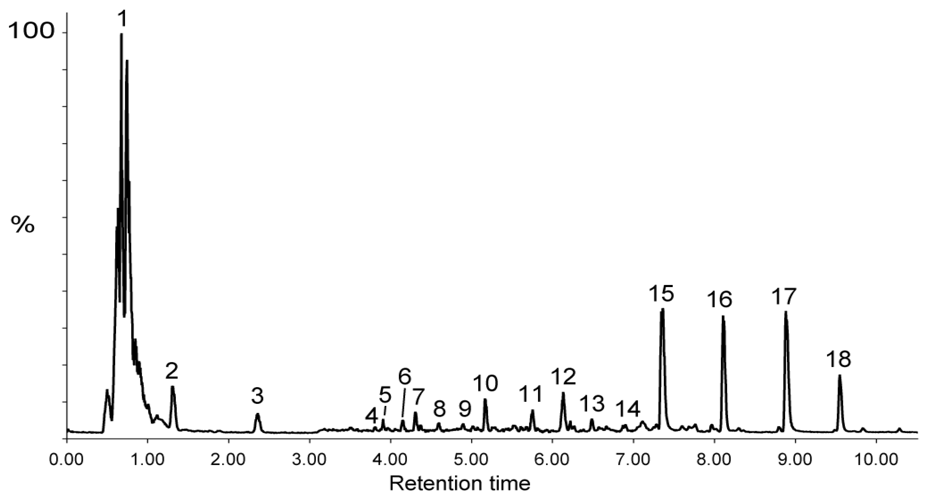
Figure 1. Total ion current chromatogram for the LC-ESI (−)-MS of the DPBR sample. Peak numbering refers to main metabolites identified by analysis of their MS/MS fragmentation patterns and reported in Table 3.
Preprints 122022 g001
Figure 2. Proposed structures (a) and names (b) of biflavonoids annotated in the DPBR sample. ESI-MS/MS spectra of [M−H] (c) and [M+H]+ (d) of the flavanone-flavone dimer volkensiflavone at m/z 539.098 and 541.114, respectively.
Preprints 122022 g002
Figure 3. UV-visible spectra recorded between λ = 230 nm and 550 nm of 0.1 mg/mL solutions of decoctions of P. butyracea stem barks (n=3). Absorbances of the different decoctions at λmax = 282 and 350 nm are reported in the inserted table.
Preprints 122022 g003
Table 1. List of plants selected.
Genus, species Family n° NHG Organ Uses Traditional indications
Aucoumea klaineana (Pierre) Burseraceae 599 Stem bark Diarrhea Macerated stem bark is used as astringent antidiarrheal agent
Pentadesma butyracea (Sabine) Clusiaceae 14802 Stem bark Diarrhea In decoction, stem bark is used as an antidiarrheal agent
Canarium schweinfurthii (Engl.) Burseraceae 1724 Stem bark Pain In decoction, the stem bark is used for stomach and intestinal pains
Scorodophloeus zenkeri (Harms) Fabaceae 1418 Stem bark Constipation Infusion of stem bark is used to treat constipation
NHG: National Herbarium of Gabon.
Table 2. Polyphenols and flavonoids contents in decoctions of P. butyracea stem barks.
Extract Total phenol content as µg GAE/mg ± SD a Total flavonoid content as µg QE/mg ± SD a
DPBY 62.1 ± 1.4 41.9 ± 4.5
DPBR 44.1 ± 2.9 83.0 ± 9.3
DPBD 129.0 ± 10.3 150.3 ± 30.3
p—value <0.001 <0.001
R2 0.9916 0.9517
a SD: Standard deviation of three independent experiments. GAE: gallic acid equivalent; QE: quercetin equivalent.
Table 3. Metabolites annotation by UHPLC-ESI-MS/MS analysis of the DPBR sample.
RT min [M−H] ̶ a
[M+HCOO] ̶ b
[M+H]+ c [M+Na]+ d Molecular formula Proposed metabolite Fragment ions in negative (−) or in positive (+) mode
Exp m/z Calc m/z
1a 0.61 209.0302 a 209.0303 a C6H10O8 D-Glucarate (−) 71/85/133/191
1b 0.61 355.0514 a 355.0518 a C11H16O13 Unknown glycan (−) 59/73/87/99/115/275/337
1c 0.62 204.9989 a 204.9992 a C6H6O8 Oxalomalic acid (−) 71/99/115/143/161
1d 0.62 179.0561a 179.0564 a 203.0528 d C6H12O6 D-Glucose (−) 59/71/89/113
1e 0.63 195.0509 a 195.0510 a 219.0470 d C6H12O7 D-Gluconic acid (−) 59/75/99/129
1f 0.63 193.0353 a 193.0353 a 217.0320 d C6H10O7 D-Glucuronic acid (−) 59/71/85/99/103/131/175
1g 0.64 223.0452 a 223.0459 a C7H12O8 Tetrahydroxy 2, 3, 4, 5 heptanedioic acid (−) 59/71/73/85/103/115/133
/149/205
1h 0.70 369.0671 a 369.0674 a 393.0646 d C12H18O13 Unknown glycan (−) 73/99/127/189
1i 0.70 105.0194 a 105.0192 a 129.0181 d C3H6O4 Glyceric acid (−) 45/59/75
1j 0.71 267.0720 a 267.0721 a 291.0692 d C9H16O9 Pentahydroxy 2, 3, 4, 6, 7 nonanedioic acid (−) 59/71/89/113/228/249
1k 0.71 351.0566 a 351.0568 a 353.0712 c C12H16O12 4-(4-Deoxy-beta-D-gluc-4-enuronosyl)-galacturonic acid (−) 59/71/83/99/143/171/189
1l 0.74 189.0040 a 189.0043 a 191.0184 c C6H6O7 Oxalosuccinic cid (−) 73/83/99/127/171
1m 0.85 133.0143 a 133.0142 a 157.0110 d C4H6O5 Malic acid (−) 71/89/115
1n 0.97 267.0720 a 267.0721 a C9H16O9 Pentahydroxy 3, 4, 5, 6, 7 nonanedioic acid (−) 59/71/89/101/119/133/249
2 1.31 191.0197 a 191.0197 a 215.0161d C6H8O7 Citric acid (−) 111/173
3 2.36 205.0352 a 205.0353 a 229.0320 d C7H10O7 Methyl citric acid (−) 71/87/101/125/187
4 3.79 153.0193 a 153.0193 a C7H6O4 Dihydroxybenzoic acid (−) 109
5 3.91 445.1348 a 445.1351 a 469.1317 d C19H26O12 Hydroxybenzoyl rhamnosylglucose (−) 59/93/137/289/307/417
6 4.15 461.1299 a 461.1300 a 463.1439 c 485.1256 d C19H26O13 Dihydroxybenzoyl rhamnosylglucose (−) 109/152
7 4.31 461.1662 a 461.1663 a 463.1827 c 485.1640 d C20H30O12 Verbasoside (−) 123/153/307
8 4.59 387.0930 a 387.0932 a 389.1080 c 411.0896 d C16H20O11 Hydroxybenzoyl pentahydroxy 2, 3, 4, 6, 7 nonanedioic acid (−) 59/93/113/137
/211/231/249/267
9 4.90 417.1035 a 417.1038 a 419.1197 c C17H22O12 Methoxyhydroxybenzoyl pentahydroxy 2, 3, 4, 6, 7 nonanedioic acid (−) 59/71/85/113/ 123/167/249/267
10 5.17 491.1768 a 537.1822 b 491.179 a
537.1824 b
493.1923 c 515.1739 d C22H34O15 Antiarol rutinoside (−) 89/101/125/153/163/247/307
11 5.75 371.0980 a 371.0983 a C16H20O10 Benzoyl pentahydroxy 2, 3, 4, 6, 7 nonanedioic acid (−) 59/71/85/113/121/231/249
12 6.13 577.1560 a 577.1562 a 579.1714 c C27H30O14 Vitexine O-rhamnoside (−) 293/413/457
(+) 283/313/415/433
13 6.48 879.1988 a 879.1989 a 881.2120 c C42H40O21 Morelloflavone diglucoside (−) 125/151/403/429/565/717
(+) 241/327/403/431/557/719
14 6.90 477.2335 a
523.2393 b
477.2341 a
523.2396 b
C23H40O13 Dimethoxyhydroxyphenyl rhamnosylglucopyranoside (-) 59/71/101/161/301/331
15 7.36 717.1460 a 717.1461 a 719.1614 c C36H30O16 Fukugiside (-) 125/151/309/403/429/565/
591 (+) 241/327/403/431/557
16 8.11 701.1511 a 701.1511 a 703.1665 c C36H30O15 Spicataside (-) 125/151/385/387/413/539
(+) 241/311/387/415/541
17 8.88 555.0928 a 555.0931 a 557.1082 c C30H20O11 Morelloflavone (-) 125/151/295/401/403/429
(+) 241/327/403/431
18 9.55 539.0981 a 539.0982 a 541.1136 c C30H20O10 Volkensiflavone (-) 107/125/151/385/387/413
(+) 241/311/387/389/415
Table 4. DPPH radical scavenging activities of decoctions of P. butyracea stem barks expressed as IC50 values. DPBR: rainy season; DPBD: dry season; DPBY: young tree in the dry season.
Extracts Free radical scavenging activity - IC50 (µg/mL ± SDa)
Ascorbic acid 6.2 ± 1.2
DPBR 23.5 ± 2.1
DPBD 8.1 ± 0.6
DPBY 11.0 ± 2.0
p—value 0.005
R2 0.7838
a SD: Standard deviation of three independent experiments.
Table 5. In vitro antidiarrheal activity of DPBR sample.
Extracts Concentration (mg/mL) % relaxation ± SDa
DPBR 1 32 ± 5
2 68 ± 1.7
4 100 ± 0
Loperamide 4 55 ± 4
a SD: Standard deviation of three independent experiments.
Table 6. Effects of the DPBR on castor oil-induced diarrhea.
Treatment Rats with diarrhea/
group
Protection
(%)
Number dried stools Number wet stools % Inhibi-tion of diarrhea WSW
(g) ± SDa
DSW
(g) ± SDa
Humidity (%) ± SDa
Control 5/5 0 3 13 0 2.54 ± 0.9 0.6 ± 0.37 75.5 ± 13.5
Loperamide
(5 mg/kg)
3/5 40 11 7 46.2 2.20 ± 0.9 0.96 ± 0.27 48.4 ± 13.5 α: **
DPBR
(250 mg/kg)
2/5 60 11 2 84.6 1.43 ± 0.65 0.57 ± 0.32 54.3 ± 12
α: *; β ns
DPBR
(500 mg/kg)
0/5 100 8 0 100 1.32 ± 1.15 0.84 ± 0.50 39 ± 7.8
α :***; β :ns
DPBR
(1,000 mg/kg)
3/5 40 9 9 30.8 4.12 ± 0.9 1.65 ± 0.27 54.8 ± 10.1
a SD: Standard deviation of three independent experiments. WSW: wet stool weight, DSW: dried stool weight. (WSW – DSW)/WSW) *100. Data were collected 4h after administration of samples or water. One-way analysis of variance (ANOVA) followed by Dunnett’s multiple comparison test were used to assess differences between groups. A value of p ˂ 0.05 was considered as statistically significant. α compared to water; β compared to loperamide, ns: no significant.
Disclaimer/Publisher’s Note: The statements, opinions and data contained in all publications are solely those of the individual author(s) and contributor(s) and not of MDPI and/or the editor(s). MDPI and/or the editor(s) disclaim responsibility for any injury to people or property resulting from any ideas, methods, instructions or products referred to in the content.
Copyright: This open access article is published under a Creative Commons CC BY 4.0 license, which permit the free download, distribution, and reuse, provided that the author and preprint are cited in any reuse.
Prerpints.org logo

Preprints.org is a free preprint server supported by MDPI in Basel, Switzerland.

Subscribe

© 2024 MDPI (Basel, Switzerland) unless otherwise stated