Preprint
Article

FAN1 Deletion Variant in Basenji Dogs with Fanconi Syndrome

Submitted:

24 October 2024

Posted:

25 October 2024

You are already at the latest version

A peer-reviewed article of this preprint also exists.

Abstract

Fanconi syndrome is a disorder of renal proximal tubule transport characterized by metabolic acidosis, amino aciduria, glucosuria, and phosphaturia. There are acquired and hereditary forms of this disorder. Fanconi syndrome in Basenjis was first described in 1976 and is now recognized as an inherited disease in these dogs. Linkage analysis within a large family of Basenjis that included both affected and unaffected individuals was performed to localize the causative variant within the genome. Significant linkage was identified between chromosome 3 (CFA3) makers and the disease phenotype. Fine mapping restricted the region to a 2.7 Mb section of CFA3. A whole genome sequence of a Basenji affected with Fanconi syndrome was generated, and the sequence data were examined for the presence of potentially deleterious homozygous variants within the mapped region. A homozygous 317 bp deletion was identified in the last exon of FAN1 of the proband. 78 Basenjis of known disease status were genotyped for the deletion variant. Among these dogs, there was almost complete concordance between genotype and phenotype. The only exception was one dog that was homozygous for the deletion variant but did not exhibit signs of Fanconi syndrome. These data indicate that the disorder is very likely the result of FAN1 deficiency. The mechanism by which this deficiency causes the disease signs remains to be elucidated. FAN1 has endonuclease and exonuclease activity that catalyzes incisions in regions of double-stranded DNA containing interstrand crosslinks. FAN1 inactivation may cause Fanconi syndrome in Basenjis by sensitization of kidney proximal tubule cells to toxin-mediated DNA crosslinking resulting in accumulation of genomic and mitochondrial DNA damage in the kidney. Differential exposure to environmental toxins that promote DNA crosslink formation may explain the wide age-at-onset variability for the disorder in Basenjis.

Keywords: 
Subject: 
Biology and Life Sciences  -   Biology and Biotechnology

1. Introduction

Fanconi syndrome (FS) is characterized by excessive frequent urination (polyuria), excessive thirst (polydipsia), bone pain and muscle weakness [1,2,3]. The disorder was first described by Dr. Guido Fanconi in 1936 [4]. FS results from impaired function of the proximal renal tubular epithelial cells leading to urinary leakage of phosphate, glucose, uric acid, amino acids, low-molecular-weight polypeptides, and other small molecules, and to proximal renal tubular acidosis [5]. While inherited forms of isolated human FS have been described [6,7], hereditary human FS usually occurs as a component of multisystem disorders such as mitochondrial cytopathies [8], Dent’s disease [9], Lowe’s syndrome [10] and cystinosis [11]. FS can also result from the toxic effects of certain drugs or heavy metals on the proximal tubules of the kidneys in individuals with no known genetic risk factors [12,13,14,15,16].
Canine FS was first reported in Basenjis by Easley and Breitschwerdt in 1976 [17]. Additional reports of FS in this dog breed have followed [18,19,20,21,22,23,24,25]. Based on these reports, the disease in Basenjis appears to be inherited as an autosomal recessive trait. Typically, the first signs of FS in Basenjis are polydipsia, polyuria, weight loss and poor hair coat [25]. In most Basenjis, the age of onset is between 4 and 7 years of age and the lifespan of affected dogs is between 11 and 12 years of age if they have been maintained with dietary management [25]. There have been reports of sporadic FS in other dog breeds, some of which have been linked to dietary factors or heavy metals [25,26,27,28,29,30,31,32,33,34,35,36,37], although these cases do not exhibit some of the laboratory findings characteristic of Basenji FS and may present with additional signs not seen in the Basenji disease.
Among Basenjis, the prevalence of FS has been estimated to be about 10% [24]. Variants in a number of genes have been associated with human FS-like disorders [38,39,40,41,42,43,44,45,46,47,48,49,50,51,52,53] , but the molecular genetic basis of the canine disorder has not been determined. Genetic linkage and whole genome sequence studies were therefore undertaken to identify the cause of the Basenji disease.

2. Materials and Methods

Genomic DNA was isolated from blood leukocytes as described previously [54]. Genetic mapping of the FS locus was performed using a 325 microsatellite marker canine linkage map to genotype a 59 member family of Basenjis including 22 afflicted with FS. Linkage analysis of the disease locus genotypes inferred from phenotypes under a completely penetrant autosomal recessive model of inheritance and marker loci was performed using Cri-map software (http://www.animalgenome.org/bioinfo/resources/manuals/Embnetut/Crimap/). In addition to the 11 canine chromosome 3 (CFA3) markers included in the original linkage map panel, we used another 29 CFA3 microsatellite markers (Supplemental Table S1) for fine mapping by haplotype homozygosity in FS-affected Basenjis but not in Basenjis with normal renal function.
For whole-genome sequencing, a 300 bp paired-end library was prepared with the Illumina TruSeq sample preparation kit and DNA from a single FS-affected Basenji. The library was sequenced in a 2 x 120 cycle run in 2 lanes of a flow cell from an Illumina Genome Analyzer II and and in a 2 x 100 cycle run in 1 lane from an Illumina HiSeq 2000. We used the same procedures to obtain whole genome sequences from 3 dogs of other breeds in unrelated projects. These dogs were not affected with FS and consequently, the produced sequences served as controls for this project. Reads from all sequences were aligned to the canine reference sequence build v2.1 using NextGENe v2.15 software. To identify candidate pathogenic mutations, we performed an exon-by-exon inspection of all genes within the fine-mapped disease associated region for potentially deleterious mutations. We also evaluated sequence gaps within the mapped region for the likelihood that they represented disease-related genomic DNA deletions. For this analysis, we generated NextGENe Expression reports with 100 bp windows to identify coverage gaps that were unique to the FS-affected Basenji and that included exonic DNA. We amplified across the single gap fulfilling these criteria with PCR primers 5’-ATATATAGTAGAGCAGTATCAGT-3’ and 5’-ATTTCCTAAAATGGCCAC-3’ and confirmed the identity of the resulting amplicons by automated Sanger sequencer (3730xl; Applied Biosystems). DNA samples from individual dogs were genotyped for the deletion allele with the same primers used to validate the deletion. These primers produce amplicons of 480 bp for the wild type allele and 163 bp for the mutant allele. Amplicon sizes were determined with a microcapillary system (QIAxel, Qiagen). An RNeasy kit (Qiagen) was used to extract total RNA from the kidney of two Dachshunds obtained after euthanasia for an unrelated health problem. Additionally, total RNA was extracted from the white blood cells and serum of an FS-affected and FS-unaffected dogs with the PAXgene Blood RNA kit (Qiagen). RT-PCR amplifications were performed with a GeneAmp®EZ rTth RNA PCT kit (Applied Biosystems) using the primer pairs in Table 1. We also performed 3’-RACE amplifications with the Invitrogen 3’ RACE System with two specific primers from exon 13; 5’-GCTGTGGACTTCCGACACT-3’ for the first amplification and 5’-CTCCCAGAGTCATCGTGTT-3’for the nested amplification. The identities of the resulting amplicons were verified by automated Sanger sequencing.

3. Results

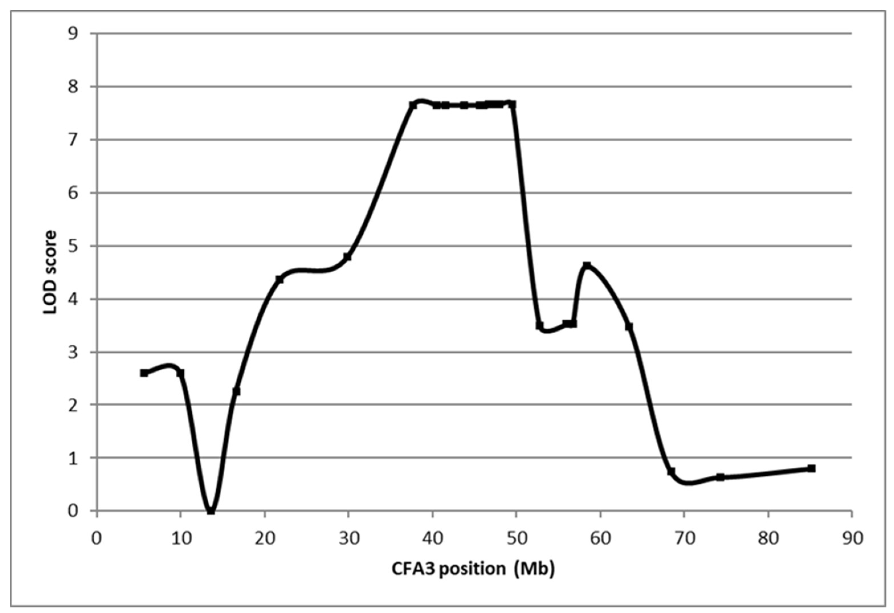
Linkage analysis was performed by genotyping DNA from a Basenji family consisting of 22 FS cases and 37 FS unaffected controls for 325 genome-wide microsatellite loci (Figure 1). The strongest associations with inferred genotypes for the FS locus and marker loci occurred on CFA3 (Figure 2). Fine mapping in 86 FS-affected Basenjis revealed that these dogs were all homozygous for the same 6-marker haplotype flanked by recombinant markers at 40,537,065 bp and 43,218,050 bp, and that none of the 11 aged Basenjis with normal renal function were homozygous for this haplotype. This analysis defined a 2.7 Mb target region of CFA3 as harboring the FS locus which contained 11 annotated genes.
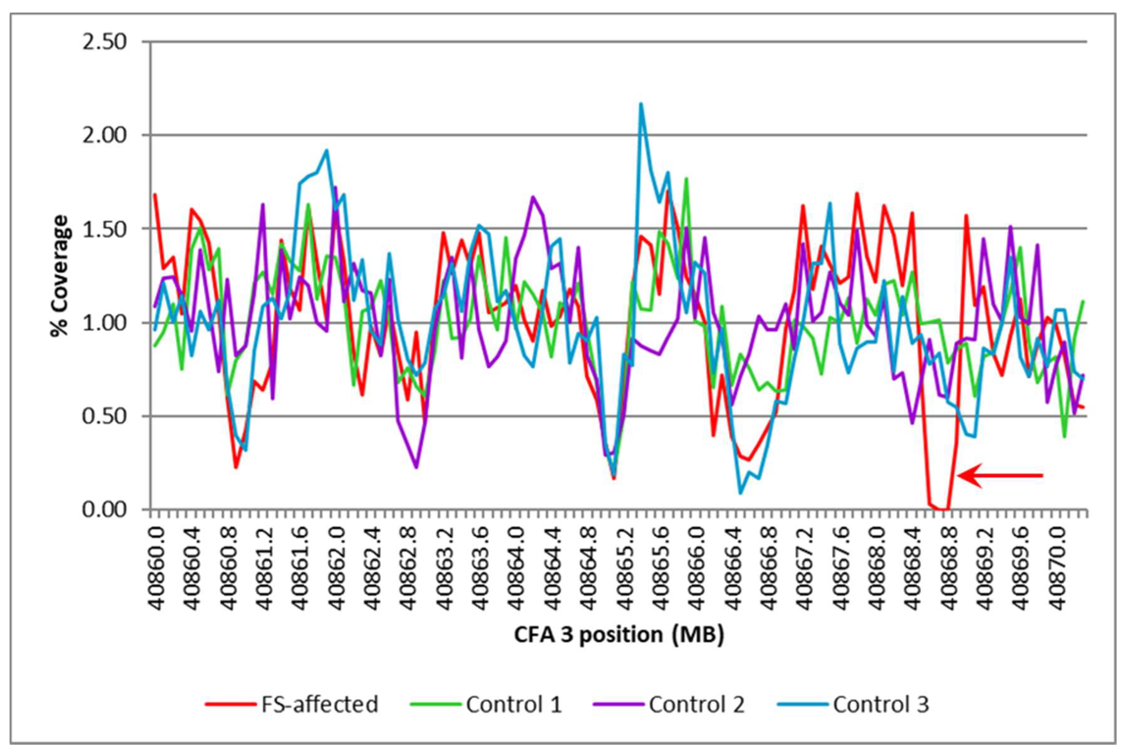
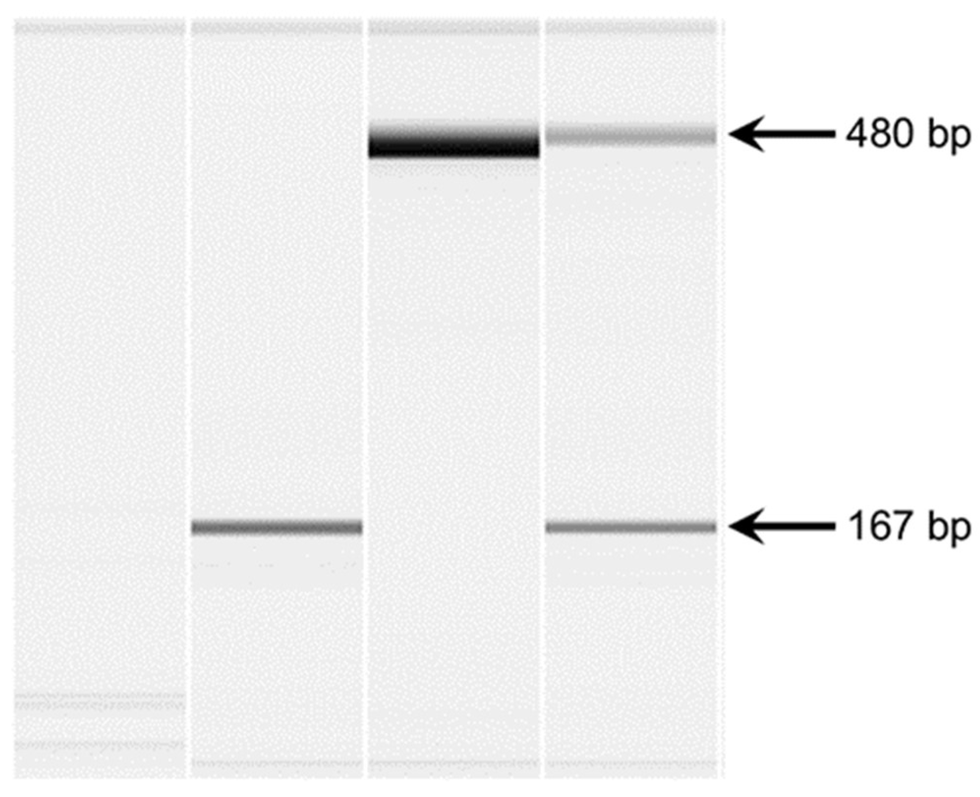
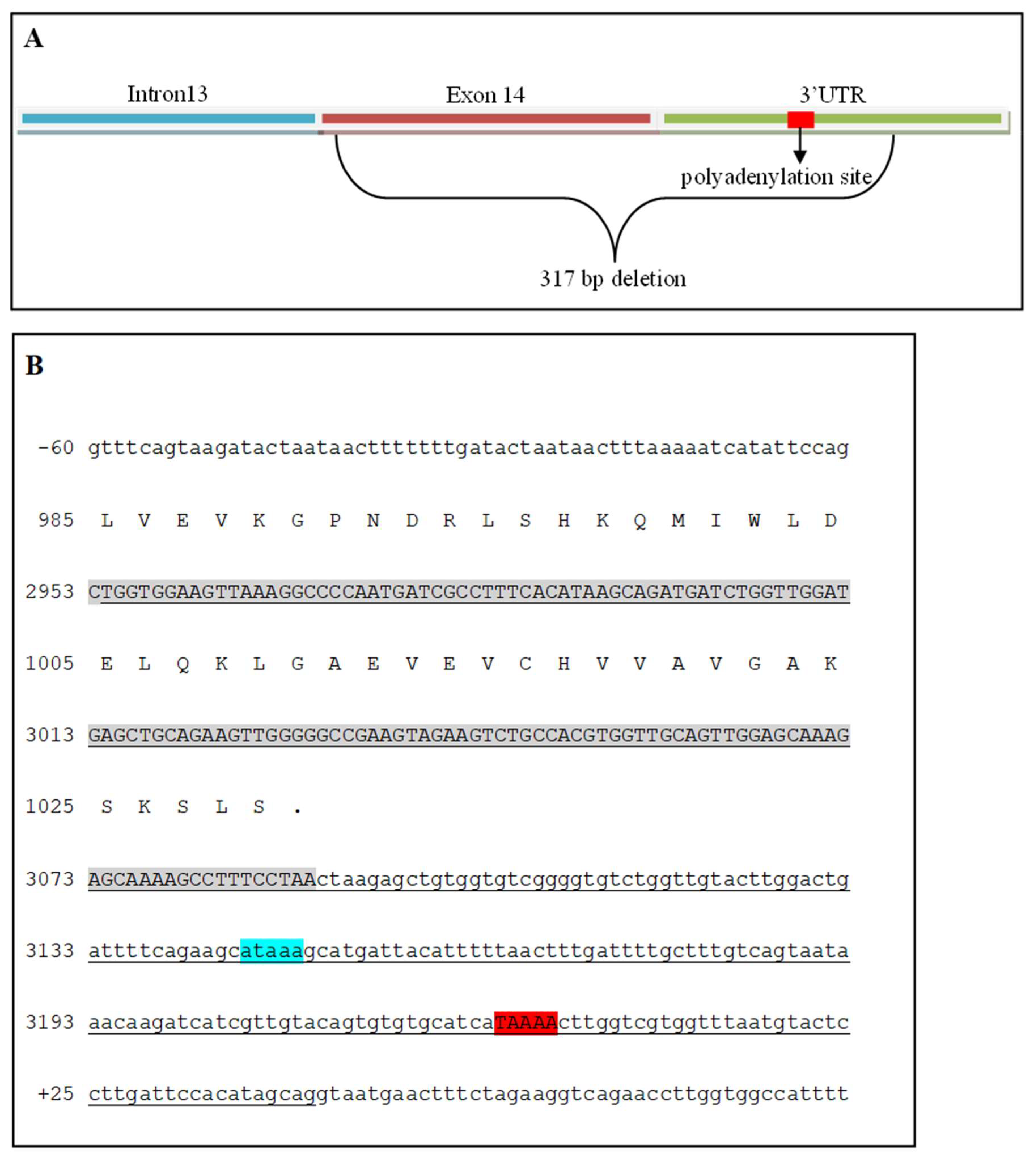
The whole-genome sequence reads from the Illumina Genome Analyzer II and Hiseq 2000 were combined and aligned to the canine genome reference to produce an aligned sequence with 12.7-fold average coverage. Exon-by-exon inspection of the sequences for the 143 annotated exons in the 11 genes within the FS region failed to reveal any sequence variants likely to alter the function of the gene products. This inspection also revealed gaps in the aligned sequence that overlapped part or all of 11 exons from within the FS target region. Comparisons of the depth of coverage in the WGS of the FS-affected Basenji with those of the WGS from 3 unaffected dogs of other breeds showed similar patterns for all but one of the sequence gaps. The exception was found only in the Basenji sequence in the vicinity of FAN1 exon 14 (Figure 3). PCR amplification with primers spanning the gap confirmed that a deletion in the genomic DNA of the FS-affected Basenji was responsible for the gap (Figure 4) and re-sequencing the amplicons produced with these primers revealed that 317 bp of exon 14 were deleted starting at the second exon14 nucleotide and extending into the 3’ untranslated region of FAN1 (Figure 5). In addition, the mutant allele has four nucleotide substitutions within the 12 nucleotides immediately 3’ to the deletion.
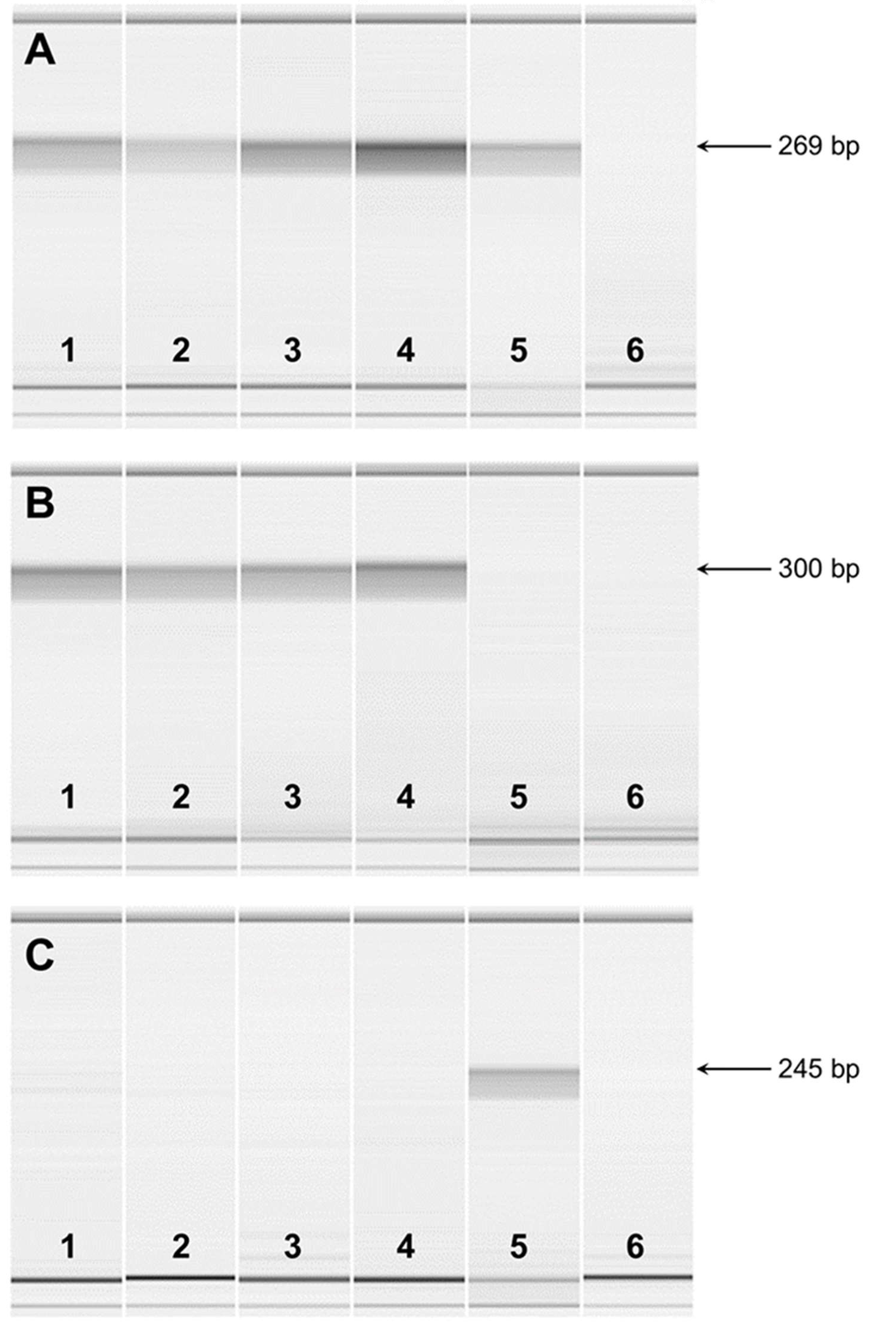
RT-PCR was used to analyze the FAN1 transcripts present in the total RNA from the kidneys of 2 unaffected dogs and from blood of FS-affected and unaffected dogs. All of the RNA preparations produced similar RT-PCR amplicons with primers designed from exon 5 and 7 sequences. Microcapillary electrophoretograms of RT-PCR amplicons demonstrated expression in all samples indicating that the mutant transcript is transcribed (Figure 6A). As expected, RT-PCR with primers designed from exon 12 and from the deleted region of exon 14 produced amplicons with RNA from normal but not affected dogs (Figure 6B). Primers designed from exon 12 and intron 13 produced amplicons with RNA from affected but not normal dogs (Figure 6C), indicating that the deletion causes intron retention in the transcript. RNA samples for both normal and affected dogs failed to produce RT-PCR amplicons with primers designed from exon 13 and sequences immediately 3’ to the deletion, suggesting that the deletion includes the polyadenylation site for normal dogs. This was confirmed with a 3’ RACE experiment which located the normal polyadenylation site 135 bp past the stop codon and 76 bp past a potential polyA signal. A similar 3’ RACE experiment revealed that the mutant RNA produces a transcript 577 bp into intron 13, 520 bp past an in-frame termination codon.
We genotyped a cohort of 78 Basenjis of known clinical status for the FAN1 deletion and found all 32 of the FS-affected Basenjis to be homozygous for the deletion allele. FS unaffected dogs tested either homozygous wild-type or heterozygous except for one unaffected dog that tested homozygous for the deletion allele. The deletion allele was highly significantly associated with the FS phenotype (p= 4.219 x 10⁻²¹, Fisher's exact test 2×2). Table 2 summarizes the genotype distribution.

4. Discussion

The data from this study indicate that FS in Basenjis is the result of FAN1 deficiency. The homozygous deletion genotype was strongly associated with FS phenotype, with only one dog that did not exhibit disease signs out of 33 that were homozygous for the deletion variant. In addition, the FAN1 risk variant was not present in 120 unaffected dogs from 81 different breeds. The whole genome sequence of the proband did not contain homozygous variants in any other genes that have been associated with the type of disease signs exhibited by the affected Basenjis. Although a functional assay of potential FAN1 enzymatic activity was not performed, the predicted translation of the variant transcript suggests a grossly altered protein structure if the transcript is synthesized and translated (Supplemental Figure 1).
The mechanism by which deficiency in FAN1 leads to the kidney pathology associated with FS remains to be fully elucidated. FAN1 was first named KIAA1018 by Nagase et al. [55] who screened brain cDNA libraries for unidentified genes. They determined that KIAA1018 was expressed at similar levels in multiple tissues. Alonso et al. [56] proposed that FAN1 was part of the myotubularin gene family of tyrosine phosphatases. They proposed a new genomic designation, MTMR15 and predicted that the encoded protein was a catalytically inactive member of the inactive MTMR family of protein tyrosine phosphatases. The inactive MTMRs have been reported to act as regulatory units for active members of the group [57]. However, no studies have been reported that directly support the hypothesis that FAN1 (MTMR15/KIAA1018) is involved in the regulation of the enzymatically active MTMRs.
Unlike the MTMR proteins, FAN1 has a ubiquitin binding domain at the N-terminus and at its C-terminus a domain with homology to bacterial and phage endonucleases, which suggested that this protein may contribute to the maintenance of genome stability [58]. FAN1has been identified as one of the proteins involved in DNA inter-strand crosslink repair [59,60,61,62,63,64]. The disease Fanconi anemia is a recessive disorder characterized by genome instability, impaired repair of DNA crosslink damage, developmental abnormalities, early-onset bone marrow failure and predisposition to cancer [65,66]. Variants in more than 20 genes, including FAN1, have been associated with Fanconi anemia [66,67,68,69,70]. The name FAN1 (Fanconi anemia-associated nuclease 1) has been proposed because this protein interacts with Fanconi anemia pathway proteins [59,60,61,62,69]. When DNA inter-strand crosslinks occur, FAN1 is recruited to the lesion sites through an interaction between its ubiquitin binding domain and the ubiquitylated complex of the Fanconi anemia pathway [59,60,61,62,69]. However, it appears that FAN1 may also mediate DNA repair independent of other proteins in this pathway [63,71]. The deleted region encodes a conserved segment of the nuclease domain, which is likely to obliterate FAN1 nuclease activity (Supplemental Figure 2) and thus its role in DNA repair. Based on the evidence that FAN1 is involved in repairing DNA inter-strand crosslinks, the proximal renal tubule pathology associated with FS may be the result of the accumulation of these crosslinks in renal tubule epithelial cells.
Support for this hypothesis comes from the finding that FAN1 deficiency sensitizes cultured cells to reagents that cause targeted DNA damage. This sensitivity can be rescued by transfection with wild-type FAN1 but not by variant constructs containing point mutations in the ubiquitin binding or endonuclease domains, which indicates that both domains are involved in DNA repair [59,60,61,62]. The nuclease domain of FAN1 has both 5’exonuclease activity and endonuclease activities that are key characteristics of DNA repair proteins [59,60,61,62].
The fact that the most apparent pathology associated with FS occurs in the proximal tubules of the kidneys suggest that this tissue may be particularly susceptible to DNA damage. Factors that promote DNA damage in this tissue may contribute to the development of FS. Consistent with this hypothesis is the fact that acquired forms of FS have been associated with heavy-metal exposure and toxicoses from drugs such as cisplatin that promote DNA damage [7,14,28,29,72,73,74,75,76]. Heavy metals, such as cadmium, may be present in plants and sea food because of contaminated soils and water [72]. Chronic environmental exposure can result in cadmium accumulation to toxic levels that cause kidney disease [72]. Cadmium toxicity can cause DNA damage, including double and single stranded breaks [77]. The kidney is particularly susceptible to cadmium and other heavy-metal toxicities. Approximately 50% of the accumulated dose of cadmium is stored in the kidney [72]. In mitochondria, cadmium inhibits the respiratory chain resulting in the generation of reactive oxygen species [78]. This leads to mitochondrial disruption with the release of cytochrome c, resulting in caspase activation causing cell death by apoptosis [79,80].
We propose that FAN1 inactivation causes FS in Basenjis by hyper-sensitization of the proximal tubule cells to toxins that mediate DNA damage, including heavy metals such as cadmium. Since FAN1 has been reported to be involved in DNA repair and the knockdown of FAN1 sensitizes cells to DNA crosslinking agents [59,60,61,62], we predict that environmental and dietary exposure to toxins that promote DNA damage leads to the accumulation of both genomic and mitochondrial DNA damage in the kidney. Differential exposure to environmental toxins may explain the wide age-at-onset variability for FS in Basenjis.

Supplementary Materials

The following supporting information can be downloaded at: Preprints.org, Figure S1: title; Table S1: title; Video S1: title.

Author Contributions

Conceptualization, G.S.J., L.H.; methodology, F.H.G.F., T.M., G.S.J.; validation, F.H.G.F., T.M., J.G.; formal analysis, F.H.G.F., T.M., G.S.J.; investigation, F.H.G.F., L.H., G.S.J.; resources, G.S.J., M.L.K.; data curation, T.M.; writing—original draft preparation, F.H.G.F., M.L.K.; writing—review and editing, F.H.G.F., T.M., M.L.K.; supervision, G.S.J., M.L.K.; project administration, G.S.J., M.L.K.; funding acquisition, G.S.J., M.L.K. All authors other than G.S.J. who was deceased prior to final manuscript preparation have read and agreed to the submitted version of the manuscript.

Funding

This research was supported by funding provided by the Orthopedic Foundation for Animals.

Institutional Review Board Statement

This study was conducted in accordance with the Declaration of Helsinki and approved by the Institutional Review Board of the University of Missouri (protocol 20520, approved 22 December 2021).

Informed Consent Statement

Not applicable.

Data Availability Statement

WGS DNA sequence data for the Basenji included in this study have been archived and deposited in the NCBI Sequence Read Archive as BioSample SAMN04196845.

Acknowledgments

Our thanks to Robert Schnabel and Jerry Taylor for assistance with whole genome sequence analyses. Our thanks also to Jon Curby of the Basenji Club of America for assisting with the recruitment of the dogs that were included in this study.

Conflicts of Interest

The Canine Genetics Laboratory at the University of Missouri provides fee-for-service genetic testing for dogs. The authors disclose no other conflict of interest.

References

  1. Foreman, J.W. Fanconi Syndrome. Pediatr Clin North Am 2019, 66, 159–167. [CrossRef]
  2. Albuquerque, A.L.B.; Dos Santos Borges, R.; Conegundes, A.F.; Dos Santos, E.E.; Fu, F.M.M.; Araujo, C.T.; Vaz de Castro, P.A.S.; Simoes E Silva, A.C. Inherited Fanconi Syndrome. World J Pediatr 2023, 19, 619–634. [CrossRef]
  3. Kunchur, M.G.; Mauch, T.J.; Parkanzky, M.; Rahilly, L.J. A Review of Renal Tubular Acidosis. J Vet Emerg Crit Care (San Antonio) 2024, 34, 325–355. [CrossRef]
  4. Lobitz, S.; Velleuer, E. Guido Fanconi (1892-1979): A Jack of All Trades. Nat Rev Cancer 2006, 6, 893–898. [CrossRef]
  5. Baum, M. The Cellular Basis of Fanconi Syndrome. Hosp Pract (Off Ed) 1993, 28, 137–138. [CrossRef]
  6. Magen, D.; Berger, L.; Coady, M.J.; Ilivitzki, A.; Militianu, D.; Tieder, M.; Selig, S.; Lapointe, J.Y.; Zelikovic, I.; Skorecki, K. A Loss-of-Function Mutation in NaPi-IIa and Renal Fanconi’s Syndrome. N Engl J Med 2010, 362, 1102–1109. [CrossRef]
  7. Lichter-Konecki, U.; Broman, K.W.; Blau, E.B.; Konecki, D.S. Genetic and Physical Mapping of the Locus for Autosomal Dominant Renal Fanconi Syndrome, on Chromosome 15q15.3. Am J Hum Genet 2001, 68, 264–268. [CrossRef]
  8. Emma, F.; Bertini, E.; Salviati, L.; Montini, G. Renal Involvement in Mitochondrial Cytopathies. Pediatr Nephrol 2012, 27, 539–550. [CrossRef]
  9. Lloyd, S.E.; Pearce, S.H.; Fisher, S.E.; Steinmeyer, K.; Schwappach, B.; Scheinman, S.J.; Harding, B.; Bolino, A.; Devoto, M.; Goodyer, P.; et al. A Common Molecular Basis for Three Inherited Kidney Stone Diseases. Nature 1996, 379, 445–449. [CrossRef]
  10. Lowe, M. Structure and Function of the Lowe Syndrome Protein OCRL1. Traffic 2005, 6, 711–719. [CrossRef]
  11. Baum, M. The Fanconi Syndrome of Cystinosis: Insights into the Pathophysiology. Pediatr Nephrol 1998, 12, 492–497. [CrossRef]
  12. Kintzel, P.E. Anticancer Drug-Induced Kidney Disorders. Drug Saf 2001, 24, 19–38. [CrossRef]
  13. Gonick, H.; Indraprasit, S.; Neustein, H.; Rosen, V. Cadmium-Induced Experimental Fanconi Syndrome. Curr Probl Clin Biochem 1975, 4, 111–118.
  14. Yui, J.C.; Geara, A.; Sayani, F. Deferasirox-Associated Fanconi Syndrome in Adult Patients with Transfusional Iron Overload. Vox Sang 2021, 116, 793–797. [CrossRef]
  15. Hall, A.M.; Trepiccione, F.; Unwin, R.J. Drug Toxicity in the Proximal Tubule: New Models, Methods and Mechanisms. Pediatr Nephrol 2022, 37, 973–982. [CrossRef]
  16. Anguissola, G.; Leu, D.; Simonetti, G.D.; Simonetti, B.G.; Lava, S.A.G.; Milani, G.P.; Bianchetti, M.G.; Scoglio, M. Kidney Tubular Injury Induced by Valproic Acid: Systematic Literature Review. Pediatr Nephrol 2023, 38, 1725–1731. [CrossRef]
  17. Easley, J.R.; Breitschwerdt, D.B. Glucosuria Associated with Renal Tubular Dysfunction in Three Basenji Dogs. J Am Vet Med Assoc 1976, 168, 938–943.
  18. Bovee, K.C.; Joyce, T.; Reynolds, R.; Segal, S. Spontaneous Fanconi Syndrome in the Dog. Metabolism 1978, 27, 45–52. [CrossRef]
  19. Bovee, K.C.; Joyce, T.; Reynolds, R.; Segal, S. The Fanconi Syndrome in Basenji Dogs: A New Model for Renal Transport Defects. Science 1978, 201, 1129–1131. [CrossRef]
  20. Bovee, K.C.; Joyce, T.; Blazer-Yost, B.; Goldschmidt, M.S.; Segal, S. Characterization of Renal Defects in Dogs with a Syndrome Similar to the Fanconi Syndrome in Man. J Am Vet Med Assoc 1979, 174, 1094–1099.
  21. Bovee, K.C.; Anderson, T.; Brown, S.; Goldschmidt, M.H.; Segal, S. Renal Tubular Defects of Spontaneous Fanconi Syndrome in Dogs. Prog Clin Biol Res 1982, 94, 435–447.
  22. Medow, M.S.; Reynolds, R.; Bovee, K.C.; Segal, S. Proline and Glucose Transport by Renal Membranes from Dogs with Spontaneous Idiopathic Fanconi Syndrome. Proc Natl Acad Sci U S A 1981, 78, 7769–7772. [CrossRef]
  23. Mainka, S.A. Fanconi Syndrome in a Basenji. Can Vet J 1985, 26, 303–305.
  24. Noonan, C.H.; Kay, J.M. Prevalence and Geographic Distribution of Fanconi Syndrome in Basenjis in the United States. J Am Vet Med Assoc 1990, 197, 345–349.
  25. Yearley, J.H.; Hancock, D.D.; Mealey, K.L. Survival Time, Lifespan, and Quality of Life in Dogs with Idiopathic Fanconi Syndrome. J Am Vet Med Assoc 2004, 225, 377–383. [CrossRef]
  26. Freeman, L.M.; Breitschwerdt, E.B.; Keene, B.W.; Hansen, B. Fanconi’s Syndrome in a Dog with Primary Hypoparathyroidism. J Vet Intern Med 1994, 8, 349–354. [CrossRef]
  27. Settles, E.L.; Schmidt, D. Fanconi Syndrome in a Labrador Retriever. J Vet Intern Med 1994, 8, 390–393. [CrossRef]
  28. Appleman, E.H.; Cianciolo, R.; Mosenco, A.S.; Bounds, M.E.; Al-Ghazlat, S. Transient Acquired Fanconi Syndrome Associated with Copper Storage Hepatopathy in 3 Dogs. J Vet Intern Med 2008, 22, 1038–1042. [CrossRef]
  29. King, J.B. Proximal Tubular Nephropathy in Two Dogs Diagnosed with Lead Toxicity. Aust Vet J 2016, 94, 280–284. [CrossRef]
  30. Yabuki, A.; Iwanaga, T.; Giger, U.; Sawa, M.; Kohyama, M.; Yamato, O. Acquired Fanconi Syndrome in Two Dogs Following Long-Term Consumption of Pet Jerky Treats in Japan: Case Report. J Vet Med Sci 2017, 79, 818–821. [CrossRef]
  31. Bommer, N.X.; Brownlie, S.E.; Morrison, L.R.; Chandler, M.L.; Simpson, J.W. Fanconi Syndrome in Irish Wolfhound Siblings. J Am Anim Hosp Assoc 2018, 54, 173–178. [CrossRef]
  32. Ahn, J.-O.; Kim, S.-M.; Song, W.-J.; Ryu, M.-O.; Li, Q.; Chung, J.-Y.; Youn, H.-Y. Transient Fanconi Syndrome After Treatment with Firocoxib, Cefadroxil, Tramadol, and Famotidine in a Maltese. J Am Anim Hosp Assoc 2019, 55, 323–327. [CrossRef]
  33. Takashima, S.; Nasu, T.; Ohata, K.; Oikawa, T.; Sugaya, T.; Kobatake, Y.; Shibata, S.; Nishii, N. Urinary Liver-Type Fatty Acid-Binding Protein in Two Dogs with Acquired Fanconi Syndrome: A Case Report. Open Vet J 2022, 12, 864–867. [CrossRef]
  34. Nybroe, S.; Bjornvad, C.R.; Hansen, C.F.H.; Andersen, T.S.L.; Kieler, I.N. Outcome of Acquired Fanconi Syndrome Associated with Ingestion of Jerky Treats in 30 Dogs. Animals (Basel) 2022, 12. [CrossRef]
  35. Hooper, A.N.; Roberts, B.K. Fanconi Syndrome in Four Non-Basenji Dogs Exposed to Chicken Jerky Treats. J Am Anim Hosp Assoc 2011, 47, e178-87. [CrossRef]
  36. Major, A.; Schweighauser, A.; Hinden, S.E.; Francey, T. Transient Fanconi Syndrome with Severe Polyuria and Polydipsia in a 4-Year Old Shih Tzu Fed Chicken Jerky Treats. Schweiz Arch Tierheilkd 2014, 156, 593–598. [CrossRef]
  37. Hill, T.L.; Breitschwerdt, E.B.; Cecere, T.; Vaden, S. Concurrent Hepatic Copper Toxicosis and Fanconi’s Syndrome in a Dog. J Vet Intern Med 2008, 22, 219–222. [CrossRef]
  38. Preston, R.; Naylor, R.W.; Stewart, G.; Bierzynska, A.; Saleem, M.A.; Lowe, M.; Lennon, R. A Role for OCRL in Glomerular Function and Disease. Pediatr Nephrol 2020, 35, 641–648. [CrossRef]
  39. Sharari, S.; Abou-Alloul, M.; Hussain, K.; Ahmad Khan, F. Fanconi-Bickel Syndrome: A Review of the Mechanisms That Lead to Dysglycaemia. Int J Mol Sci 2020, 21. [CrossRef]
  40. Govers, L.P.; Toka, H.R.; Hariri, A.; Walsh, S.B.; Bockenhauer, D. Mitochondrial DNA Mutations in Renal Disease: An Overview. Pediatr Nephrol 2021, 36, 9–17. [CrossRef]
  41. Khandelwal, P.; Mahesh, V.; Mathur, V.P.; Raut, S.; Geetha, T.S.; Nair, S.; Hari, P.; Sinha, A.; Bagga, A. Phenotypic Variability in Distal Acidification Defects Associated with WDR72 Mutations. Pediatr Nephrol 2021, 36, 881–887. [CrossRef]
  42. Sheppard, S.E.; Barrett, B.; Muraresku, C.; McKnight, H.; De Leon, D.D.; Lord, K.; Ganetzky, R. Heterozygous Recurrent HNF4A Variant p.Arg85Trp Causes Fanconi Renotubular Syndrome 4 with Maturity Onset Diabetes of the Young, an Autosomal Dominant Phenocopy of Fanconi Bickel Syndrome with Colobomas. Am J Med Genet A 2021, 185, 566–570. [CrossRef]
  43. Duan, N.; Huang, C.; Pang, L.; Jiang, S.; Yang, W.; Li, H. Clinical Manifestation and Genetic Findings in Three Boys with Low Molecular Weight Proteinuria - Three Case Reports for Exploring Dent Disease and Fanconi Syndrome. BMC Nephrol 2021, 22, 24. [CrossRef]
  44. Elsayed, A.K.; Al-Khawaga, S.; Hussain, K.; Abdelalim, E.M. An Induced Pluripotent Stem Cell Line Derived from a Patient with Neonatal Diabetes and Fanconi-Bickel Syndrome Caused by a Homozygous Mutation in the SLC2A2 Gene. Stem Cell Res 2021, 54, 102433. [CrossRef]
  45. Grunert, S.C.; Schumann, A.; Baronio, F.; Tsiakas, K.; Murko, S.; Spiekerkoetter, U.; Santer, R. Evidence for a Genotype-Phenotype Correlation in Patients with Pathogenic GLUT2 (SLC2A2) Variants. Genes (Basel) 2021, 12. [CrossRef]
  46. Kanako, K.-I.; Sakakibara, N.; Murayama, K.; Nagatani, K.; Murata, S.; Otake, A.; Koga, Y.; Suzuki, H.; Uehara, T.; Kosaki, K.; et al. BCS1L Mutations Produce Fanconi Syndrome with Developmental Disability. J Hum Genet 2022, 67, 143–148. [CrossRef]
  47. Sharari, S.; Aouida, M.; Mohammed, I.; Haris, B.; Bhat, A.A.; Hawari, I.; Nisar, S.; Pavlovski, I.; Biswas, K.H.; Syed, N.; et al. Understanding the Mechanism of Dysglycemia in a Fanconi-Bickel Syndrome Patient. Front Endocrinol (Lausanne) 2022, 13, 841788. [CrossRef]
  48. Agakidou, E.; Agakidis, C.; Kambouris, M.; Printza, N.; Farini, M.; Vourda, E.; Gerou, S.; Sarafidis, K. A Novel Mutation of VPS33B Gene Associated with Incomplete Arthrogryposis-Renal Dysfunction-Cholestasis Phenotype. Case Rep Genet 2020, 2020, 8872294. [CrossRef]
  49. Forst, A.-L.; Reichold, M.; Kleta, R.; Warth, R. Distinct Mitochondrial Pathologies Caused by Mutations of the Proximal Tubular Enzymes EHHADH and GATM. Front Physiol 2021, 12, 715485. [CrossRef]
  50. Guan, Y.-J.; Guo, Y.-N.; Peng, W.-T.; Liu, L.-L. Case Report: Cystinosis in a Chinese Child With a Novel CTNS Pathogenic Variant. Front Pediatr 2022, 10, 860990. [CrossRef]
  51. Kudo, H.; Suzuki, R.; Kondo, A.; Nozu, K.; Nakamura, Y.; Mikami, H.; Soma, J.; Nakaya, I. Association of Familial Fanconi Syndrome with a Novel GATM Variant. Tohoku J Exp Med 2023, 260, 337–340. [CrossRef]
  52. Sivaji, V.; Raju, P.; Marimuthu, S.; Sundar, S. Familial Renal Glycosuria Identified in an Indian Family. BMJ Case Rep 2024, 17. [CrossRef]
  53. Saiteja, P.; Krishnamurthy, S.; Deepthi, B.; Krishnasamy, S.; Sravani, M. Distal Renal Tubular Acidosis as Presenting Manifestation of Wilson Disease in a 11-Year-Old Girl. CEN Case Rep 2024, 13, 93–97. [CrossRef]
  54. Katz, M.L.; Khan, S.; Awano, T.; Shahid, S.A.; Siakotos, A.N.; Johnson, G.S. A Mutation in the CLN8 Gene in English Setter Dogs with Neuronal Ceroid-Lipofuscinosis. Biochem Biophys Res Commun 2005, 327. [CrossRef]
  55. Nagase, T.; Ishikawa, K.; Suyama, M.; Kikuno, R.; Hirosawa, M.; Miyajima, N.; Tanaka, A.; Kotani, H.; Nomura, N.; Ohara, O. Prediction of the Coding Sequences of Unidentified Human Genes. XIII. The Complete Sequences of 100 New CDNA Clones from Brain Which Code for Large Proteins in Vitro. DNA Res 1999, 6, 63–70. [CrossRef]
  56. Alonso, A.; Sasin, J.; Bottini, N.; Friedberg, I.; Friedberg, I.; Osterman, A.; Godzik, A.; Hunter, T.; Dixon, J.; Mustelin, T. Protein Tyrosine Phosphatases in the Human Genome. Cell 2004, 117, 699–711. [CrossRef]
  57. Azzedine, H.; Bolino, A.; Taieb, T.; Birouk, N.; Di Duca, M.; Bouhouche, A.; Benamou, S.; Mrabet, A.; Hammadouche, T.; Chkili, T.; et al. Mutations in MTMR13, a New Pseudophosphatase Homologue of MTMR2 and Sbf1, in Two Families with an Autosomal Recessive Demyelinating Form of Charcot-Marie-Tooth Disease Associated with Early-Onset Glaucoma. Am J Hum Genet 2003, 72, 1141–1153. [CrossRef]
  58. Yoshikiyo, K.; Kratz, K.; Hirota, K.; Nishihara, K.; Takata, M.; Kurumizaka, H.; Horimoto, S.; Takeda, S.; Jiricny, J. KIAA1018/FAN1 Nuclease Protects Cells against Genomic Instability Induced by Interstrand Cross-Linking Agents. Proc Natl Acad Sci U S A 2010, 107, 21553–21557. [CrossRef]
  59. Liu, T.; Ghosal, G.; Yuan, J.; Chen, J.; Huang, J. FAN1 Acts with FANCI-FANCD2 to Promote DNA Interstrand Cross-Link Repair. Science 2010, 329, 693–696. [CrossRef]
  60. MacKay, C.; Declais, A.-C.; Lundin, C.; Agostinho, A.; Deans, A.J.; MacArtney, T.J.; Hofmann, K.; Gartner, A.; West, S.C.; Helleday, T.; et al. Identification of KIAA1018/FAN1, a DNA Repair Nuclease Recruited to DNA Damage by Monoubiquitinated FANCD2. Cell 2010, 142, 65–76. [CrossRef]
  61. Smogorzewska, A.; Desetty, R.; Saito, T.T.; Schlabach, M.; Lach, F.P.; Sowa, M.E.; Clark, A.B.; Kunkel, T.A.; Harper, J.W.; Colaiacovo, M.P.; et al. A Genetic Screen Identifies FAN1, a Fanconi Anemia-Associated Nuclease Necessary for DNA Interstrand Crosslink Repair. Mol Cell 2010, 39, 36–47. [CrossRef]
  62. Kratz, K.; Schopf, B.; Kaden, S.; Sendoel, A.; Eberhard, R.; Lademann, C.; Cannavo, E.; Sartori, A.A.; Hengartner, M.O.; Jiricny, J. Deficiency of FANCD2-Associated Nuclease KIAA1018/FAN1 Sensitizes Cells to Interstrand Crosslinking Agents. Cell 2010, 142, 77–88. [CrossRef]
  63. Jin, H.; Cho, Y. Structural and Functional Relationships of FAN1. DNA Repair (Amst) 2017, 56, 135–143. [CrossRef]
  64. Thongthip, S.; Bellani, M.; Gregg, S.Q.; Sridhar, S.; Conti, B.A.; Chen, Y.; Seidman, M.M.; Smogorzewska, A. Fan1 Deficiency Results in DNA Interstrand Cross-Link Repair Defects, Enhanced Tissue Karyomegaly, and Organ Dysfunction. Genes Dev 2016, 30, 645–659. [CrossRef]
  65. Hoover, A.; Turcotte, L.M.; Phelan, R.; Barbus, C.; Rayannavar, A.; Miller, B.S.; Reardon, E.E.; Theis-Mahon, N.; MacMillan, M.L. Longitudinal Clinical Manifestations of Fanconi Anemia: A Systematized Review. Blood Rev 2024, 101225. [CrossRef]
  66. Woo, A.Y.-H.; Jia, L. ALDH2 Mutations and Defense against Genotoxic Aldehydes in Cancer and Inherited Bone Marrow Failure Syndromes. Mutat Res 2024, 829, 111870. [CrossRef]
  67. Parsa, F.G.; Nobili, S.; Karimpour, M.; Aghdaei, H.A.; Nazemalhosseini-Mojarad, E.; Mini, E. Fanconi Anemia Pathway in Colorectal Cancer: A Novel Opportunity for Diagnosis, Prognosis and Therapy. J Pers Med 2022, 12. [CrossRef]
  68. Mehta, P.A.; Ebens, C. Fanconi Anemia. 1993.
  69. Gwon, G.H.; Kim, Y.; Liu, Y.; Watson, A.T.; Jo, A.; Etheridge, T.J.; Yuan, F.; Zhang, Y.; Kim, Y.; Carr, A.M.; et al. Crystal Structure of a Fanconi Anemia-Associated Nuclease Homolog Bound to 5’ Flap DNA: Basis of Interstrand Cross-Link Repair by FAN1. Genes Dev 2014, 28, 2276–2290. [CrossRef]
  70. Zhang, J.; Walter, J.C. Mechanism and Regulation of Incisions during DNA Interstrand Cross-Link Repair. DNA Repair (Amst) 2014, 19, 135–142. [CrossRef]
  71. Jin, H.; Roy, U.; Lee, G.; Scharer, O.D.; Cho, Y. Structural Mechanism of DNA Interstrand Cross-Link Unhooking by the Bacterial FAN1 Nuclease. J Biol Chem 2018, 293, 6482–6496. [CrossRef]
  72. Johri, N.; Jacquillet, G.; Unwin, R. Heavy Metal Poisoning: The Effects of Cadmium on the Kidney. Biometals 2010, 23, 783–792. [CrossRef]
  73. Johri, N.; Jacquillet, G.; Unwin, R. Heavy Metal Poisoning: The Effects of Cadmium on the Kidney. Biometals 2010, 23, 783–792. [CrossRef]
  74. Chakraborty, S.; Dutta, A.R.; Sural, S.; Gupta, D.; Sen, S. Ailing Bones and Failing Kidneys: A Case of Chronic Cadmium Toxicity. Ann Clin Biochem 2013, 50, 492–495. [CrossRef]
  75. Senthilnathan, S.; Nallusamy, G.; Varadaraj, P. An Interesting Case of Acquired Renal Fanconi Syndrome. Cureus 2024, 16, e65208. [CrossRef]
  76. Sheridan, R.; Mirabile, J.; Hafler, K. Determination of Six Illegal Antibiotics in Chicken Jerky Dog Treats. J Agric Food Chem 2014, 62, 3690–3696. [CrossRef]
  77. Pal, S.; Kim, J.Y.; Park, S.H.; Lim, H. Bin; Lee, K.-H.; Song, J.M. Quantitative Classification of DNA Damages Induced by Submicromolar Cadmium Using Oligonucleotide Chip Coupled with Lesion-Specific Endonuclease Digestion. Environ Sci Technol 2011, 45, 4460–4467. [CrossRef]
  78. Wang, Y.; Fang, J.; Leonard, S.S.; Rao, K.M.K. Cadmium Inhibits the Electron Transfer Chain and Induces Reactive Oxygen Species. Free Radic Biol Med 2004, 36, 1434–1443. [CrossRef]
  79. Thevenod, F.; Friedmann, J.M.; Katsen, A.D.; Hauser, I.A. Up-Regulation of Multidrug Resistance P-Glycoprotein via Nuclear Factor-KappaB Activation Protects Kidney Proximal Tubule Cells from Cadmium- and Reactive Oxygen Species-Induced Apoptosis. J Biol Chem 2000, 275, 1887–1896. [CrossRef]
  80. Tzirogiannis, K.N.; Panoutsopoulos, G.I.; Demonakou, M.D.; Hereti, R.I.; Alexandropoulou, K.N.; Basayannis, A.C.; Mykoniatis, M.G. Time-Course of Cadmium-Induced Acute Hepatotoxicity in the Rat Liver: The Role of Apoptosis. Arch Toxicol 2003, 77, 694–701. [CrossRef]
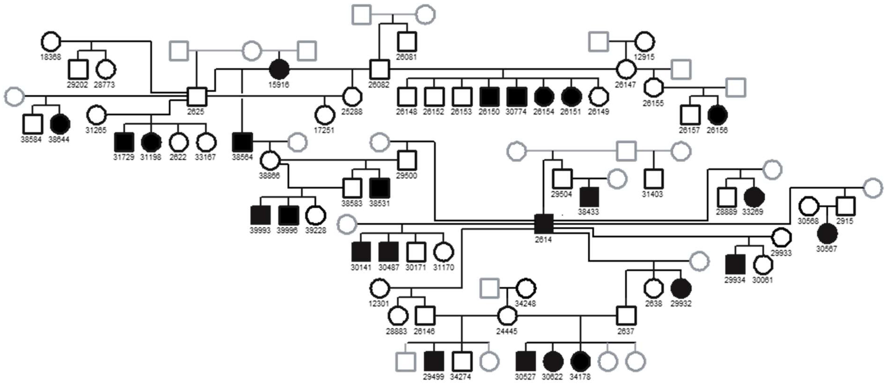
Figure 1. Pedigree of the Basenji family used for linkage mapping of the FS locus.
Preprints 137417 g001
Figure 2. Plot of linkage results of the Fanconi Syndrome Basenji pedigree (22 cases and 37 controls).
Preprints 137417 g002
Figure 3. Comparison of percent coverage in a part of the CFA3 target region between an FS-affected dog and 3 controls. The region represented in this graph starts at 40,860,000 to 40,870,300 bp of CFA3 in intervals of 100 bp. The percent coverage is the average coverage of the interval normalized for the average genome coverage for the individual dog. Arrow indicates the gap in coverage that was unique to the affected Basenji.
Preprints 137417 g003
Figure 4. Microcapillary electrophoretograms of PCR amplicons produced with primers spanning the gap in the FAN1. Lane 1 represents a negative control. PCR was performed with DNA from an FS-affected dog (2) and two FS-unaffected dogs (lanes 3 and 4). The FS-affected dog produced an 167 bp amplicon which is smaller than the expected band. One of the FS-unaffected dogs (3) produced the expected band and the other dog (lane 4) produced the expected band and the deletion allele band.
Preprints 137417 g004
Figure 5. The deletion boundaries represented by illustration and genomic sequences of the FAN1. (A) Illustration of the 3’ end of the FAN1 gene. The deletion starts after the first base of exon 14 and goes into the 3’ UTR past the primary polyadenylation site. (B) Sequences for the end of intron 13, exon 14 and the 3’UTR. Gray shaded sequences correspond to exon 14, blue shaded represents the potential poly signal and red shaded is the polyadenylation site. The 317 deleted bases are underlined.
Preprints 137417 g005
Figure 6. Microcapillary electrophoretograms of RT-PCR amplicons from FAN1 mRNA. RT-PCR was performed with total RNA extracted from kidney of two FS-unaffected dogs (1 and 2), blood of two FS-unaffected dogs (3 and 4) and one FS-affected dog (5). Lane 6 represents a negative control. (A) RT-PCR was performed with primers from exon 5 to exon 7 of the FAN1 gene. The expected amplicon size was 269 bp. (B) RT-PCR was performed with primers from exon 12 to exon 14 of the FAN1 gene. The expected amplicon size was 300 bp. (C) RT-PCR was performed with primers from exon 12 to intron 13 of the FAN1 gene. The expected amplicon size was 245 bp.
Preprints 137417 g006
Table 1. RT-PCR primer sequences for FAN1 mRNA.
Preprints 137417 i001
Table 2. Distribution of genotypes among healthy and FS-affected Basenjis.
Phenotype Del/Del Genotypes
Del/Wt
Wt/Wt Total
FS affected 32 0 0 32
FS unaffected 1 33 12 46
Total 33 33 12 78
Disclaimer/Publisher’s Note: The statements, opinions and data contained in all publications are solely those of the individual author(s) and contributor(s) and not of MDPI and/or the editor(s). MDPI and/or the editor(s) disclaim responsibility for any injury to people or property resulting from any ideas, methods, instructions or products referred to in the content.
Copyright: This open access article is published under a Creative Commons CC BY 4.0 license, which permit the free download, distribution, and reuse, provided that the author and preprint are cited in any reuse.
Alerts
Prerpints.org logo

Preprints.org is a free preprint server supported by MDPI in Basel, Switzerland.

Subscribe

© 2025 MDPI (Basel, Switzerland) unless otherwise stated