Preprint
Review

On the Similarity and Differences between NELIBS & NELIPS in Laser Induced Nanomaterials Plasma

Submitted:

25 October 2024

Posted:

25 October 2024

You are already at the latest version

A peer-reviewed article of this preprint also exists.

Abstract
The interaction of pulsed lasers with matter involving nanomaterials as a pure target or thin layer deposited on a target, initiates transient plasma which shows strong enhancement in a spectral line emission. This domain of research has been explored via two well established techniques dubbed NELIBS and NELIPS. These Nano-Enhanced Laser Induced Breakdown or Plasma Spectroscopy techniques entail similarities as well as differences. Thereupon, certain confusion has arisen from various aspects of the similarities as well as differences between the two techniques. In this article, we will investigate the application of either technique to retrieve relevant data about enhanced spectral line plasma emission phenomenon. To discriminate between these two techniques, survey on the nature of target, the origin of enhancement and prevalent theoretical approaches is presented. Finally, potential achievements, challenges, and the expected prospective are highlighted.
Keywords: 
Subject: 
Physical Sciences  -   Atomic and Molecular Physics

1. Introduction

1.1. Plasma from the Thermodynamical Point of View

Plasma is the fourth state of matter after solid, liquid and gas states [1]. Thermodynamically, it can be regarded as an ensemble that comprises four different types of species. These include three corpuscular atoms, ions and electrons, while the fourth component is radiation. It exists at relatively large temperatures, therefore, a small fraction of atoms are typically ionized creating a finite number of ions and electrons [1,2,3]. Some of these electrons escape from the plasma active volume while the residue constitutes a cloud of quasi-free electrons oscillating in a collective manner about the heavy-positive ionic background [1,2,3]. Each kind of species maintains a local thermodynamical equilibrium with their own species and hence, each one component species are distributed according to its proper equilibrium distribution function characterized by certain temperature [1,2,3,4,5,6,7]. A compact summary of the proper equilibrium distribution functions correlated with each of the plasma constituents is given in Table 1.

1.2. Plasma States of Equilibrium

It is a common practice that plasma can be fully characterized on account of the values of two measurable parameters namely electron density n e ( c m 3 ) and temperature T e ( e V ) [1,2,3,4,5,6,7]. Both of them should be measured experimentally with sufficient accuracy in a process called plasma diagnostics. The values of these parameters determine the thermodynamical state of plasma and hence the set of distribution functions that can be applied. In plasmas, the collisional processes rather than radiative one control the thermodynamical state [3,4,5,6,7]. The crucial parameter is the electron-atom collisions frequency which is correlated to plasma parameters via ( f c n e T e 1.5 ) [7].
At relatively large electron density regimes n e 10 19 c m 3 , the collision frequency is large enough to maintain complete thermodynamical equilibrium (CTE) between the different plasma species and the radiation field density.
Hence, one should expect that plasma to behave like a gray body emitter with equal temperatures of the different species. Accordingly, a continuous radiation field characterizes the emission spectrum. As the electron density decreases to the range of 10 18 10 16 ( c m 3 ) due to plasma expansion and recombination processes, the low collision frequency imparts a drastic decrease in continuous radiation emission accompanied with the appearance of spectral lines [2,3,5,6,7]. These feature a plasma state of local thermodynamical equilibrium (LTE) which maintains equilibrium between the species of corpuscular nature (electrons, atoms and ions) while the radiation field temperature is far from equilibrium. The other states of equilibrium can be found in different texts (which is out of the scope of this article) [3,4,5,6,7]. In Table 2, a compact list of the dominant states of equilibrium of plasma is presented together with approximate value ranges of electron density and the valid set of equilibrium distributions [7].
Briefly, the electron density determines the state of equilibrium, while the electron temperature vitally determines the strength of the variation of these distribution functions [7]. Therefore, these parameters must be measured precisely using different techniques [3].
The measurements of those two parameters (called plasma diagnostics) can be carried out by particle diagnostics utilizing the Langmuir probes and/or faraday cups (which is out of the scope of this article) [8].
Otherwise, accurate analysis of the emitted light from plasma in the so called optical plasma spectroscopy OES-Technique is usually conducted assuming that the emitted light is sufficiently influenced by plasma parameters [3,7]. Fortunately, plasmas produced by interaction of pulsed lasers with matter are often in the state of the LTE [5].
However, in very special cases after a few tenths of nanoseconds of termination of laser pulse, the plasma emits like a gray body due to relatively large values of plasma parameters and the plasma state is very close to CTE [3,5,9]. Nevertheless, at relatively longer delay times (10th of s), the plasma cools fairly enough that PLTE is the pronounced state of equilibrium [3]. On the other hand, as the plasma is getting even colder, the electron density drops down to 109 cm-3, as given in Table 2. In this case, there is a competition between the collisional processes leading to excitation and de-excitation, ionization, recombination and the radiative processes (especially at the lower atomic laying states).
This promptly determines the atomic state distribution function (ASDF) in the so called collisional-radiative (CR) model. All the collisional and radiative processes leading to population and de-population of energy states should be taken into consideration (with its proper rate) for each level. Often, the radiative processes are fast enough to control the ASDF at lower states, and less likely the upper states [10,11].

1.3. Plasma Spectroscopy

Light from plasma is resolved into its inherent spectral contents using versatile monochromotors, spectrometers and/or spectrographs with suitable resolving power. These dispersive systems are recently equipped with a suitable detector; - charge coupled device CCD; camera. A time controlled intensified CCD-cameras which converts the optical signals into electric one and graphical output reading unit (Analog to digital ATD interfaces and/or controller should be employed in conjunction with suitable PC. Suitable software can routinely control and correlate the functional dependence of the emitted spectral radiance (or intensity) to wavelength at the readout unit. The whole experimental setup should be synchronized, wavelength calibrated using standard low pressure lamps, and, if possible, absolutely calibrated using standard radiometric light source e.g. DH CAL-2000 lamps [9].
Eventually, careful processing of plasma emission spectrum helps glean useful information about the values of plasma parameters ( n e , T e ) and one can specify the thermodynamic state of plasma as given in Table 2 and consequently the applicable distribution function(s) according to Table 1. Nevertheless, the application of the proper distribution allows for theoretically predicting some important quantities e.g. ( N j , N m + , N 0 , N 0 + ) , and consequently, the theoretical spectral intensity I T h ( λ , T e ) at each emitted wavelength. In addition, the direct comparison with the corresponding measured intensity I E x p ( λ , T e ) permits to assess the deviation of plasma state from equilibrium. These procedures provide the basic aims of basic science of laser induced plasma spectroscopy (LIPS) [2,3,5,9].
In parallel, the target material ingredients as well as the relative concentration of the different elements in target matrix can be obtained through elemental (as well as ionic) analysis correlated with intrinsic emission spectral lines. Although this pertains to the domain of analytical spectrochemistry, the whole was named as laser induced breakdown spectroscopy (LIBS) with its versatile range of applications [3,5,12].
Quantitatively, in LIPS and LIBS-Technique, the electron density ( n e ) is normally evaluated via direct measurement of spectral line width (the full width at half maximum -FWHM), which relates directly to electron density in the non-hydrogenic atoms ( n e Δ λ S t a r k ) [3,5,12,13]. This intrinsic Stark broadening of the upper emitting state is produced by the micro-electric fields created by the fast-moving electron cloud in plasma (impact approximation) [2,3,12,13,14].
On other hand, the electron temperature ( T e ) is measured in terms of the relative spectral line intensity of two or more lines, belonging to the same ionization stage. Otherwise, in general Boltzmann plot method is carried out which would reproduce a perfect straight-line with negative-slope reminiscent of the electron temperature [2,3,12,13,14,15]. This would feature plasma in LTE-equilibrium (which enables the use of Boltzmann distribution function in Table 1) and optically thin spectral line emission [3,15]. This would require good knowledge of inherent atomic quantities as to energy of excited state, statistical weight and transition probability, as well as Stark broadening parameters, which can be found in different standard international tables [13,16].
But unfortunately, from the point of view of plasma spectroscopy, the spectral lines emitted from plasma produced by focusing of pulsed laser light on targets suffer too many problems of physical nature. One of them is the distortion of spectral line shape by effect of plasma inhomogeneity [17], which manifests itself via self-absorption [18] and/or self-reversal [19]. The first acting to enlarge the measured FWHM of emission lines decreases the line height, while the later one is characterized by a clear dip at the transition wavelength [19,20] and consequently both effects leading to large errors in the measured plasma parameters [18]. Corrections against self-absorption can be done via utilizing the bench mark (optically thin) H- spectral line at 656.27nm [21], but both of self-reversal and spectral line asymmetry needs more efforts [22]. Nonetheless, a very confusing problem exists concerning the observed spectral line shift. This shift proves beneficial to physicists, because they can use it to measure the plasma electron density (the amount of shift depends directly on electron density) [13,17]. Nevertheless, there is a relatively large uncertainty and/or "sometimes" absence of certain lines transition properties e.g. transition probability [23] and/or Stark broadening parameters [24] which also preclude reliable results. Meanwhile, when the target material contains a complex matrix of elements of unknown ratios (Matrix effect) the situation tends to be exacerbated. This pesky recurrent overlapping of two or more lines originated from different elements at nearly the same wavelength poses a real problem to achieve fair resolution spectrometers [25]. In such cases, the researchers feel uncertain about existence (or absence) of some elements while diagnosing some complex matrixes, like elemental analysis of malignant tissues [26], identifying certain elements of unknown Meteorites [27], or in forensic investigations [28] as well as recently discovered natural mines by geologists [29].
Moreover, in the LIBS-Technique, the relatively poor level of the limit of detection (LOD) would prevent differentiating minor element weak signal from the large matrix target background noise [30]. Normally, the LOD of the LIBS-Technique is typically around few ppm ( μ g m / K g ) [31] with a limited improvement upon the use of expensive techniques e.g. double pulse lasers technique (with different configurations) [32,33], femtosecond laser pulses [34].
Therefore, it was clear that there is a need to introduce new idea or technique that can help to enhance the weak signals from the minor concentration elements. This was achieved with the help of the unique properties of nanomaterials. Recently, strong enhanced spectral line emission from plasmas has been produced upon irradiation of pure-nanomaterials targets by pulsed laser [35] and/or when tested samples of different materials are covered by a thin layer of Nobel nanomaterials [36]. These remarkable preliminary studies [35,36] are followed by a series of publications as will be shown within this context. However, the exact physical mechanism of which is still under ongoing investigation.

1.4. Enhanced Emission from Plasmas Induced by Laser Interaction with Nanomaterials

Nanomaterial is that class of materials with maximum geometric size lower than 100 nm. This idea was initiated after the famous talk by R. Feynman [37] who indicated that; as one can go by matter size from atomic to nano-dimensions; the physical properties should show peculiar changes.
Thereafter, it was approved that the physical and chemical properties of such class of materials show substantial changes [38]. These changes were attributed to the large ratio of the surface area to volume [39] which leads to increase of number of atoms at the surface with respect to the total number of atoms in the nanoparticle [38,39]. Also, being directly exposed to external effects, these atoms maintain quantum effects [40] as well as higher surface energy to maintain the particle shape [41]. Indeed, noticeable changes in thermal, mechanical, electrical and magnetic properties and consequently the optical properties of matter show up drastically upon reaching such small dimensions [42].
The first attempt of employment of nanoparticles in the process of enhanced emission using laser induced plasma was reported by Ohta et al [43] after addition of thin layer of Nobel nanoparticles (silver and gold) to the surface of plant leaves. The observed enhanced emission from plant leaves was attributed to the process of localized surface plasmon resonance (LSPR). This phenomenon has been extensively studied under controlled laboratory conditions after deposition of a thin layer of Nobel nanomaterials to variety of tested samples by De Giacomo et al [36]. Accordingly, this process has been recalled "for first time" by De Giacomo et al, as Nano-Enhanced Laser Induced Breakdown Spectroscopy NELIBS [36]. A strong enhanced emission from the surface of metallic and non-metallic and organic materials was observed after addition of thin layer of different nanoparticles in a variety of published articles under the acronym NELIBS [43,44,45,46,47,48,49,50,51,52,53,54,55,56,57,58,59,60,61]. This observed enhanced emission was attributed to occurrence of resonance between the localized surface plasmons of nanoparticles (LSPR) with incident laser light.
Indeed, enhanced emission after addition of thin layer of nanomaterial calls for an investigation of the interaction of laser with pure-nanomaterial targets in comparison to the corresponding bulk material under the same conditions. Nonetheless, the first reported enhancement from pure-nanomaterials was communicated by EL Sherbini [35].
For discrimination reasons, the observed enhanced emission from pure-nanomaterials was pertinently considered to be recalled as Nano-Enhanced Laser Induced Plasma Spectroscopy NELIPS. Still, it is worth noting that both approaches (NELIPS and NELIBS) still share similar basic experimental setup (OES-Technique) and call for the same procedures of measurements which attest for the strong enhanced emission from nanomaterial plasmas.
However, an overall review of the published articles in different journals about NELIBS and NELIPS approaches reveals some differences regarding the nature of targets material (pure nanomaterial or a thin layer) and the origin of enhancement (whether from nanomaterial or from the substrate). In addition, there remains the different aspects of tackling theoretical approach from a point of view of physics of interaction or field of applications, the recommended basic theory (Thermal processes or the foundation of EM theory), achievements of each approach, as well as challenges concerning problems that need to be studied and finally the expected prospective in future.

2. Materials and Methods in NELIBS and NELIPS

Figure 1 shows schematic diagrams of the basic experimental setup and detection technique used in NELIPS and NELIBS. It is almost the same basic structure except for the heterogeneous fabric of the targets under investigation in NELIBS as well as the specific devices or facilities used by different researchers in different countries. The usual procedures of measurement of plasma parameters are carried as mentioned before. The enhanced emissions are usually recorded but from different sources and conditions as will be given in Table 3.

3. Results

Compactly, a summary of the basic differences between the NELIPS and NELIBS is wrapped up in Table 3. It presents a succinct description "without going into the fine technical details or lengthy derivations" of both NELIPS and NELIBS approaches including, the way the nanomaterials are employed, the source of enhanced emission, aims, the adopted theoretical approach to explain the enhanced emission of light, the main achievements in both approaches, challenges and the expected prospects for future.

4. Discussion

In the discussion, to avoid any probable confusion, we shall attempt to resolve the apparent overlapping between the different terms or acronyms NELIBS and NELIPS.
As to the first two items, both approaches have employed nanomaterials in the process of laser induced plasma. In NELIPS, the target is made of pure-nanomaterials [35,62,63,64,65,66,67,68] while in NELIBS; a very thin layer of nanomaterials is deposited on the target surface of examined sample material [36,43,44,45,46,47,48,49,50,51,52,53,54,55,56,57,58,59,60,61].
The enhanced emission was observed in both approaches, but in NELIBS the enhanced emission originated from the examined sample substrate material [36,43,44,45,46,47,48,49,50,51,52,53,54,55,56,57,58,59,60,61] and rarely from the deposited nanomaterial layer. This is in contrast with NELIPS where the enhanced emission was found inherent to the pure nanomaterials [35,62,63,64,65,66,67,68].
In item #3, the NELIBS candidates concern with the field of applications to improve the limit of detection (LOD) of the LIBS-Technique via enhancement of the spectral lines intensities emerged from the minor concentration elements in a tested sample matrix [35,62,63,64,65,66,67,68]. On the other hand, the NELIPS-approach aims at modelling of the emission enhancement of plasmas produced by pulsed lasers interaction with pure-nanomaterials [35,62,63,64,65,66,67,68], in particular, at low laser irradiance levels [64,68] and short laser wavelengths [64,68] hopefully to attain the outcomes in item # 8.
In order to fulfil item # 8 in both NELIPS and NELIBS approaches, there are actually two viable ways based on two different points of view as mentioned in item # 4. in the NELIPS, the thermal processes in conjunction with (OES) measurements were taken into consideration, since pulsed laser induced plasmas from bulk and nanomaterials features a photo-thermal process. Otherwise, in the NELIBS approach, the E.M. theory was recommended to explain the results of enhanced emission from the sample substrates, with a quantitative processing as given in item #5. Here, it was assumed that the incident laser light induced a resonance excitation of surface plasmons of the in between the added Nobel nanoparticles and the examined metallic substrate sample. Consequently, strong coupling of the incident laser light to the examined material [36,53] manifests itself in the form of sharp enhancement to the incident laser associated electric field E T o t a l [53]. The induced field between the nanoparticles rises by a factors up to several orders of magnitude as reported in ref. [36,53] accompanied by staggeringly large increase of incident laser irradiance level I L a s e r | E T o t a l | 2 . A strong field ionization process (tunneling effect) was assumed in response to field enhancement leads to very luminous plasma emerging from the substrate and strong enhanced emission of spectral lines from the tested sample.
According to items # 6 and #8, the limit of detection LOD-Technique was remarkably improved to the range of pbb (part per billion) in verification to item #8.
On the other hand, in the NELIPS approach and according to item #4, the results of experimental work using OES-technique in conjunction with the theory of thermal balancing was considered adequate to establish the assumptions of modeling [64,68]. Concerning item # 5, the results of OES-technique was the point of start [35], which leads to experimental findings [35,62,63,64,65,66,67,68] as following:
  • The threshold of plasma ignition of the pure nanomaterial by laser is much smaller than the corresponding bulk counterpart ( φ T h N a n o φ T h B u l k ) .
  • The plasma ignition threshold of the pure nanomaterial is in direct relation with the diameter of the nanoparticles ( φ T h N a n o D N a n o ) [64,65,68].
  • The plasma thermal-ignition threshold of the pure nanomaterial as well as the bulk counterpart depends on the inverse square of laser excitation wavelength ( φ T h N a n o & φ T h B u l k ) λ L a s e r 2 [64,68].
  • The strong reduction of plasma ignition threshold of the nanomaterial was practically found to match the ratio of the nanoparticle diameter to the theoretically calculated thermal diffusion length of the bulk material [64,68] i.e. ( φ T h N a n o / φ T h B u l k D N a n o / T B u l k ) 1 , with thermal conduction length expressed as: ( T B u l k = K T B u l k τ L a s e r / ρ B u l k C p B u l k ) [64,68]
  • The amount of enhanced emission by the pure-nanomaterials was practically independent of the relative electron density and electron temperature [35,64], but directly proportional to the relative concentration [ ]
These experimental findings (as mentioned also in item #6) provide the bases of the thermal modeling, by assumption of strong reduction of thermal conduction length of the nanoparticles to the diameter of the nanoparticle i.e. T N a n o D N a n o [64,68].
The complete details of experimental setup, data processing, and findings mentioned at item # 6 – NELIPS-Approach, as well as the lengthy theoretical derivations based on theory of thermal balancing are referred to in corresponding publications. Moreover, part of the experimental findings of the NELIPS-Approach was considered as achievements as mentioned in item #6 since they were found for the first time [35].
However, certain limitations referred to as in item #7 for the NELIBS approach; have to do with delicate measures of experimental procedure to reproduce optimal um enhancement levels. Consistent laser beam parameters variation to facilitate enhancement process by different perspective nanomaterials lay ahead for more brilliant outcomes. Laser fluence and different laser wavelengths under proper detection delay modality are required to achieve this goal. Besides, some of technical problems about the spectral shift of the peak absorption wavelength of the nanoparticles with the inter-nanoparticle distance have not been taken into consideration [53] in addition. There are possible obvious problems concerning the wetting of both of nanoparticles and the examined substrates by the solution containing the Nobel-nanomaterial. Finally, relying on Keldysh parameter ( γ ) [69] which specifies the dividing line between the two mechanisms of photoionization of atoms is likely irrelevant to yield addition to the light enhanced emission in NELIBS. However, item # 8 was perfectly verified and opened the door to the extra fine, simple and cheap useful micro-analytical chemistry promoting the potential use of LIBS-technique in a wide variety of biological, industrial, material science applications [43,44,45,46,47,48,49,50,51,52,53,54,55,56,57,58,59,60,61].
On the other hand, NELIPS approach faces challenges mentioned in item # 7. At first, unavailability of basic thermal quantities at Nano-scales for the different nanomaterials e.g. latent heat of vaporization, coefficient of thermal conductivity, specific heats…etc. is circumvented in favour of the validity of the concept of conservation of energy. This relies on the energy density per unit volume ( ρ B u l k L V B u l k ) ( J / m 3 ) being constant, regardless of the class of the material used (from bulk to nanomaterials) i.e. one can assume the validity of the law of conservation of energy per unit volume, regardless of the class of irradiated material, as: ( ρ B u l k L V B u l k ) ( ρ N a n o L V N a n o ) = ( ρ L V ) ( J / m 3 ) = C o n s t . Meanwhile, this fortunately helps establish a theoretical relation regarding nanomaterial plasma ignition threshold [64,68].
However, a separate experimental measurement of these thermal quantities in a direct way at the very short Nano-scales is considered as a prospective for NELIPS as mention in item #8.
Furthermore, as to NELIBS approach concerning item # 7, the theory of localized surface plasmon resonance disregards the analysis of parallel and vertical components of focusing lens induced conical shape of incident laser field (TE or TM-polarization states of laser) at the interaction spot. Besides, interplay of state of polarization of the laser light (linear "TE and/or TM", circular or elliptical) with conducting nanoparticles as well as in-between them should be taken into consideration. Finally, the enhanced light emission from plasmas emerged from substrates should be expressed in a clear manner in terms of enhancement of the total electric field caused by the nanoparticles.
Besides, in item # 7, concerning the NELIPS-Approach, the observed variation of the amount of enhancement over the different emitted wavelengths which requires a reliable time-dependant collisional-radiative model, as well as the observed temporal variation of enhancement with delay time deserved to be seriously studied in a separate publication. Finally, the probable distortion of the nanoparticle shapes (or inter-nanoparticle distance) by the sintering effect during target preparation by compression of nanoparticles powder into a tablet form should be monitored as well.

5. Conclusions

The interaction of pulsed lasers with matter involving nanomaterials as a pure target or thin layer deposited on a target, initiates transient plasma which shows strong enhancement of spectral line emission. This domain of research has been explored via two well established techniques dubbed NELIBS and NELIPS entailing similarities as well as differences. Subtle differentiation between these two techniques was clearly presented, depending on the nature of target, the origin of enhancement and prevalent theoretical approaches. The potential achievements, challenges, and the expected prospective were highlighted as possible. These two approaches are basically complementary, but further effort should be carried out looking for common challenges before these two approaches. This would pave the way for a more rigorously robust theory of interaction of pulsed lasers with nanomaterials.

Author Contributions

All authors contributed equally to all activities related to this article. Allauthors have read and agreed to the published version of the manuscript.

Funding

This research received no external funding.

Data Availability Statement

The data presented in this study concerning NELIPS are available on request from the corresponding author on reasonable request.

Acknowledgments

We express our deepest gratitude to the wholeheartedness of Quantum Beam Sci. (MDPI) to publish this work. Our thankful appreciation is due to members of the Department of Physics, Cairo University as well. This work is dedicated In the Memory of Prof. H.-J Kunze-Ruhr-Universität Bochum RUB· Fakultät für Physik und Astronomie, Germany.

Conflicts of Interest

The authors declare no conflicts of interest.

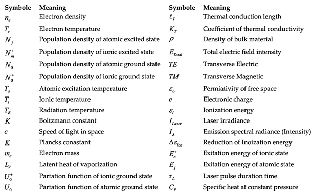
List of Symbols

Preprints 137499 i001

References

  1. Chen, Francis F. Introduction to Plasma Physics and Controlled Fusion, 2016. [CrossRef]
  2. Fujimoto, T. “Plasma Spectroscopy.” In Plasma Polarization Spectroscopy, edited by Takashi Fujimoto and Atsushi Iwamae, 44:29–49. Berlin, Heidelberg: Springer Berlin Heidelberg, 2008. [CrossRef]
  3. Kunze, Hans-Joachim. Introduction to Plasma Spectroscopy. Vol. 56. Springer Series on Atomic, Optical, and Plasma Physics. Berlin, Heidelberg: Springer Berlin Heidelberg, 2009. [CrossRef]
  4. Hora, H. Plasmas at High Temperature and Density Applications and Implications of Laser-Plasma Interaction; Lecture Notes in Physics Monographs; Softcover reprint of the original 1st ed. 1991. 2014. [Google Scholar]
  5. Miziolek, Andrzej W. , V. Palleschi, and Israel Schechter, eds. Laser-Induced Breakdown Spectroscopy (LIBS): Fundamentals and Applications. Cambridge, UK; New York: Cambridge University Press, 2006; ISBN 9780521852746. [Google Scholar]
  6. Bellan, P.M. Fundamentals of Plasma Physics; Cambridge University Press: Cambridge ; New York, 2006; ISBN 9780511160967.
  7. Linne, M. Spectroscopic Measurement An Introduction to the Fundamentals; An Elsevier Science Imprint: London, 2002; ISBN 0-12-451071-X. [Google Scholar]
  8. Ussenov, Y. A., T. S. Ramazanov, K. N. Dzhumagulova, and M. K. Dosbolayev. Application of Dust Grains and Langmuir Probe for Plasma Diagnostics. EPL (Europhysics Letters) 2014, 105, 15002. [Google Scholar] [CrossRef]
  9. El Sherbini, Ashraf M. , Abdelnasser M. Aboulfotouh, and Christian G. Parigger. Electron Number Density Measurements Using Laser-Induced Breakdown Spectroscopy of Ionized Nitrogen Spectral Lines. Spectrochimica Acta Part B: Atomic Spectroscopy 2016, 125, 152–58. [Google Scholar] [CrossRef]
  10. Ralchenko, Yuri, ed. Modern Methods in Collisional-Radiative Modeling of Plasmas. Vol. 90. Springer Series on Atomic, Optical, and Plasma Physics. Cham: Springer International Publishing, 2016. [CrossRef]
  11. Van Sijde, B. Der, J. J. A. M. Van Der Mullen, and D. C. Schram. Collisional Radiative Models in Plasmas. Beiträge Aus Der Plasmaphysik 1984, 24, 447–73. [Google Scholar] [CrossRef]
  12. Cremers, David A., and Leon J. Radziemski. Handbook of Laser-Induced Breakdown Spectroscopy. 1st ed. Wiley, 2013. [CrossRef]
  13. Konjević, N.; Dimitrijević, M.S.; Wiese, W.L. Experimental Stark Widths and Shifts for Spectral Lines of Neutral Atoms (A Critical Review of Selected Data for the Period 1976 to 1982). Journal of Physical and Chemical Reference Data 1984, 13, 619–647. [Google Scholar] [CrossRef]
  14. Griem, H.R. (1964) Plasma spectroscopy. McGrow-Hill, Inc.
  15. Fikry M, Alhijry IA, Aboulfotouh AM, El Sherbini AM. Feasibility of Using Boltzmann Plots to Evaluate the Stark Broadening Parameters of Cu(I) Lines. Applied Spectroscopy. 2021, 75, 1288–1295. [Google Scholar] [CrossRef]
  16. Kramida, Alexander, and Yuri Ralchenko. “NIST Atomic Spectra Database, NIST Standard Reference Database 78.” National Institute of Standards and Technology, 1999. [CrossRef]
  17. Konjević, N. Plasma Broadening and Shifting of Non-Hydrogenic Spectral Lines: Present Status and Applications. Physics Reports 1999, 316, 339–401. [Google Scholar] [CrossRef]
  18. El Sherbini, A.M.; El Sherbini, Th.M.; Hegazy, H.; Cristoforetti, G.; Legnaioli, S.; Palleschi, V.; Pardini, L.; Salvetti, A.; Tognoni, E. Evaluation of Self-Absorption Coefficients of Aluminum Emission Lines in Laser-Induced Breakdown Spectroscopy Measurements. Spectrochimica Acta Part B: Atomic Spectroscopy 2005, 60, 1573–1579. [Google Scholar] [CrossRef]
  19. El Sherbini, A.M.; El Sherbini, A.E.; Parigger, C.G. Measurement of Electron Density from Stark-Broadened Spectral Lines Appearing in Silver Nanomaterial Plasma. Atoms 2018, 6, 44. [Google Scholar] [CrossRef]
  20. Holstein, T. Imprisonment of Resonance Radiation in Gases. Phys. Rev. 1947, 72, 1212–1233. [Google Scholar] [CrossRef]
  21. El Sherbini, A.M.; Hegazy, H.; El Sherbini, Th.M. Measurement of Electron Density Utilizing the Hα-Line from Laser Produced Plasma in Air. Spectrochimica Acta Part B: Atomic Spectroscopy 2006, 61, 532–539. [Google Scholar] [CrossRef]
  22. Chan, G.C.-Y.; Hieftje, G.M.; Omenetto, N.; Axner, O.; Bengtson, A.; Bings, N.H.; Blades, M.W.; Bogaerts, A.; Bolshov, M.A.; Broekaert, J.A.C.; et al. EXPRESS: Landmark Publications in Analytical Atomic Spectrometry: Fundamentals and Instrumentation Development. Appl Spectrosc 2024, 00037028241263567. [Google Scholar] [CrossRef] [PubMed]
  23. Alhijry, I.A.; El Sherbini, A.M.; El Sherbini, T.M. Measurement of Deviations of Transition Probability of the Neutral Silver Lines at 827.35 and 768.77 Nm Using OES-Technique. Journal of Quantitative Spectroscopy and Radiative Transfer 2020, 245, 106922. [Google Scholar] [CrossRef]
  24. Sherbini, A.M.E.; Aboulfotouh, A.-N.; Rashid, F.; Allam, S.H.; Al-Kaoud, A.M.; Dakrouri, A.E.; Sherbini, T.M.E. Spectroscopic Measurement of Stark Broadening Parameter of the 636.2 Nm Zn I-Line. NS 2013, 5, 501–507. [Google Scholar] [CrossRef]
  25. Grünberger, S.; Ehrentraut, V.; Eschlböck-Fuchs, S.; Hofstadler, J.; Pissenberger, A.; Pedarnig, J.D. Overcoming the Matrix Effect in the Element Analysis of Steel: Laser Ablation-Spark Discharge-Optical Emission Spectroscopy (LA-SD-OES) and Laser-Induced Breakdown Spectroscopy (LIBS). Analytica Chimica Acta 2023, 1251, 341005. [Google Scholar] [CrossRef]
  26. Wang, Q.; Xiangli, W.; Teng, G.; Cui, X.; Wei, K. A Brief Review of Laser-Induced Breakdown Spectroscopy for Human and Animal Soft Tissues: Pathological Diagnosis and Physiological Detection. Applied Spectroscopy Reviews 2021, 56, 221–241. [Google Scholar] [CrossRef]
  27. Senesi, G.S.; Tempesta, G.; Manzari, P.; Agrosì, G. An Innovative Approach to Meteorite Analysis by Laser-Induced Breakdown Spectroscopy. Geostandard Geoanalytic Res 2016, 40, 533–541. [Google Scholar] [CrossRef]
  28. Jantzi, S.C.; Almirall, J.R. Characterization and Forensic Analysis of Soil Samples Using Laser-Induced Breakdown Spectroscopy (LIBS). Anal Bioanal Chem 2011, 400, 3341–3351. [Google Scholar] [CrossRef]
  29. Fabre, C. Advances in Laser-Induced Breakdown Spectroscopy Analysis for Geology: A Critical Review. Spectrochimica Acta Part B: Atomic Spectroscopy 2020, 166, 105799. [Google Scholar] [CrossRef]
  30. Khan, Z.H.; Ullah, M.H.; Rahman, B.; Talukder, A.I.; Wahadoszamen, Md.; Abedin, K.M.; Haider, A.F.M.Y. Laser-Induced Breakdown Spectroscopy (LIBS) for Trace Element Detection: A Review. Journal of Spectroscopy 2022, 2022, 1–25. [Google Scholar] [CrossRef]
  31. Ismail, M.A.; Imam, H.; Elhassan, A.; Youniss, W.T.; Harith, M.A. LIBS Limit of Detection and Plasma Parameters of Some Elements in Two Different Metallic Matrices. J. Anal. At. Spectrom. 2004, 19, 489. [Google Scholar] [CrossRef]
  32. Gautier, C.; Fichet, P.; Menut, D.; Dubessy, J. Applications of the Double-Pulse Laser-Induced Breakdown Spectroscopy (LIBS) in the Collinear Beam Geometry to the Elemental Analysis of Different Materials. Spectrochimica Acta Part B: Atomic Spectroscopy 2006, 61, 210–219. [Google Scholar] [CrossRef]
  33. Scaffidi, J.; Angel, S.M.; Cremers, D.A. Emission Enhancement Mechanisms in Dual-Pulse LIBS. Anal. Chem. 2006, 78, 24–32. [Google Scholar] [CrossRef] [PubMed]
  34. Rohwetter, Ph.; Yu, J.; Méjean, G.; Stelmaszczyk, K.; Salmon, E.; Kasparian, J.; Wolf, J.-P.; Wöste, L. Remote LIBS with Ultrashort Pulses: Characteristics in Picosecond and Femtosecond Regimes. J. Anal. At. Spectrom. 2004, 19, 437–444. [Google Scholar] [CrossRef]
  35. EL Sherbini, A.M.; Aboulfotouh, A.; Rashid, F.F.; Allam, S.H.; Dakrouri, A.E.; EL Sherbini, Th.M. Observed Enhancement in LIBS Signals from Nano vs. Bulk ZnO Targets: Comparative Study of Plasma Parameters. WJNSE 2012, 2, 181–188. [Google Scholar] [CrossRef]
  36. De Giacomo, A.; Gaudiuso, R.; Koral, C.; Dell’Aglio, M.; De Pascale, O. Nanoparticle-Enhanced Laser-Induced Breakdown Spectroscopy of Metallic Samples. Anal. Chem. 2013, 85, 10180–10187. [Google Scholar] [CrossRef]
  37. Feynman, R.P.; Robbins, J. The Pleasure of Finding Things out: The Best Short Works of Richard P. Feynman; Helix books; Perseus Books: Cambridge, Mass, 1999; ISBN 9780738201085. [Google Scholar]
  38. Asha, A.B.; Narain, R. Nanomaterials Properties. In Polymer Science and Nanotechnology; Elsevier, 2020; pp. 343–359 ISBN 9780128168066. [CrossRef]
  39. Joudeh, N.; Linke, D. Nanoparticle Classification, Physicochemical Properties, Characterization, and Applications: A Comprehensive Review for Biologists. J Nanobiotechnol 2022, 20, 262. [Google Scholar] [CrossRef]
  40. Campos, A.; Troc, N.; Cottancin, E.; Pellarin, M.; Weissker, H.-C.; Lermé, J.; Kociak, M.; Hillenkamp, M. Plasmonic Quantum Size Effects in Silver Nanoparticles Are Dominated by Interfaces and Local Environments. Nat. Phys. 2019, 15, 275–280. [Google Scholar] [CrossRef]
  41. Vollath, D.; Fischer, F.D.; Holec, D. Surface Energy of Nanoparticles – Influence of Particle Size and Structure. Beilstein J. Nanotechnol. 2018, 9, 2265–2276. [Google Scholar] [CrossRef]
  42. Li, S.; Meng Lin, M.; Toprak, M.S.; Kim, D.K.; Muhammed, M. Nanocomposites of Polymer and Inorganic Nanoparticles for Optical and Magnetic Applications. Nano Reviews 2010, 1, 5214. [Google Scholar] [CrossRef]
  43. Ohta, T.; Ito, M.; Kotani, T.; Hattori, T. Emission Enhancement of Laser-Induced Breakdown Spectroscopy by Localized Surface Plasmon Resonance for Analyzing Plant Nutrients. Appl Spectrosc 2009, 63, 555–558. [Google Scholar] [CrossRef]
  44. De Giacomo, A.; Gaudiuso, R.; Koral, C.; Dell’Aglio, M.; De Pascale, O. Nanoparticle Enhanced Laser Induced Breakdown Spectroscopy: Effect of Nanoparticles Deposited on Sample Surface on Laser Ablation and Plasma Emission. Spectrochimica Acta Part B: Atomic Spectroscopy 2014, 98, 19–27. [Google Scholar] [CrossRef]
  45. De Giacomo, A.; Dell’Aglio, M.; Gaudiuso, R.; Koral, C.; Valenza, G. Perspective on the Use of Nanoparticles to Improve LIBS Analytical Performance: Nanoparticle Enhanced Laser Induced Breakdown Spectroscopy (NELIBS). J. Anal. At. Spectrom. 2016, 31, 1566–1573. [Google Scholar] [CrossRef]
  46. Koral, C.; De Giacomo, A.; Mao, X.; Zorba, V.; Russo, R.E. Nanoparticle Enhanced Laser Induced Breakdown Spectroscopy for Improving the Detection of Molecular Bands. Spectrochimica Acta Part B: Atomic Spectroscopy 2016, 125, 11–17. [Google Scholar] [CrossRef]
  47. Gaudiuso, R.; Koral, C.; Dell’Aglio, M.; De Pascale, O.; De Giacomo, A. Fundamental Study and Analytical Applications of Nanoparticle-Enhanced Laser-Induced Breakdown Spectroscopy (NELIBS) of Metals, Semiconductors and Insulators. In Nano-Optics: Principles Enabling Basic Research and Applications; Di Bartolo, B., Collins, J., Silvestri, L., Eds.; Springer Netherlands: Dordrecht, 2017; ISBN 9789402408485. [Google Scholar]
  48. Dell’Aglio, M.; Alrifai, R.; De Giacomo, A. Nanoparticle Enhanced Laser Induced Breakdown Spectroscopy (NELIBS), a First Review. Spectrochimica Acta Part B: Atomic Spectroscopy 2018, 148, 105–112. [Google Scholar] [CrossRef]
  49. Koral, C.; Dell’Aglio, M.; Gaudiuso, R.; Alrifai, R.; Torelli, M.; De Giacomo, A. Nanoparticle-Enhanced Laser Induced Breakdown Spectroscopy for the Noninvasive Analysis of Transparent Samples and Gemstones. Talanta 2018, 182, 253–258. [Google Scholar] [CrossRef]
  50. El Farash, A.; El Sherbini, A.; Helal, O.; El-Sherif, A. Enhanced Ti I Spectral Intensity Using NELIBS Technique. Eng. Sci. and Milit. Techno. 2019, 3, 84–90. [Google Scholar] [CrossRef]
  51. Tang, H.; Hao, X.; Hu, X. Spectral Enhancement Effect of LIBS Based on the Combination of Au Nanoparticles with Magnetic Field. Optik 2019, 179, 1129–1133. [Google Scholar] [CrossRef]
  52. Palásti, D.J.; Albrycht, P.; Janovszky, P.; Paszkowska, K.; Geretovszky, Z.; Galbács, G. Nanoparticle Enhanced Laser Induced Breakdown Spectroscopy of Liquid Samples by Using Modified Surface-Enhanced Raman Scattering Substrates. Spectrochimica Acta Part B: Atomic Spectroscopy 2020, 166, 105793. [Google Scholar] [CrossRef]
  53. De Giacomo, A.; Alrifai, R.; Gardette, V.; Salajková, Z.; Dell’Aglio, M. Nanoparticle Enhanced Laser Ablation and Consequent Effects on Laser Induced Plasma Optical Emission. Spectrochimica Acta Part B: Atomic Spectroscopy 2020, 166, 105794. [Google Scholar] [CrossRef]
  54. De Giacomo, A.; Dell’Aglio, M. Nanoparticle-Enhanced Laser Induced Breakdown Spectroscopy (NELIBS) on Biological Samples. In Laser-Induced Breakdown Spectroscopy in Biological, Forensic and Materials Sciences; Galbács, G., Ed.; Springer International Publishing: Cham, 2022; ISBN 9783031145018. [Google Scholar]
  55. Khan, M.R.; Haq, S.U.; Abbas, Q.; Nadeem, A. Improvement in Signal Sensitivity and Repeatability Using Copper Nanoparticle-Enhanced Laser-Induced Breakdown Spectroscopy. Spectrochimica Acta Part B: Atomic Spectroscopy 2022, 195, 106507. [Google Scholar] [CrossRef]
  56. Dell’Aglio, M.; Di Franco, C.; De Giacomo, A. Different Nanoparticle Shapes for Nanoparticle-Enhanced Laser-Induced Breakdown Spectroscopy: Nanosphere and Nanorod Effects. J. Anal. At. Spectrom. 2023, 38, 766–774. [Google Scholar] [CrossRef]
  57. Dell’Aglio, M.; Mallardi, A.; Gaudiuso, R.; Giacomo, A.D. Plasma Parameters During Nanoparticle-Enhanced Laser-Induced Breakdown Spectroscopy (NELIBS) in the Presence of Nanoparticle–Protein Conjugates. Appl Spectrosc 2023, 77, 1253–1263. [Google Scholar] [CrossRef] [PubMed]
  58. Awan, R.A.; Siraj, K.; Haq, S.U.; Abbas, Q.; Rahim, M.S.A.; Younas, Q.; Fareed, S.; Ahsen, R.; Ahmad, Z.; Irshad, M.; et al. Laser Induced Breakdown Spectroscopy of Aluminum Incorporated with Metallic Nanoparticles. Opt Quant Electron 2023, 55, 73. [Google Scholar] [CrossRef]
  59. Salajková, Z.; Dell’Aglio, M.; Gardette, V.; De Giacomo, A. Nanoparticle-Enhanced Laser-Induced Breakdown Spectroscopy. In Laser Induced Breakdown Spectroscopy (LIBS); Singh, V.K., Tripathi, D.K., Deguchi, Y., Wang, Z., Eds.; Wiley, 2023; pp. 165–182 ISBN 9781119758402.
  60. Narlagiri, L.M.; Soma, V.R. Nanoparticle-Enhanced Laser-Induced Breakdown Spectroscopy for Sensing Applications. In Laser Induced Breakdown Spectroscopy (LIBS); Singh, V.K., Tripathi, D.K., Deguchi, Y., Wang, Z., Eds.; Wiley, 2023; pp. 183–210 ISBN 9781119758402.
  61. Safi, A.; Landis, J.E.; Adler, H.G.; Khadem, H.; Eseller, K.E.; Markushin, Y.; Honarparvaran, S.; De Giacomo, A.; Melikechi, N. Enhancing Biomarker Detection Sensitivity through Tag-Laser Induced Breakdown Spectroscopy with NELIBS. Talanta 2024, 271, 125723. [Google Scholar] [CrossRef] [PubMed]
  62. Rashid, F.F.; ELSherbini, A.M.; Al-Muhamady, A. Strong Emission from Nano-Iron Using Laser-Induced Breakdown Spectroscopy Technique. Appl. Phys. A 2014, 115, 1395–1399. [Google Scholar] [CrossRef]
  63. EL Sherbini, A.M.; Galil, A.A.; Allam, S.H; EL Sherbini, Th.M. Nanomaterials Induced Plasma Spectroscopy. J. Phys.: Conf. Ser. 2014, 548, 012031. [Google Scholar] [CrossRef]
  64. El Sherbini, A.M.; Parigger, C.G. Wavelength Dependency and Threshold Measurements for Nanoparticle-Enhanced Laser-Induced Breakdown Spectroscopy. Spectrochimica Acta Part B: Atomic Spectroscopy 2016, 116, 8–15. [Google Scholar] [CrossRef]
  65. El Sherbini, A.M.; Parigger, C.G. Nano-Material Size Dependent Laser-Plasma Thresholds. Spectrochimica Acta Part B: Atomic Spectroscopy 2016, 124, 79–81. [Google Scholar] [CrossRef]
  66. El Sherbini, A.M.; El Sherbini, A.E.; Parigger, C.G. Measurement of Electron Density from Stark-Broadened Spectral Lines Appearing in Silver Nanomaterial Plasma. Atoms 2018, 6, 44. [Google Scholar] [CrossRef]
  67. Sherbini, A.M.E.; Sherbini, A.E.E.; Parigger, C.G.; Sherbini, T.M.E. Nano-Particle Enhancement of Diagnosis with Laser-Induced Plasma Spectroscopy. J. Phys.: Conf. Ser. 2019, 1253, 012002. [Google Scholar] [CrossRef]
  68. El Sherbini, A.M.; Hagrass, M.M.; Rizk, M.R.M.; El-Badawy, E.A. Plasma Ignition Threshold Disparity between Silver Nanoparticle-Based Target and Bulk Silver Target at Different Laser Wavelengths. Plasma Sci. Technol. 2019, 21, 015502. [Google Scholar] [CrossRef]
  69. Keldysh. L, Ionization in the field of a strong electromagnetic wave, Sov. Phys. J. Exp.Theory Phys. 1965, 20, 1307–1314. [Google Scholar]
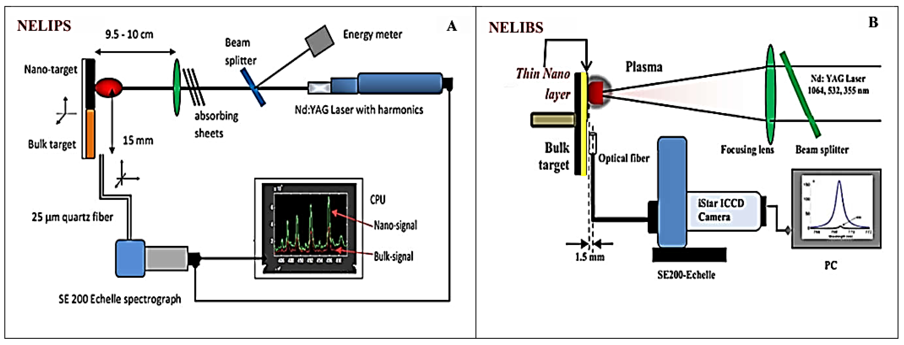
Figure 1. The basic schematics of the basic experimental setup in NELIPS (A), and NELIBS (B).
Preprints 137499 g001
Table 1. Proper equilibrium distribution functions; different symbol meaning at the end.
Species Proper distribution Proper expression
Atoms Boltzmann N j g j = N 0 U 0 exp ( E j K T a )
Electrons Maxwell f ( v ) d v = 4 π v 2 ( m e 2 π K T e ) 1.5 exp ( m e v 2 2 K T e ) d v
Ions Saha-Boltzmann N n + g n = n e N 0 + 2 U 0 + ( h 2 2 π m e K T i ) 1.5 exp ( ε i o n Δ ε i o n E n + 2 K T i )
Radiation Planck I ( λ , T R ) = 2 h c 2 λ 5 ( exp ( h c λ K T R ) 1 ) 1
Table 2. The plasma states of equilibrium.
Electron density
( c m 3 )
State of equilibrium Conditions on temperatures Applicable distribution functions
n e 10 18 19 Complete Thermodynamical Equilibrium
(CTE)
T a T e T i T R Boltzmann
Saha-Boltzmann
Maxwell
Planck
10 19 18 n e 10 16 15 Local Thermodynamical Equilibrium
(LTE)
T a T e T i T R Boltzmann
Saha-Boltzmann
Maxwell
10 16 15 n e 10 11 10 Partial
Local Thermodynamical
Equilibrium
(PLTE)
T a T e T i T R Boltzmann
Maxwell
n e 10 9 Corona state
(Equilibrium)
T a T e T i T R None of these distribution functions is applicable and Collisional-Radiative modeling should be constructed.
Table 3. Differences between the NELIPS and NELIBS-Approaches.
NELIPS NELIBS
  • Nature of target
Pure-nanomaterial. [35,62,63,64,65,66,67,68]. Thin layer of nanomaterial deposited on the surface of the analysed sample [36,43–61].
2.
Source of enhanced emission
From the pure-nanomaterial [35,62–68]. From the analysed sample material [36,43–61].
3.
Aims
Modelling of the enhanced emission from pure-nanomaterials [35,64,65,68]. Reduction of limit of detection LOD of the LIBS-spectrochemical technique [36,43,44,45,46,47,48,49,53,54].
4.
Recommended theory
Thermodynamics and plasma spectroscopy [64,65,66,67,68]. Electromagnetic theory and plasma spectroscopy [36,44,53,56,61].
5.
Suggested approach
  • Experimentally; the OES-technique was employed, followed by careful handling of spectral data under a variety of experimental conditions including laser fluence, delay time, laser wavelength, and different types of pure-nanomaterials which leads to important outcomes (as given in item #6).[36,43,44,45,46,47,48,49,50,51,52,53,54,55,56,57,58,59,60,61]
  • Theoretically, suggests a balance between the incident laser fluence and the plasma ignition, within the framework the variety of the experimental findings.
  • The thermal modeling suggests strong reduction of thermal conduction length of the nanomaterial to the limit of nanoparticle diameter i.e. ( T N D N ) [65].
Suggested a resonance between the localized surface plasmons (LSPR) with frequency of the incident laser light, which enhances coupling of laser energy to substrate material [36,53].
6.
Achievements
  • Enhanced emission from plasma induced by the interaction of pulsed lasers with different pure-nanomaterials is a real phenomenon [35,62,63,64,65,66,67,68].
  • Enhancement is rapidly declines in exponential manner with laser fluence [63].
  • Also it manifestly increases with delay time1.
  • Enhancement is not constant over the different emitted spectral wavelengths2.
  • Enhanced plasma emission from pure-nanomaterial is larger by UV laser irradiation, moderate at VIS and relatively small at IR [64].
  • Analysis approved that plasma ignition threshold from pure nanomaterials is much lower than that from bulk counterpart ( φ T h N a n o / φ T h B u l k 1 ) [64,65,66,67,68].
  • The measured ratio of plasma ignition threshold of nanomaterial to that of the bulk match the ratio of the used nanoparticle diameter to the theoretically calculated thermal conduction length of the bulk material counterpart ( φ T h N a n o / φ T h B u l k D N a n o / T B u l k ) [64,65,66,67,68].
  • The plasma ignition thresholds from the pure-nonmaterial and the bulk counterpart ( φ T h N a n o & B u l k ) was found to depend on several experimental parameters including;
  • The laser wavelength ( φ T h N a n o & B u l k λ L a s e r 2 ) [64,68].
  • The nanoparticle diameter ( φ T h N a n o D N a n o ) [65].
  • The type of nonmaterial [35].
9.
The amount of enhanced emission was found;
  • Independent of the plasma parameters ( n e & T e ) [35,64,65,66,67,68]
  • Depends on the relative concentration ( E n h λ = I N / I B N 0 N / N 0 B ) [35,63,68].
10.
Theoretically, the dependence of plasma ignition threshold from the bulk and pure-nanomaterial on the various measured quantities was explicitly derived respectively as [64,65,66,67,68]; φ T h B u l k = ( ρ B u l k L V B u l k + 4 π 2 m e ε o c 2 e 2 ε i λ L a s e r 2 ) T φ T h N a n o = ( ρ N a n o L V N a n o + 4 π 2 m e ε o c 2 e 2 ε i λ L a s e r 2 ) D N a n o With thermal conduction length ( T B u l k = K T B u l k τ L a s e r / ρ T B u l k C p T B u l k )
  • Improvement to the limit of detection LOD of the LIBS-technique down to the range of ppb (nano-gram/gram) of the examined materials [36,43,44,45,46,47,48,49,50,51,52,53,54,55,56,57,58,59,60,61].
  • A rigours theoretical derivation based on EM Theory about the strong enhancement of the electric field in between the nanoparticles and the surface of metallic substrate taking excitation of surface plasmon resonance with incident laser frequency and hence the enhanced coupling of laser light energy to substrate material is presented at ref [53].
7.
Challenges
  • Basic thermal quantities are unavailable at Nano scales e.g. latent heat of vaporization, coefficient of thermal conductivity, specific heats…etc.
  • The inconsideration of the theory of localized surface plasmon resonance in the process of enhanced emission.
  • The amount of enhancement is not constant over the different emitted wavelengths.
  • Tentative theoretical assessment of the measured linear regression of amount of enhanced emission with delay time.
  • The problem of the distortion of the nanoparticles "sintering" upon compression technique used to put the nanoparticles powders in a tablet form.
  • There is an excessive use of optimization parameters from one published article to another to reach the maximum enhancement in emission from the substrate material; (e.g. the arbitrary chosen delay and gate times, laser wavelength, laser energy, thickness of nanomaterial layer, different concentrations, different inter-particles distance…etc.), which poses serious difficulties on the reproducibility of results. [43,44,45,46,47,48,49,50,51,52,53,54,55,56,57,58,59,60,61]
  • There is a need for much better regular theoretical approach than that given at ref [53] based on the EM theory of LSPR in addition to thermal processes (Thermodynamics) and leading to direct formal expression about enhanced emission from the plasma originated from the target substrate material.
8.
Expected prospective
  • The principle of the invariance of the basic physical thermal constants at the very short Nano scales must be revisited e.g. the latent heat of vaporization, coefficient of thermal conductivity, etc.) .
  • The role of LSPR will be considered in the process of enhanced emission in conjunction with the thermal processes. (Hybrid model) will be considered. The modeling of NELIPS results validation to reproduce NELIBS experiment findings.
The extra fine micro-analytical chemistry promoting the potential use of LIBS-technique in a wide variety of biological, industrial, material science applications. [43,44,45,46,47,48,49,50,51,52,53,54,55,56,57,58,59,60,61]
1,2 Means: that these pieces of work are not yet been published.
Disclaimer/Publisher’s Note: The statements, opinions and data contained in all publications are solely those of the individual author(s) and contributor(s) and not of MDPI and/or the editor(s). MDPI and/or the editor(s) disclaim responsibility for any injury to people or property resulting from any ideas, methods, instructions or products referred to in the content.
Copyright: This open access article is published under a Creative Commons CC BY 4.0 license, which permit the free download, distribution, and reuse, provided that the author and preprint are cited in any reuse.
Alerts
Prerpints.org logo

Preprints.org is a free preprint server supported by MDPI in Basel, Switzerland.

Subscribe

© 2025 MDPI (Basel, Switzerland) unless otherwise stated