Preprint
Article

Role of Chikungunya E2 Protein in Human Cell

This version is not peer-reviewed.

Submitted:

25 October 2024

Posted:

28 October 2024

You are already at the latest version

Abstract

Chikungunya is mosquito born disease that is spread across continents. This research provides valuable insights into the potential functional significance of the hydrophobic tail and cysteine-rich regions in E2 proteins. These findings lay the groundwork for future investigations into the specific roles of the C-terminal domain and post-translational modifications, which could ultimately lead to a better understanding of viral mechanisms and potential therapeutic targets.

Keywords: 
Subject: 
Biology and Life Sciences  -   Virology

1. Introduction

Chikungunya virus is an RNA virus that belongs to alphavirus genus. Chikungunya affects a large part of Asia, Africa and Europe(World Health Organization, 2015). The symptoms of chikungunya include rashes, fever, muscles pain and joint pain. The pain in the muscles and joint is the signature of the disease. The exact figures of morbidity and mortality not clear due to current challenges in reporting during the outbreaks and diagnosis (Deoshatwar et al., 2021; Gopalkrishna et al., 2019; A. Kumar et al., 2016, 2022). Though chikungunya related mortality rare and associated with co-morbidity with other diseases (Gill et al., 2009; Roesch et al., 2018; Shahid et al., 2023; Shikha et al., 2023). The diagnosis of chikungunya is mainly through RT-PCR and also through antibodies. For RT-PCR, target gene includes non-structural protein 2 (nsP2) and the E1 and nsp4 genes. Currently, many vaccine candidates are in the pipelines against chikungunya using the regions/proteins of chikungunya. E2 proteins are one the vaccine candidate. E2 proteins is glycoprotein that facilitates binding of chikungunya virus to host cells (Gill et al., 2022). Chikungunya virus attacks the host cell by receptor mediated endocytosis mechanism. For the same even E1-E2 heterodimer complexes undergoes an irreversible conformational change leading to the opening of viral capsid and entry of viral genomic content inside the host cell. E2 protein exists as membrane protein during viral replication. Protein sequence based and network-based analysis can provide important insight on the role of E2 protein in the host cell (Garg et al., 2023; Kaur et al., 2023). In this piece of work, we have tried to investigate the relevance of post-translational modification of E2 proteins and its effect on viral cycle on human host.

2. Materials and Methods

In silico sequence-based prediction method was used. The E2 protein sequence was retrieved from NCBI. Potential globular domain as well as transmembrane domain was determined using GLOBPLOT2 and TMHMM-2.0 respectively. Hyrodopathycity was determined using Protscale of Expasy. Post-translation modification were determined using NetNGlyc - 1.0 (Duvaud et al., 2021; P. Kumar, Rani, et al., 2022).

3. Results

Prediction of Transmembrane Domain

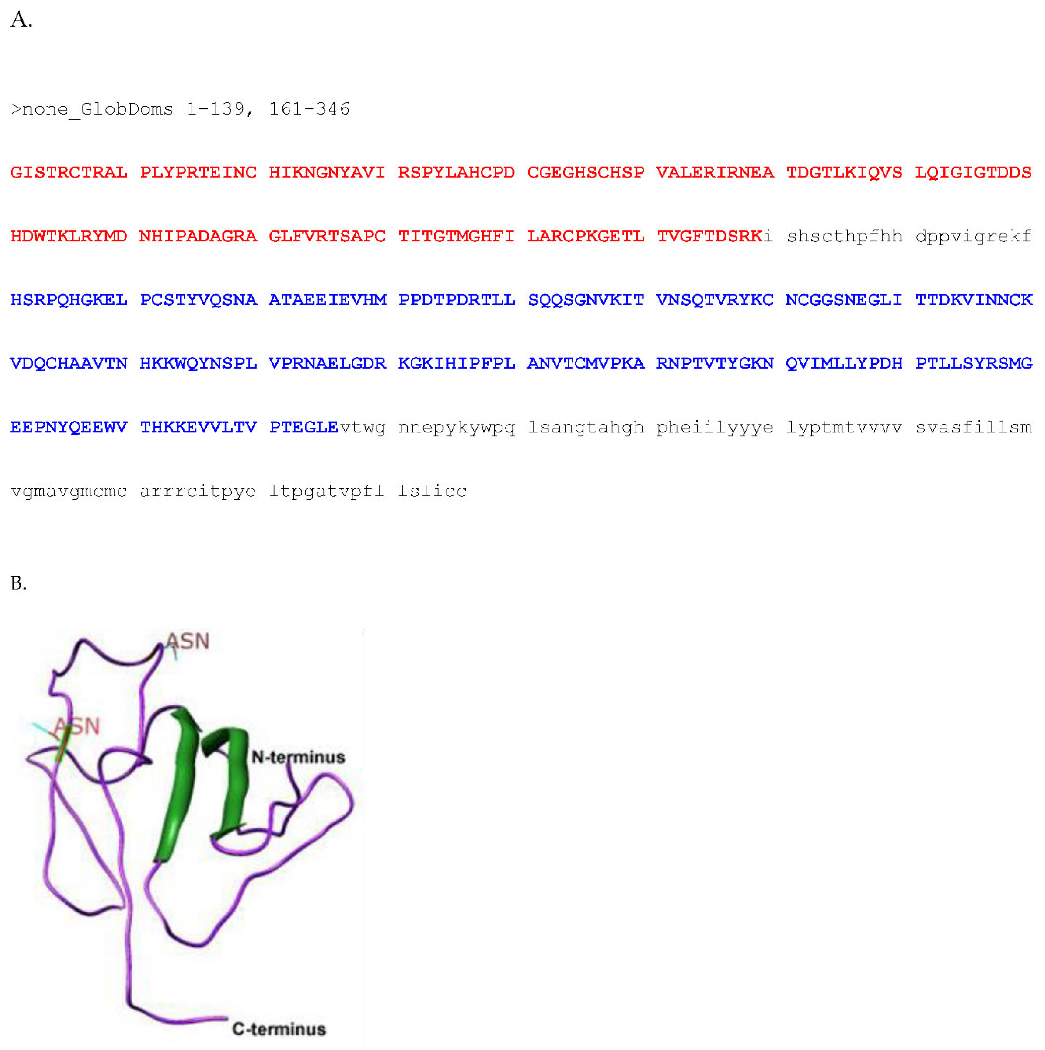
Transmembrane region was predicted from 384 to 406 amino acids (Figure 1). Hydrophobic region of C-terminal supports the presence two transmembrane domains in precursor E2(Bogunia & Makowski, 2020). Second transmembrane come out of membrane after post-transnational modification. This hydrophobic region is also seems important interaction between E1 and E2 (Figure 1). It contains three domains. N-terminal region, transmembrane region and cytoplasmic region. E2 N-terminal region is approx 350 amino acid long, followed by 20 aa transmembrane region and then cytoplasmic region of again 20 amino acids. Transmembrane domain and C-terminal region are more hydrophobic in comparison to N-terminal region. E2 have 45% conserved protein. The percentage of conversion drop down from 360-380 aa which is the predicted transmembrane region. At the C-terminal only that E2 protein interact with E1 by this C-terminal tail. Thus, might be the interaction between E1 and E2 doesn’t depend on specific motif. Predicting the fold of second globular domain (structure of 183-280 aa out of 161-346 aa)(Dobhal et al., 2024; A. Kumar et al., 2016).

Phylogenetic Analysis

Phylogenetic analysis of less conserved proteins like E2 is difficult. Phylogenetic tree is prepared from E2 protein, it showed more similarity between E2 proteins of chikungunya virus is and O’ nyong-nyong virus, whereas E2 proteins two viruses have significant phylogenetic distance with other alphaviruses (Figure 2). This distance is also significant with Semliki forest virus (SFV). Even though chikungunya and O’ nyong-nyong belong to Semliki forest virus class. This result is important in illustrating high mortality in Chikungunya infection in comparison to other alpha viruses.

Post-Transnational Modification

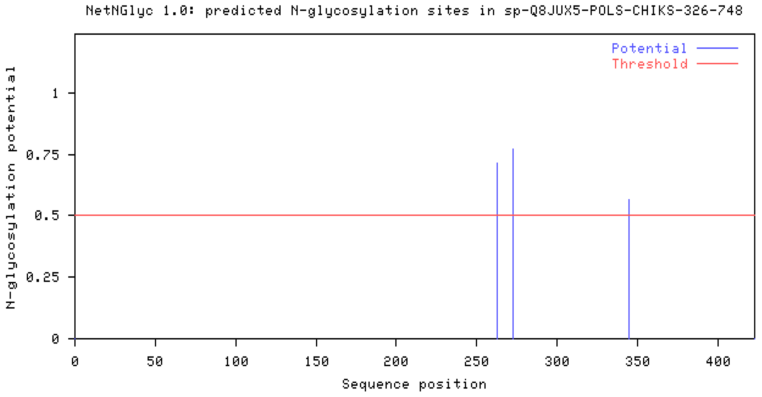
Post-translation modification are the key player in regulating the protein structure-function (Gill et al., 2010; A. Kumar et al., 2012; Mishra et al., 2009). Alpha virus E2 are heavily palmitoylated containing 5 to 6 palmitic acid. These lipid molecules are present in cytosolic tail. In case of chikungunya we are not able to satisfactorily determine the reason of palmitoylation site (Figure 3).

4. Discussion

Presence of the hydrophobic tail and large number of cysteines in the proteins could be play role in the E2 function inside the host cell. Further studies are required to understand the role of C-terminal domain and type of post-translation modifications. N-terminus region plays very important role in binding to host cell surface. In 404 amino acid chikungunya E2 protein N-terminus length is approx 350 aa (Figure 4). This is region which defines E2 glycoprotein domain. HMM logo of the region showed large number of conserved Cysteines (almost all cysteines are conserved). I fail to determine the importance of this fact. Look at globular domain in this region protein contain two such domain(P. Kumar, Dhingra, et al., 2022; Sajid et al., 2021).

Author Contributions

Conceptualization, A.K.; methodology, A.K.; writing—review and editing, A.K. All authors have read and agreed to the published version of the manuscript.

Funding

This research received no external funding.

Institutional Review Board Statement

Not applicable.

Informed Consent Statement

Not applicable.

Data Availability Statement

no new data were created.

Conflicts of Interest

The author declare no conflicts of interest.

References

  1. Bogunia, M., & Makowski, M. (2020). Influence of Ionic Strength on Hydrophobic Interactions in Water: Dependence on Solute Size and Shape. The Journal of Physical Chemistry B, 124(46), 10326–10336. [CrossRef]
  2. Deoshatwar, A., Salve, D., Gopalkrishna, V., Kumar, A., Barve, U., Joshi, M., Katendra, S., Dhembre, V., Maheshwari, S., & Viswanathan, R. (2021). Evidence-Based Health Behavior Interventions for Cholera: Lessons from an Outbreak Investigation in India. The American Journal of Tropical Medicine and Hygiene, 106(1), 229–232. [CrossRef]
  3. Dobhal, S., Chauhan, K., Kumar, S., Shikha, S., Jogi, M., Kumar, D., Kumar, A., Jaiswal, V., & Kumar, P. (2024). In silico Identification of MHC Displayed Tumor Associated Peptides in Ovarian Cancer for Multi-Epitope Vaccine Construct. Endocrine, Metabolic & Immune Disorders Drug Targets, 24. [CrossRef]
  4. Duvaud, S., Gabella, C., Lisacek, F., Stockinger, H., Ioannidis, V., & Durinx, C. (2021). Expasy, the Swiss Bioinformatics Resource Portal, as designed by its users. Nucleic Acids Research, 49(W1). [CrossRef]
  5. Garg, K., Kumar, A., Kizhakkethil, V., Kumar, P., & Singh, S. (2023). Overlap in oncogenic and pro-inflammatory pathways associated with areca nut and nicotine exposure. Cancer Pathogenesis and Therapy. [CrossRef]
  6. Gill, J., Kumar, A., & Sharma, A. (2022). Structural comparisons reveal diverse binding modes between nucleosome assembly proteins and histones. Epigenetics & Chromatin, 15(1), 20. [CrossRef]
  7. Gill, J., Kumar, A., Yogavel, M., Belrhali, H., Jain, S. K., Rug, M., Brown, M., Maier, A. G., & Sharma, A. (2010). Structure, localization and histone binding properties of nuclear-associated nucleosome assembly protein from Plasmodium falciparum. Malaria Journal, 9(1), 90. [CrossRef]
  8. Gill, J., Yogavel, M., Kumar, A., Belrhali, H., Jain, S. K., Rug, M., Brown, M., Maier, A. G., & Sharma, A. (2009). Crystal structure of malaria parasite nucleosome assembly protein: Distinct modes of protein localization and histone recognition. Journal of Biological Chemistry, 284(15), 10076–10087. [CrossRef]
  9. Gopalkrishna, V., Joshi, M., Viswanathan, R., Malu, G., Ganorkar, N., Chavan, N., Shinde, M., Kumar, A., & Dhurandhare, S. (2019). Cholera outbreak in Aurangabad, Maharashtra, western India. Indian Journal of Medical Research, 150(6), 640. [CrossRef]
  10. Kaur, R., Kumar, P., & Kumar, A. (2023). Insights on the nuclear shuttling of H2A-H2B histone chaperones. Nucleosides, Nucleotides & Nucleic Acids, 1–13. [CrossRef]
  11. Kumar, A., Barve, U., Gopalkrishna, V., Tandale, B. V, Katendra, S., Joshi, M. S., Salve, D., & Viswanathan, R. (2022). Outbreak of cholera in a remote village in western India. The Indian Journal of Medical Research, 156(3), 442–448. [CrossRef]
  12. Kumar, A., Kashyap, M., Bhavesh, N. S., Yogavel, M., & Sharma, A. (2012). Structural delineation of histone post-translation modifications in histone-nucleosome assembly protein complex. Journal of Structural Biology, 180(1), 1–9. [CrossRef]
  13. Kumar, A., Sharma, D., Aggarwal, M. L., Chacko, K. M., & Bhatt, T. K. (2016). Cancer/testis antigens as molecular drug targets using network pharmacology. Tumor Biology, 37(12), 15697–15705. [CrossRef]
  14. Kumar, P., Dhingra, A., Sharma, D., Kumar, A., & Singh, S. (2022). Microbiome and Development of Ovarian Cancer. Endocrine, Metabolic & Immune Disorders Drug Targets. [CrossRef]
  15. Kumar, P., Rani, A., Singh, S., & Kumar, A. (2022). Recent advances on DNA and omics-based technology in Food testing and authentication: A review. Journal of Food Safety, 42(4). [CrossRef]
  16. Mishra, P. C., Kumar, A., & Sharma, A. (2009). Analysis of small nucleolar RNAs reveals unique genetic features in malaria parasites. BMC Genomics, 10(1), 68. [CrossRef]
  17. Roesch, C., Popovici, J., Bin, S., Run, V., Kim, S., Ramboarina, S., Rakotomalala, E., Rakotoarison, R. L., Rasoloharimanana, T., Andriamanantena, Z., Kumar, A., Guillotte-Blisnick, M., Huon, C., Serre, D., Chitnis, C. E., Vigan-Womas, I., & Menard, D. (2018). Genetic diversity in two Plasmodium vivax protein ligands for reticulocyte invasion. PLOS Neglected Tropical Diseases, 12(10), e0006555. [CrossRef]
  18. Sajid, M., Srivastava, S., Kumar, A., Kumar, A., Singh, H., & Bharadwaj, M. (2021). Bacteriome of Moist Smokeless Tobacco Products Consumed in India With Emphasis on the Predictive Functional Potential. Frontiers in Microbiology, 12, 784841. [CrossRef]
  19. Shahid, M., Srivastava, S., Shukla, P., Yadav, R., Sajid, M., Kumar, A., Singh, S., & Bharadwaj, M. (2023). Characterization of physiochemical parameters & their effect on microbial content of smokeless tobacco products marketed in north India. The Indian Journal of Medical Research, 158(5 & 6), 542–551. [CrossRef]
  20. Shikha, S., Jogi, M. K., Jha, R., Kumar, R. A., Sah, T., Singh, P., Sagar, R., Kumar, A., Marwal, R., Ponnusamy, K., Agarwal, S. M., Kumar, R. S., Arif, N., Bharadwaj, M., Singh, S., & Kumar, P. (2023). Genome sequencing of SARS-CoV-2 omicron variants in Delhi reveals alterations in immunogenic regions in spike glycoprotein. Frontiers in Immunology, 14, 1209513. [CrossRef]
  21. World Health Organization. (2015). WHO Chikungunya Fact Sheet. World Health Organization Media Centre.
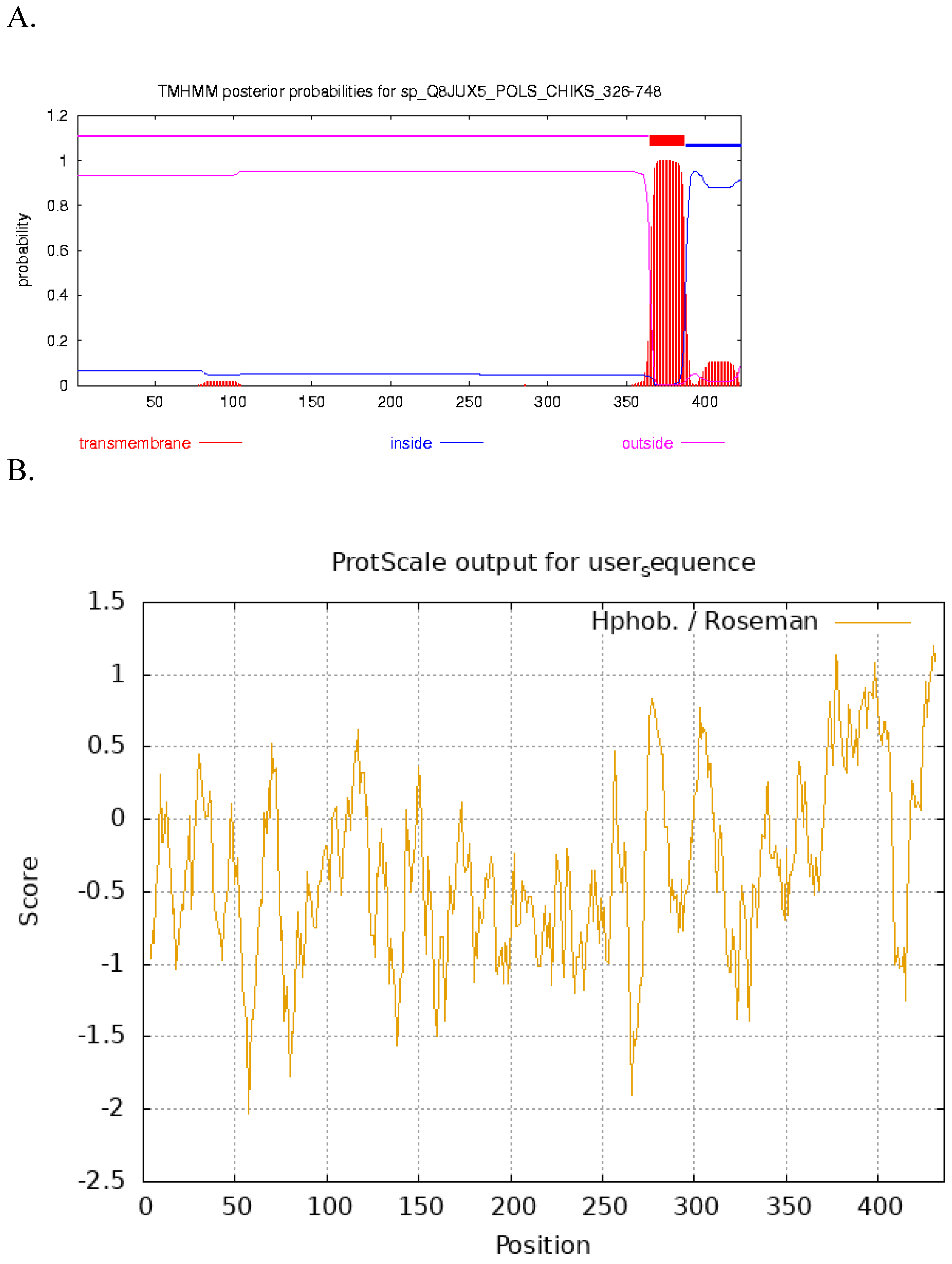
Figure 1. A. Prediction of transmembrane helices in E2 proteins using TMHMM. B. Prediction of hydropathcity of E2 protein determined using ProtScale Expasy.
Preprints 137490 g001
Figure 2. Phylogenetic tree prepared based on E2 protein of chikungunya.
Preprints 137490 g002
Figure 3. Prediction of glycosylation sites using NetNGlyc - 1.0. Three potential glycosylation site were predicted out which one site is near the transmembrane site.
Preprints 137490 g003
Figure 4. A. Potential globular domains (GlobDoms) by Russell/Linding definition was determine. B. Predicted structure of E2 protein.
Preprints 137490 g004
Disclaimer/Publisher’s Note: The statements, opinions and data contained in all publications are solely those of the individual author(s) and contributor(s) and not of MDPI and/or the editor(s). MDPI and/or the editor(s) disclaim responsibility for any injury to people or property resulting from any ideas, methods, instructions or products referred to in the content.
Copyright: This open access article is published under a Creative Commons CC BY 4.0 license, which permit the free download, distribution, and reuse, provided that the author and preprint are cited in any reuse.
Alerts
Prerpints.org logo

Preprints.org is a free preprint server supported by MDPI in Basel, Switzerland.

Subscribe

© 2025 MDPI (Basel, Switzerland) unless otherwise stated