Preprint
Review

This version is not peer-reviewed.

Vascular Endothelial Growth Factor (VEGF) Family and the Immune System: Activators or Inhibitors?

A peer-reviewed article of this preprint also exists.

Submitted:

20 November 2024

Posted:

20 November 2024

You are already at the latest version

Abstract

The vascular endothelial growth factor (VEGF) family includes key mediators of vasculogenesis and angiogenesis. VEGFs are secreted by various cells of epithelial and mesenchymal origin and by some immune cells in response to physiological and pathological stimuli. In addition, immune cells express VEGF receptors and/or coreceptors and can respond to VEGFs in an autocrine or paracrine manner. This immunological role of VEGFs has opened the possibility to use the VEGF inhibitors already developed to inhibit tumor angiogenesis also in combination approaches with different immunotherapies to enhance the action of effector T lymphocytes against tumor cells. This review aims at analyzing the current knowledge on the crosstalk between VEGFs and the immune system and at highlighting those aspects that still need to be further investigated.

Keywords: 
;  ;  ;  ;  

1. Introduction

Blood vessel formation and organization occurs through two processes defined as vasculogenesis and angiogenesis. Vasculogenesis is the development of a capillary network due to differentiation of pluripotent mesenchymal cells into hemangioblasts, while angiogenesis is the formation of new blood vessels from preexisting ones [1]. The importance of vasculogenesis and angiogenesis is linked to their homeostatic role of suppliers of oxygen and nutrients to tissues and organs, and of removing, at the same time, discarded metabolites. Therefore, vasculogenesis and angiogenesis represent crucial points of physiological processes such as embryonic development, growth, hematopoiesis, tissue remodeling and wound healing, but also of pathological conditions, such as cancer, inflammation, atherosclerosis, or diabetic retinopathy [1,2] These processes are coordinated both by the interplay between different cell types of endothelial and non-endothelial origin, and by cell responses to angiogenic or anti-angiogenic factors [3].
The vascular endothelial growth factor (VEGF) family comprises key mediators in the vasculogenesis, and angiogenesis processes as highlighted by studies with knockout mice [4]. Since 1983, with the isolation of the vascular permeability factor VPF/VEGF-A [5], the most studied member of the family, the role of the diverse VEGFs and their receptors has been deeply characterized either in physiological or pathological angiogenesis [6].
Due to the possibility to block tumor angiogenesis by acting on VEGF-A, pharmacological studies have led to the approval of a monoclonal antibody against VEGF-A (Bevacizumab/Avastin) in cancer therapy. Thereafter, tyrosine kinase inhibitors against the VEGFRs (such as Sorafenib or Sunitinib) have been approved for the treatment of different types of cancer either as monotherapy or in association with chemotherapy or radiotherapy [7]. Anti-VEGF-A antibodies, specifically formulated for the use in the eye (Ranibizumab, Aflibercept, and Brolucizumab), have been also approved for the treatment of maculopathies [8].
Following his intuition on tumor angiogenesis, Judah Folkman was also the pioneer who suggested that immune cells may influence angiogenesis and vice versa [9]. In fact, immune cells produce several growth factors and cytokines that modulate both angiogenesis and lymphangiogenesis [10]. In addition, the role of angiogenic growth factors and in particular of the VEGF family members in the modulation of anti-tumor immunity has been investigated [11,12]. Application of immunotherapy in diverse types of cancer, such as renal cell carcinoma, previously treated with angiogenic inhibitors targeting the VEGF family, has led to the proposal of combined therapeutic protocols [13,14]. Therefore, understanding the functional connections among VEGFs, VEGF receptors (VEGFR) and co-receptors, and the immune system has become more and more important.
This review intends to recapitulate available knowledge in this field and underline the aspects that still need deeper investigation.

2. VEGF Family Members and Immune Cells
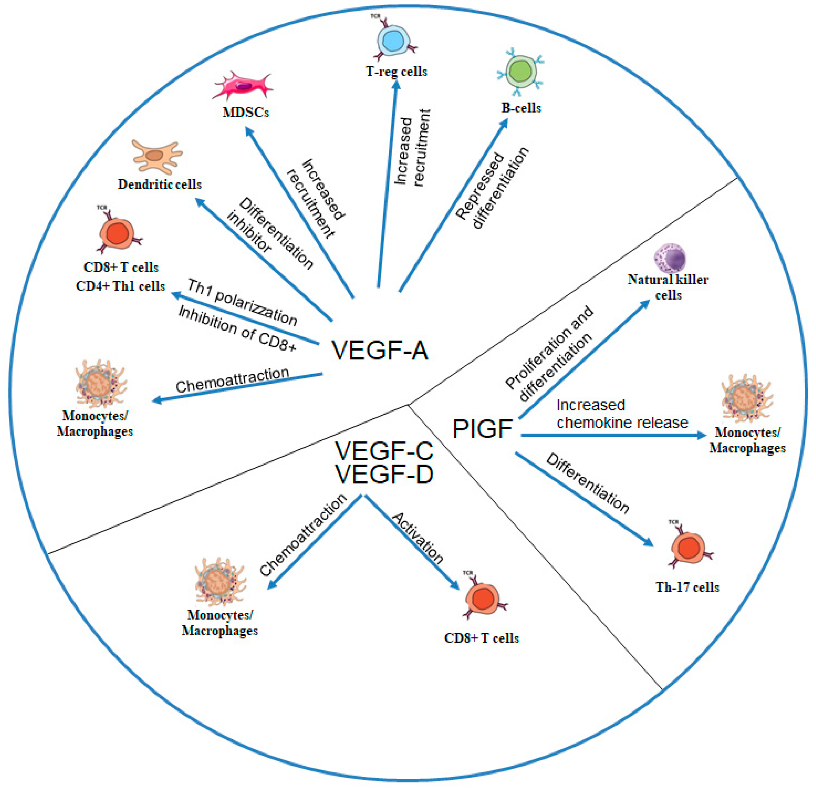
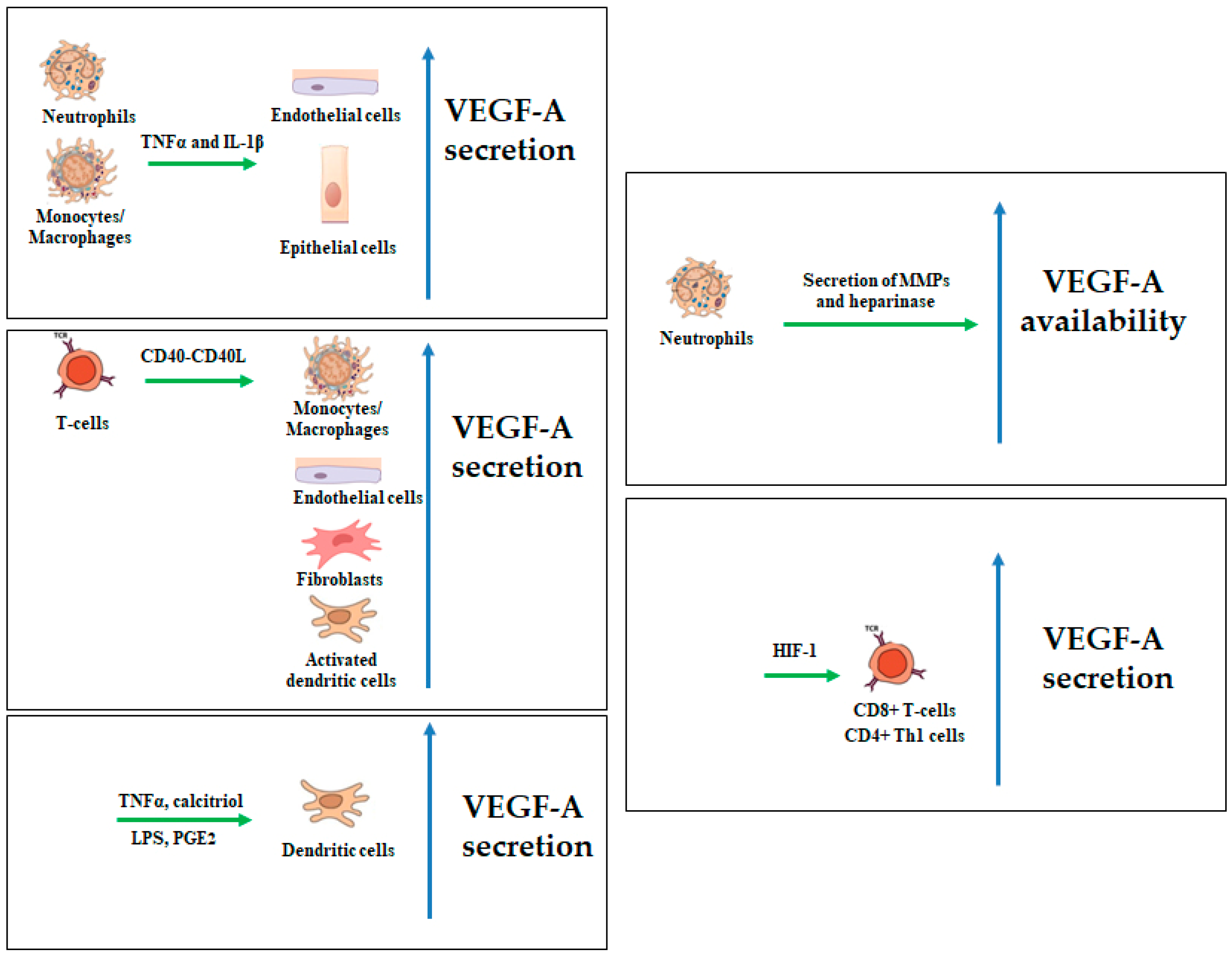
The mammalian VEGF family is composed of five growth factors, VEGF-A, VEGF-B, placenta growth factor (PlGF), VEGF-C, and VEGF-D, that recognize, with different affinity, the cellular receptors VEGFR (VEGFR-1, VEGFR-2, and VEGFR-3) [15]. Membrane VEGFRs present three different domains: an extracellular domain that binds to the growth factor, a transmembrane domain, and an intracellular domain with tyrosine kinase activity. VEGF-A binds to both VEGFR-1 and VEGFR-2, VEGF-B and PlGF bind only to VEGFR-1, and VEGF-C and VEGF-D bind to VEGFR-3. Some isoforms of these growth factors, derived from alternative splicing of the same gene, recognize, and bind to the non-tyrosine kinase receptors neuropilin-1 (NRP-1) and neuropilin-2 (NRP-2). Other molecules can interact with the VEGFs, such as integrins, cadherins, or heparin sulfate proteoglycans, but their specific role and effects have been less characterized. A summary of the cross talks among VEGFs and immune cells is shown in Figure 1 and described in the text.

2.1. VEGF-A

VEGF-A was firstly characterized as a permeability factor and then for its actions on endothelial cell proliferation, vasodilation, and inhibition of endothelial cell apoptosis [16]. It plays a crucial role in the maintenance of physiological vascular homeostasis in different cells and tissues, and its involvement in the pathogenesis of diseases such as tumor growth and metastases, diabetic and hypertensive retinopathy, has been demonstrated.
The different VEGF-A structures derived from alternative splicing determine protein-protein interactions that can modulate growth factor functions. Exons 6 and 7, which are lacked in some isoforms, are responsible for binding to heparin and extracellular matrix heparin-sulfate proteoglycans, and exon 7 for VEGF-A interaction with NRP-1 and NRP-2.
VEGF-A is secreted by different cell types of epithelial and mesenchymal origin, and its expression is mainly modulated by hypoxia [25]. Several growth factors and inflammatory mediators can also induce VEGF-A expression, including platelet-derived growth factor-BB, epidermal growth factor, transforming growth factor (TGF)-β1, interleukin (IL)-1β, and tumor necrosis factor (TNF)-α [26].
In the presence of proinflammatory molecules, such as lipopolysaccharide (LPS), TNF-α, and IL-1β, classically activated dendritic cells (DCs) express VEGF-A together with high levels of anti-angiogenic molecules. In contrast, alternatively activated DCs matured in the presence of anti-inflammatory molecules, such as calcitriol or prostaglandin E2 (PGE2), secrete high levels of VEGF-A, mediating vascular growth at the site of tissue inflammation or wound, and in the reactive lymph nodes [20]. These findings indicate that the presence of pro- or anti-inflammatory mediators in the tissue microenvironment is the principal responsible for production of VEGF-A by DCs.
Immune cell contact-mediated activation can lead to VEGF-A induction. CD40L expressed on T cells engages CD40 on endothelial cells, monocytes, and fibroblast-like synoviocytes, resulting in strong VEGF-A induction [27,28] (Figure 2).
VEGF-A mainly interacts with immune cells by promoting a chemotactic response that recruits dendritic cells, myeloid-derived suppressor cells, and macrophages/monocytes to the damaged tissue where they contribute in maintaining local inflammatory processes by increasing the release of proinflammatory cytokines [16,17].
VEGF-A is also involved in the recruitment of precursor DCs and in their trans-differentiation within the tissue into endothelial-like cells [20].
Besides its role as chemoattractant, VEGF-A is secreted by T cells on stimulation by IL-2 or hypoxia. Hypoxia also induced the expression of VEGFR-2 in T cells, suggesting that T cells might also respond to VEGF-A. Indeed, VEGF-A augmented interferon (IFN)-γ and inhibited IL-10 secretion together with enhancing T cells polarization towards a T helper (h)1 phenotype [19]. Neutrophils can modulate VEGF-A bioavailability and bioactivity via secretion of matrix metalloproteinases and heparinase [29].

2.2. VEGF-B and PlGF

VEGF-B is expressed in early embryonic stages contributing to the development of cardiovascular system and myocardium, but its role in vasculogenesis is not essential as demonstrated by the VEGF-B-/- homozygotic mice that are viable at birth and show moderate cardiovascular system defects. Two VEGF-B isoforms are derived from alternative splicing: VEGF-B167 that binds to ECM and interacts with VEGFR-1 [32,33,43] and NRP-1 and VEGF-B186 which interacts with VEGFR-1 only and, after cleavage, gains the ability to bind NRP-1. In adults, VEGF-B is found in different tissues such as myocardium, pancreas, and skeletal muscle, but its role as angiogenetic factors is still debated. In fact, VEGF-B is principally related with the survival of smooth muscle cells, pericytes, neurons and cardiomyocytes.
PlGF has been originally identified in the human placental tissue and its expression correlated with trophoblast growth and differentiation, trophoblast invasion and blastocyst implantation. In the adult, PlGF is found in four isoforms (PlGF-1, PlGF-2, PlGF-3 and PlGF-4) with different structure and activity. All isoforms bind and activate VEGFR-1; the PlGF-2 isoform also interacts with NRP-1, NRP-2 and heparin.
PlGF significantly induces expression of inflammatory chemokines, such as TNF-α, IL-1β, monocyte chemotactic protein (MCP)-1, IL-8, and VEGF-A, by monocytes, contributing in sustaining monocyte activation [30,31]. PlGF specifically induces the production of TGF-β1 by tumor-associated macrophages [21].
Analyzing the implantation site in PlGF null mice, Tayade et al. found a role for PlGF in uterine natural killer (NK) cell proliferation and differentiation [22]. Uterine NK cells are major responsible for early vasculature changes in pregnant endometrium. They also express VEGF-A. However, different studies did not succeed in demonstrating the presence of VEGFR-1 on uterine NK cells and the mechanism of PlGF- or VEGF-A-dependent activation of uterine NK cells is not clear yet.
The analyses of splenocytes from PlGF null mice showed significantly lower levels of IL-17 compared to wild-type animals. Subsequent analyses indicated that PlGF selectively addresses T helper (h) cell fate towards the Th17 lineage [23]. T cells express VEGFR-1 and PlGF transduces its differentiation program through activation of this tyrosine kinase receptor.
No data specifically highlighting the role of VEGF-B in immune cells has been reported so far.

2.3. VEGF-C and VEGF-D

VEGF-C is expressed in the embryonic tissues where it regulates lymphangiogenesis by binding and activating VEGFR-3. Upon processing, VEGF-C is also able to bind to VEGFR-2. VEGF-C activity can be increased by binding to NRP-2 which acts as a co-receptor for VEGFR-3 [32]. VEGF-C inactivation impairs lymphatic vessel development resulting in lethal accumulation of interstitial fluids in the tissues. In adult life, VEGF-C is involved in lymphangiogenesis in both physiological and pathological situations.
VEGF-D is highly expressed in the embryonic lungs where it also participates to the development of the lymphatic vessels. Like VEGF-C, VEGF-D binds to VEGFR-3 and NRP-2. However, VEGF-D inactivation is not lethal, resulting into a moderate atrophy of lymphatic circulation.
In the tumor microenvironment, the presence of VEGF-C or VEGF-D could be linked to migration of tumor cells into lymphatic vessels, and the formation of lymph node metastasis.
Neutrophils contribute to lymphangiogenesis by secretion of VEGF-C [29]. VEGF-C is highly induced in mesenchymal stromal cells (MSCs) by steroids and tumor necrosis factor-α and VEGF-C in turn promotes CD8+ T cell responses. This immune promoting effect is abolished by blockade or specific genetic ablation of VEGFR3 in CD8+ T cells [24].

3. Receptors of the VEGF Family and Their Expression on IMMUNE cells

3.1. VEGFR-1

VEGFR-1, previously known as Fms-like tyrosine kinase (Flt)-1 receptor, is essential for development and differentiation of the embryonic vasculature. In fact, embryos in which VEGFR-1 has been knocked-out died in utero between day 8.5 and 9.0 [33]. The defect was later ascribed to an increased outgrowth of endothelial cells and angioblast commitment, which inhibited a proper organization of vascular network, probably due by the lack of the inhibitory activity exerted by VEGFR-1 in respect to VEGF-A [34]. In fact, VEGF-A reacts with higher affinity with VEGFR-1, without inducing a clear tyrosine kinase activation of the receptor, but impeding VEGF-A to properly interact with its functional VEGFR-2 [35]. Besides the membrane-bound form, endothelial cells also produce a soluble VEGFR-1 (sVEGFR-1) isoform that, by binding members of the vascular endothelial growth factor (VEGF) family, reduces the amounts of VEGFs available for the interaction with their membrane receptors, negatively regulating VEGFR-mediated signaling. sVEGFR-1 has an additional role in angiogenesis: it is deposited in the extracellular matrix, and interacts with cell membrane components [36]. Interaction of sVEGFR-1 with α5β1 integrin on endothelial cells regulates vessel growth, triggering a dynamic, pro-angiogenic phenotype [37]. Therefore, the amount of and the location of sVEGFR-1 in the tissue microenvironment regulate the activation or inhibition of cells expressing membrane VEGFR-1 (mVEGFR-1) or α5β1 integrin.
Different immune cells express mVEGFR-1 and reacts to VEGFR-1-binding growth factors such as VEGF-A, VEGF-B, and PlGF.
Human DC precursor cells express mVEGFR-1 and DC maturation is inhibited by a VEGFR-1-dependent signaling triggered by either VEGF-A or PlGF [20]. No data about a similar action of VEGF-B has been reported so far.
Acute lung injury is characterized by hyperinflammation and involves reduced levels of alveolar macrophage and recruitment of monocyte-derived macrophages in a VEGFR-1-dependent manner. The monocyte-derived macrophages that express VEGFR-1 displayed an anti-inflammatory phenotype [38]. Conversely, VEGFR-1 expression is up-regulated in tumor-associated macrophages and VEGFR-1 activation sustains polarization toward the M2 phenotype [39].
VEGFR-1 is expressed not only in monocytes/macrophages but also in bone marrow-derived hematopoietic progenitor cells, mobilized in response to VEGFs and other tumor factors [40]. These cells are able to migrate to distant sites before the arrival of metastatic tumor cells and are essential in the formation and maintenance of pre-metastatic niches [41].
So far, no data are available on sVEGFR-1 secretion by the different immune cells described above and on the possible role of sVEGFR-1 in blocking VEGF-mediated signaling in these cells. In addition, different immune cells express the α5β1 integrin [42] and could respond to the stimulation of sVEGFR-1 in the extracellular matrix. This aspect still requires additional investigation.

3.2. VEGFR-2.

VEGFR-2, previously known as the kinase insert domain receptor (KDR), is predominantly expressed on endothelial cells of both blood and lymphatic vessels.
The importance of VEGFR-2 was revealed by knockout experiments in which the inactivation of VEGFR-2 in mice resulted into lack of vasculogenesis leading to embryonic death. In the adult, VEGFR-2 regulates angiogenesis, through VEGF-A-mediated signal transduction, and lymphangiogenesis, by interacting with VEGF-C and VEGF-D.
Monocyte-derived mature DCs express both VEGFR-1 and VEGFR-2, but VEGF-A inhibits the antigen-presenting function of mature DCs through the interaction with VEGFR-2 [43,44]. Similarly, myeloid-derived suppressor cell (MDSC) are recruited by VEGF-A in a VEGFR-2-dependent manner [18].
So far, VEGF-A is considered the major responsible for the immunosuppressive environment in tumors [45] even if the expression of VEGFR-2 on T cells is controversial. In vitro activated T-cells but also tumor infiltrating T cells express VEGFR-2 [46]. VEGF-A/VEGFR-2 axis is involved in induction of either T cell proliferation or tumor-induced T-cell exhaustion, this latter through increased expression of immune checkpoint molecules [47,48].
VEGF-A induces Tregs proliferation through binding and activating VEGFR-2, but only certain Treg subclasses express VEGFR-2 [18]. It would be of interest to evaluate the prognostic role of tumor-infiltrating VEGFR-2 positive Tregs. This selective evaluation could be more accurate for patient prognosis than considering all Tregs.

3.3. VEGFR-3

VEGFR-3, or Fms-like tyrosine kinase (Flt)-4, plays a crucial role both in the formation of the lymphatic vessel network during embryonic development and in the formation of adult lymphatic vessels. A soluble sVEGFR-3 was also found and inhibited lymphatic vessel development by interfering with the signals triggered by VEGF-C and VEGF-D [49].
In the immune system, VEGFR-3 is expressed by macrophages and promotes cell chemotaxis and activation [24,50].
VEGFR-3 signaling modulates allergic airway inflammation. During acute inflammation, VEGFR-3 blocking decreased immune cell recruitment into the lungs. In contrast, the memory response to allergen is significantly exacerbated by a lack of VEGFR-3. This memory response is dependent on the absence of both VEGF-C and VEGF-D, suggesting that these two growth factors may compensate for each other in allergic airway inflammation [51].

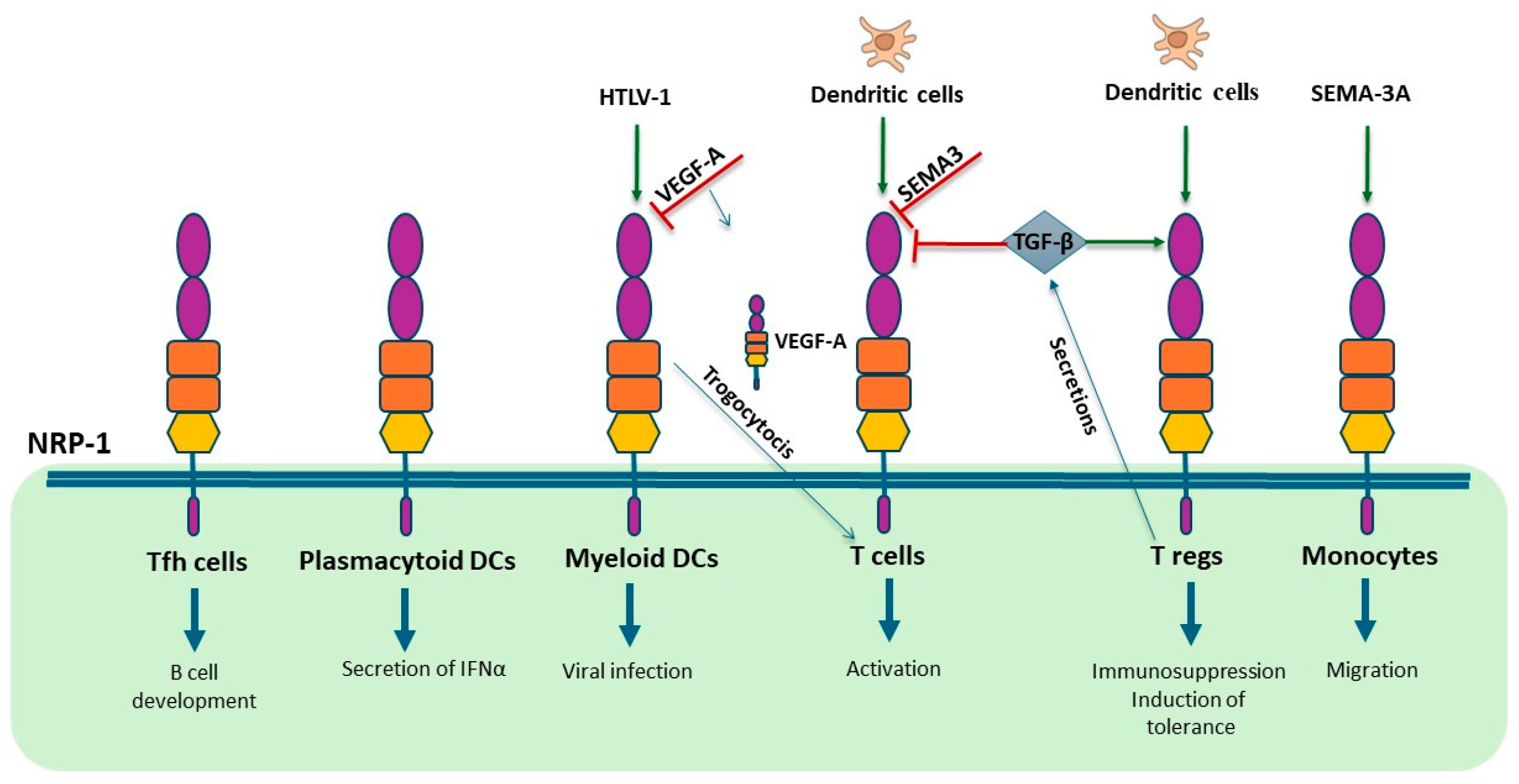
3.4. NRP-1 and NRP-2

NRP-1 and NRP-2 were initially identified as receptors for semaphorins (SEMA) involved in neuron system development [52]. NRP-1 promotes the interaction of SEMA3 with plexins, mediating axon guidance during embryonic development. Subsequently, additional growth factors have been shown to bind NRP-1 and NRP-2, such as transforming growth factor (TGF)-β, hepatocyte growth factor, or platelet-derived growth factor, as well as integrins and small molecules such as synectin [53], but the respective roles in angiogenesis have not been fully characterized yet.
NRPs are non-tyrosine kinase transmembrane receptors and act as co-receptors for different isoforms of VEGFs [54]. Both NRP-1 and NRP-2 exist as either membrane bound or soluble forms, that display decoy functions to membrane NRPs [55]. Endothelial cells of arteries express primarily NRP-1, which possess a selective binding site for VEGF-A165, PlGF-2, and VEGF-B167. The co-expression of NRP-1 and VEGFR-2 enhances the binding of VEGF-A165 to VEGFR-2 and increases the efficacy of its signaling. Endothelial cells veins and lymphatic vessels express predominantly NRP-2, with binding sites for VEGF-C and VEGF-D, thus enhancing the signaling through VEGFR-3. More recently, NRP-1 has been involved in the interaction between integrin α5β1 on endothelial cells and the sVEGFR-1 present in the extracellular matrix leading to angiogenesis [56]. VEGF-A can also directly act on neuron-expressing NRP-1, but the NRP-1 signaling co-receptors have not been identified in these cases [52].
NRP-1 contributes to immunity in different ways [57]. NRP-1 has been involved in immune system development and thymocyte differentiation, mainly through binding to SEMA [58,59]. It has also been involved in mediating antigen presentation to T cells by antigen-presenting cells [60]. NRP-1 promoted cell–cell adhesion via homophilic interactions and colocalized with CD3 at the contact zone, indicating a potential role for NRP-1 in the initiation of primary immune responses. However, only a few effector T cells and plasmacytoid DCs express NRP-1. NRP-1 action in immune cells is reported to be mediated by SEMA/plexin binding and it is mainly inhibitory [61]. In fact, SEMA3A secreted from activated DC and T cell can also bind to NRP1 on T cells and inhibit T cell proliferation by blocking actin cytoskeleton reorganization. NRP-1 can exert immunoinhibitory activity also through binding to TGF-β, a well-known immunosuppressive cytokine, also secreted by regulatory T cells (Tregs) [62]. NRP-1 on Tregs can bind the latent form of TGF-β from the surrounding tissue fluid or plasma and induce further immunosuppression.
NRP-1 is expressed by Tregs but not naive T helper (Th) cells [63] and plays a key role in promoting long interactions between Treg cells and DCs [64]. Nrp-1 expression on Treg cells gives them an advantage over naive Th cells in the absence of proinflammatory stimuli. Even if VEGFR-1 is present on DCs, neither VEGFR-1 nor VEGFR-2 is expressed on Tregs, excluding the direct involvement of VEGF-A, VEGF-B, and PlGF in immune synapse maintenance.
NRP-1 is expressed by a subpopulation of T follicular helper (Tfh) cells in the secondary lymphoid organs in humans and its expression can be induced in vitro by interaction with autologous memory B cells and correlated with the plasma B cell precursors. This indicated a role for NRP-1 on Tfh cells in B cell differentiation [65].
NRP-1 is expressed on plasmacytoid DCs. Blocking of NRP-1 reduces the production of IFN-α by plasmacytoid DCs, although the exact underlying mechanism is unclear [66].
NRP1 expressed on myeloid DCs can be transferred to T cells by trogocytosis [67]. VEGF-A secreted by human DCs can bind to NRP1 captured by T lymphocytes. Therefore, NRP-1 transfer to T cells during the immune synapse can convert T lymphocytes into VEGF-A-carrying cells. Together with the enhanced signaling of VEGFR-2 on endothelial cells in the presence, in trans, of the NRP-1–VEGF-A complex, intercellular transfer of NRP-1 might participate in the remodeling of endothelial vessels in secondary lymphoid organs during inflammation [67].
Myeloid DCs are susceptible to infection by the human T-cell lymphotropic virus type 1 (HTLV-1) and NRP-1 has been demonstrated to be essential for viral infection [68]. Interestingly, VEGF-A acts as a selective competitor of HTLV-1 entry into the cells by binding to the same binding site on NRP-1. In fact, HTLV-1 contains in its envelop protein a structural motif homologous to VEGF-A165.
NRP-1 has been reported on tissue-resident macrophages. NRP-1 is also expressed on tumor-associated macrophages (TAMs) and is crucial for their migration to the hypoxic niche of the tumor in response to SEMA3A. TAMs express VEGFR-1 and a role for VEGFs in recruiting macrophages has been already underlined. However, no data are available so far to sustain a collaboration of VEGFR-1 and NRP-1 in transducing this chemotactic signal. Carrer et al. identified a novel subset of bone marrow-derived monocytes, which were NRP-1 positive. When injected into tumors, these NRP-1-expressing monocytes promoted tumor vasculature normalization [69]. Similarly, NRP1 expressed in glioma TAMs has been associated with tumor promotion. Indeed, mice with a TAM-specific deletion of NRP1 resulted in slower tumor growth, reduced tumor vascularity and increased survival [70].
Differently from NRP-1, NRP-2 seems to be more broadly expressed in macrophages, during the differentiation from monocytes towards DCs, and in T cell subsets [71]. However, The NRP-2 role in immune cells has not been completely defined.
Altogether, current literature data indicate a role for NRPs in the homeostasis and pathological states of immune cells, but information are still fragmentary and not all the aspects have been considered, especially the involvement of the VEGFs in activation or repression of immune responses and the possible crosstalk between NRPs and VEGFRs.
Figure 3. NRP-1 roles in immune cells. NRP-1 is expressed in different cells of the immune system. When expressed in T follicular helper (Tfh) cells, NRP-1 mediates B cell differentiation [65], whereas in plasmacytoid DCs, it induces the secretion of interferon (IFN)α [66]. Human T-cell lymphotropic virus type 1 (HTLV-1) enters myeloid DCs through NRP-1 and VEGF-A can block this infection [68]. Myeloid DCs can transfer NRP-1 and VEGF-A to T cells by trogocytosis [67]. DCs directly activate T cells through NRP-1 but also block T cell activation by secretion of SEMA3 [62]. Regulatory T cells (Tregs) are also directly activated by DCs but also indirectly by secretion of TGF-β that also blocks T cell activation [64]. SEMA-3A/NRP-1 interaction leads to increased migration of monocytes [57]. .
Preprints 140287 g003

4. Clinical Advancements and Challenges in Combining VEGF-Targeted Anti-Angiogenic Therapy with Checkpoint Inhibitor Immunotherapy

Due to the amount of data that connect tumor angiogenesis, expression of VEGFs and VEGFRs in the tumor microenvironment, and their role in modulating tumor immune responses, the possibility of combining anti-VEGFs together with anti-checkpoint inhibitors (ICIs) in tumor therapy has been evaluated. Indeed, anti-VEGFs have a broad range of effects on the immune system and effectively contribute to reverting the immunosuppressive environment of the tumor [72]. Nevertheless, anti-VEGFs as a single therapeutic approach are likely insufficient to generate a complete or robust immune response against cancer, especially in patients with advanced-stage disease. Therefore, anti-VEGFs have been proposed in combination with various immunotherapeutic strategies that boost adaptive immune responses, such as ICIs. An accumulating number of clinical trials have been conducted to explore the efficacy of the combination (Table 1).
The presence of high levels of VEGF-A in the serum of patients with melanoma before treatment with the anti-CTLA-4 antibody ipilimumab was found to correlate with decreased overall survival compared with that of patients with low VEGF-A expression, providing an additional rationale for targeting VEGF-A in these patients. Therefore, a clinical trial has tested the combination of Ipilimumab and Bevacizumab in melanoma patients. This treatment led to an increased infiltration of lymphocytes in the tumor tissue [73]. The same combination later showed to have clinical benefits. Results from the phase I and II clinical trial (NCT00790010) showed that Ipilimumab plus Bevacizumab in patients with metastatic melanoma had favorable clinical outcomes (disease control rate 67.4%), increasing tumor vascular expression of ICAM-1 and VCAM-1 and lymphocyte infiltration in the tumors [74].
The most successful results of combination therapies have been reported in renal cell carcinoma (RCC) and hepatocellular carcinoma (HCC). To explore the efficacy of anti-PD-L1 combined with anti-VEGF-A, the phase I study NCT01633970 was proposed and aimed at investigating the safety of Atezolizumab plus Bevacizumab or chemotherapy in metastatic RCC [75]. Compared with tumor samples from patients at baseline or post Bevacizumab monotherapy, expression of CD8, PD-L1, and major histocompatibility complex-I (MHC-I) markedly increased after combination therapy. The transformation to hot tumor was associated with increased expression of CX3CL1 which participated in the recruitment of peripheral CD8+ T cells [75]. Subsequently, the results of a randomized phase II clinical trial evaluating the efficacy of Atezolizumab plus Bevacizumab versus Sunitinib as a first line of treatment in RCC were reported (NTC01984242). The combination had a marked benefit over Sunitinib alone in patients with tumors showing high numbers of MDSCs and in patients with tumors harboring high numbers of effector T cells and high levels PD-L1 expression (≥ 5%), whereas Sunitinib had greater efficacy than the combination in patients with tumors with high levels of angiogenesis [13]. A pivotal phase III trial (NCT02420821) is currently underway to confirm the results of this phase II study [76]. Furthermore, a phase III trial comparing Avelumab and Axitinib combination therapy against Sunitinib monotherapy in patients with metastatic RCC in a first line setting (NCT02684006) revealed that Avelumab/Axitinib combination significantly prolonged progression-free survival (PFS) compared to Sunitinib [77]. Additional clinical trials are ongoing in patients with advanced RCC that showed promising results with PD-1/PD-L1 inhibitors and anti-angiogenic agent combination treatments (Table 1).
In patients with advanced HCC, a first randomized phase III clinical trial, IMBRAVE 150 (NCT03434379), showed a remarkable improvements in co-primary endpoints, PFS and overall survival (OS), using the combination of Atezolizumab and Bevacizumab compared to Sorafenib. It exhibited, in terms of patient-reported outcomes, a delayed deterioration of quality of life, with tolerated and controllable toxicity, in the combination group compared to the monotherapy group [77]. The safety and efficacy of the combination of Pembrolizumab and Lenvatinib were evaluated in patients with unresectable HCC in a phase Ib KEYNOTE-524 study, which led to the design of another study, LEPP-002 (NCT03713593), a phase III trial to evaluate Pembrolizumab in combination with Lenvatinib as a potential first-line treatment for patients with advanced HCC [78].
In non-squamous non-small cell lung cancer (NSCLC), a phase III clinical trial (IMpower150, NCT02366143) comparing Atezolizumab, Bevacizumab and Carboplatin/Paclitaxel combination therapy (ABCP group) against Bevacizumab and Carboplatin/Paclitaxel combination therapy showed significant improvement in PFS and OS in the ABCP group compared to the control group [79]. Based on these results, Atezolizumab was approved by FDA for use in combination with Bevacizumab, Paclitaxel, and Carboplatin as first-line treatment for patients with metastatic NSCLC.
In these different combination therapeutic approaches, the rationale for using a selective VEGF-A inhibitor such as Bevacizumab or the tyrosine kinase inhibitors Sunitinib or Sorafenib, which block different VEGFRs along with other receptors, is not clear. In addition to the clinical aspects, the results of the clinical trials should also be analyzed from a molecular point of view to better understand which anti-angiogenic drug would be more beneficial.
Table 1. Clinical trials investigating anti-angiogenic therapy in combination with immune checkpoint inhibitors.
Clinical trial ID Study phase Agent(s) Anti-Angiogenic target Anti-tumor immunity target Cancer types Ref.
NCT00790010 I Bevacizumab + Ipilimumab VEGF-A CTLA-4 Melanoma [80]
NCT01950390 II Bevacizumab + Ipilimumab VEGF-A CTLA-4 Melanoma [80]
NCT01633970 I Bevacizumab + Atezolizumab VEGF-A PD-L1 RCC [75]
NCT01984242 II Bevacizumab + Atezolizumab vs Sunitinib VEGF-A, VEGFR PD-L1 RCC [13,76]
NCT02420821 III Bevacizumab + Atezolizumab vs Sunitinib VEGF-A, VEGFR PD-L1 RCC [81,82]
NCT02231749 III Nivolumab + Ipilimumab vs Sunitinib VEGFR, PDGFR PD-1, CTLA-4 RCC [83,84]
NCT02493751 I Axitinib + Avelumab VEGFR PD-L1 RCC [85,86]
NCT02684006 III Axitinib + Avelumab vs Sunitinib VEGFR, PDGFR PD-L1 RCC [77,78]
NCT02724878 II Bevacizumab + Atezolizumab VEGF-A PD-L1 RCC [87]
NCT02853331 III Pembrolizumab + Axitinib vs Sunitinib VEGFR, PDGFR PD-1 RCC [81,88]
NCT02811861 III Pembrolizumab + Lenvatinib vs Sunitinib VEGFR, PDGFR PD-1 RCC [79]
NCT03721653 II Bevacizumab + Atezolizumab + FOLFOXIRI VEGF-A PD-L1 CRC [89,90]
NCT03434379 II Bevacizumab + Atezolizumab VEGF-A PD-L1 HCC [91,92]
NCT03006926 I Lenvatinib + Pembrolizumab VEGFR, PDGFR PD-1 HCC [93,94]
NCT03713593 III Lenvatinib + Pembrolizumab vs Lenvatinib VEGFR, PDGFR PD-1 HCC [95]
NCT02873962 II Bevacizumab + Nivolumab VEGF-A PD-1 OC [96]
NCT03038100 III Bevacizumab + Atezolizumab and Chemiotherapy VEGF-A PD-L1 OC [97]
NCT03170960 I Cabozantinib + Atezolizumab VEGFR PD-L1 UC, RCC, NSCLC, HCC, [98,99]
NCT02366143 III Atezolizumab + Bevacizumab + Paclitaxel/Carboplatin VEGF-A PD-L1 NSCLC [100]
NCT02443324 I Ramucirumab + Pembrolizumab VEGFR PD-1 G/GEJ, NSCLC, UC, BTC [101]
NCT02572687 I Ramucirumab + Durvalumab VEGFR PD-L1 NSCLC, G/GEJ, HCC [102]
NCT02856425 I Nintedanib + Pembrolizumab PDGFR, VEGFR PD-1 Advanced solid tumors [103]
NCT03377023 I/II Nintedanib + Nivolumab + Ipilimumab PDGFR, VEGFR PD-1, CTLA-4 NSCLC [104]
Abbreviations: CRC, colorectal cancer, G/GEJ, gastric or gastroesophageal junction adenocarcinoma, HCC, hepatocellular carcinoma, NSCLC, non-small cell lung cancer, OC, ovarian cancer, RCC, renal cell cancer, UC, urothelial carcinoma, BTC, biliary tract cancer.

5. Innovative Approaches Targeting VEGFs and Tumor-Mediated Immune Suppression

Besides approved clinical trials, additional promising molecules targeting the VEGFs have been developed and are now in the preclinical phase. Among these, the monoclonal antibody (mAb) D16F7 has been developed against VEGFR-1 that selectively blocks VEGFR-1 dimerization and signal transduction upon binding with VEGFs. This antibody showed efficacy in improving anti-PD-1 responses in melanoma [105]. D16F7 mAb decreases in vitro and in vivo chemotaxis of activated M2 macrophages, thus contributing in the reduction of the immunosuppressive microenvironment of the tumor.
For VEGF-A ligands, and to a limited extent also for PlGF, a transition is now observed towards the developing of smaller ligands like nanobodies, single-chain monoclonal antibodies and peptides [106]. These also include addition of unnatural amino acids and chemical modifications for designed and improvement of the properties, such as serum stability and greater affinity, of previously identified molecules [107].
Additional strategies such as vaccine combination with anti-angiogenic agents have also been followed. The combination of a vaccine composed of peptides of the tumor antigen suvivin (SVX vaccine) with sunitinib in a colorectal carcinoma model has been proposed [108]. The therapeutic synergy between SVX vaccine and sunitinib have been highlighted when the vaccine was administered at the end of anti-angiogenic treatment. The initial sunitinib treatment resulted into the promotion of an immune-favorable tumor microenvironment, with pericyte coverage of tumor vessels, rich in NK cells and tumor-infiltrating effector T cells, but poor in myeloid-derived suppressor cells. This new immune environment can reinforce the activity of the vaccine. With the aim of investigating whether a positive synergy between lymphangiogenesis and cancer immunotherapy could be exploited in a cancer vaccine, lethally irradiated tumor cells overexpressing VEGF-C and topical adjuvants were used. The “VEGF-C vax” induced extensive local lymphangiogenesis and promoted stronger T cell activation in the murine model, both at the intradermal vaccine site and in the draining lymph nodes, resulting in higher frequencies of antigen-specific T cells. In fact, the lethally irradiated tumor cells undergo radiation-induced cell death and provide a source of tumor-associated antigens, whereas VEGF-C overexpression activates local lymphatics to proliferate and increase antigen transport to the distal lymph nodes [109].
Preclinical applications of small interfering RNAs (siRNAs) indicate that the next-generation anti-angiogenic compounds could be represented by this class of molecules [110]. However, siRNAs are unstable, and delivery systems should be developed for they usage in clinical applications.

6. Conclusions

Different immune cells secrete VEGFs and present the VEGFRs or the coreceptor NRPs. However, literature data are still fragmentary and do not consider the co-expression of diverse receptors and co-receptors on the same cell type or the presence of different VEGFs in the cell milieu, which could lead to opposite results depending on the relative abundance of one VEGF compared to the others. In addition, the investigation of other VEGFs besides VEGF-A should be encouraged for the development of novel anti-angiogenic and anti-inflammatory agents. These new therapeutics may be more useful in the treatment of specific pathologies, alone or in combination with immunotherapies.

Author Contributions

“Conceptualization, CMF and AO; investigation, MLC; data curation, EB, CR, MLC; writing—original draft preparation, CMF and AO; writing—review and editing, all authors. All authors have read and agreed to the published version of the manuscript.”.

Funding

This research was funded by the Italian Ministry of Health, RC2021-2024.

Conflicts of Interest

The authors declare no conflicts of interest.

References

  1. Venkatakrishnan, G.; Parvathi, V.D. Decoding the mechanism of vascular morphogenesis to explore future prospects in targeted tumor therapy. Medical Oncology 2022, 39, 178. [Google Scholar] [CrossRef] [PubMed]
  2. Ribatti, D.; Pezzella, F. Overview on the Different Patterns of Tumor Vascularization. Cells 2021, 10, 639. [Google Scholar] [CrossRef]
  3. Lorenc, P.; Sikorska, A.; Molenda, S.; Guzniczak, N.; Dams-Kozlowska, H.; Florczak, A. Physiological and tumor-associated angiogenesis: Key factors and therapy targeting VEGF/VEGFR pathway. Biomed Pharmacother 2024, 180, 117585. [Google Scholar] [CrossRef] [PubMed]
  4. Ferrara, N.; Gerber, H.; LeCourter, J. The biology of VEGF and its receptors. Nat Med 2003, 9, 669–676. [Google Scholar] [CrossRef] [PubMed]
  5. Dvorak, H.F. Reconciling VEGF With VPF: The Importance of Increased Vascular Permeability for Stroma Formation in Tumors, Healing Wounds, and Chronic Inflammation. Front Cell Dev Biol 2021, 9, 660609. [Google Scholar] [CrossRef]
  6. Pérez-Gutiérrez, L.; Ferrara, N. Biology and therapeutic targeting of vascular endothelial growth factor A. Nat Rev Mol Cell Biol 2023, 24, 816–834. [Google Scholar] [CrossRef]
  7. Ahmad, A.; Nawaz, M.I. Molecular mechanism of VEGF and its role in pathological angiogenesis. J Cell Biochem 2022, 123, 1938–1965. [Google Scholar] [CrossRef]
  8. Deng, J.; Qin, Y. Advancements and emerging trends in ophthalmic anti-VEGF therapy: a bibliometric analysis. Int Ophthalmol 2024, 44, 368. [Google Scholar] [CrossRef]
  9. Folkman, J. Angiogenesis in cancer, vascular, rheumatoid and other disease. Nat Med 1995, 1, 27–31. [Google Scholar] [CrossRef]
  10. Yang, F.; Lee, G.; Fan, Y. Navigating tumor angiogenesis: therapeutic perspectives and myeloid cell regulation mechanism. Angiogenesis 2024, 27, 333–349. [Google Scholar] [CrossRef]
  11. Voron, T.; Marcheteau, E.; Pernot, S.; Colussi, O.; Tartour, E.; Taieb, J.; Terme, M. Control of the immune response by pro-angiogenic factors. Front Oncol 2014, 4, 70. [Google Scholar] [CrossRef] [PubMed]
  12. Yang, J.; Yan, J.; Liu, B. Targeting VEGF/VEGFR to modulate antitumor immunity. Front Immunol 2018, 9, 978. [Google Scholar] [CrossRef] [PubMed]
  13. McDermott, D.F.; Huseni, M.A.; Atkins, M.B.; Motzer, R.J.; Rini, B.I.; Escudier, B.; Fong, L.; Joseph, R.W.; Pal, S.K.; Reeves, J.A.; et al. Clinical activity and molecular correlates of response to atezolizumab alone or in combination with bevacizumab versus sunitinib in renal cell carcinoma. Nat Med 2018, 24, 749–757. [Google Scholar] [CrossRef]
  14. Zeng, H.; Xu, Q.; Wang, J.; Xu, X.; Luo, J.; Zhang, L.; Luo, C.; Ying, J.; Li, J. The effect of anti-PD-1/PD-L1 antibodies combined with VEGF receptor tyrosine kinase inhibitors versus bevacizumab in unresectable hepatocellular carcinoma. Front Immunol 2023, 14, 1073133. [Google Scholar] [CrossRef]
  15. Cébe-Suarez, S.; Zehnder-Fjallman, A.; Ballmer-Hofer, K. The role of VEGF receptors in angiogenesis; complex partnerships. Cell Mol Life Sci 2006, 63, 601–615. [Google Scholar] [CrossRef] [PubMed]
  16. Shibuya, M. VEGF-VEGFR System as a Target for Suppressing Inflammation and other Diseases. Endocr Metab Immune Disord Drug Targets 2015, 15, 135–144. [Google Scholar] [CrossRef]
  17. Barleon, B.; Sozzani, S.; Zhou, D.; Weich, H.A.; Mantovani, A.; Marmé, D. Migration of human monocytes in response to vascular endothelial growth factor (VEGF) is mediated via the VEGF receptor flt-1. Blood 1996, 87, 3336–3343. [Google Scholar] [CrossRef] [PubMed]
  18. Bourhis, M.; Palle, J.; Galy-Fauroux, I.; Terme, M. Direct and Indirect Modulation of T Cells by VEGF-A Counteracted by Anti-Angiogenic Treatment. Frontiers in Immunology 2021, 12. [Google Scholar] [CrossRef]
  19. Mor, F.; Quintana, F.J.; Cohen, I.R. Angiogenesis-inflammation cross-talk: vascular endothelial growth factor is secreted by activated T cells and induces Th1 polarization. J Immunol 2004, 172, 4618–4623. [Google Scholar] [CrossRef]
  20. Sozzani, S.; Rusnati, M.; Riboldi, E.; Mitola, S.; Presta, M. Dendritic cell-endothelial cell cross-talk in angiogenesis. Trends Immunol 2007, 28, 385–392. [Google Scholar] [CrossRef]
  21. Kong, X.; Bu, J.; Chen, J.; Ni, B.; Fu, B.; Zhou, F.; Pang, S.; Zhang, J.; Xu, S.; He, C. PIGF and Flt-1 on the surface of macrophages induces the production of TGF-β1 by polarized tumor-associated macrophages to promote lung cancer angiogenesis. Eur J Pharmacol 2021, 912, 174550. [Google Scholar] [CrossRef]
  22. Tayade, C.; Hilchie, D.; He, H.; Fang, Y.; Moons, L.; Carmeliet, P.; Foster, R.A.; Croy, B.A. Genetic depletion of placenta growth factor in mice alters uterine NK cells. J Immunol 2007, 178, 4267–4275. [Google Scholar] [CrossRef] [PubMed]
  23. Yoo, S.-A.; Kim, M.; Kang, M.-C.; Kong, J.-S.; Kim, K.-M.; Lee, S.; Hong, B.-K.; Jeong, G.H.; Lee, J.; Shin, M.-G.; et al. Placental growth factor regulates the generation of T H 17 cells to link angiogenesis with autoimmunity. Nat Immunol 2019, 20, 1348–1359. [Google Scholar] [CrossRef] [PubMed]
  24. Gan, Y.; Zhang, T.; Chen, X.; Cao, W.; Lin, L.; Du, L.; Wang, Y.; Zhou, F.; He, X.; He, Y.; et al. Steroids Enable Mesenchymal Stromal Cells to Promote CD8(+) T Cell Proliferation Via VEGF-C. Adv Sci (Weinh) 2021, 8, 2003712. [Google Scholar] [CrossRef] [PubMed]
  25. Kimura, H.; Esumi, H. Reciprocal regulation between nitric oxide and vascular endothelial growth factor in angiogenesis. Acta Biochim Pol 2003, 50, 49–59. [Google Scholar] [CrossRef] [PubMed]
  26. Kang, Y.; Li, H.; Liu, Y.; Li, Z. Regulation of VEGF-A expression and VEGF-A-targeted therapy in malignant tumors. J Cancer Res Clin Oncol 2024, 150, 221. [Google Scholar] [CrossRef]
  27. Melter, M.; Reinders, M.E.; Sho, M.; Pal, S.; Geehan, C.; Denton, M.D.; Mukhopadhyay, D.; Briscoe, D.M. Ligation of CD40 induces the expression of vascular endothelial growth factor by endothelial cells and monocytes and promotes angiogenesis in vivo. Blood 2000, 96, 3801–3808. [Google Scholar] [CrossRef]
  28. Cho, C.S.; Cho, M.L.; Min, S.Y.; Kim, W.U.; Min, D.J.; Lee, S.S.; Park, S.H.; Choe, J.; Kim, H.Y. CD40 engagement on synovial fibroblast up-regulates production of vascular endothelial growth factor. J Immunol 2000, 164, 5055–5061. [Google Scholar] [CrossRef]
  29. Tan, K.W.; Chong, S.Z.; Wong, F.H.; Evrard, M.; Tan, S.M.; Keeble, J.; Kemeny, D.M.; Ng, L.G.; Abastado, J.P.; Angeli, V. Neutrophils contribute to inflammatory lymphangiogenesis by increasing VEGF-A bioavailability and secreting VEGF-D. Blood 2013, 122, 3666–3677. [Google Scholar] [CrossRef]
  30. Perelman, N.; Selvaraj, S.K.; Batra, S.; Luck, L.R.; Erdreich-Epstein, A.; Coates, T.D.; Kalra, V.K.; Malik, P. Placenta growth factor activates monocytes and correlates with sickle cell disease severity. Blood 2003, 102, 1506–1514. [Google Scholar] [CrossRef]
  31. Selvaraj, S.K.; Giri, R.K.; Perelman, N.; Johnson, C.; Malik, P.; Kalra, V.K. Mechanism of monocyte activation and expression of proinflammatory cytochemokines by placenta growth factor. Blood 2003, 102, 1515–1524. [Google Scholar] [CrossRef]
  32. Parker, M.W.; Linkugel, A.D.; Goel, H.L.; Wu, T.; Mercurio, A.M.; Vander Kooi, C.W. Structural basis for VEGF-C binding to neuropilin-2 and sequestration by a soluble splice form. Structure 2015, 23, 677–687. [Google Scholar] [CrossRef] [PubMed]
  33. Fong, G.; Rossant, J.; Gertsenstein, M.; Breitman, M. Role of the Flt-1 receptor tyrosine kinase in regulating the assembly of vascular endothelium. Nature 1995, 376, 66–70. [Google Scholar] [CrossRef] [PubMed]
  34. Fong, G.; Zhang, L.; Bryce, D.; Peng, J. Increased hemangioblast commitment, not vascular disorganization, is the primary defect in flt-1 knock-out mice. Development 1999, 126, 3015–3025. [Google Scholar] [CrossRef] [PubMed]
  35. Failla, C.M.; Carbo, M.; Morea, V. Positive and Negative Regulation of Angiogenesis by Soluble Vascular Endothelial Growth Factor Receptor-1. Int J Mol Sci 2018, 19. [Google Scholar] [CrossRef] [PubMed]
  36. Orecchia, A.; Lacal, P.M.; Schietroma, C.; Morea, V.; Zambruno, G.; Failla, C.M. Vascular endothelial growth factor receptor-1 is deposited in the extracellular matrix by endothelial cells and is a ligand for the  integrin. J Cell Sci 2003, 116, 3479–3489. [Google Scholar] [CrossRef]
  37. Orecchia, A.; Mettouchi, A.; Uva, P.; Simon, G.C.; Arcelli, D.; Avitabile, S.; Ragone, G.; Meneguzzi, G.; Pfenninger, K.H.; Zambruno, G.; et al. Endothelial cell adhesion to soluble vascular endothelial growth factor receptor-1 triggers a cell dynamic and angiogenic phenotype. Faseb J 2014, 28, 692–704. [Google Scholar] [CrossRef]
  38. Osada, M.; Yamashita, A.; Akinaga, S.; Hosono, K.; Ito, Y.; Shibuya, M.; Asari, Y.; Amano, H. VEGFR1 TK signaling protects the lungs against LPS-induced injury by suppressing the activity of alveolar macrophages and enhancing the anti-inflammatory function of monocyte-derived macrophages. Toxicol Appl Pharmacol 2024, 492, 117083. [Google Scholar] [CrossRef]
  39. Jetten, N.; Verbruggen, S.; Gijbels, M.J.; Post, M.J.; De Winther, M.P.; Donners, M.M. Anti-inflammatory M2, but not pro-inflammatory M1 macrophages promote angiogenesis in vivo. Angiogenesis 2014, 17, 109–118. [Google Scholar] [CrossRef]
  40. Lasser, S.A.; Ozbay Kurt, F.G.; Arkhypov, I.; Utikal, J.; Umansky, V. Myeloid-derived suppressor cells in cancer and cancer therapy. Nat Rev Clin Oncol 2024, 21, 147–164. [Google Scholar] [CrossRef]
  41. Meng, D.; Meng, M.; Luo, A.; Jing, X.; Wang, G.; Huang, S.; Luo, M.; Shao, S.; Zhao, X.; Liu, R. Effects of VEGFR1(+) hematopoietic progenitor cells on pre-metastatic niche formation and in vivo metastasis of breast cancer cells. J Cancer Res Clin Oncol 2019, 145, 411–427. [Google Scholar] [CrossRef] [PubMed]
  42. Dalpati, N.; Rai, S.K.; Dash, S.P.; Kumar, P.; Singh, D.; Sarangi, P.P. Integrins α5β1 and αvβ3 Differentially Participate in the Recruitment and Reprogramming of Tumor-associated Macrophages in the In Vitro and In Vivo Models of Breast Tumor. J Immunol 2024. [Google Scholar] [CrossRef] [PubMed]
  43. Mimura, K.; Kono, K.; Takahashi, A.; Kawaguchi, Y.; Fujii, H. Vascular endothelial growth factor inhibits the function of human mature dendritic cells mediated by VEGF receptor-2. Cancer Immunol Immunother 2007, 56, 761–770. [Google Scholar] [CrossRef] [PubMed]
  44. Dikov, M.M.; Ohm, J.E.; Ray, N.; Tchekneva, E.E.; Burlison, J.; Moghanaki, D.; Nadaf, S.; Carbone, D.P. Differential roles of vascular endothelial growth factor receptors 1 and 2 in dendritic cell differentiation. J Immunol 2005, 174, 215–222. [Google Scholar] [CrossRef] [PubMed]
  45. Geindreau, M.; Ghiringhelli, F.; Bruchard, M. Vascular Endothelial Growth Factor, a Key Modulator of the Anti-Tumor Immune Response. International Journal of Molecular Sciences 2021, 22, 4871. [Google Scholar] [CrossRef]
  46. Voron, T.; Colussi, O.; Marcheteau, E.; Pernot, S.; Nizard, M.; Pointet, A.L.; Latreche, S.; Bergaya, S.; Benhamouda, N.; Tanchot, C.; et al. VEGF-A modulates expression of inhibitory checkpoints on CD8+ T cells in tumors. J Exp Med 2015, 212, 139–148. [Google Scholar] [CrossRef]
  47. Meder, L.; Schuldt, P.; Thelen, M.; Schmitt, A.; Dietlein, F.; Klein, S.; Borchmann, S.; Wennhold, K.; Vlasic, I.; Oberbeck, S.; et al. Combined VEGF and PD-L1 Blockade Displays Synergistic Treatment Effects in an Autochthonous Mouse Model of Small Cell Lung Cancer. Cancer Res 2018, 78, 4270–4281. [Google Scholar] [CrossRef]
  48. Kim, C.G.; Jang, M.; Kim, Y.; Leem, G.; Kim, K.H.; Lee, H.; Kim, T.S.; Choi, S.J.; Kim, H.D.; Han, J.W.; et al. VEGF-A drives TOX-dependent T cell exhaustion in anti-PD-1-resistant microsatellite stable colorectal cancers. Sci Immunol 2019, 4. [Google Scholar] [CrossRef]
  49. Pavlakovic, H.; Becker, J.; Albuquerque, R.; Wilting, J.; Ambati, J. Soluble VEGFR-2: an antilymphangiogenic variant of VEGF receptors. Ann N Y Acad Sci 2010, 1207 Suppl 1, E7–15. [Google Scholar] [CrossRef]
  50. Kannan, S.; Rutkowski, J.M. VEGFR-3 signaling in macrophages: friend or foe in disease? Front Immunol 2024, 15, 1349500. [Google Scholar] [CrossRef]
  51. Maisel, K.; Hrusch, C.L.; Medellin, J.E.G.; Potin, L.; Chapel, D.B.; Nurmi, H.; Camacho, D.F.; Gleyzer, R.; Alitalo, K.; Sperling, A.I.; et al. Pro-lymphangiogenic VEGFR-3 signaling modulates memory T cell responses in allergic airway inflammation. Mucosal Immunol 2021, 14, 144–151. [Google Scholar] [CrossRef] [PubMed]
  52. Raimondi, C.; Ruhrberg, C. Neuropilin signalling in vessels, neurons and tumours. Semin Cell Dev Biol 2013, 24, 172–178. [Google Scholar] [CrossRef] [PubMed]
  53. Valdembri, D.; Caswell, P.T.; Anderson, K.I.; Schwarz, J.P.; Konig, I.; Astanina, E.; Caccavari, F.; Norman, J.C.; Humphries, M.J.; Bussolino, F.; et al. Neuropilin-1/GIPC1 signaling regulates  integrin traffic and function in endothelial cells. PLOS Biol 2009, 7, 115–132. [Google Scholar] [CrossRef]
  54. Prud'homme, G.J.; Glinka, Y. Neuropilins are multifunctional coreceptors involved in tumor initiation, growth, metastasis and immunity. Oncotarget 2012, 3, 921–939. [Google Scholar] [CrossRef] [PubMed]
  55. Rossignol, M.; Gagnon, M.L.; Klagsbrun, M. Genomic organization of human neuropilin-1 and neuropilin-2 genes: identification and distribution of splice variants and soluble isoforms. Genomics 2000, 70, 211–222. [Google Scholar] [CrossRef] [PubMed]
  56. Colotti, G.; Failla, C.M.; Lacal, P.M.; Ungarelli, M.; Ruffini, F.; Di Micco, P.; Orecchia, A.; Morea, V. Neuropilin-1 is required for endothelial cell adhesion to soluble vascular endothelial growth factor receptor 1. Febs j 2022, 289, 183–198. [Google Scholar] [CrossRef] [PubMed]
  57. Roy, S.; Bag, A.K.; Singh, R.K.; Talmadge, J.E.; Batra, S.K.; Datta, K. Multifaceted role of neuropilins in the immune system: potential targets for immunotherapy. Front Immunol 2017, 8, 1228. [Google Scholar] [CrossRef]
  58. Romeo, P.H.; Lemarchandel, V.; Tordjman, R. Neuropilin-1 in the immune system. Adv Exp Med Biol 2002, 515, 49–54. [Google Scholar] [CrossRef]
  59. Mendes-da-Cruz, D.A.; Lepelletier, Y.; Brignier, A.C.; Smaniotto, S.; Renand, A.; Milpied, P.; Dardenne, M.; Hermine, O.; Savino, W. Neuropilins, semaphorins, and their role in thymocyte development. Ann N Y Acad Sci 2009, 1153, 20–28. [Google Scholar] [CrossRef]
  60. Tordjman, R.; Lepelletier, Y.; Lemarchandel, V.; Cambot, M.; Gaulard, P.; Hermine, O.; Roméo, P.H. A neuronal receptor, neuropilin-1, is essential for the initiation of the primary immune response. Nat Immunol 2002, 3, 477–482. [Google Scholar] [CrossRef]
  61. Catalano, A.; Caprari, P.; Moretti, S.; Faronato, M.; Tamagnone, L.; Procopio, A. Semaphorin-3A is expressed by tumor cells and alters T-cell signal transduction and function. Blood 2006, 107, 3321–3329. [Google Scholar] [CrossRef] [PubMed]
  62. Solomon, B.D.; Mueller, C.; Chae, W.J.; Alabanza, L.M.; Bynoe, M.S. Neuropilin-1 attenuates autoreactivity in experimental autoimmune encephalomyelitis. Proc Natl Acad Sci U S A 2011, 108, 2040–2045. [Google Scholar] [CrossRef] [PubMed]
  63. Bruder, D.; Probst-Kepper, M.; Westendorf, A.M.; Geffers, R.; Beissert, S.; Loser, K.; von Boehmer, H.; Buer, J.; Hansen, W. Neuropilin-1: a surface marker of regulatory T cells. Eur J Immunol 2004, 34, 623–630. [Google Scholar] [CrossRef] [PubMed]
  64. Sarris, M.; Andersen, K.G.; Randox, F.; Mayr, L.; Betz, A.G. Neuropilin-1 expression on regulatory T cells enhances their interactions with dendritic cells during antigen recognition. Immunity 2008, 28, 402–413. [Google Scholar] [CrossRef]
  65. Renand, A.; Milpied, P.; Rossignol, J.; Bruneau, J.; Lemonnier, F.; Dussiot, M.; Coulon, S.; Hermine, O. Neuropilin-1 expression characterizes T follicular helper (Tfh) cells activated during B cell differentiation in human secondary lymphoid organs. PLoS One 2013, 8, e85589. [Google Scholar] [CrossRef]
  66. Grage-Griebenow, E.; Löseke, S.; Kauth, M.; Gehlhar, K.; Zawatzky, R.; Bufe, A. Anti-BDCA-4 (neuropilin-1) antibody can suppress virus-induced IFN-alpha production of plasmacytoid dendritic cells. Immunol Cell Biol 2007, 85, 383–390. [Google Scholar] [CrossRef]
  67. Bourbié-Vaudaine, S.; Blanchard, N.; Hivroz, C.; Roméo, P.H. Dendritic cells can turn CD4+ T lymphocytes into vascular endothelial growth factor-carrying cells by intercellular neuropilin-1 transfer. J Immunol 2006, 177, 1460–1469. [Google Scholar] [CrossRef]
  68. Lambert, S.; Bouttier, M.; Vassy, R.; Seigneuret, M.; Petrow-Sadowski, C.; Janvier, S.; Heveker, N.; Ruscetti, F.W.; Perret, G.; Jones, K.S.; et al. HTLV-1 uses HSPG and neuropilin-1 for entry by molecular mimicry of VEGF165. Blood 2009, 113, 5176–5185. [Google Scholar] [CrossRef]
  69. Carrer, A.; Moimas, S.; Zacchigna, S.; Pattarini, L.; Zentilin, L.; Ruozi, G.; Mano, M.; Sinigaglia, M.; Maione, F.; Serini, G.; et al. Neuropilin-1 identifies a subset of bone marrow Gr1- monocytes that can induce tumor vessel normalization and inhibit tumor growth. Cancer Res 2012, 72, 6371–6381. [Google Scholar] [CrossRef]
  70. Miyauchi, J.T.; Chen, D.; Choi, M.; Nissen, J.C.; Shroyer, K.R.; Djordevic, S.; Zachary, I.C.; Selwood, D.; Tsirka, S.E. Ablation of Neuropilin 1 from glioma-associated microglia and macrophages slows tumor progression. Oncotarget 2016, 7, 9801–9814. [Google Scholar] [CrossRef]
  71. Schellenburg, S.; Schulz, A.; Poitz, D.M.; Muders, M.H. Role of neuropilin-2 in the immune system. Mol Immunol 2017, 90, 239–244. [Google Scholar] [CrossRef] [PubMed]
  72. Ribatti, D. Immunosuppressive effects of vascular endothelial growth factor. Oncol Lett 2022, 24, 369. [Google Scholar] [CrossRef] [PubMed]
  73. Hodi, F.S.; Lawrence, D.; Lezcano, C.; Wu, X.; Zhou, J.; Sasada, T.; Zeng, W.; Giobbie-Hurder, A.; Atkins, M.B.; Ibrahim, N.; et al. Bevacizumab plus ipilimumab in patients with metastatic melanoma. Cancer Immunol Res 2014, 2, 632–642. [Google Scholar] [CrossRef] [PubMed]
  74. Wang, H.; Hu, L.; Zhang, F.; Fang, M.; Xu, J.; Li, M.; Chen, Z. An investigative meta-analysis on the effectiveness and safety of integrating VEGF/VEGFR inhibitors with PD-1/PD-L1 inhibitors in cases with R/M HNSCC. Oral Oncol 2024, 153, 106814. [Google Scholar] [CrossRef] [PubMed]
  75. Wallin, J.J.; Bendell, J.C.; Funke, R.; Sznol, M.; Korski, K.; Jones, S.; Hernandez, G.; Mier, J.; He, X.; Hodi, F.S.; et al. Atezolizumab in combination with bevacizumab enhances antigen-specific T-cell migration in metastatic renal cell carcinoma. Nat Commun 2016, 7, 12624. [Google Scholar] [CrossRef]
  76. Pal, S.K.; McDermott, D.F.; Atkins, M.B.; Escudier, B.; Rini, B.I.; Motzer, R.J.; Fong, L.; Joseph, R.W.; Oudard, S.; Ravaud, A.; et al. Patient-reported outcomes in a phase 2 study comparing atezolizumab alone or with bevacizumab vs sunitinib in previously untreated metastatic renal cell carcinoma. BJU Int 2020, 126, 73–82. [Google Scholar] [CrossRef]
  77. Motzer, R.J.; Penkov, K.; Haanen, J.; Rini, B.; Albiges, L.; Campbell, M.T.; Venugopal, B.; Kollmannsberger, C.; Negrier, S.; Uemura, M.; et al. Avelumab plus Axitinib versus Sunitinib for Advanced Renal-Cell Carcinoma. N Engl J Med 2019, 380, 1103–1115. [Google Scholar] [CrossRef]
  78. Choueiri, T.K.; Motzer, R.J.; Rini, B.I.; Haanen, J.; Campbell, M.T.; Venugopal, B.; Kollmannsberger, C.; Gravis-Mescam, G.; Uemura, M.; Lee, J.L.; et al. Updated efficacy results from the JAVELIN Renal 101 trial: first-line avelumab plus axitinib versus sunitinib in patients with advanced renal cell carcinoma. Ann Oncol 2020, 31, 1030–1039. [Google Scholar] [CrossRef]
  79. Motzer, R.J.; Porta, C.; Eto, M.; Powles, T.; Grünwald, V.; Hutson, T.E.; Alekseev, B.; Rha, S.Y.; Merchan, J.; Goh, J.C.; et al. Lenvatinib Plus Pembrolizumab Versus Sunitinib in First-Line Treatment of Advanced Renal Cell Carcinoma: Final Prespecified Overall Survival Analysis of CLEAR, a Phase III Study. J Clin Oncol 2024, 42, 1222–1228. [Google Scholar] [CrossRef]
  80. Wu, X.; Li, J.; Connolly, E.M.; Liao, X.; Ouyang, J.; Giobbie-Hurder, A.; Lawrence, D.; McDermott, D.; Murphy, G.; Zhou, J.; et al. Combined Anti-VEGF and Anti-CTLA-4 Therapy Elicits Humoral Immunity to Galectin-1 Which Is Associated with Favorable Clinical Outcomes. Cancer Immunol Res 2017, 5, 446–454. [Google Scholar] [CrossRef]
  81. Rini, B.I.; Powles, T.; Atkins, M.B.; Escudier, B.; McDermott, D.F.; Suarez, C.; Bracarda, S.; Stadler, W.M.; Donskov, F.; Lee, J.L.; et al. Atezolizumab plus bevacizumab versus sunitinib in patients with previously untreated metastatic renal cell carcinoma (IMmotion151): a multicentre, open-label, phase 3, randomised controlled trial. Lancet 2019, 393, 2404–2415. [Google Scholar] [CrossRef] [PubMed]
  82. Motzer, R.J.; Powles, T.; Atkins, M.B.; Escudier, B.; McDermott, D.F.; Alekseev, B.Y.; Lee, J.L.; Suarez, C.; Stroyakovskiy, D.; De Giorgi, U.; et al. Final Overall Survival and Molecular Analysis in IMmotion151, a Phase 3 Trial Comparing Atezolizumab Plus Bevacizumab vs Sunitinib in Patients With Previously Untreated Metastatic Renal Cell Carcinoma. JAMA Oncol 2022, 8, 275–280. [Google Scholar] [CrossRef] [PubMed]
  83. Motzer, R.J.; Rini, B.I.; McDermott, D.F.; Arén Frontera, O.; Hammers, H.J.; Carducci, M.A.; Salman, P.; Escudier, B.; Beuselinck, B.; Amin, A.; et al. Nivolumab plus ipilimumab versus sunitinib in first-line treatment for advanced renal cell carcinoma: extended follow-up of efficacy and safety results from a randomised, controlled, phase 3 trial. Lancet Oncol 2019, 20, 1370–1385. [Google Scholar] [CrossRef] [PubMed]
  84. Motzer, R.J.; McDermott, D.F.; Escudier, B.; Burotto, M.; Choueiri, T.K.; Hammers, H.J.; Barthélémy, P.; Plimack, E.R.; Porta, C.; George, S.; et al. Conditional survival and long-term efficacy with nivolumab plus ipilimumab versus sunitinib in patients with advanced renal cell carcinoma. Cancer 2022, 128, 2085–2097. [Google Scholar] [CrossRef] [PubMed]
  85. Choueiri, T.K.; Larkin, J.; Oya, M.; Thistlethwaite, F.; Martignoni, M.; Nathan, P.; Powles, T.; McDermott, D.; Robbins, P.B.; Chism, D.D.; et al. Preliminary results for avelumab plus axitinib as first-line therapy in patients with advanced clear-cell renal-cell carcinoma (JAVELIN Renal 100): an open-label, dose-finding and dose-expansion, phase 1b trial. Lancet Oncol 2018, 19, 451–460. [Google Scholar] [CrossRef]
  86. Larkin, J.; Oya, M.; Martignoni, M.; Thistlethwaite, F.; Nathan, P.; Ornstein, M.C.; Powles, T.; Beckermann, K.E.; Balar, A.V.; McDermott, D.; et al. Avelumab Plus Axitinib as First-Line Therapy for Advanced Renal Cell Carcinoma: Long-Term Results from the JAVELIN Renal 100 Phase Ib Trial. Oncologist 2023, 28, 333–340. [Google Scholar] [CrossRef]
  87. McGregor, B.A.; McKay, R.R.; Braun, D.A.; Werner, L.; Gray, K.; Flaifel, A.; Signoretti, S.; Hirsch, M.S.; Steinharter, J.A.; Bakouny, Z.; et al. Results of a Multicenter Phase II Study of Atezolizumab and Bevacizumab for Patients With Metastatic Renal Cell Carcinoma With Variant Histology and/or Sarcomatoid Features. J Clin Oncol 2020, 38, 63–70. [Google Scholar] [CrossRef]
  88. Plimack, E.R.; Powles, T.; Stus, V.; Gafanov, R.; Nosov, D.; Waddell, T.; Alekseev, B.; Pouliot, F.; Melichar, B.; Soulières, D.; et al. Pembrolizumab Plus Axitinib Versus Sunitinib as First-line Treatment of Advanced Renal Cell Carcinoma: 43-month Follow-up of the Phase 3 KEYNOTE-426 Study. Eur Urol 2023, 84, 449–454. [Google Scholar] [CrossRef]
  89. Antoniotti, C.; Rossini, D.; Pietrantonio, F.; Catteau, A.; Salvatore, L.; Lonardi, S.; Boquet, I.; Tamberi, S.; Marmorino, F.; Moretto, R.; et al. Upfront FOLFOXIRI plus bevacizumab with or without atezolizumab in the treatment of patients with metastatic colorectal cancer (AtezoTRIBE): a multicentre, open-label, randomised, controlled, phase 2 trial. Lancet Oncol 2022, 23, 876–887. [Google Scholar] [CrossRef]
  90. Antoniotti, C.; Rossini, D.; Pietrantonio, F.; Salvatore, L.; Lonardi, S.; Tamberi, S.; Marmorino, F.; Moretto, R.; Prisciandaro, M.; Tamburini, E.; et al. Upfront Fluorouracil, Leucovorin, Oxaliplatin, and Irinotecan Plus Bevacizumab With or Without Atezolizumab for Patients With Metastatic Colorectal Cancer: Updated and Overall Survival Results of the ATEZOTRIBE Study. J Clin Oncol 2024, 42, 2637–2644. [Google Scholar] [CrossRef]
  91. Galle, P.R.; Finn, R.S.; Qin, S.; Ikeda, M.; Zhu, A.X.; Kim, T.Y.; Kudo, M.; Breder, V.; Merle, P.; Kaseb, A.; et al. Patient-reported outcomes with atezolizumab plus bevacizumab versus sorafenib in patients with unresectable hepatocellular carcinoma (IMbrave150): an open-label, randomised, phase 3 trial. Lancet Oncol 2021, 22, 991–1001. [Google Scholar] [CrossRef] [PubMed]
  92. Cheng, A.L.; Qin, S.; Ikeda, M.; Galle, P.R.; Ducreux, M.; Kim, T.Y.; Lim, H.Y.; Kudo, M.; Breder, V.; Merle, P.; et al. Updated efficacy and safety data from IMbrave150: Atezolizumab plus bevacizumab vs. sorafenib for unresectable hepatocellular carcinoma. J Hepatol 2022, 76, 862–873. [Google Scholar] [CrossRef] [PubMed]
  93. Finn, R.S.; Ikeda, M.; Zhu, A.X.; Sung, M.W.; Baron, A.D.; Kudo, M.; Okusaka, T.; Kobayashi, M.; Kumada, H.; Kaneko, S.; et al. Phase Ib Study of Lenvatinib Plus Pembrolizumab in Patients With Unresectable Hepatocellular Carcinoma. J Clin Oncol 2020, 38, 2960–2970. [Google Scholar] [CrossRef]
  94. Lee, M.M.P.; Chan, L.L.; Chan, S.L. The role of lenvatinib in the era of immunotherapy of hepatocellular carcinoma. J Liver Cancer 2023, 23, 262–271. [Google Scholar] [CrossRef] [PubMed]
  95. Llovet, J.M.; Kudo, M.; Merle, P.; Meyer, T.; Qin, S.; Ikeda, M.; Xu, R.; Edeline, J.; Ryoo, B.Y.; Ren, Z.; et al. Lenvatinib plus pembrolizumab versus lenvatinib plus placebo for advanced hepatocellular carcinoma (LEAP-002): a randomised, double-blind, phase 3 trial. Lancet Oncol 2023, 24, 1399–1410. [Google Scholar] [CrossRef] [PubMed]
  96. Liu, J.F.; Herold, C.; Gray, K.P.; Penson, R.T.; Horowitz, N.; Konstantinopoulos, P.A.; Castro, C.M.; Hill, S.J.; Curtis, J.; Luo, W.; et al. Assessment of Combined Nivolumab and Bevacizumab in Relapsed Ovarian Cancer: A Phase 2 Clinical Trial. JAMA Oncol 2019, 5, 1731–1738. [Google Scholar] [CrossRef]
  97. Moore, K.N.; Bookman, M.; Sehouli, J.; Miller, A.; Anderson, C.; Scambia, G.; Myers, T.; Taskiran, C.; Robison, K.; Mäenpää, J.; et al. Atezolizumab, Bevacizumab, and Chemotherapy for Newly Diagnosed Stage III or IV Ovarian Cancer: Placebo-Controlled Randomized Phase III Trial (IMagyn050/GOG 3015/ENGOT-OV39). J Clin Oncol 2021, 39, 1842–1855. [Google Scholar] [CrossRef]
  98. Pal, S.K.; McGregor, B.; Suárez, C.; Tsao, C.K.; Kelly, W.; Vaishampayan, U.; Pagliaro, L.; Maughan, B.L.; Loriot, Y.; Castellano, D.; et al. Cabozantinib in Combination With Atezolizumab for Advanced Renal Cell Carcinoma: Results From the COSMIC-021 Study. J Clin Oncol 2021, 39, 3725–3736. [Google Scholar] [CrossRef]
  99. Li, D.; Loriot, Y.; Burgoyne, A.M.; Cleary, J.M.; Santoro, A.; Lin, D.; Aix, S.P.; Garrido-Laguna, I.; Sudhagoni, R.; Guo, X.; et al. Cabozantinib plus atezolizumab in previously untreated advanced hepatocellular carcinoma and previously treated gastric cancer and gastroesophageal junction adenocarcinoma: results from two expansion cohorts of a multicentre, open-label, phase 1b trial (COSMIC-021). EClinicalMedicine 2024, 67, 102376. [Google Scholar] [CrossRef]
  100. Socinski, M.A.; Jotte, R.M.; Cappuzzo, F.; Orlandi, F.; Stroyakovskiy, D.; Nogami, N.; Rodríguez-Abreu, D.; Moro-Sibilot, D.; Thomas, C.A.; Barlesi, F.; et al. Atezolizumab for First-Line Treatment of Metastatic Nonsquamous NSCLC. N Engl J Med 2018, 378, 2288–2301. [Google Scholar] [CrossRef]
  101. Herbst, R.S.; Arkenau, H.T.; Santana-Davila, R.; Calvo, E.; Paz-Ares, L.; Cassier, P.A.; Bendell, J.; Penel, N.; Krebs, M.G.; Martin-Liberal, J.; et al. Ramucirumab plus pembrolizumab in patients with previously treated advanced non-small-cell lung cancer, gastro-oesophageal cancer, or urothelial carcinomas (JVDF): a multicohort, non-randomised, open-label, phase 1a/b trial. Lancet Oncol 2019, 20, 1109–1123. [Google Scholar] [CrossRef]
  102. Bang, Y.J.; Golan, T.; Dahan, L.; Fu, S.; Moreno, V.; Park, K.; Geva, R.; De Braud, F.; Wainberg, Z.A.; Reck, M.; et al. Ramucirumab and durvalumab for previously treated, advanced non-small-cell lung cancer, gastric/gastro-oesophageal junction adenocarcinoma, or hepatocellular carcinoma: An open-label, phase Ia/b study (JVDJ). Eur J Cancer 2020, 137, 272–284. [Google Scholar] [CrossRef] [PubMed]
  103. Baldini, C.; Danlos, F.X.; Varga, A.; Texier, M.; Halse, H.; Mouraud, S.; Cassard, L.; Champiat, S.; Signolle, N.; Vuagnat, P.; et al. Safety, recommended dose, efficacy and immune correlates for nintedanib in combination with pembrolizumab in patients with advanced cancers. J Exp Clin Cancer Res 2022, 41, 217. [Google Scholar] [CrossRef] [PubMed]
  104. Liang, H.; Wang, M. Prospect of immunotherapy combined with anti-angiogenic agents in patients with advanced non-small cell lung cancer. Cancer Manag Res 2019, 11, 7707–7719. [Google Scholar] [CrossRef]
  105. Lacal, P.M.; Atzori, M.G.; Ruffini, F.; Scimeca, M.; Bonanno, E.; Cicconi, R.; Mattei, M.; Bernardini, R.; D'Atri, S.; Tentori, L.; et al. Targeting the vascular endothelial growth factor receptor-1 by the monoclonal antibody D16F7 to increase the activity of immune checkpoint inhibitors against cutaneous melanoma. Pharmacol Res 2020, 159, 104957. [Google Scholar] [CrossRef] [PubMed]
  106. Ye, X.; Gaucher, J.F.; Vidal, M.; Broussy, S. A Structural Overview of Vascular Endothelial Growth Factors Pharmacological Ligands: From Macromolecules to Designed Peptidomimetics. Molecules 2021, 26. [Google Scholar] [CrossRef] [PubMed]
  107. Zeng, J.; Deng, Q.; Chen, Z.; Yan, S.; Dong, Q.; Zhang, Y.; Cui, Y.; Li, L.; He, Y.; Shi, J. Recent development of VEGFR small molecule inhibitors as anticancer agents: A patent review (2021-2023). Bioorg Chem 2024, 146, 107278. [Google Scholar] [CrossRef] [PubMed]
  108. Mougel, A.; Méjean, F.; Tran, T.; Adimi, Y.; Galy-Fauroux, I.; Kaboré, C.; Mercier, E.; Urquia, P.; Terme, M.; Tartour, E.; et al. Synergistic effect of combining sunitinib with a peptide-based vaccine in cancer treatment after microenvironment remodeling. Oncoimmunology 2022, 11, 2110218. [Google Scholar] [CrossRef]
  109. Sasso, M.S.; Mitrousis, N.; Wang, Y.; Briquez, P.S.; Hauert, S.; Ishihara, J.; Hubbell, J.A.; Swartz, M.A. Lymphangiogenesis-inducing vaccines elicit potent and long-lasting T cell immunity against melanomas. Sci Adv 2021, 7. [Google Scholar] [CrossRef]
  110. Shamshiripour, P.; Hajiahmadi, F.; Lotfi, S.; Esmaeili, N.R.; Zare, A.; Akbarpour, M.; Ahmadvand, D. Next-Generation Anti-Angiogenic Therapies as a Future Prospect for Glioma Immunotherapy; From Bench to Bedside. Front Immunol 2022, 13, 859633. [Google Scholar] [CrossRef]
Figure 1. Schematic representation of the different interactions among VEGFs and immune cells. VEGF-A has been reported to interact with monocytes/macrophages, dendritic cells, mesenchymal-derived suppressor cells (MDSCs), and regulatory T cells (T-reg) by acting as a chemoattractant factor [16,17,18]. VEGF-A increases polarization of T cells towards the T helper (h) 1 phenotype [19], inhibits differentiation of dendritic cells and B cells [20]. PlGF increases chemokine release by monocytes/macrophages, induces differentiation of Natural killer cells and Th-17 lymphocytes [21,22,23]. VEGF-C and, probably, also VEGF-D act as chemoattractants for monocytes/macrophages and induce activation of CD8+ T cells [24].
Preprints 140287 g001
Figure 2. Mechanisms of regulation of VEGF-A expression in immune cells. Neutrophils and monocytes/macrophages secrete inflammatory cytokine, such as tumor necrosis factor (TNF)α and interleukin (IL)-1β, that induce VEGF-A secretion by endothelial and not-endothelial cells [26]. CD40-CD40L mediated cell-cell contact can induce VEGF-A secretion by different cell types [27,28]. VEGF-A secretion by dendritic cells is induced by molecules such as tumor necrosis factor (TNF)α, calcitriol, lipopolysaccharide (LPS), or prostaglandin (PG)E2. Neutrophils augment VEGF-A availability by secretion of matrix metalloproteinases (MMPs) and heparinases [29] and hypoxia can induce T cells to secrete VEGF-A through hypoxia inducible factor (HIF)-1 [19].
Preprints 140287 g002
Disclaimer/Publisher’s Note: The statements, opinions and data contained in all publications are solely those of the individual author(s) and contributor(s) and not of MDPI and/or the editor(s). MDPI and/or the editor(s) disclaim responsibility for any injury to people or property resulting from any ideas, methods, instructions or products referred to in the content.
Copyright: This open access article is published under a Creative Commons CC BY 4.0 license, which permit the free download, distribution, and reuse, provided that the author and preprint are cited in any reuse.
Prerpints.org logo

Preprints.org is a free preprint server supported by MDPI in Basel, Switzerland.

Subscribe

Disclaimer

Terms of Use

Privacy Policy

Privacy Settings

© 2026 MDPI (Basel, Switzerland) unless otherwise stated