Preprint
Article

Simultaneous Determination of the Main Aromatic Com-pounds and Divanillin in Vanilla planifolia Jacks. ex Andrews by HPLC-DAD

Submitted:

23 November 2024

Posted:

26 November 2024

You are already at the latest version

Abstract

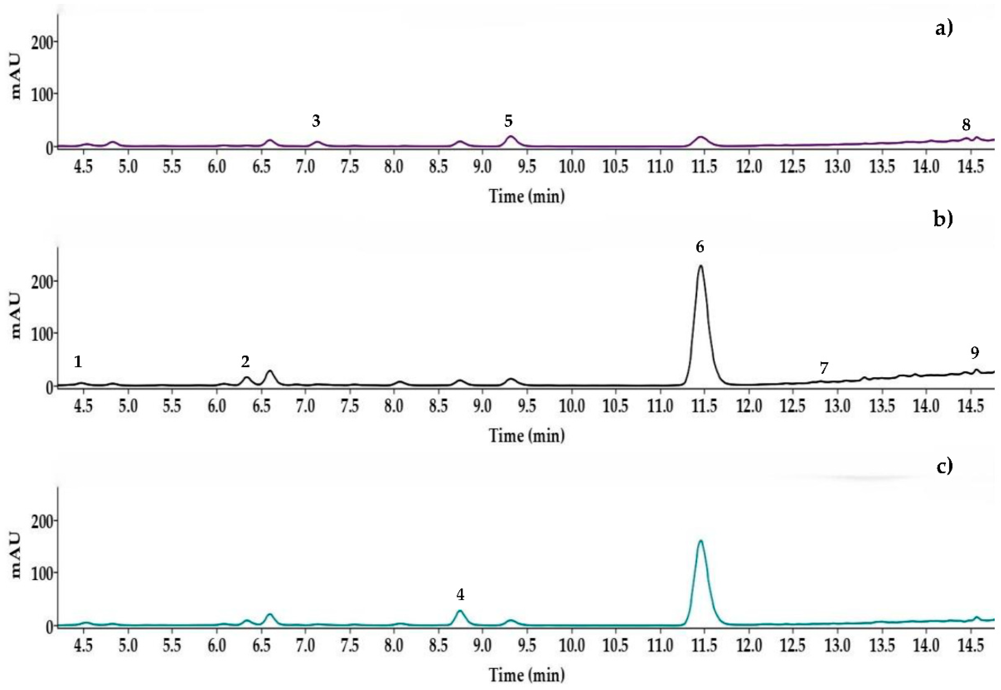
Vanilla planifolia Jacks. ex Andrews pods can lose up to 50% of their vanillin during curing. One explanation for this is the transformation of vanillin due to enzyme action, such as peroxidases, which generate the formation of dimers like divanillin. Therefore, in this work, a simultaneous high-performance liquid chromatography method (HPLC-DAD) was developed and validated for the separation and quantification of the main compounds present in Vanilla planifolia Jacks. ex Andrews and divanillin. The separation of 9 compounds of interest was achieved within 15 minutes using a Zorbax Eclipse XDB-C18 column (250 mm x 4.6 mm i.d., 5 μm particle size). The variables optimized included the mobile phase (water as solvent A, methanol as solvent B, and acidified water, 10–2 M H3PO4, as solvent C), the separation gradient, and the column tempera-ture (40-60 °C). The maximum divanillin content was 0.02 g/100 g d.w. in a sample from Papantla de Olarte. Chromatographic performance evaluation revealed excellent resolution, retention factor, and selectivity. The method was successfully validated in terms of limits of detection and quantification, linearity, and precision, as well as its application to cured pods, with evidence of divanillin presence in all analyzed sample.

Keywords: 
;  ;  ;  ;  ;  

1. Introduction

The perennial vine, Vanilla planifolia Jacks. ex Andrews, is characterized by its substantial, green, succulent stem. To develop the characteristic aroma of vanilla, mature green fruits must undergo a curing process consisting of four stages: wilting, sweating, drying, and conditioning [1,2,3,4,5]. During this process, glycosylated precursors, such as glucovanillin, are enzymatically hydrolyzed to their free forms [3,6,7,8]. Over a period of 120 to 180 days, a complex sequence of enzymatic, chemical, and biochemical reactions occurs during the curing of vanilla. The fruits, which initially have a high moisture content (80%), transform into pods with a low moisture content (20-30%). This process prevents the proliferation of microorganisms, especially molds, resulting in highly aromatic pods with dark brown shades [9,10,11]. Various enzymes, including glucosidases, synthases, oxidases, proteases, methyltransferases and phenylalanine ammonia-lyase, are involved in the biogenesis of the vanilla aroma. The involvement of the enzyme β-glucosidase in the formation of the vanilla aroma has been extensively researched [3,6,7,8]. The activities of oxidative enzymes, notably peroxidases and polyphenol oxidase, are closely monitored during the growth and maturation of vanilla pods, though the precise reactions contributing to flavor development are not fully understood [11,12,13]. Specifically, peroxidases participate in the oxidation of vanillin to generate dimers, with divanillin being the most dominant [14,15,16]. However, various authors have reported that during the curing process of vanilla, there is a reduction in the vanillin potential of up to 50% [2,3,9]. To explain the decrease in vanillin content during the curing process, different hypotheses have been proposed. One hypothesis suggests that due to the aldehyde group of vanillin, it may participate in Maillard reactions by reacting with free amino groups present in the pods [17]. However, these studies do not account for the high percentage of reduction, which, as previously mentioned, can be up to 50% [2,3,9]. The most probable explanation is the transformation of vanillin by oxidative enzymes such as peroxidases, leading to the formation of dimers like divanillin [14,15,16,18] (Figure 1). The presence of this compound has been described due to its generation from vanillin and the action of peroxidases present in the milk of various dairy products such as ice cream, creams, smoothies, among others [19].
Divanillin is approved for use in food (FEMA GRAS #4107) in USA [20], is used as a flavor enhancer in dairy products, as it imparts a pleasant and creamy sensation to foods when added at concentrations of 5-50 mg/L [21,22]. Sensory studies revealed human recognition thresholds for the velvety mouth-coating sensation between 1.0 μmol/kg for divainillin [23]. In addition to being an excellent antioxidant, it has demonstrated antitumor capabilities [24,25]. Moreover, there are various patents describing the production of this compound for commercial purposes from vanillin, lignin, and other sources [21]. The presence of divanillin has been identified in commercial extracts from various countries such as Madagascar, Mexico, and Tahiti in vanilla beans [14,21]. However, there is no validated method described in the literature for its quantification both individually and simultaneously with the other compounds present in Vanilla planifolia Jacks. ex Andrews.
Although vanillin represents 85% of the volatile compounds in Vanilla planifolia Jacks. ex Andrews, the exquisite natural aroma of vanilla is highly complex and attributed to a diverse mixture of different volatile compounds, not solely to the presence of vanillin [26,27,28]. Among the compounds with higher concentrations are vanillin, vanillic acid, p-hydroxybenzaldehyde and p-hydroxybenzoic acid [3,26,29,30,31,32] (Figure 1). For the identification and analysis of these compounds, several instrumental methods have been used, being high performance liquid chromatography coupled to ultraviolet-visible (HPLC-UV-Vis or HPLC-DAD) the most used. These techniques are specifically described for Vanilla planifolia Jacks. ex Andrews pods in the standards NF-ISO-5565-2-1999; NMX-FF-074-SCFI-2009, and NOM-182-SCFI-2011 [33,34,35], following Soxhlet extraction or other extraction methods [29,30,31,32]. Alternative techniques employed for analyzing these compounds include high-performance liquid chromatography coupled with mass spectrometry (HPLC-MS) [32,36], gas chromatography coupled with mass spectrometry (GC-MS) [14,26,37,38] and nuclear magnetic resonance (NMR) [14,15,36]. Therefore, the objective of the present study is to develop and validate a reliable HPLC-DAD method that allows for the simultaneous separation and quantification not only of the main aromatic compounds but also of the divanillin dimer. Additionally, various samples of cured pods were selected to demonstrate the applicability of the method.

2. Materials and Methods

2.1. Reagents and Solvents

The standards p-hydroxybenzyl alcohol (1), vanillyl alcohol (2), p-hydroxybenzaldehyde (3), p-hydroxybenzoic acid (4), vanillic acid (5), vanillin (6), anisyl alcohol (7), and anisic acid (8) were purchased from Sigma-Aldrich Chemical Co (St Louis, MO, USA), while divainillin (9) was acquired from BOC Sciences (Shirley, NY, USA). Water and methanol (HPLC grade) were purchased from Tedia (Fairfield, OH, USA), dimethyl sulfoxide (DMSO), and phosphoric acid (H3PO4, HPLC grade) were purchased from Sigma-Aldrich Chemical Co.

2.2. Preparation of Standard Solutions

The stock solutions were prepared using a 1:1 mixture of DMSO:(MeOH/Water acidified (100 mM, H3PO4) with a 3:7 ratio) employing different concentrations (0.5, 1, 2, 10, 25, 50, 100 mg/L) of each compound. Calibration curves were obtained for each compound of interest. Regarding the development of the HPLC method and its validation, a single solution containing all 9 compounds of interest was used.

2.3. Raw Material

Seven batches of cured Vanilla planifolia Jacks. ex Andrews pods from the 2020-2021 harvest were analyzed, sourced from different regions of the state of Veracruz (Mexico) at varying altitudes. Among these, three batches were from the Totonacapan region. The origin locations of the cured pods were: Tecolutla (VTEC, 20 masl), Papantla de Olarte (VPAP, 80 masl), Misantla (VMST, 300 masl), Zongolica (VZGL, 1200 masl), and Atzalan (VATZ, 1676 masl). These samples were used for method validation.

2.4. Sample Preparation

The extractions were performed following the method established by Pérez Silva et al. [3] with some modifications. The cured vanilla pods were ground using a Chefman MEC-5256U mill. 300 mg of the sample was weighed and then, 20 mL of a 1:1 mixture of DMSO:(MeOH/acidified water (3:7) (100 mM, H3PO4)) was added. The sample was immersed in an Elmasonic P D78224 ultrasonic bath (Elma Schmidbauer GmbH, Singen, Germany) for 10 min at 37 kHz and room temperature. The supernatant was filtered using a No. 4 filter paper and and stored at 4 °C until further use.

2.5. Instrument and Development of Chromatographic Conditions

The samples were analyzed using an Agilent model 1260 Infinity II apparatus (Agilent Technologies, Santa Clara, CA, USA), equipped with a photodiode array detector (G7117C), multicolumn thermostat (G7116A), quaternary pump (G7111B), and an autosampler (G7129A) controlled by OpenLAB CDS software (Agilent Technologies, Version 2.5). The photodiode array detector (DAD) was set to three wavelengths for detecting the compounds of interest at 230 nm (vanillyl alcohol, p-hydroxybenzyl alcohol, vanillin and anisyl alcohol), 254 nm (vanillic acid, p-hydroxybenzoic acid, anisic acid and divanillin), and 280 nm (p-hydroxybenzaldehyde). A Zorbax Eclipse XDB-C18 column (Agilent Technologies, 250 mm in length, 4.6 mm internal diameter, and 5 µm particle size) was used.
A stock solution containing the 9 compounds of interest was the sample used for all chromatographic tests, which consisted of a solvent gradient of solvent A (HPLC-grade water), solvent B (HPLC-grade methanol), and solvent C (acidified water, 10–2 M H3PO4), temperatures (40-60 °C), and flow rates (1.5–2.25 mL/min). The criteria for choosing the most effective chromatographic separation were determined by evaluating key chromatographic properties: retention time (tR), resolution (Rs; Equation 1), retention factor (k; Equation 2), and selectivity (α; Equation 3). The calculation of these chromatographic properties were performed using the equations outlined below:
R s = 2 ( t R ( B ) t R ( A ) ) W b ( A ) W b ( B )
k = t ' R t M
α = t ' R ( B ) t ' R ( A ) = K ( B ) K ( A )
Wb(A) and Wb(B) denote the widths at the base of the adjacent peaks A and B, tR(A) and tR(B) denote the retention times of these adjacent peaks, tM stands for the dead time of the column and t'R is the adjusted retention time (calculated as t'R = tR-tM). These parameters were determined using OpenLAB Data Analysis software (Agilent Technologies, Version 2.5).

2.6. Validation of the Chromatographic Method

The chromatographic method was validated according to ICH Guideline Q2 (R1). Validation included appropriate detection, identification, and quantification of 9 compounds of interest. Evaluated parameters included: linearity, limit of detection (LOD) and quantification (LOQ), precision and robustness.

2.6.1. Linearity

Linearity was assessed by constructing calibration curves for each compound of interest based on their standard solutions. Different concentrations (0.5, 1, 2, 10, 25, 50, 100 mg/L) of each standard were used. The coefficients of determination (R2) were calculated to indicate the degree of linear correlation. This specific parameter is crucial for confirming the linear correlation between the obtained results and the actual concentration of each compound within the analyzed range.

2.6.2. Detection and Quantification Limits

LOD and LOQ have been determined based on the ICH Q2 guideline. Specific calibration curves have been developed with concentrations close to the LOD and LOQ ranges. LOD and LOQ have been validated with samples with concentrations of the compounds of interest close to the LOD and LOQ limits. In this way, concentrations of 0.1, 0.5, 1, 2, 5, and 10 mg/L of each of the compounds of interest have been employed to obtain the limits of detection (LOD; Equation 4) and quantification (LOQ; Equation 5), using the standard deviation of the response and the regression slope.
L O D = 3 σ S
L O Q = 10 σ S
where, σ denotes the standard deviation of the blank response, while S refers to the slope of the calibration curve. All analyses were performed in duplicate, and calculations were carried out using Microsoft Office Excel 365 (Version 2401, Microsoft, Redmond, WA, USA).

2.6.3. Precision

The precision of the method was assessed by examining repeatability and intermediate precision concerning the following chromatographic properties: retention time, peak area, peak width, peak height, and resolution for the 9 compounds of interest. Thirty independent HPLC analyses were conducted over three consecutive days, with 10 analyses performed each day. Intermediate precision was evaluated by computing the coefficient of variation (CV) for each parameter using data from 30 tests. Repeatability was assessed by calculating the average CV from analyses conducted on the same day. According to the AOAC Manual for the Peer-Verified Methods Program (AOAC, 2012), a CV below 10% was used as a reference to confirm the method's precision.

2.6.4. Robustness

Robustness refers to a method's ability to remain unaffected by small, intentional variations in its parameters. The robustness of the method was assessed by introducing a ±5% variation in column temperature, flow rate, and injection volume. For each parameter, three different levels were tested, with four repetitions at each level.
To evaluate the impact of these variations on the chromatographic parameters (retention time, peak area and peak resolution) an analysis of variance and a Tukey test were performed, assuming a significance level of 0.05. This procedure was conducted using Minitab version 17.1.0 (Minitab, Inc., State College, PA, The Plains, VA, USA).

3. Results and Discussion

3.1. Development of the HPLC Method

For the development and validation of the HPLC-DAD method, a mixture of 9 standards corresponding to the 9 compounds studied in this work was prepared. The criterion for selecting the most effective chromatographic separation was achieved through optimal chromatographic conditions. The evaluated properties included retention time (tR), selectivity (α), retention factor (k), and resolution (Rs). Most studies set a minimum target resolution of greater than 1.5 [39]; resolution of 1 indicates a 4% overlap between adjacent peaks, while a resolution of 1.5 reduces this overlap to 0.3%. Any value below 1 signifies poor separation. The retention factor (k) is generally sought to be between 1 and 10, though it can sometimes be extended to a range of 0.5 to 20.65 [40]. Selectivity is typically targeted to be between 1 and 2 [41]. Various trial and error experiments were conducted to find the optimal chromatographic conditions that yield well-defined peaks with moderate separation times and column pressure.

3.1.1. Optimization of the Separation Gradient

The gradient was optimized by several trial-and-error experiments, involving multiple chromatographic separations by varying the percentages of solvent A to solvent B (90-35%). The first gradient tested showed difficulties in the separation of of p-hydroxybenzaldehyde and vanillic acid. These compounds exhibit nearly identical retention times, despite differing in their UV-Vis spectra (280 and 254 nm respectively).
Finally, the best separation of the compounds of interest was achieved with the following separation gradient: 0 min, 9% B; 2 min 9% B; 7 min 19% B; 10 min 19% B; 15 min 80% B; 18 min 80% B; 23 min 9% B; 29 min 9% B; solvent C remains constant (10%) during the whole chromatographic separation.

3.1.2. Study of the Effect of Column Temperature

The next step in the development of the method involved determining the effect of column temperature on the separation of compounds, specifically analyzing the influence of temperature on retention time and peak resolution. Various separations were performed with the column at the following temperatures: 40, 45, 50, 55, and 60 °C.
A trend was observed where higher column temperatures led to some compounds beginning to overlap. Notably, starting at 50 °C, vanillyl alcohol and p-hydroxybenzoic acid did not separate successfully, exhibiting overlapping peaks. Therefore, the final temperature selected for the development of the chromatographic method was 45 °C.

3.1.3. Study of the Effect of Flow Rate

After determining the optimal column operating temperature, tests were conducted at various flow rates (1.5, 1.75, 2.0, and 2.25 mL/min) to achieve the best separation of the compounds of interest. The flow rate that provided the best separation for p-hydroxybenzaldehyde and vanillic acid was 2.25 mL/min. Higher flow rates were not used because the pressure generated compromised the integrity of the column.

3.2. Characteristics of the Developed Method

Finally, the optimal gradient for the HPLC-DAD method developed in this study is as follows: 0 min, 9% B; 2 min 9% B; 7 min 19% B; 10 min 19% B; 15 min 80% B; 18 min 80% B; 23 min 9 % B; 29 min 9% B; solvent C remains constant at 10% throughout the chromatographic separation. The column temperature is set to 45 °C, and the flow rate is 2.25 mL/min. Separation of the 9 studied compounds was achieved in less than 15 minutes, with a total analysis time of 29 minutes, including re-equilibration to initial conditions. The chromatogram obtained under optimal conditions is shown in Figure 2.
The chromatographic properties of the developed method are described in Table 1. The results indicate that the compounds are correctly separated with appropriate values for peak width, retention factor, selectivity, and resolution, despite the challenges in separating these compounds. The method validation was carried out in accordance with the ICH Guideline Q2 (R1) [42], evaluating linearity, precision, detection limits, and quantification limits.
Several studies report the quantification of vanillin by HPLC [33,34,35], but very few validated these methods for the quantification of vanillin [43,44], along with other volatile phenols in Vanilla planifolia Jacks. ex Andrews [45,46,47] and anisic compounds in Vanilla pompona ssp. grandiflora (Lindl.) Soto-Arenas [48]. However, in this proposed method, the quantification of a dimer (divanillin) for the first time, and 8 volatile phenols in Vanilla planifolia Jacks. ex Andrews was achieved, improving the retention times of all the compounds reported in the literature [44,45,46,47,48], except for the tR of 9.90 min [43], where only vanillin was determined (Table 2). To date, no validated method for the quantification of divanillin and other phenols in cured vanilla beans and alcoholic vanilla extracts has been reported in the literature.

3.2.1. Linearity

The linearity of the developed chromatographic method was confirmed by the coefficients of determination (R²) of the calibration curves obtained for the studied compounds. Good linearity was observed for all 9 compounds of interest within the studied range (Table 2), as all compounds exhibited R² values of 0.9996 or higher.

3.2.2. Detection and Quantification Limits

The limit of quantification (LOQ) and the limit of detection (LOD) (Table 3) for the analyzed compounds were calculated by multiplying the standard deviation of the blank by 3 and 10, respectively, and dividing by the regression slope of each compound, as described in the Materials and Methods section. These values fall within the parts per million (ppm) range, indicating the ability to detect very small quantities of the analyte.

3.2.3. Precision

Retention time, peak area, and resolution were assessed to evaluate the precision of the method (repeatability and intermediate precision), expressed in terms of coefficient of variation (CV). All CV values were below 6% for all peaks and for all evaluated chromatographic properties, as shown in Table 4. The precision obtained was below the acceptable limits defined by AOAC, i.e., ±10% according to the AOAC manual for the Verified Pairwise Methods Program [49]. Therefore, it can be confirmed that the developed method is precise, with retention time being the parameter showing the highest precision.

3.2.4. Robustness

The robustness of a method refers to its capacity to remain unaffected by minor intentional variations in its variables. In this study, robustness was evaluated by applying a ±5% variation in column temperature, flow rate, and injection volume. The impact of these changes on three properties—retention time, peak area, and chromatographic resolution—was assessed. Statistical comparisons were performed using Tukey’s test, with the results presented in Table 5. Different letters in the same row for each parameter indicate statistically significant differences (p < 0.05)."
The developed method demonstrated full robustness in terms of retention times and resolution when the injection volume was varied between 9.5 and 10.5 μL. The peak area was not considered as it is dependent on the injected volume. Accurate control of the sample injection volume is therefore crucial. However, variations in column temperature and flow rate resulted in significant differences (p < 0.05) in retention times and peak areas, indicating that the method is highly sensitive to changes in these parameters. Consequently, strict control of these variables is required, and the method documentation should include a precautionary note. Nevertheless, with proper tuning and equipment calibration, the method has demonstrated its capacity for precise and well-defined chromatographic peak separation [50].

3.3. Application to Real Samples

Once the optimal method for analyzing the compounds of interest present in vanilla was obtained, an additional study was conducted to quantify the compounds in Vanilla planifolia Jacks. ex Andrews pods from Mexico, specifically from the 2020-2021 harvest in the state of Veracruz. The analysis results are summarized in Table 6, as well as the chromatogram of a sample is shown in Figure 3, where the analyzed compounds in a vanilla sample can be identified, along with the specific wavelengths at which each compound absorbs. In summary, of the 9 compounds identified in the Mexican vanilla pods, 7 are phenols (p-hydroxybenzyl alcohol, vanillyl alcohol, p-hydroxybenzoic acid, p-hydroxybenzaldehyde, vanillic acid, vanillin, and divanillin), 1 alcohol (anisyl alcohol), and 1 aromatic acid (anisic acid) [26]. The compounds identified in all samples were vanillyl alcohol, p-hydroxybenzoic acid, p-hydroxybenzaldehyde, vanillic acid, vanillin, and divanillin. Notably, anisic acid and anisyl alcohol were detected and quantified in all samples, except anisic acid in VTEC. Both compounds have been previously identified and quantified in Vanilla planifolia Jacks. ex Andrews using GC-MS [26,37,51], as they are not major compounds like in cured pods of Vanilla xtahitensis J.W. Moore and Vanilla pompona (ssp. grandiflora (Lindl.) Soto-Arenas and Schiede) [48,52,53,54,55], which can be quantified using HPLC-UV-Vis. The major characteristic compounds of Vanilla planifolia (p-hydroxybenzoic acid, p-hydroxybenzaldehyde, vanillic acid, and vanillin), as documented in previous studies [26,29,56,57], were identified and quantified in this work, with VPAP02G showing the highest vanillin content (3.57 g/100 g d.w.). The presence of divanillin in commercial alcoholic extracts from Tahiti, Madagascar, and Mexico has been previously described [14], as well as in dairy products, due to the oxidation of vanillin by peroxidase enzymes present in milk [13,19,58]. Additionally, the natural generation of the dimer is significant due to its effect on food products; divanillin has been reported to enhance sensory properties at low concentrations (5-50 mg L–1) in dairy products [14], influencing not only the fatty and creamy flavor properties but also significantly improving fullness and mouthfeel [13,14]. The sample with the highest divanillin content was VPAP05 (0.02 g/100 g d.w.), followed by VAZN03 (0.01 g/100 g d.w.), while the other 5 samples, although from different localities at different altitudes, showed no significant difference in divanillin content.
These results indicate that the proposed HPLC method is applicable for analyzing compounds present in vanilla. This is of great interest because it allows for the quantification of both major and minor compounds, as well as the dimer, with high precision.

4. Conclusions

A rapid and reproducible HPLC-DAD method was developed for the separation and quantification of the 9 major aromatic compounds present in vanilla with an analysis time of 15 min, achieving high precision (CV < 6%). This results in a major enhancement and a considerable decrease in the time needed for analysis. The optimal chromatographic conditions were: column temperature of 45 °C, solvent flow rate of 2.25 mL min–1 and 10 µL injection volume in addition to excellent validation results (linearity, sensitivity, LOD and LOQ). The method was effectively applied to various batches of cured Vanilla planifolia pods, allowing the analysis and quantification of the most important compounds. Furthermore, the proposed HPLC-DAD method offers significant advantages as a practical technique for the quantification of the main aromatic compounds, including a dimer present in cured Vanilla planifolia pods as well as analysis in a shorter time. It can also be applied to the analysis of other species in the Vanilla genus, where anisic compounds are of greater importance, such as in Vanilla x tahitensis J.W. Moore and Vanilla pompona (ssp. grandiflora (Lindl.) Soto-Arenas and Schiede), making the developed method potentially useful as a fundamental reference.

Author Contributions

Conceptualization, G.F.B. and A.P.-S; methodology, Y.E.T.-Z., G.F.B. and A.P.-S.; software, Y.E.T.-Z.; validation, G.F.B., A.P.-S. and G.d.C.R.-J.; formal analysis, Y.E.T.-Z.; investigation, G.F.B., A.P.-S. and G.d.C.R.-J.; resources, A.P.-S.; data curation, Y.E.T.-Z., G.d.C.R.-J. and G.F.B.; writing—original draft preparation, Y.E.T.-Z.; writing—review and editing, A.P.-S., G.d.C.R.-J. and G.F.B.; visualization, G.F.B.; supervision, G.F.B., A.P.-S. and G.d.C.R.-J.; project administration, A.P.-S.; funding acquisition, A.P.-S. and G.F.B. All authors have read and agreed to the published version of the manuscript.

Funding

This research was funded by Consejo Nacional de Humanidades, Ciencia y Tecnología (CONAHCyT), project “297484” (Strategies for adaptation and mitigation to climate change necessary for the rescue of vanilla cultivation in Mexico). The authors express their gratitude to CONAHCyT for the scholarship provided to Yunuen Erandy Torres Zamudio (No. CVU: 784608).

Data Availability Statement

The original contributions presented in the study are included in the article/supplementary material, further inquiries can be directed to the corresponding author/s.

Acknowledgments

We would like to thank TecNM Campus Tuxtepec for providing the facilities and equipment, as well as the University of Cadiz (UCA) and TecNM Campus Veracruz for their support.

Conflicts of Interest

The authors declare no conflicts of interest.

References

  1. Odoux, E. Changes in Vanillin and Glucovanillin Concentrations during the Various Stages of the Process Traditionally Used for Curing Vanilla fragrans Beans in Reunion. Fruits 2000, 55, 119–125. [Google Scholar] [CrossRef]
  2. Dunphy, P.; Bala, K. Vanilla Curing. Perfum. flavorist 2009, 34, 34–40. [Google Scholar]
  3. Pérez Silva, A.; Gunata, Z.; Lepoutre, J.P.; Odoux, E. New Insight on the Genesis and Fate of Odor-Active Compounds in Vanilla Beans (Vanilla Planifolia G. Jackson ) during Traditional Curing. Food Res. Int. 2011, 44, 2930–2937. [Google Scholar] [CrossRef]
  4. Januszewska, R.; Giret, E.; Clement, F.; Van Leuven, I.; Goncalves, C.; Vladislavleva, E.; Pradal, P.; Nåbo, R.; Landuyt, A.; D’Heer, G.; et al. Impact of Vanilla Origins on Sensory Characteristics of Chocolate. Food Res. Int. 2020, 137, 109313. [Google Scholar] [CrossRef]
  5. Singletary, K.W. Vanilla: Potential Health Benefits. Nutr. Today 2020, 55, 186–196. [Google Scholar] [CrossRef]
  6. Odoux, E.; Escoute, J.; Verdeil, J.L. The Relation between Glucovanillin, β-D-Glucosidase Activity and Cellular Compartmentation during the Senescence, Freezing and Traditional Curing of Vanilla Beans. Ann. Appl. Biol. 2006, 149, 43–52. [Google Scholar] [CrossRef]
  7. Brillouet, J.M.; Odoux, E.; Conejero, G. A Set of Data on Green, Ripening and Senescent Vanilla Pod (Vanilla Planifolia ; Orchidaceae): Anatomy, Enzymes, Phenolics and Lipids. Fruits 2010, 65, 221–235. [Google Scholar] [CrossRef]
  8. Khoyratty, S.; Kodja, H.; Verpoorte, R. Vanilla Flavor Production Methods: A Review. Ind. Crops Prod. 2018, 125, 433–442. [Google Scholar] [CrossRef]
  9. Odoux, E. Glucosylated Aroma Precursors and Glucosidase(s) in Vanilla Bean (Vanilla Planifolia G. Jackson). Fruits 2006, 60, 171–183. [Google Scholar] [CrossRef]
  10. Dunphy, P.; Bala, K. Review: A Flavor of Vanilla. Perfum. flavorist 2010, 35, 42–42. [Google Scholar]
  11. Buitimea-Cantúa, G. V.; Chávez-Leal, V.; Soto-Caballero, M.C.; Tellez-Medina, D.I.; Welti-Chanes, J.; Escobedo-Avellaneda, Z. Enzymatic Activity and Its Relationships with the Total Phenolic Content and Color Change in the High Hydrostatic Pressure-Assisted Curing of Vanilla Bean (Vanilla Planifolia). Molecules 2023, 28. [Google Scholar] [CrossRef] [PubMed]
  12. Ranadive, A. Vanilla- inside Look: Chemistry and Biochemistry of Vanilla Flavor. Perfum. Flavourist 2006, 31, 38–44. [Google Scholar]
  13. Pardío, V.T.; Flores, A.; López, K.M.; Martínez, D.I.; Márquez, O.; Waliszewski, K.N. Effect of Endogenous and Exogenous Enzymatic Treatment of Green Vanilla Beans on Extraction of Vanillin and Main Aromatic Compounds. J. Food Sci. Technol. 2018, 55, 2059–2067. [Google Scholar] [CrossRef] [PubMed]
  14. Gatfield, I.; Rei, I.; Krammer, G.; Schmidt, C.O.; Kindel, G.; Bertram, H. Divainillin Novel Taste-Active Component during the Traditional Curing Process. Perfum. flavorist 2006, 31, 18–20. [Google Scholar]
  15. Hernández-Ramos, D. Estudio de La Degradación Química de La Vainillina. Tesis de Maestría en Ciencias en Alimentos. Instituto Tecnológico de Tuxtepec. Oaxaca, México. 2009.
  16. Peña-Mojica, E. Estudio Del Efecto Enzimático, Térmico y Fotoquímico En La Transformación de La Vainilla, Tesis de Maestría en Ciencias en Alimentos. Instituto Tecnológico Tuxtepec. Oaxaca, México., 2019.
  17. Frenkel, C.; Havkin-Frenkel, D. Material Properties: The Physics and Chemistry of Vanillin. Perfum. Flavourist 2006, 31, 28–36. [Google Scholar]
  18. Li, Z.; Jiang, Y.; Wust, K.; Callari, M.; Stenzel, M.H. Crosslinking of Self-Assembled Protein-Polymer Conjugates with Divanillin. Aust. J. Chem. 2020, 73, 1034–1041. [Google Scholar] [CrossRef]
  19. Anklam, E.; Gaglione, S.; Müller, A. Oxidation Behaviour of Vanillin in Dairy Products. Food Chem. 1997, 60, 43–51. [Google Scholar] [CrossRef]
  20. Smith, R.L.; Cohen, S.M.; Doull, J.; Feron, V.J.; Goodman, J.I.; Marnett, L.J.; Portoghese, P.S.; Waddell, W.J.; Wagner, B.M.; Adams, T.B. GRAS Flavoring Substances 22. Food Technol. 2005, 59, 24–62. [Google Scholar]
  21. Reiss, I.; Gatfield, I.-L.; Krammer, G.; Clerc, A.; Kindel, G. Use of Divanillin as a Flavouring Agent (U.S. Patent Application No. 10/547,981.) 2006.
  22. Krings, U.; Esparan, V.; Berger, R.G. The Taste Enhancer Divanillin: A Review on Sources and Enzymatic Generation. Flavour Fragr. J. 2015, 30, 362–365. [Google Scholar] [CrossRef]
  23. Schwarz, B.; Hofmann, T. Identification of Novel Orosensory Active Molecules in Cured Vanilla Beans (Vanilla Planifolia ). J. Agric. Food Chem. 2009, 57, 3729–3737. [Google Scholar] [CrossRef]
  24. Oliveira, G.S.N. de; Furlaneto, C.G.; Tokuhara, C.K.; Ventura, T.M.O.; Pessôa, A. de S.; Fakhoury, V.S.; Pagnan, A.L.; Inacio, K.K.; Rovis Sanches, M.L.; Buzalaf, M.; et al. Antitumor Effect of Vanillin and Divanillin on Murine Osteosarcoma Cells. SSRN Electron. J. 2022. [Google Scholar] [CrossRef]
  25. Jantaree, P.; Lirdprapamongkol, K.; Kaewsri, W.; Thongsornkleeb, C.; Choowongkomon, K.; Atjanasuppat, K.; Ruchirawat, S.; Svasti, J. Homodimers of Vanillin and Apocynin Decrease the Metastatic Potential of Human Cancer Cells by Inhibiting the FAK/PI3K/Akt Signaling Pathway. J. Agric. Food Chem. 2017, 65, 2299–2306. [Google Scholar] [CrossRef] [PubMed]
  26. Pérez-Silva, A.; Odoux, E.; Brat, P.; Ribeyre, F.; Rodriguez-Jimenes, G.; Robles-Olvera, V.; García-Alvarado, M.A.; Günata, Z. GC-MS and GC-Olfactometry Analysis of Aroma Compounds in a Representative Organic Aroma Extract from Cured Vanilla (Vanilla Planifolia G. Jackson) Beans. Food Chem. 2006, 99, 728–735. [Google Scholar] [CrossRef]
  27. Brunschwig, C.; Collard, F.X.; Bianchini, J. P. Raharivelomanana, P. Evaluation of Chemical Variability of Cured Vanilla Beans (Vanilla Tahitensis and Vanilla Planifolia). Nat. Prod. Commun. 2009, 4, 1393–1400. [Google Scholar] [CrossRef]
  28. Peña-Barrientos, A.; Perea-Flores, M. de J.; Martínez-Gutiérrez, H.; Patrón-Soberano, O.A.; González-Jiménez, F.E.; Vega-Cuellar, M.; Dávila-Ortiz, G. Physicochemical, Microbiological, and Structural Relationship of Vanilla Beans (Vanilla Planifolia Andrews) during Traditional Curing Process and Use of Its Waste. J. Appl. Res. Med. Aromat. Plants 2023, 32. [Google Scholar] [CrossRef]
  29. Gassenmeier, K.; Riesen, B.; Magyar, B.; Barzalona, M.; Casanova, J. Commercial Quality and Analytical Parameters of Cured Vanilla Beans (Vanilla Planifolia) from Different Origins from the 2006--2007 Crop. Flavour Fragr. J. 2008, 23, 194–201. [Google Scholar] [CrossRef]
  30. Zhang, S.; Mueller, C. Comparative Analysis of Volatiles in Traditionally Cured Bourbon and Ugandan Vanilla Bean (Vanilla Planifolia) Extracts. J. Agric. Food Chem. 2012, 60, 10433–10444. [Google Scholar] [CrossRef]
  31. Pérez-Silva, A.; Nicolás-García, M.; Petit, T.; Dijoux, J.B.; Vivar-Vera, M. de los Á.; Besse, P.; Grisoni, M.; de los Ángeles Vivar-Vera, M.; Besse, P.; Grisoni, M. Quantification of the Aromatic Potential of Ripe Fruit of Vanilla Planifolia (Orchidaceae) and Several of Its Closely and Distantly Related Species and Hybrids. Eur. Food Res. Technol. 2021, 247, 1489–1499. [Google Scholar] [CrossRef]
  32. Da Silva Oliveira, J.P.; Garrett, R.; Bello Koblitz, M.G.; Furtado Macedo, A. Vanilla Flavor: Species from the Atlantic Forest as Natural Alternatives. Food Chem. 2021, 375, 131891. [Google Scholar] [CrossRef]
  33. NF-ISO-5565-2. Vanille[Vanilla Fragans (Salisbury) Ames]— Partie 2:Méthodes d’essai. 1999.
  34. NMX-FF-074-SCFI-2009. Norma Mexicana “Productos No Industrializados Para Uso Humano-Vainilla (Vanilla fragrans (Salisbury) Ames) Especificaciones y Métodos de Prueba” 2009.
  35. NOM-182-SCFI-2011. Norma Oficial Mexicana, Vainilla de Papantla, Extractos y Derivados. Especificaciones, Información Comercial y Métodos de Ensayo (Prueba). 2011.
  36. De Jager, L.S.; Perfetti, G.A.; Diachenko, G.W. Comparison of Headspace-SPME-GC-MS and LC-MS for the Detection and Quantification of Coumarin, Vanillin, and Ethyl Vanillin in Vanilla Extract Products. Food Chem. 2008, 107, 1701–1709. [Google Scholar] [CrossRef]
  37. Sabik, H.; Pérez-Silva, A.; Bélanger, D.; Vivar-Vera, M.D.L.Á.; Nicolás-García, M.; Reyes López, D. Identification of Volatile Compounds in Cured Mexican Vanilla (Vanilla Planifolia G. Jackson) Beans Using Headspace Solid-Phase Microextraction with Gas Chromatography-Mass Spectrometry. Fruits 2016, 71, 407–418. [Google Scholar] [CrossRef]
  38. Mourtzinos, I.; Konteles, S.; Kalogeropoulos, N.; Karathanos, V.T. Thermal Oxidation of Vanillin Affects Its Antioxidant and Antimicrobial Properties. Food Chem. 2009, 114, 791–797. [Google Scholar] [CrossRef]
  39. Zheng, J.; Polyakova, Y.; Row, K.H. Retention Factors and Resolutions of Amino Benzoic Acid Isomers with Some Lonic Liquids. Biotechnol. Bioprocess Eng. 2006, 11, 477–483. [Google Scholar] [CrossRef]
  40. Snyder, L.R.; Dolan, J.W. High-Performance Gradient Elution the Practical Application of the Linear-Solvent-Strength Model.; 1th ed.; John Wiley & Sons. New Jersey, USA, 2006. [CrossRef]
  41. Vázquez-Espinosa, M.; González-de-Peredo, A. V.; Espada-Bellido, E.; Ferreiro-González, M.; Barbero, G.F.; Palma, M. Simultaneous Determination by UHPLC-PDA of Major Capsaicinoids and Capsinoids Contents in Peppers. Food Chem. 2021, 356, 1–8. [Google Scholar] [CrossRef] [PubMed]
  42. Ich ICH Topic Q2 (R1) Validation of Analytical Procedures : Text and Methodology (Vol. 1994). Geneva. 2005.
  43. Sharma, A.; Verma, S.C.; Saxena, N.; Chadda, N.; Singh, N.P.; Sinha, A.K. Microwave- and Ultrasound-Assisted Extraction of Vanillin and Its Quantification by High-Performance Liquid Chromatography in Vanilla Planifolia. J. Sep. Sci. 2006, 29, 613–619. [Google Scholar] [CrossRef]
  44. Ferreira, M.; Schwanz, M.; Cosenza, G.; Henriques, A.; Soares, L.A.L. Analysis of Vanillin by TLC and HPLC-PDA in Herbal Material and Tincture from Vanilla Planifolia Jacks Ex. Andrews. Drug Anal. Res. 2018, 2, 1–7. [Google Scholar] [CrossRef]
  45. Sinha, A.K.; Verma, S.C.; Sharma, U.K. Development and Validationof an RP-HPLC Method for Quantitative Determination of Vanillin and Related Phenolic Compunds in Vanilla Planifolia. J. Sep. Sci. 2007, 30, 15–20. [Google Scholar] [CrossRef]
  46. Cicchetti, E.; Chaintreau, A. Quantitation of the Main Constituents of Vanilla by Reverse Phase HPLC and Ultra-High-Pressure-Liquid-Chromatography with UV Detection: Method Validation and Performance Comparison. J. Sep. Sci. 2009, 32, 3043–3052. [Google Scholar] [CrossRef]
  47. Takahashi, M.; Sakamaki, S.; Fujita, A. Simultaneous Analysis of Guaiacol and Vanillin in a Vanilla Extract by Using High-Performance Liquid Chromatography with Electrochemical Detection. Biosci. Biotechnol. Biochem. 2013, 77, 595–600. [Google Scholar] [CrossRef]
  48. Maruenda, H.; Vico, M.D.L.; Householder, J.E.; Janovec, J.P.; Cañari, C.; Naka, A.; Gonzalez, A.E. Exploration of Vanilla Pompona from the Peruvian Amazon as a Potential Source of Vanilla Essence: Quantification of Phenolics by HPLC-DAD. Food Chem. 2013, 138, 161–167. [Google Scholar] [CrossRef]
  49. AOAC AOAC Peer Verified Methods Program, Manual on Policies and Procedures. (AOAC Inter). Maryland (USA). 2012.
  50. Shabir, G.A. Validation of High-Performance Liquid Chromatography Methods for Pharmaceutical Analysis. J. Chromatogr. A 2003, 987, 57–66. [Google Scholar] [CrossRef]
  51. Adedeji, J.; Hartman, T.G.; Ho, C.T. Flavor Characterization of Different Varieties of Vanilla Beans. Perfum. Flavorist 1993, 18, 25–33. [Google Scholar]
  52. Ehlers, D.; Pfister, M. Compounds of Vanillons (Vanilla Pompona Schiede). J. Essent. Oil Res. 1997, 9, 427–431. [Google Scholar] [CrossRef]
  53. Brunschwig, C.; Senger-Emonnot, P.; Aubanel, M.L.; Pierrat, A.; George, G.; Rochard, S.; Raharivelomanana, P. Odor-Active Compounds of Tahitian Vanilla Flavor. Food Res. Int. 2012, 46, 148–157. [Google Scholar] [CrossRef]
  54. Brunschwig, C.; Rochard, S.; Pierrat, A.; Rouger, A.; Senger-Emonnot, P.; George, G.; Raharivelomanana, P. Volatile Composition and Sensory Properties of Vanilla ×tahitensis Bring New Insights for Vanilla Quality Control. J. Sci. Food Agric. 2015, 96, 848–858. [Google Scholar] [CrossRef] [PubMed]
  55. Tavares de Oliveira, R.; da Silva Oliveira, J.P.; Furtado Macedo, A. Vanilla beyond Vanilla Planifolia and Vanilla × tahitensis: Taxonomy and Historical Notes, Reproductive Biology, and Metabolites. Plants 2022, 11. [Google Scholar] [CrossRef]
  56. Ranadive, A.S. Vanilla-Cultivation, Curing, Chemistry, Technology and Commercial Products. In Spices, herbs and edible fungi; Charalambous, G., Ed.; Elsevier Science: Amsterdam Netherlands, 1994; pp. 517–576. ISBN 9780444817617. [Google Scholar]
  57. Saltron, F.; Langella, C.; Guerere, M. Évaluation de La Qualité de La Vanille Malgache. Ann. des Falsif. l’expertise Chim. Toxicol. 2002, 95, 79–105. [Google Scholar]
  58. Homayouni, A.; Javadi, M.; Ansari, F.; Pourjafar, H.; Jafarzadeh, M.; Barzegar, A. Advanced Methods in Ice Cream Analysis: A Review. Food Anal. Methods 2018, 11, 3224–3234. [Google Scholar] [CrossRef]
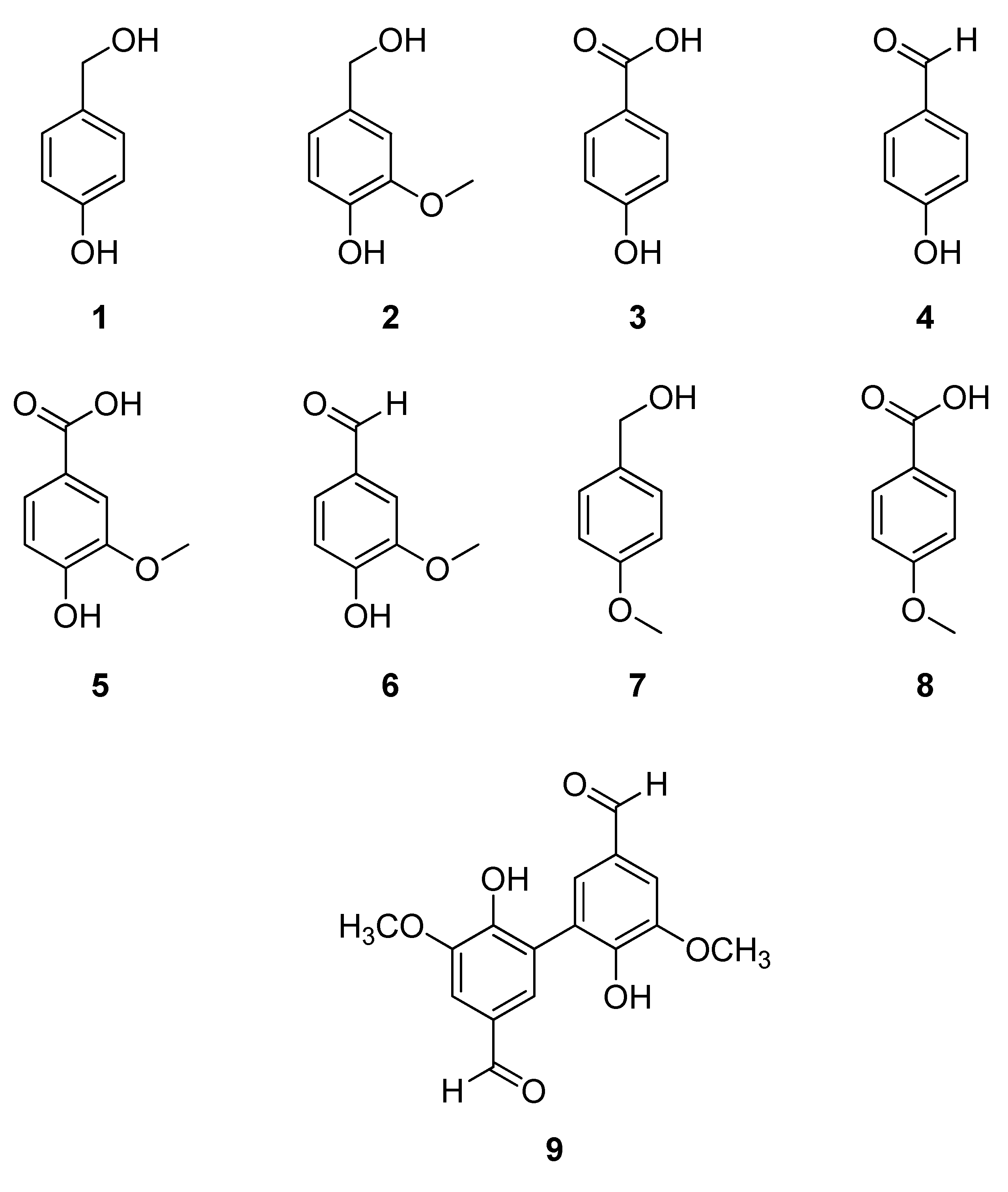
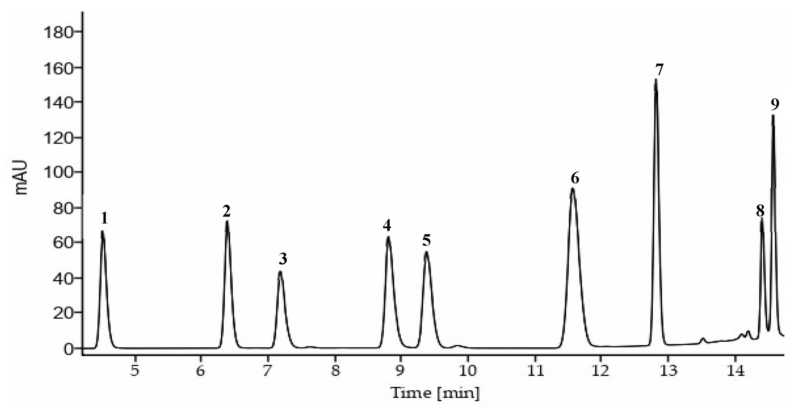
Figure 1. Structures of the compounds of interest: 1-p-hydroxybenzyl alcohol; 2-vanillyl alcohol; 3-p-hydroxybenzoic acid; 4-p-hydroxybenzaldehyde; 5-vanillic acid; 6-vanillin; 7-anisyl alcohol; 8-anisic acid and 9-divanillin.
Preprints 140611 g001
Figure 2. Chromatogram of the compounds: 1-p-hydroxybenzyl alcohol; 2-vanillyl alcohol; 3-p-hydroxybenzoic acid; 4-p-hydroxybenzaldehyde; 5-vanillic acid; 6-vanillin; 7-anisyl alcohol; 8-anisic acid and 9-divanillin, obtained under optimal conditions (λ = 230 nm).
Preprints 140611 g002
Figure 3. HPLC-DAD chromatogram of a sample of Vanilla planifolia Jacks. ex Andrews at a) 230 nm; 3-p-hydroxybenzoic acid, 5-vanillic and 8-anisic acid b) 254 nm; 1-p-hydroxybenzyl alcohol, 2-vanillyl alcohol, 6-vanillin; 7-anisyl alcohol and 9-divanillin and b) 280 nm; 4-p-hydroxybenzaldehyde obtained under optimal conditions.
Preprints 140611 g003
Table 1. Chromatographic characteristics of the method developed by HPLC-DAD.
# Compound Retention time (min) Width (seg) Retention factor (k) Selectivity (α) Resolution (Rs)
1 p-hydroxybencyl alcohol 4.45 0.27 3.86 1.53 6.51
2 Vanillyl alcohol 6.31 0.30 5.89 1.14 2.50
3 p-hydroxybenzoic acid 7.09 0.34 6.74 1.26 4.54
4 p-hydroxybenzaldehyde 8.71 0.37 8.50 1.07 1.49
5 Vanillinic acid 9.27 0.38 9.11 1.26 4.72
6 Vanillin 11.41 0.53 11.45 1.13 3.58
7 Anisyl alcohol 12.75 0.22 12.91 1.14 8.83
8 Anisic acid 14.38 0.15 14.69 1.01 1.08
9 Divanillin 14.54 0.17 14.87 - -
Table 2. Comparison of retention times in the literature with those obtained in this work.
# Compound Retention time (min)
This work [43] [45] [46] [47] [48] [44]
1 p-hydroxybencyl alcohol 4.45 - 5.9 - - 5.34 -
2 Vanillyl alcohol 6.31 - 8.05 - - 7.41 -
3 p-hydroxybenzoic acid 7.09 - 10.27 7.4 - 8.17 -
4 p-hydroxybenzaldehyde 8.71 - 12.33 9.9 - 9.93 -
5 Vanillinic acid 9.27 - 11.33 9.1 - 11.43 -
6 Vanillin 11.41 9.90 13.9 11.8 12.5 14.29 13.0
7 Anisyl alcohol 12.75 - - - - 15.10 -
8 Anisic acid 14.38 - - - - - -
9 Divanillin 14.54 - - - - - -
Table 3. Validation parameters of the HPLC method developed.
# Compound Calibration curve R2 LOD (mg/L) LOQ (mg/L)
1 p-hydroxybencyl alcohol y = 8.838x + 4.2881 0.9995 0.0387 0.1291
2 Vanillyl alcohol y = 9.6997x + 1.0834 0.9998 0.0371 0.1238
3 p-hydroxybenzoic acid y = 27.764x + 1.4178 1 0.0131 0.0436
4 p-hydroxybenzaldehyde y = 30.9288x - 1.4969 0.9999 0.0119 0.0397
5 Vanillinic acid y = 14.9978x + 0.3961 0.9999 0.0243 0.0810
6 Vanillin y = 24.3273x - 11.1028 0.9992 0.0161 0.0535
7 Anisyl alcohol y = 17.515x - 0.2164 1 0.0209 0.0698
8 Anisic acid y = 27.6438x + 2.1590 0.9997 0.0131 0.0436
9 Divanillin y = 8.6675x + 0.568 0.9999 0.0418 0.1395
Table 4. Coefficient of variance of repeatability and intermediate precision with respect to different chromatographic properties.
# Compound Retention time Peak area Peak resolution
Repe.a IPb Repe.a IPb Repe.a IPb
1 p-hydroxybencyl alcohol 0.04 0.47 3.83 4.16 4.09 4.56
2 Vanillyl alcohol 0.03 1.02 4.78 5.32 3.74 4.49
3 p-hydroxybenzoic acid 0.03 0.42 4.38 4.89 3.76 4.23
4 p-hydroxybenzaldehyde 0.02 0.32 5.13 5.52 3.00 4.59
5 Vanillinic acid 0.02 0.41 3.66 4.27 2.62 3.60
6 Vanillin 0.02 0.42 4.11 4.53 5.08 5.53
7 Anisyl alcohol 0.01 0.13 5.50 5.74 3.58 4.23
8 Anisic acid 0.01 0.01 3.20 5.12 1.95 5.98
9 Divanillin 0.01 0.03 3.68 4.37 - -
a Repeatability coefficient of variance (%) (n = 10). b Intermediate precision coefficient of variance (%) (n = 30).
Table 5. Robustness of the developed method.
# Compound Temperature (°C) Flow rate (mL/min) Injection volume (μL)
42 45 48 2.15 2.25 2.35 9.5 10 10.5
Retention time (min) 1 p-hydroxybencyl alcohol 4.65a 4.51b 4.38c 4.71a 4.53b 4.35c 4.54a 4.50a 4.54a
2 Vanillyl alcohol 6.53a 6.38b 6.23c 6.60a 6.40b 6.20c 6.41a 6.41a 6.40a
3 p-hydroxybenzoic acid 7.44a 7.21b 6.98c 7.48a 7.23b 7.01c 7.25a 7.26a 7.25a
4 p-hydroxybenzaldehyde 9.05a 8.81b 8.59c 9.11a 8.83b 8.58c 8.85a 8.86a 8.85a
5 Vanillinic acid 9.68a 9.40b 9.15c 9.71a 9.43b 9.17c 9.45a 9.46a 9.45a
6 Vanillin 11.87a 11.59b 11.25c 11.92a 11.61b 11.26c 11.63a 11.64a 11.63a
7 Anisyl alcohol 12.91a 12.81b 12.69c 13.01a 12.81b 12.63c 12.82a 12.83a 12.84a
8 Anisic acid 14.16a 14.40b 14.33c 14.53a 14.40b 14.28c 14.41a 14.41a 14.41a
9 Divanillin 14.61a 14.56b 14.51c 14.67a 14.56b 14.47c 14.56a 14.56a 14.57a
Peak area 1 p-hydroxybencyl alcohol 868.32a 866.74a 866.98a 908.24a 869.09b 831.72c 825.80c 870.78b 912.76a
2 Vanillyl alcohol 975.67a 973.17b 971.51b 1019.89a 973.46b 933.88c 925.89c 975.69b 1023.98a
3 p-hydroxybenzoic acid 2855.06a 2850.69a 2847.27a 2986.38a 2854.19b 2735.78c 2713.27c 2860.93b 3001.60a
4 p-hydroxybenzaldehyde 3314.75a 3307.27a 3306.63a 3466.72a 3312.86b 3177.17c 3148.89c 3317.92b 3481.35a
5 Vanillinic acid 1573.53a 1573.36a 1573.58a 16447.76a 1573.89b 1510.19c 1498.03c 1578.22b 1656.57a
6 Vanillin 2217.02a 2211.84ab 2207.45b 2317.35a 2214.87b 2114.51c 2104.80c 2218.34b 2327.29a
7 Anisyl alcohol 1687.10a 1685.42a 1683.46a 1761.03a 1685.47b 1615.21c 1603.78c 1688.79b 1771.34a
8 Anisic acid 2644.18a 2636.90ab 2631.36b 2757.75a 2638.10b 2523.55c 2507.05c 2642.36b 2771.19a
9 Divanillin 1056.07b 1053.30c 1089.0a 1128.90a 1053.28b 1036.16c 998.23c 1052.72b 1108.32a
Peak
resolution
1 p-hydroxybencyl alcohol 6.20a 6.56a 6.56a 6.37a 6.29a 6.33a 6.13a 6.31a 6.21a
2 Vanillyl alcohol 2.67a 2.45b 2.41b 2.58a 2.41ab 2.38b 2.34a 2.42a 2.44a
3 p-hydroxybenzoic acid 4.06a 3.96a 4.25a 3.89a 3.78a 3.87a 3.80a 3.83a 3.80a
4 p-hydroxybenzaldehyde 1.59a 1.49ab 1.47b 1.44a 1.44a 1.46a 1.45a 1.48a 1.47a
5 Vanillinic acid 5.23a 4.58b 4.83ab 5.58a 4.90b 4.67b 4.79a 4.69 a 4.63a
6 Vanillin 3.06b 2.95b 3.77a 3.34a 3.28a 3.47a 3.15a 2.97a 3.07a
7 Anisyl alcohol 7.56b 7.73ab 8.26a 8.07a 7.65a 7.46a 7.79a 7.57a 7.41a
8 Anisic acid 0.98ab 1.02a 0.95b 0.98ab 1.06a 0.96b 1.04a 1.07a 1.05a
*The same letter within a column indicates no significant differences, as determined by Tukey's test (p < 0.05).
Table 6. Quantification of compounds in Vanilla planifolia Jacks. ex Andrews.
Sample Compounds (g/100 g d.w.)
p-hydroxybencyl alcohol Vanillyl alcohol p-hydroxybenzoic acid p-hydroxybenzaldehyde Vanillic acid, Vainillin Anisyl alcohol Anisic acid Divainillin
VAZN02 0.09 ± 0.00c 0.05 ± 0.01c 0.02 ± 0.00d 0.12 ± 0.01b 0.15 ± 0.01b 2.74 ± 0.08d 0.02 ± 0.00ab 0.003± 0.00b 0.002 ± 0.01c
VAZN03 0.06 ±0.00d 0.05 ± 0.00c 0.04 ± 0.00b 0.09 ± 0.00c 0.16 ± 0.01b 3.37 ± 0.01b 0.004 ± 0.00b 0.003 ± 0.00ab 0.01 ± 0.00b
VMST01G 0.15 ± 0.00a 0.12 ± 0.00a 0.04 ± 0.00b 0.08 ± 0.00c 0.17 ± 0.01ab 1.41 ± 0.02f 0.02 ± 0.01ab 0.01 ± 0.00ab 0.005 ± 0.00c
VPAP02G 0.03 ± 0.00e 0.09 ± 0.02abc 0.01 ± 0.00e 0.07 ± 0.00d 0.19 ± 0.01a 3.57 ± 0.01a 0.01 ± 0.00ab 0.01 ± 0.01ab 0.003 ± 0.00c
VPAP05 0.05± 0.01de 0.07± 0.01bc 0.02 ± 0.00d 0.06 ± 0.00e 0.09 ± 0.01c 0.93 ± 0.03g 0.004 ± 0.00b 0.01 ± 0.00a 0.02 ± 0.00a
VTEC01 0.14 ± 0.00a 0.07 ± 0.01bc 0.02 ± 0.00c 0.13 ± 0.00a 0.19 ± 0.01a 3.08 ± 0.00c 0.03 ± 0.00a N.D. 0.002± 0.00c
VZGL01 0.12 ± 0.01b 0.1 ± 0.01ab 0.04 ± 0.00a 0.12 ± 0.00b 0.16 ± 0.01b 2.5 ± 0.01e 0.02 ± 0.01a 0.01 ± 0.00ab 0.01 ± 0.00c
a Compounds expressed as the mean of three replicates ± standard deviation (g/100 g dry weight (d.w.) ± S.D.). N.D.= Not detected. Different uppercase letters in the same column indicate significant difference (p < 0.05) between samples.
Disclaimer/Publisher’s Note: The statements, opinions and data contained in all publications are solely those of the individual author(s) and contributor(s) and not of MDPI and/or the editor(s). MDPI and/or the editor(s) disclaim responsibility for any injury to people or property resulting from any ideas, methods, instructions or products referred to in the content.
Copyright: This open access article is published under a Creative Commons CC BY 4.0 license, which permit the free download, distribution, and reuse, provided that the author and preprint are cited in any reuse.

Downloads

80

Views

37

Comments

0

Subscription

Notify me about updates to this article or when a peer-reviewed version is published.

Email

Prerpints.org logo

Preprints.org is a free preprint server supported by MDPI in Basel, Switzerland.

Subscribe

© 2025 MDPI (Basel, Switzerland) unless otherwise stated