
Submitted:
01 December 2024
Posted:
02 December 2024
You are already at the latest version
Abstract
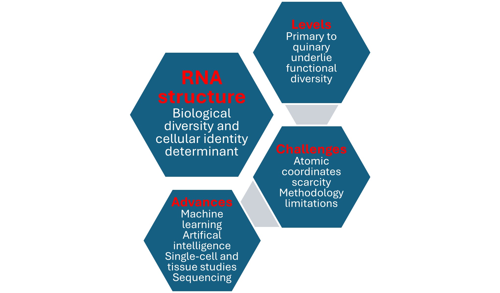
First believed to be a simple intermediary between the information encoded in deoxyribonucleic acid and that functionally displayed in proteins, ribonucleic acid (RNA) is now known to have many functions through its abundance and intricate, ubiquitous, diverse, and dynamic structure. About 70-90% of the human genome is transcribed into protein-coding and noncoding RNAs as main determinants along with regulatory sequences of cellular to populational biological diversity. From the nucleotide sequence or primary structure, through Watson-Crick pairing self-folding or secondary structure, to compaction via longer distance Watson-Crick and non-Watson-Crick interactions or tertiary structure, and interactions with RNA or other biopolymers or quaternary structure, or with metabolites and biomolecules or quinary structure, RNA structure plays a critical role in RNA’s lifecycle from transcription to decay and many cellular processes. In contrast to success with 3-dimensional protein structure prediction using AlphaFold, RNA tertiary and beyond structures prediction remains challenging. However, approaches involving machine learning and artificial intelligence, sequencing of RNA and its modifications, and structural analyses at the single-cell and intact tissue levels, among others, provide an optimistic outlook for the continued development and refinement of RNA-based applications. Here, we highlight those in gene therapy.
Keywords:
RNA structure
; tertiary structure
; helix structure
; gene therapy
; messenger RNA
; small interfering RNA
; long non-coding RNA
1. RNA as an Organic Code
The seven-note musical alphabet played with variations in number, tempo, intensity, rhythm, pitch, or instrument has produced myriads of musical melodies over millennia. Similarly, the organic codes of nucleic acid, protein, polysaccharide, and lipid biomolecules [1,2,3] use relatively few symbols to generate vast information-storing combinations with context-dependent meanings, underlying the diversity and complexity of Earthly life [3,4,5]. Underscoring the analogy between the human-made music code and organic codes, sonification tools provide auditory displays of individual and collective biomolecule sequence information as an adjunct to visual and analytical bioinformatics tools [6-8].
The hypothesis of the emergence of overlapping organic codes heralding the living organisms (biotic) era is in line with the intricate interdependence between deoxyribonucleic acid [6–8(DNA), ribonucleic acid (RNA), proteins, carbohydrates, lipids, and metabolites in cell- or capsid (viruses)-based organisms [9,10,11,12].
The genetic code is the core of life [13], and DNA is its blueprint [14]. The 1800s saw crucial developments in this field. Justus Liebig reported an acidic material in a beef muscle filtrate. Friedrich Miescher discovered ‘nuclein’ in leukocyte nuclei as a protein-degradation-resistant, phosphorus-rich, natural living system’s chemical and chromosome structural component [15]. Richard Altmann coined the term nucleic acid [16].
By 1910, two kinds of nucleic acids were distinguished based on sources and isolation methods: the thymonucleic or zoonucleic acid, now termed DNA, from thymus or animals, and the phytonucleic acid, now termed RNA, from yeast and plants [17,18]. Later, both were found ubiquitously in living organisms. Levene and collaborators [19,20,21] identified the planar aromatic ring structures of DNA’s constituent purine (adenine [A] and guanine [G]) and pyrimidine (thymine [T] and cytosine [C]) nitrogenous bases, and that RNA has the pyrimidine uracil [U], instead of thymine, and pentose instead of hexose as carbohydrate.
In 1944, Oswald Avery [22] proposed that DNA is the genetic information carrier. In 1950, Erwin Chargaff [23] deciphered the consistent proportions of DNA’s constituent bases and the A-T and C-G base pairing rules. In 1953, Maurice Wilkins [24,25] and Rosalind Franklin with Raymond Gosling [26,27] conducted the X-ray diffraction and crystallography studies that led James Watson and Francis Crick to discover the double-helical structure of DNA in 1953 [28,29,30] as the foundation for the DNA theory of inheritance [31]. Each nucleotide interacts with water, ions, amino acids, small molecules, and every other nucleotide, stabilizing the structure [32].
The Watson-Crick right-handed helical B-DNA is the native form of DNA in cells. However, DNA’s helical structure and biological properties can vary transiently along short repetitive tracts, as in left-handed Z-DNA [33,34,35,36], or reversibly en masse, as in the transition between B- and A-DNA in microorganisms in extreme temperatures and pH [37,38]. Other higher-order variations include supercoils (double helix ends join in bacterial genomes), bubbles, hairpins and cruciforms (when palindromes are present), slipped loops, three-stranded triple helices (H-DNA), and tetrameric i-motifs (over 50,000 in the human genome) and related four-stranded G-quadruplexes [39,40,41]. Tertiary DNA structures vary from person to person in critical genes like the insulin gene, constituting therapeutic targets [42].
First proposed by Mitsui et al. in 1970 [43] and later proven by Wang AH et al. [33], Z-DNA is a left-handed helix in equilibrium with the lower energy right-handed B-DNA. Flipons, typically involving an alternating purine/pyrimidine motif, can flip between B- and Z-DNA conformations under physiological conditions aided by binding proteins, introducing diversity to transcriptomes, particularly in immunity and transcription functions [35,36,44].
Discovered by Franklin and Gosling in 1953 [45] in DNA crystals after dehydration, A-DNA, also derived from protein binding to DNA, is a right-handed double helix but with a shorter and more compact helical structure than B-DNA, resulting in slightly more base pairs per turn, a smaller twist angle, and a shorter rise per base pair. The major groove of A-DNA is deep and narrow, the minor groove is wide and shallow, and the base pairs are not perpendicular to the helix-axis as in B-DNA. A-DNA can occur in DNA-RNA hybrid double helices and double-stranded RNAs. RNA can only form an A-type double helix because of the steric restrictions imposed on ribose by the 2’ hydroxyl residue [40].
After Z-DNA was discovered and named after its sugar-phosphate backbone’s zig-zag course as an alternative to the more common Watson-Crick B-DNA, nuclear magnetic resonance, and other studies showed that the common A-RNAs, particularly those with higher Guanine/Cytosine content, could similarly undergo the right-to-left-handed conformational change to the higher energy Z-RNA [44,46,47,48]. Z-binding proteins specifically recognize and bind Z-DNA [49,50,51] and Z-RNA [52,53]. Z-DNA and Z-RNA encoded by flipons under physiological conditions are implicated in various biological processes, including transcription and immunity [44]. Z-RNA has been studied less than Z-DNA and both are challenging to detect in vivo.
DNA organizes in the cellular nucleus into nucleosomes. Nucleosomes are the basic units of eukaryotic chromatin, the 3D structure of tightly folded chromosomes to fit into cellular nuclei. Nucleosomes are formed by 147 bp of duplex DNA wrapped around an octamer of histones [54], followed by a linker DNA bound by histone (H1) in complex eukaryotes [55]. Nucleosomes reduce access to >95% of the DNA [56], and maintain a defined architecture along the genome, with certain positions with well-positioned nucleosomes [57,58,59,60,61,62,63]. The most significant nucleosome-free regions are associated with gene promoter regions upstream of the transcription start sites, replication origins, and transcription termination sites [64,65]. The widths of nucleosome-free regions correlate with gene expression [66]. Nucleosome architecture perturbation associated with stress, cell cycle phase changes, nutrient sources, or the cell metabolic cycle underscores the association between nucleosome architecture and gene activity [60,64,67,68].
Structural similarities between RNA and DNA allow the formation of RNA-DNA hybrids, such as the R loops, which also include a displaced single-stranded DNA [69]. Antisense noncoding RNAs may form R lops. R loops accumulate throughout the genome in pericentromeric DNA, telomeres, ribosomal DNA, or transcription termination regions, among others, and are involved in transcription and chromatin structure. Because they can also adversely affect genome stability and replication, several DNA and RNA metabolism factors, such as ribonucleases, RNA-DNA helicases, RNA processing factors, and topoisomerase I, degrade R-loops or prevent their formation [69].
As an example of a protein that interacts with DNA and RNA, the topoisomerase I enzyme prevents genomic instability by alleviating DNA torsional strain. Topoisomerase I introduces transient single-strand breaks that prevent the accumulation of supercoiling and torsional stress, which could otherwise lead to damage and instability of DNA, and cell death [70]. Interactions between RNA and Topoisomerase I regulate DNA during transcription by modulating Topoisomerase I-mediated relaxation. In cancer cells, for instance, DNA transcription is often elevated, necessitating increased levels of Topoisomerase I activity to relax the DNA and maintain proper gene expression. RNA opposes Topoisomerase I activity. Inhibiting RNA binding of Topoisomerase I may work similarly to antineoplastic Topoisomerase I inhibitors like camptothecin by increasing Topoisomerase I catalytic complexes on DNA [70].
Beyond cancer, dysfunction of R loop-interacting factors in several genetic diseases leads to replication stress, genome instability, chromatin alterations, or gene silencing [69]. Furthermore, many chromatin-associated complexes, including histone modifiers, transcription factors, and DNA methyltransferase, interact with RNA [71]. RNA can also promote the repair of double-strand breaks in DNA, by helping position and holding the broken DNA ends in place and guiding the cellular repair machinery, thereby contributing to genome integrity [72].
2. RNA Has Many Functions Through Its Intricate, Ubiquitous, Diverse, and Dynamic Structure
RNA has emerged as a central biomolecule in the multidirectional flow of genetic information for phenotype and biological diversity generation [12,73]. RNA is no longer considered simply an intermediary between the data stored in DNA and that functionally displayed in proteins. Although about 70–90% of the human [74] and 85–90% of the yeast genome [75] are transcribed into RNA, much remains unknown about RNA functions in cells [76].
RNA structure plays critical roles in every step of RNA’s lifecycle, including transcription, splicing, localization [77,78], translation [79,80], and RNA decay [81]. However, RNA structure differs among individual cells and provides an additional layer of information in defining cellular identities by, for instance, informing RNA-binding protein binding and gene regulation [82]. To this end, overall RNA structure profiles better discriminate cell type identity and differentiation stage than gene expression profiles alone. For instance, RNA structure is more homogeneous in human embryonic stem cells than differentiating neurons, with the greatest homogeneity found in coding regions. More extensive heterogeneity is found within 3’ untranslated regions and is determined by specific RNA-binding proteins. Moreover, the cell-type variable region of 18S ribosomal RNA is associated with cell cycle and translation control. It is therefore important to systematically characterize RNA structure-function relationships at single-cell resolution using approaches such as single-cell structure probing of RNA transcripts [82].
RNA accomplishes many functions through various structural levels beyond its primary and secondary structural ones defined by nucleotide sequence and Watson-Crick pairing-based folding, respectively [83,84,85,86]. Along with the staggering number of noncoding RNA genes, RNA structural versatility underlies biological diversity from the organismal to population levels.
RNA’s bases closely stack on each other like ‘coins in a roll’ via noncovalent interactions, exposing their charged exocyclic groups to water molecules and ions, underlying RNA’s solvability and helical conformations unrelated to Watson-Crick pairing [87]. RNA’s conformation also varies with environmental changes, liquid-liquid phase separation, or interactions with other biomolecules [88,89,90].
Underlying its compactness, RNA intrinsically tends to form A-U, G-C, and G-U Watson-Crick base pairs in short and long-range structures, higher-order architectures, and RNA-RNA interactions in picoseconds to seconds [91,92,93,94], which are fundamental to its diverse functions [95]. As many as 40% of the nucleotides of an RNA molecule can be part of hairpins and multi-helix junction loops [96], and 30% to 40% of RNA duplexes in living cells involve sequences over 200 nucleotides apart [95].
3. RNA’s Structure Is Defined At Primary, Secondary, Tertiary, Quaternary, and Quinary Levels
3.1. Primary, Secondary, and Tertiary RNA Structures
The primary structure is the RNA’s linear nucleotide sequence (Figure 1). The secondary structure describes the paired and unpaired elements of stems, loops, and bulges that form as the single-stranded RNA molecule folds back on itself via Watson-Crick pairs and interacts via hydrogen bonding and stacking as soon as it is synthesized [89,93] (Figure 1).
The tertiary or 3D structure, which typically compacts the RNA, is achieved by longer-distance Watson-Crick and non-Watson-Crick interactions of elements within the preformed secondary structures [85] (Figure 1). These interactions give rise to structural elements, including pseudoknots, which lock together two stem-loops by base pairing and sugar-phosphate interactions, often in a so-called kissing interaction.
Many RNA loops are characterized by structural modules with highly organized networks of noncanonical interactions comprising ordered non-Watson-Crick base pairs embedded between Watson-Crick base pairs [97]. Non-Watson–Crick pairs are key for folding and binding to proteins or other ligands [98,99,100].
RNA three-dimensional (3D) motifs occupy places in structured RNA molecules corresponding to the hairpin, internal, and multi-helix junction loops of their 2D structure representations [96]. These 3D structural RNA modules, with specific loop geometries, contribute to structural stability, have central roles as architectural organizers of catalytic activity and ligand binding sites in RNA molecules, and are recurrently observed in RNA families throughout phylogeny [96,101,102,103,104,105,106].
Among RNA 3D motifs are pseudoknots, which are minimally composed of two helical segments connected by single-stranded regions or loops (Figure 2). Pseudoknots form the catalytic core of various ribozymes, self-splicing introns, and telomerase, and alter gene expression by inducing ribosomal frameshifting in many viruses (reviewed in [107]). The best characterized is the H-type pseudoknot (Figure 2).
Guanine-rich regions in RNA and DNA can form noncanonical G-quadruplex structures encompassing stacked guanine tetrads, a square planar structure formed by four guanine residues [108] (Figure 3). RNA G-quadruplexes participate in translation, splicing, RNA stability, and cellular stress responses, among other functions mediated by the RNA binding proteins with which they interact [108].
3.2. Quaternary RNA Structure
Similar to the interactions of DNA and histones, RNA’s quaternary structures result from a folded RNA’s interaction with other biopolymers, such as proteins and RNAs. The diverse tertiary structures of transfer RNA deciphered in 1974, the hammerhead ribozyme in 1993, the P4-P6 domain of the group I intron in 1996, the Hepatitis Delta Virus ribozyme in 1998, and the hairpin ribozyme in 2001 do not oligomerize into symmetric quaternary structures, as do many proteins [109,110,111,112,113]. Even the ribosome, an RNA heterotrimer comprising 5S, 16S, and 23S rRNAs in addition to numerous ribosomal proteins, exhibits no point-group symmetry [114,115,116,117] except for the peptidyl transferase center which exhibits local pseudosymmetry [118]. However, several biological RNAs, such as the bacteriophage ϕ29 prohead RNA, exhibit global symmetry at the tertiary and quaternary structural levels. Global symmetry stabilizes the RNA fold, coordinates ligand-RNA interactions, and facilitates association with symmetric binding partners [119].
3.3. Quinary RNA Structure
The quinary structure of RNA results from its weak and nonspecific interaction with cellular metabolites, such as osmolytes, accumulated by cells in response to osmotic stress [90]. Understanding the effects of osmolytes on RNA’s tertiary structure, whether stabilizing or destabilizing, is crucial to comprehend the intricacies of RNA [120]. For instance, as hydrated magnesium ions neutralize a notable fraction of the negative charge of an RNA tertiary structure, the RNA becomes less responsive to stabilizing osmolytes and may even be destabilized [120].
Real-time in-cell nuclear magnetic spectroscopy reveals that RNA can also modulate protein quinary structure. This is exemplified by quinary interactions between the thioredoxin protein and messenger RNA fostered by antibiotics, such as tetracycline and streptomycin, that bind the bacterial small ribosomal (30S) subunit [121]. Messenger RNA and ribosomes, representing up to 90% of the total RNA in the cell, prominently mediate these weak, cytosolic quinary protein interactions, which affect protein stability, substrate binding, and activity [122,123,124,125,126,127,128,129,130,131,132]. As another example, the enzymes adenylate kinase and dihydrofolate reductase, and the respective coenzymes, ATP and NADPH, bind to ribosomes with micromolar affinity, suppressing both enzymes’ activities [130].
4. Determining RNA Tertiary and Beyond Structures Remains Challenging
The complex biological functions of RNA molecules are underpinned by their specific sustained 3D structures, with or without the help of proteins or other RNAs in multimolecular complexes [133]. However, the study of RNA 3D structure is often hindered by the scarcity of atomic coordinates, a significant challenge in the field. These determinations are typically low-resolution or miss atoms due to the limitations of the low-throughput and costly structure determination methods, i.e., X-ray crystallography, nuclear magnetic resonance, and cryo-electron microscopy [134], which also creates a significant gap between the number of RNAs sequenced and the number of structures defined. Moreover, RNA’s shifting into diverse forms according to environmental conditions renders structural studies challenging. Traditional imaging methods, such as cryo-electron microscopy single-particle averaging analysis, rely on averaging data from thousands of selected molecules with common shapes, making it difficult to capture the unique shapes of individual RNA molecules.
Developed during the last two decades [135], some RNA 3D structure prediction computational tools use high-resolution homologs’ more precise structural information to annotate the base-pairing interactions in low-resolution structures in coarse-grained models/simulations [136,137] or in imaging data missing atoms [138]. Moreover, a machine-learning approach identifies accurate structural models without assumptions about their defining characteristics despite being trained with the atomic coordinates of only 18 known RNA structures [139].
Structural imaging studies are complemented by gel or capillary electrophoresis based on in-line probing, i.e., structural sensitivity to spontaneous degradation, nucleases targeting either single- or double-stranded regions, or chemical probes, such as dimethyl sulfoxide (DMS). DMS, for instance, is used to probe unpaired adenines and cytidines, and 1- metho-p-toluenesulfonate (CMCT) to probe unpaired uridines in chemical inference of RNA structures sequencing (CRIS-seq) [140,141,142]. DMS is also used in RNA structure sequencing (Structure-seq and STRucture-seq2) [143,144,145,146], dimethyl sulfate-modified RNA sequencing (DMS-seq) [147], dimethyl sulfate mutational profiling with sequencing (DMS-MaPseq) [148], and transfer RNA structure sequencing (tRNA structure-seq) [149]. Pyrdiostatin, the chemical probe in RNA GQ sequencing (rG4-seq) [150], and selective 2’-hydroxyl acylation analyzed by primer extension (SHAPE) [151,152,153,154,155,156] have also extended chemical probing to the entire transcriptome [85,157,158,159,160].
Even if RNA structures are accurately determined, they may not represent the one(s) relevant in vivo. Many factors influence RNA structure in the living cell, including variations in organelle environments and interactions with proteins or other macromolecules, which render the elucidation of RNA structure in vivo particularly challenging. For instance, in silico modeling provides the most thermodynamically stable structure of an RNA sequence, while RNAs can become trapped in vivo in alternative structures [85,161]. Moreover, processing the low abundance, long nascent, or precursor RNAs, including splicing and polyadenylation, entails pathway networks that determine mature isoform composition and control gene expression, further adding to the complexity of studying RNA structure [162].
Alphafold’s success [163,164] in predicting protein 3D structures has not yet extended to RNA [165]. This is due to differences in building blocks (amino acids vs. nucleotides), diversity of sequence range (up to tens of thousands of nucleotides for RNA vs. a few hundred amino acids for proteins), number of available structure data (orders of magnitude greater for proteins), and folding stability (multiple conformations for RNA vs. usually one for proteins) [135].
Readily available RNA 3D structural prediction tools often rely on the primary sequence and canonical 2D structures formed by A-U, G-C, and G-U Watson-Crick pairs to detect structural RNA modules from primary sequence data and identify recurrent interaction networks [166,167,168,169,170,171]. Several databases contain RNA structural information [106,172,173,174].
During the last decade, computational RNA structure predictions have evolved from the earliest thermodynamic and molecular dynamic-based approaches to deep learning-based conformation approaches [175]. Earlier deep learning models for RNA structure have been competitive but not consistently better than traditional 3D structure prediction methods, including ab initio physics-based methods using various levels of granularity in nucleotide representation, template-based methods that try to map sequences to structural motifs before merging them into a whole structure, or hybrid methods, combining ab initio and template-based methods [135,176]. However, platforms such as the RNA3DB dataset [177], which arranges the RNA 3D chains into distinct non-redundant groups (Components), and Dfold, which combines an autoregressive Deep Generative Model, Monte Carlo Tree Search, and a scoring model [178] have been developed to improve RNA 3D structure prediction.
An innovative technique to study the 3D structure of individual molecules without averaging builds on advanced Individual-Particle cryo-Electron Tomography (IPET) to focus on single-molecule 3D imaging in cryopreserved samples. IPET captures a snapshot of RNA’s folding landscape by capturing molecules in various stages of folding, from immature states to their optimal shape. This approach may allow the folding engineering of more effective RNA vaccines and dynamic sensors for molecular medicine [179], as well as advances in RNA-based gene therapy.
5. RNA in Gene Therapy: An Example of Structure-Function Knowledge Application
RNA-based therapeutics have emerged as a powerful subset of gene therapy, offering significant advantages in targeting previously considered “undruggable” pathways and providing versatile therapeutic options. RNA therapeutics have shown promise in treating multiple conditions, including genetic disorders, cancers, and infectious diseases [180]. In regenerative medicine, messenger (m)RNA-based approaches have demonstrated potential for cell reprogramming and targeted tissue restoration [181].
The evolution of RNA in gene therapy has been marked by significant structural-functional discoveries and technological advances that have enhanced our understanding and therapeutic potential [182]. Key milestones include the development of antisense oligonucleotides (ASOs) in the early 1980s and the proposal of RNA interference (RNAi) in the 2000s [182,183].
5.1. Types of RNA Used in Gene Therapy
5.1.1. Messenger RNA
mRNA is a powerful tool in gene therapy, particularly for protein replacement and vaccination strategies [184]. This approach directly delivers synthetic mRNA-encoding therapeutic proteins into cells [185]. Once there, cellular machinery translates it into functional proteins [184]. The rapid development and success of mRNA vaccines against SARS-CoV-2 have showcased the potential of this technology, accelerating research into mRNA therapeutics for other diseases [186].
The therapeutic use of mRNA offers several advantages, including transient expression without genomic integration, which enhances safety [184]; the ability to produce almost any functional protein or peptide in the human body [185,186]; faster design and production compared to conventional approaches [185]; cost-effectiveness and flexibility [185]; and higher transfection efficiency and lower toxicity compared to DNA-based drugs [185,186].
The history of mRNA therapeutics dates back to the early 1960s when mRNA was discovered as a critical player in genetic information flow [184,185]. However, it was in the late 1980s that researchers began exploring mRNA as a therapeutic tool [185]. In 1987, Robert Malone from the Salk Institute demonstrated that synthetic mRNA strands mixed with lipid particles could transfect human cells to express proteins of interest [185].
As the field continues to evolve, over 54 mRNA vaccines and drugs are currently in various stages of clinical testing for multiple diseases, from infectious to cardiovascular conditions [185]. The versatility and rapid production capabilities of mRNA therapeutics position them as a promising tool in the future of precision medicine.
5.1.2. siRNA
Small interfering RNA (siRNA) operates through the RNA interference (RNAi) pathway to silence specific genes [183,187-189]. This mechanism involves double-stranded RNA molecules that bind to complementary mRNA sequences, inducing their degradation and allowing for targeted gene knockdown [183,187,188]. siRNA therapeutics offer several advantages, including targeting almost any gene with high precision [190], [183,187–189and silencing genes previously considered “undruggable” [183].
Andrew Fire and Craig Mello discovered RNAi in 1998, changing our understanding of gene regulation and opening new avenues for therapeutic intervention [183,188,190]. As research progresses, innovations in chemical modifications and delivery systems continue to improve the efficacy and safety of siRNA therapeutics, making them a promising tool in treating various diseases [189].
5.1.3. miRNA
MicroRNA (miRNA) therapeutics have emerged as a promising approach in gene therapy [191]. They offer unique advantages in modulating gene expression patterns associated with various diseases [192]. Two main strategies have been developed to manipulate miRNA activity: miRNA mimics and inhibitors.
miRNA mimics are synthetic double-stranded RNA molecules designed to replicate the function of endogenous miRNAs [193]. They supplement downregulated or non-functional miRNAs, recruit the RNA-induced silencing complex (RISC) to complementary mRNA sequences, and facilitate targeted RNA interference [193]. Advantages of miRNA mimics include the potential to restore tumor suppressor miRNA function in cancer therapy [14], and the versatility in targeting multiple genes simultaneously [193].
Conversely, miRNA inhibitors, also known as anti-miRNAs or antagomiRs, are chemically modified, single-stranded oligonucleotides designed to bind to and inhibit endogenous miRNAs [193]. This mechanism upregulates target mRNA translation and alleviates the effects caused by the overexpression of malignant miRNAs [14]. Advantages of miRNA inhibitors include the ability to target specific miRNAs [193], and the potential for long-lasting effects due to chemical modifications [192].
miRNAs have shown promise in therapeutic applications, particularly in cancer treatment and other disorders characterized by dysregulated gene expression [191,193]. Their endogenous nature and ability to regulate multiple genes within a pathway offer potential advantages over traditional single-target therapies [193].
5.1.4. Long Non-Coding RNAs
Long non-coding RNAs (lncRNAs) are becoming significant targets and tools in gene therapy. Their potential applications are expanding rapidly [194]. Typically, longer than 200 nucleotides, lncRNAs play crucial roles in gene regulation and various cellular processes, influencing complex genetic networks [194,195]. Manipulating them through gene therapy may offer new avenues for treating complex genetic disorders and cancers.
The therapeutic targeting of lncRNAs has gained traction over the past decade as their diverse functions in gene regulation have been uncovered [194,195]. LncRNAs can modulate chromatin structure, influence transcriptional and post-transcriptional processes, and interact with proteins and other RNAs, making them integral to cellular function [194,195]. Their involvement in critical biological processes and disease mechanisms positions lncRNAs as promising candidates for therapeutic intervention.
Advantages of Targeting lncRNAs include their tissue- or cell-type-specific expression patterns [195]; acting as scaffolds for protein complexes, enhancers of gene expression, or decoys that inhibit oncogenic pathways [194]; and usefulness as diagnostic and prognostic biomarkers [195]. Ongoing research is exploring various strategies for effectively targeting lncRNAs. These strategies include transcriptional inhibition, post-transcriptional modulation, and using CRISPR technology to edit lncRNA expression patterns or genomic loci [194,195].
The potential of lncRNAs as therapeutic targets is underscored by their involvement in numerous diseases. For instance, studies have shown that targeting specific oncogenic lncRNAs can inhibit tumor growth and metastasis in preclinical models [194]. Furthermore, restoring downregulated or lost lncRNAs presents an exciting opportunity for therapeutic development [194].
5.2. Molecular and Cellular Mechanisms
5.2.1. Antisense Oligonucleotides (ASOs)
ASOs are short, synthetic strands of nucleic acids that bind to complementary mRNA sequences [189,195,196]. This binding can either induce degradation of the target mRNA through RNase H-mediated cleavage or modulate splicing, effectively altering protein production [195,196]. By targeting specific mRNAs, ASOs can reduce the expression of disease-causing proteins [190]. As such, they are valuable tools in treating genetic disorders.
5.2.2. RNA Interference (RNAi)
RNAi employs small interfering RNAs (siRNAs) and microRNAs (miRNAs) to silence specific genes [183]. siRNAs are designed to match complementary mRNA sequences, leading to their degradation by the RNA-induced silencing complex (RISC) [197]. RNAi provides a powerful means to regulate gene expression and has significant therapeutic potential in conditions such as cancer and viral infections [183,191].
5.2.3. mRNA Delivery
Synthetic mRNAs can be introduced into cells to produce therapeutic proteins [184,185]. This approach has gained significant attention with the development of mRNA vaccines, particularly during the COVID-19 pandemic [186]. Synthetic mRNAs can stimulate an immune response or replace defective proteins in genetic disorders by encoding specific proteins [198]. The ability to rapidly design and produce mRNAs allows for flexible responses to emerging health threats [185,186].
5.2.4. CRISPR-Cas Systems
RNA guides are crucial in CRISPR-Cas technology, enabling precise genome editing [185,190,194,199]. In this system, a guide RNA (gRNA) directs the Cas enzyme to specific DNA sequences within the genome, allowing for targeted modifications such as gene knockouts or insertions [199]. This capability represents a significant advancement in gene therapy, offering potential cures for genetic defects rather than merely treating symptoms.
5.3. RNA Delivery Methods
5.3.1. Viral Vectors
Viral vectors, particularly adeno-associated viruses (AAVs) and lentiviruses have become essential tools for delivering RNA-based therapies in gene therapy [200]. These vectors are adept at transducing target cells and facilitating the long-term expression of therapeutic RNAs. This makes them valuable for treating various genetic disorders and diseases. Their ability to integrate into host genomes allows for sustained therapeutic effects [200]. This ability is particularly beneficial in chronic conditions requiring ongoing protein expression.
Adeno-associated viruses (AAVs) are favored for their low immunogenicity and capacity to transduce both dividing and non-dividing cells [200]. This versatility enables AAVs to be used in a wide range of tissues, including those that are difficult to target with other delivery methods [200]. On the other hand, lentiviruses are capable of stable integration into the host genome, which is advantageous for achieving persistent gene expression [200]. These properties make viral vectors a cornerstone in developing RNA-based gene therapies.
5.3.2. Non-Viral Vectors
Non-viral delivery systems have gained significant prominence in RNA-based gene therapy, mainly through lipid nanoparticles (LNPs) [185,201]. LNPs are designed to effectively encapsulate and protect RNA molecules, facilitating cellular uptake and enhancing endosomal escape [201]. This capability ensures therapeutic RNA reaches its intended target within the cell, maximizing its efficacy.
The development of lipid nanoparticles has been transformative for RNA delivery. This is especially true in the context of mRNA therapeutics [201]. LNPs protect RNA from degradation and promote efficient cellular uptake [185]. The protection and promotions work to address critical challenges related to RNA stability and delivery [186]. The success of LNP-delivered mRNA vaccines against COVID-19 has further validated this approach, demonstrating the potential of non-viral vectors in achieving effective therapeutic outcomes [184,185,186].
5.4. RNA-Based Gene Therapy Applications
5.4.1. Genetic Disorders
Antisense oligonucleotides (ASOs) and small interfering RNAs (siRNAs) are critical approaches developed to silence mutant genes responsible for conditions such as hereditary transthyretin-mediated amyloidosis (hATTR) [190,191]. The approval of the first siRNA drug for hATTR in 2018 marked a milestone in RNA-based gene therapy, demonstrating the potential of these therapies to address previously challenging genetic diseases [190].
ASOs bind to complementary mRNA sequences [189]. This leads to either degradation of the target mRNA or modulation of splicing processes. It allows for correcting RNA processing errors and restoring average protein production [189]. For instance, nusinersen, an ASO approved for spinal muscular atrophy (SMA), enhances exon inclusion in the SMN2 gene to compensate for the loss of function in the SMN1 gene [202].
SiRNAs operate through RNA interference (RNAi) pathways, effectively silencing specific genes by targeting their mRNA for degradation [183,189]. This approach is particularly beneficial for conditions characterized by toxic protein accumulation, such as hATTR, where siRNAs can selectively inhibit harmful gene expression [190].
In addition to ASOs and siRNAs, mRNA-based therapies are being explored for protein replacement in disorders like cystic fibrosis and ornithine transcarbamylase deficiency [186]. These therapies involve delivering synthetic mRNA that encodes functional proteins directly into cells, enabling the production of therapeutic proteins that restore normal cellular function [184]. The mutation-agnostic nature of mRNA therapies allows them to be applicable across various genetic mutations, presenting a versatile option for treating monogenic diseases [185,186].
5.4.2. Cancer
RNA-based gene therapies offer innovative strategies for cancer treatment, including silencing oncogenes, restoring tumor suppressor functions, and modulating immune responses [194]. Small interfering RNAs (siRNAs) and antisense oligonucleotides (ASOs) are also prominent RNA-based strategies for cancer [183,190,191]. They have shown promise in preclinical and early clinical studies [190]. SiRNAs can specifically target and degrade mRNA transcripts from oncogenic fusion proteins, effectively silencing genes that drive tumor growth [185]. Similarly, ASOs can bind to complementary mRNA sequences to restore standard gene expression patterns [203].
mRNA-based therapies are also being explored for their potential in cancer treatment [185,186]. These therapies encode functional proteins that restore normal cellular functions or stimulate immune responses against tumors [194]. For instance, mRNA vaccines can elicit robust immune responses by encoding tumor-associated antigens (TAAs), allowing simultaneous delivery of multiple antigens to enhance both humoral and cell-mediated immunity [185,186].
5.4.3. Infectious Diseases
Beyond vaccines, RNA therapeutics are being explored for direct antiviral effects [204]. Small interfering RNAs can target specific viral genes, effectively inhibiting viral replication by silencing essential genes required for the virus’s life cycle [204]. Research indicates that siRNAs can significantly reduce viral loads across various infection models [190]. This is paving the way for therapeutic applications against viruses.
The versatility of mRNA technology allows the rapid design of constructs tailored to specific pathogens, enabling quick responses to emerging infectious threats [185]. Additionally, lipid nanoparticles have been instrumental in the success of mRNA vaccines by protecting fragile mRNA from degradation and facilitating cellular uptake [184].
5.4.4. Protein Malfunction Diseases
RNA-based therapies are emerging as practical solutions for diseases caused by protein malfunction [205]. They deliver functional RNA to produce missing or defective proteins. A notable example is MRT5005, an mRNA therapy designed for cystic fibrosis (CF), which has entered clinical trials as a protein replacement therapy targeting the cystic fibrosis transmembrane conductance regulator (CFTR) protein [205].
MRT5005 is a codon-optimized mRNA therapy delivered via aerosolized lipid nanoparticles directly into the lungs [186]. This genotype-agnostic approach aims to re-store CFTR protein production, essential for maintaining proper ion transport and flu-id balance in epithelial cells. Thus, it improves lung function in CF patients [205].
The development of mRNA therapies like MRT5005 reflects a broader trend toward addressing genetic disorders directly through protein replacement strategies. Unlike traditional small molecule drugs that may only target specific mutations or pathways, mRNA therapies can theoretically encode any missing protein, offering a more comprehensive solution [185,186].
5.4.5. Cell Reprogramming and Tissue Restoration
mRNA tools have demonstrated significant utility in cell reprogramming approaches within regenerative medicine [184,186]. By delivering specific mRNAs that induce crucial factors necessary for reprogramming or tissue restoration, researchers can achieve greater control and safety than traditional gene therapy techniques since mRNA is transient and does not integrate into the genome [184]. This method has emerged as a promising strategy for generating induced pluripotent stem cells (iPSCs) without leaving residual genetic material after achieving desired cellular states [186].
mRNA tools enable researchers to precisely adjust the expression levels of reprogramming factors, which enhances their use in diverse cell types and conditions within regenerative medicine [185]. Beyond generating induced pluripotent stem cells (iPSCs), these mRNA tools are also being investigated for tissue engineering applications. They deliver growth factors or signaling molecules directly to damaged tissues, stimulating regeneration and promoting healing.
5.5. Challenges and Limitations
5.5.1. RNA Stability
A primary challenge in RNA-based gene therapy is the inherent instability of RNA molecules, which are susceptible to degradation by endogenous nucleases [185,186,206]. To address this issue, researchers have developed various chemical modifications and advanced delivery systems to enhance RNA stability and protect therapeutic RNA from degradation [207]. These improvements are crucial for maximizing the pharmacological efficacy of RNA therapies and ensuring their successful application in clinical settings.
5.5.2. Off-Target Effects
5.5.3. Immune Responses
Another limitation of RNA-based therapies is the potential for immune responses against the delivered RNA molecules or their delivery vehicles [194]. The immune system may recognize foreign RNA as a threat, leading to inflammatory reactions that can diminish therapeutic efficacy or cause adverse effects [194]. Strategies to mitigate these immune responses, such as employing modified nucleotides or optimizing delivery formulations, are critical for improving the safety profile of RNA therapeutics.
5.5.4. Delivery
Effective delivery of RNA therapeutics to target cells remains a significant hurdle [183,185,186,193]. Traditional methods, such as viral vectors, pose risks of immunogenicity and insertional mutagenesis [200]. Consequently, non-viral delivery systems, including lipid nanoparticles (LNPs) and polymer-based carriers, are optimized to enhance cellular uptake and stability while minimizing potential side effects [185,201]. Developing these delivery mechanisms is essential for achieving the desired therapeutic outcomes in various applications.
5.5.5. Expression Duration
The transient nature of mRNA therapies can be both an advantage and a limitation [185]. While the lack of genomic integration reduces long-term risks associated with permanent alterations, it also necessitates repeated administrations to maintain therapeutic effects. Developing sustained-release formulations or alternative strategies that extend the duration of action without compromising safety is an ongoing area of research [208].
5.6. Future Gene Therapy Perspectives
The field of RNA therapeutics is rapidly evolving, with emerging technologies poised to enhance the potency and specificity of RNA-based therapies. Innovations such as untranslated region optimization and machine learning-based synthetic RNA motif design are expected to improve therapeutic outcomes significantly. Additionally, the future of RNA-based gene therapy may increasingly rely on combination approaches that integrate multiple RNA modalities or combine RNA therapeutics with traditional small molecule drugs and immunotherapies [193]. These strategies could provide synergistic effects, enabling more effective treatment of complex diseases.
Advances in RNA engineering and synthetic biology are expanding the toolkit for gene therapy. For instance, CRISPR-Cas systems using guide RNAs offer unprecedented precision in genome editing, opening new possibilities for treating genetic disorders [194,199]. These technologies enhance the specificity of gene manipulation and allow for targeted interventions to address the underlying causes of diseases.
6. Conclusion
Since the central molecular biology dogma was formulated almost seven decades ago, mounting evidence revealed that RNA is the main determinant of biological diversity. This is driven by RNA’s abundance, modifications, and structural versatility of its coding and noncoding versions, which occasionally overlap. RNA structure-function relationships also vary among cells in an organism, determining cellular identity. However, understanding RNA’s tertiary, quaternary, and quinary structures and their functional relationships, remains challenging. Advances in approaches including the use of machine learning and artificial intelligence [139] and data gatherings, such as the United States National Institutes of Health’s and National Academies of Sciences’ RNA sequencing initiative, provide an optimistic outlook for the continued development and refinement of disruptive RNA-based approaches for medical therapy, diagnosis, and prevention, and agriculture and industrial applications [209], as illustrated here for gene therapy. To this end, novel imaging methods enable detailed RNA analysis within single cells and intact tissues [210].
The evolution of RNA-based gene therapy has been characterized by remarkable advancements that have greatly enhanced our understanding of RNA biology, novel delivery technologies, and their therapeutic applications. Over the past few years, researchers have made significant strides in unraveling the complexities of RNA and its various forms, such as mRNA, siRNA, and miRNA. This deeper knowledge has allowed scientists to develop more effective and targeted treatments.
As efforts continue to address and overcome existing challenges, such as improving delivery mechanisms, ensuring stability, and minimizing potential off-target effects, there is growing optimism about the potential of RNA-based therapies. Researchers are actively exploring innovative approaches to maximize the efficacy of these treatments, broadening their application to a wide range of diseases, including genetic disorders, cancer, and viral infections. Moreover, the rise of personalized medicine relies heavily on the advancements in RNA therapeutics. By tailoring treatments to the unique genetic makeup of individual patients, we can potentially enhance therapeutic outcomes and reduce adverse effects, ushering in a new era of healthcare.
With ongoing research and an increasing number of clinical trials demonstrating positive results, RNA therapeutics are on the verge of making significant contributions to regenerative medicine. These therapies hold the potential not only to treat diseases that were once deemed incurable but also to improve the quality of life for many individuals. As we look forward to the future of RNA-based gene therapy (Figure 4), it is clear that continued investment and exploration in this field will likely lead to transformative solutions in medicine.
Author Contributions
Both authors (W.A.H., K.H., and R.P.) contributed to writing—original draft preparation, writing—review, and editing. Both authors have read and agreed to the published version of the manuscript.
Funding
This review received no external funding.
Conflicts of Interest
The authors declare no conflict of interest.
References
- Maraldi, N.M. In search of a primitive signaling code. Biosystems 2019, 183, 103984. [Google Scholar] [CrossRef] [PubMed]
- Melero, A.; Jiménez-Rojo, N. Cracking the membrane lipid code. Curr Opin Cell Biol 2023, 83, 102203. [Google Scholar] [CrossRef]
- Gabius, H.J.; Cudic, M.; Diercks, T.; Kaltner, H.; Kopitz, J.; Mayo, K.H.; Murphy, P.V.; Oscarson, S.; Roy, R.; Schedlbauer, A.; Toegel, S.; Romero, A. What is the Sugar Code? Chembiochem 2022, 23, e202100327. [Google Scholar] [CrossRef] [PubMed]
- Chatterjee, S.; Yadav, S. The Origin of Prebiotic Information System in the Peptide/RNA World: A Simulation Model of the Evolution of Translation and the Genetic Code. Life (Basel) 2019, 9, 25. [Google Scholar] [CrossRef]
- Chatterjee, S.; Yadav, S. The Coevolution of Biomolecules and Prebiotic Information Systems in the Origin of Life: A Visualization Model for Assembling the First Gene. Life (Basel) 2022, 12, 834. [Google Scholar] [CrossRef] [PubMed]
- Riego, E.; Silva, A.; De la Fuente, J. The sound of the DNA language. Biol Res 1995, 28, 197–204. [Google Scholar]
- Sánchez Sousa, A.; Baquero, F.; Nombela, C. The making of “The Genoma Music”. Rev Iberoam Micol 2005, 22, 242–248. [Google Scholar] [CrossRef]
- Temple, M.D. An auditory display tool for DNA sequence analysis. BMC Bioinformatics 2017, 18, 221. [Google Scholar] [CrossRef] [PubMed]
- de Farias, S.T.; Prosdocimi, F.; Caponi, G. Organic Codes: A Unifying Concept for Life. Acta Biotheor 2021, 69, 769–782. [Google Scholar] [CrossRef] [PubMed]
- Kondratyeva, L.G.; Dyachkova, M.S.; Galchenko, A.V. The Origin of Genetic Code and Translation in the Framework of Current Concepts on the Origin of Life. Biochemistry (Mosc) 2022, 87, 150–169. [Google Scholar] [CrossRef] [PubMed]
- Pavlinova, P.; Lambert, C.N.; Malaterre, C.; Nghe, P. Abiogenesis through gradual evolution of autocatalysis into template-based replication. FEBS Lett 2023, 597, 344–379. [Google Scholar] [CrossRef] [PubMed]
- Haseltine, W.A.; Patarca, R. The RNA revolution in the central molecular biology dogma evolution. Int J Mol. Sci 2024, 25, 12695. [Google Scholar] [CrossRef]
- Crick, F.H.; Barnett, L.; Brenner, S.; Watts-Tobin, R.J. General nature of the genetic code for proteins. Nature 1961, 192, 1227–1232. [Google Scholar] [CrossRef]
- Portin, P. The birth and development of the DNA theory of inheritance: Sixty years since the discovery of the structure of DNA. J Genet 2014, 93, 293–302. [Google Scholar] [CrossRef]
- Dahm, R. Friedrich Miescher and the discovery of DNA. Dev Biol 2005, 278, 274–288. [Google Scholar] [CrossRef]
- Altmann, R. Ueber nucleinsäuren. Arch. f. Anat. u. Physiol. Physiol. Abt. 1889, 524–536. [Google Scholar]
- Levene, P.A. On the biochemistry of nucleic acids. J Am Chem Soc 1910, 32, 231–240. [Google Scholar] [CrossRef]
- Frixione, E.; Ruiz-Zamarripa, L. The “scientific catastrophe” in nucleic acids research that boosted molecular biology. J Biol Chem 2019, 294, 2249–2255. [Google Scholar] [CrossRef]
- Levene, P.A.; Bass, L.W. Nucleic Acids; Chemical Catalog Company: New York, 1931; Available online: https://babel.hathitrust.org/cgi/pt?id=uc1.b4165245;view=1up;seq=5 (accessed on 8 August 2024).
- Levene, P.A.; Tipson, R.S. The ring structure of adenosine. Science 1931, 74, 521. [Google Scholar] [CrossRef]
- Levene, P.A.; Tipson, R.S. The ring structure of thymidine. Science 1935, 81, 98. [Google Scholar] [CrossRef] [PubMed]
- Avery, O.T.; Macleod, C.M.; McCarty, M. Studies on the chemical nature of the substance inducing transformation of Pneumococcal types: Induction of transformation by a desoxyribonucleic acid fraction isolated from Pneumococcus type III. J Exp Med 1944, 79, 137–158. [Google Scholar] [CrossRef]
- Chargaff, E. Chemical specificity of nucleic acids and mechanism of their enzymatic degradation. Experientia 1950, 6, 201–209. [Google Scholar] [CrossRef] [PubMed]
- Wilkins, M.H.; Stokes, A.R.; Wilson, H.R. Molecular structure of deoxypentose nucleic acids. Nature 1953, 171, 738–740. [Google Scholar] [CrossRef] [PubMed]
- Wilkins, M.H.; Stokes, A.R.; Wilson, H.R. Molecular structure of nucleic acids. Molecular structure of deoxypentose nucleic acids. Ann N Y Acad Sci 1995, 758, 13–16. [Google Scholar] [CrossRef] [PubMed]
- Franklin, R.E.; Gosling, R.G. Molecular configuration in sodium thymonucleate. Nature 1953, 171, 740–741. [Google Scholar] [CrossRef]
- Franklin, R.E.; Gosling, R.G. Evidence for 2-chain helix in crystalline structure of sodium deoxyribonucleate. Nature 1953, 172, 156–157. [Google Scholar] [CrossRef] [PubMed]
- Watson, J.D.; Crick, F.H. Molecular structure of nucleic acids; a structure for deoxyribose nucleic acid. Nature 1953, 171, 737–738. [Google Scholar] [CrossRef]
- Watson, J.D.; Crick, F.H. The structure of DNA. Cold Spring Harb Symp Quant Biol 1953, 18, 123–131. [Google Scholar] [CrossRef]
- Watson, J.D. The Double Helix: A Personal Account of the Discovery of the Structure of DNA; Weidenfield and Nicolson: London, 1981. [Google Scholar]
- Watson, J.D.; Crick, F.H. Genetical implications of the structure of deoxyribonucleic acid. Nature 1953, 171, 964–967. [Google Scholar] [CrossRef]
- Martel, P. Base crystallization and base stacking in water. Eur J Biochem 1979, 96, 213–219. [Google Scholar] [CrossRef] [PubMed]
- Wang, A.H.; Quigley, G.J.; Kolpak, F.J.; Crawford, J.L.; van Boom, J.H.; van der Marel, G.; Rich, A. Molecular structure of a left-handed double helical DNA fragment at atomic resolution. Nature 1979, 282, 680–686. [Google Scholar] [CrossRef]
- Wing, R.; Drew, H.; Takano, T.; Broka, C.; Tanaka, S.; Itakura, K.; Dickerson, R.E. Crystal structure analysis of a complete turn of B-DNA. Nature 1980, 287, 755–758. [Google Scholar] [CrossRef] [PubMed]
- Herbert, A. A Genetic Instruction Code Based on DNA Conformation. Trends Genet 2019, 35, 887–890. [Google Scholar] [CrossRef] [PubMed]
- Herbert, A. The ancient Z-DNA and Z-RNA specific Zα fold has evolved modern roles in immunity and transcription through the natural selection of flipons. R Soc Open Sci 2024, 11, 240080. [Google Scholar] [CrossRef] [PubMed]
- Whelan, D.R.; Hiscox, T.J.; Rood, J.I.; Bambery, K.R.; McNaughton, D.; Wood, B.R. Detection of an en masse and reversible B- to A-DNA conformational transition in prokaryotes in response to desiccation. J R Soc Interface 2014, 11, 20140454. [Google Scholar] [CrossRef]
- DiMaio, F.; Yu, X.; Rensen, E.; Krupovic, M.; Prangishvili, D.; Egelman, E.H. Virology. A virus that infects a hyperthermophile encapsidates A-form DNA. Science 2015, 348, 914–917. [Google Scholar] [CrossRef]
- Choi, J.; Majima, T. Conformational changes of non-B DNA. Chem Soc Rev 2011, 40, 5893–5909. [Google Scholar] [CrossRef]
- Travers, A.; Muskhelishvili, G. DNA structure and function. FEBS J 2015, 282, 2279–2295. [Google Scholar] [CrossRef]
- Peña Martinez, C.D.; Zeraati, M.; Rouet, R.; Mazigi, O.; Henry, J.Y.; Gloss, B.; Kretzmann, J.A.; Evans, C.W.; Ruggiero, E.; Zanin, I.; Marušič, M.; Plavec, J.; Richter, S.N.; Bryan, T.M.; Smith, N.M.; Dinger, M.E.; Kummerfeld, S.; Christ, D. Human genomic DNA is widely interspersed with i-motif structures. The EMBO J 2024. [Google Scholar] [CrossRef]
- Guneri, D.; Alexandrou, E.; El Omari, K.; Dvořáková, Z.; Chikhale, R.V.; Pike, D.T.S.; Waudby, C.A.; Morris, C.J.; Haider, S.; Parkinson, G.N.; Waller, Z.A.E. Structural insights into i-motif DNA structures in sequences from the insulin-linked polymorphic region. Nat Commun 2024, 15, 7119. [Google Scholar] [CrossRef]
- Mitsui, Y.; Langridge, R.; Shortle, B.E.; Cantor, C.R.; Grant, R.C.; Kodama, M.; Wells, R.D. “Physical and enzymatic studies on poly d(I–C)·poly d(I–C), an unusual double-helical DNA”. Nature 1970, 228, 1166–1169. [Google Scholar] [CrossRef] [PubMed]
- Krall, J.B.; Nichols, P.J.; Henen, M.A.; Vicens, Q.; Vögeli, B. Structure and Formation of Z-DNA and Z-RNA. Molecules 2023, 28, 843. [Google Scholar] [CrossRef] [PubMed]
- Franklin, R.E.; Gosling, R.G. The structure of sodium thermonucleate fibres. I. The influence of water content. Acta Cryst 1953, 6, 673–377. [Google Scholar] [CrossRef]
- Hall, K.; Cruz, P.; Tinoco, I., Jr.; Jovin, T.M.; van de Sande, J.H. ‘Z-RNA’--a left-handed RNA double helix. Nature 1984, 311, 584–586. [Google Scholar] [CrossRef] [PubMed]
- Davis, P.W.; Adamiak, R.W.; Tinoco, I. Jr. Z-RNA: The solution NMR structure of r(CGCGCG). Biopolymers 1990, 29, 109–122. [Google Scholar] [CrossRef] [PubMed]
- Popenda, M.; Milecki, J.; Adamiak, R.W. High salt solution structure of a left-handed RNA double helix. Nucleic Acids Res 2004, 32, 4044–4054. [Google Scholar] [CrossRef]
- Herbert, A.; Alfken, J.; Kim, Y.G.; Mian, I.S.; Nishikura, K.; Rich, A. A Z-DNA binding domain present in the human editing enzyme, double-stranded RNA adenosine deaminase. Proc Natl Acad Sci U S A 1997, 94, 8421–8426. [Google Scholar] [CrossRef] [PubMed]
- Kim, Y.G.; Lowenhaupt, K.; Oh, D.B.; Kim, K.K.; Rich, A. Evidence that vaccinia virulence factor E3L binds to Z-DNA in vivo: Implications for development of a therapy for poxvirus infection. Proc Natl Acad Sci U S A 2004, 101, 1514–1518. [Google Scholar] [CrossRef] [PubMed]
- Schwartz, T.; Rould, M.A.; Lowenhaupt, K.; Herbert, A.; Rich, A. Crystal structure of the Zalpha domain of the human editing enzyme ADAR1 bound to left-handed Z-DNA. Science 1999, 284, 1841–1845. [Google Scholar] [CrossRef]
- Brown, B.A., 2nd; Lowenhaupt, K.; Wilbert, C.M.; Hanlon, E.B.; Rich, A. The zalpha domain of the editing enzyme dsRNA adenosine deaminase binds left-handed Z-RNA as well as Z-DNA. Proc Natl Acad Sci U S A 2000, 97, 13532–13536. [Google Scholar] [CrossRef]
- Placido, D.; Brown, B.A., 2nd; Lowenhaupt, K.; Rich, A.; Athanasiadis, A. A left-handed RNA double helix bound by the Z alpha domain of the RNA-editing enzyme ADAR1. Structure 2007, 15, 395–404. [Google Scholar] [CrossRef]
- Richmond, T.J.; Davey, C.A. The structure of DNA in the nucleosome core. Nature 2003, 423, 145–150.htpps. [Google Scholar] [CrossRef]
- Izzo, A.; Kamieniarz, K.; Schneider, R. The histone H1 family: Specific members, specific functions? Biol Chem 2008, 389, 333–343. [Google Scholar] [CrossRef] [PubMed]
- Kornberg, R.D. Chromatin structure: A repeating unit of histones and DNA. Science 1974, 184, 868–871. [Google Scholar] [CrossRef] [PubMed]
- Yuan, G.C.; Liu, Y.J.; Dion, M.F.; Slack, M.D.; Wu, L.F.; Altschuler, S.J.; Rando, O.J. Genome-scale identification of nucleosome positions in S. cerevisiae. Science 2005, 309, 626–630. [Google Scholar] [CrossRef]
- Mavrich, T.N.; Ioshikhes, I.P.; Venters, B.J.; Jiang, C.; Tomsho, L.P.; Qi, J.; Schuster, S.C.; Albert, I.; Pugh, B.F. A barrier nucleosome model for statistical positioning of nucleosomes throughout the yeast genome. Genome Res 2008, 18, 1073–1083. [Google Scholar] [CrossRef] [PubMed]
- Mavrich, T.N.; Jiang, C.; Ioshikhes, I.P.; Li, X.; Venters, B.J.; Zanton, S.J.; Tomsho, L.P.; Qi, J.; Glaser, R.L.; Schuster, S.C.; Gilmour, D.S.; Albert, I.; Pugh, B.F. Nucleosome organization in the Drosophila genome. Nature 2008, 453, 358–362. [Google Scholar] [CrossRef] [PubMed]
- Shivaswamy, S.; Bhinge, A.; Zhao, Y.; Jones, S.; Hirst, M.; Iyer, V.R. Dynamic remodeling of individual nucleosomes across a eukaryotic genome in response to transcriptional perturbation. PLoS Biol 2008, 6, e65. [Google Scholar] [CrossRef]
- Vaillant, C.; Palmeira, L.; Chevereau, G.; Audit, B.; d’Aubenton-Carafa, Y.; Thermes, C.; Arneodo, A. A novel strategy of transcription regulation by intragenic nucleosome ordering. Genome Res 2010, 20, 59–67. [Google Scholar] [CrossRef]
- Valouev, A.; Johnson, S.M.; Boyd, S.D.; Smith, C.L.; Fire, A.Z.; Sidow, A. Determinants of nucleosome organization in primary human cells. Nature 2011, 474, 516–520. [Google Scholar] [CrossRef]
- Baldi, S.; Krebs, S.; Blum, H.; Becker, P.B. Genome-wide measurement of local nucleosome array regularity and spacing by nanopore sequencing. Nat Struct Mol Biol 2018, 25, 894–901. [Google Scholar] [CrossRef] [PubMed]
- Deniz, O.; Flores, O.; Battistini, F.; Pérez, A.; Soler-López, M.; Orozco, M. Physical properties of naked DNA influence nucleosome positioning and correlate with transcription start and termination sites in yeast. BMC Genomics 2011, 12, 489. [Google Scholar] [CrossRef] [PubMed]
- Deniz, Ö.; Flores, O.; Aldea, M.; Soler-López, M.; Orozco, M. Nucleosome architecture throughout the cell cycle. Sci Rep 2016, 6, 19729. [Google Scholar] [CrossRef]
- Weiner, A.; Hughes, A.; Yassour, M.; Rando, O.J.; Friedman, N. High-resolution nucleosome mapping reveals transcription-dependent promoter packaging. Genome Res 2010, 20, 90–100. [Google Scholar] [CrossRef]
- Kaplan, N.; Moore, I.K.; Fondufe-Mittendorf, Y.; Gossett, A.J.; Tillo, D.; Field, Y.; LeProust, E.M.; Hughes, T.R.; Lieb, J.D.; Widom, J.; Segal, E. The DNA-encoded nucleosome organization of a eukaryotic genome. Nature 2009, 458, 362–366. [Google Scholar] [CrossRef] [PubMed]
- Nocetti, N.; Whitehouse, I. Nucleosome repositioning underlies dynamic gene expression. Genes Dev 2016, 30, 660–672. [Google Scholar] [CrossRef]
- Santos-Pereira, J.M.; Aguilera, A. R loops: New modulators of genome dynamics and function. Nat Rev Genet 2015, 16, 583–597. [Google Scholar] [CrossRef] [PubMed]
- Bhola, M.; Abe, K.; Orozco, P.; Rahnamoun, H.; Avila-Lopez, P.; Taylor, E.; Muhammad, N.; Liu, B.; Patel, P.; Marko, J.F.; Starner, A.C.; He, C.; Van Nostrand, E.L.; Mondragón, A.; Lauberth, S.M. RNA interacts with topoisomerase I to adjust DNA topology. Mol Cell 2024, 84, 3192–3208.e11. [Google Scholar] [CrossRef] [PubMed]
- Song, J.; Gooding, A.R.; Hemphill, W.O.; Love, B.D.; Robertson, A.; Yao, L.; Zon, L.I.; North, T.E.; Kasinath, V.; Cech, T.R. Structural basis for inactivation of PRC2 by G-quadruplex RNA. Science 2023, 381, 1331–1337. [Google Scholar] [CrossRef] [PubMed]
- Jeon, Y.; Lu, Y.; Ferrari, M.M.; Channagiri, T.; Xu, P.; Meers, C.; Zhang, Y.; Balachander, S.; Park, V.S.; Marsili, S.; Pursell, Z.F.; Jonoska, N.; Storici, F. RNA-mediated double-strand break repair by end-joining mechanisms. Nat Commun 2024, 15, 7935. [Google Scholar] [CrossRef] [PubMed]
- Jones, C.H.; Androsavich, J.R.; So, N.; Jenkins, M.P.; MacCormack, D.; Prigodich, A.; Welch, V.; True, J.M.; Dolsten, M. Breaking the mold with RNA-a “RNAissance” of life science. NPJ Genom Med 2024, 9, 2. [Google Scholar] [CrossRef] [PubMed]
- Pertea, M. The human transcriptome: An unfinished story. Genes (Basel) 2012, 3, 344–360. [Google Scholar] [CrossRef]
- David, L.; Huber, W.; Granovskaia, M.; Toedling, J.; Palm, C.J.; Bofkin, L.; Jones, T.; Davis, R.W.; Steinmetz, L.M. A high-resolution map of transcription in the yeast genome. Proc Natl Acad Sci U S A 2006, 103, 5320–5325. [Google Scholar] [CrossRef]
- Kazimierczyk, M.; Kasprowicz, M.K.; Kasprzyk, M.E.; Wrzesinski, J. Human Long Noncoding RNA Interactome: Detection, Characterization and Function. Int J Mol Sci 2020, 21, 1027. [Google Scholar] [CrossRef] [PubMed]
- Martin, K.C.; Ephrussi, A. mRNA localization: Gene expression in the spatial dimension. Cell 2009, 136, 719–730. [Google Scholar] [CrossRef] [PubMed]
- Wan, Y.; Kertesz, M.; Spitale, R.C.; Segal, E.; Chang, H.Y. Understanding the transcriptome through RNA structure. Nat Rev Genet 2011, 12, 641–655. [Google Scholar] [CrossRef]
- Mao, Y.; Liu, H.; Liu, Y.; Tao, S. Deciphering the rules by which dynamics of mRNA secondary structure affect translation efficiency in Saccharomyces cerevisiae. Nucleic Acids Res 2014, 42, 4813–4822. [Google Scholar] [CrossRef]
- Clancy, S.; Brown, W. Translation: DNA to mRNA to protein. Nat Educ 2008, 1, 101. [Google Scholar]
- Garneau, N.L.; Wilusz, J.; Wilusz, C.J. The highways and byways of mRNA decay. Nat Rev Mol Cell Biol 2007, 8, 113–126. [Google Scholar] [CrossRef]
- Wang, J.; Zhang, Y.; Zhang, T.; Tan, W.T.; Lambert, F.; Darmawan, J.; Huber, R.; Wan, Y. RNA structure profiling at single-cell resolution reveals new determinants of cell identity. Nat Methods 2024, 21, 411–422. [Google Scholar] [CrossRef]
- Tomezsko, P.J.; Corbin, V.D.A.; Gupta, P.; Swaminathan, H.; Glasgow, M.; Persad, S.; Edwards, M.D.; McIntosh, L.; Papenfuss, A.T.; et al. Determination of RNA structural diversity and its role in HIV-1 RNA splicing. Nature 2020, 582, 438–442. [Google Scholar] [CrossRef] [PubMed]
- Vicens, Q.; Kieft, J.S. Thoughts on how to think (and talk) about RNA structure. Proc Natl Acad Sci U S A 2022, 119, e2112677119. [Google Scholar] [CrossRef]
- Assmann, S.M.; Chou, H.L.; Bevilacqua, P.C. Rock, scissors, paper: How RNA structure informs function. Plant Cell 2023, 35, 1671–1707. [Google Scholar] [CrossRef] [PubMed]
- Shine, M.; Gordon, J.; Schärfen, L.; Zigackova, D.; Herzel, L.; Neugebauer, K.M. Co-transcriptional gene regulation in eukaryotes and prokaryotes. Nat Rev Mol Cell Biol 2024, 25, 534–554. [Google Scholar] [CrossRef] [PubMed]
- Westhof, E.; Fritsch, V. RNA folding: Beyond Watson-Crick pairs. Structure 2000, 8, R55–R65. [Google Scholar] [CrossRef] [PubMed]
- Cruz, J.A.; Westhof, E. The dynamic landscapes of RNA architecture. Cell 2009, 136, 604–609. [Google Scholar] [CrossRef]
- Ganser, L.R.; Kelly, M.L.; Herschlag, D.; Al-Hashimi, H.M. The roles of structural dynamics in the cellular functions of RNAs. Nat Rev Mol Cell Biol 2019, 20, 474–489. [Google Scholar] [CrossRef]
- Alfano, C.; Fichou, Y.; Huber, K.; Weiss, M.; Spruijt, E.; Ebbinghaus, S.; De Luca, G.; Morando, M.A.; Vetri, V.; Temussi, P.A.; Pastore, A. Molecular Crowding: The History and Development of a Scientific Paradigm. Chem Rev 2024, 124, 3186–3219. [Google Scholar] [CrossRef]
- Schultes, E.A.; Spasic, A.; Mohanty, U.; Bartel, D.P. Compact and ordered collapse of randomly generated RNA sequences. Nat Struct Mol Biol 2005, 12, 1130–1136. [Google Scholar] [CrossRef]
- Dethoff, E.A.; Chugh, J.; Mustoe, A.M.; Al-Hashimi, H.M. Functional complexity and regulation through RNA dynamics. Nature 2012, 482, 322–330. [Google Scholar] [CrossRef]
- Mustoe, A.M.; Brooks, C.L.; Al-Hashimi, H.M. Hierarchy of RNA functional dynamics. Annu Rev Biochem 2014, 83, 441–466. [Google Scholar] [CrossRef] [PubMed]
- Bose, R.; Saleem, I.; Mustoe, A.M. Causes, functions, and therapeutic possibilities of RNA secondary structure ensembles and alternative states. Cell Chem Biol 2024, 31, 17–35. [Google Scholar] [CrossRef] [PubMed]
- Lu, Z.; Zhang, Q.C.; Lee, B.; Flynn, R.A.; Smith, M.A.; Robinson, J.T.; Davidovich, C.; Gooding, A.R.; Goodrich, K.J.; Mattick, J.S.; et al. RNA Duplex Map in Living Cells Reveals Higher-Order Transcriptome Structure. Cell 2016, 165, 1267–1279. [Google Scholar] [CrossRef]
- Parlea, L.G.; Sweeney, B.A.; Hosseini-Asanjan, M.; Zirbel, C.L.; Leontis, N.B. The RNA 3D Motif Atlas: Computational methods for extraction, organization and evaluation of RNA motifs. Methods 2016, 103, 99–119. [Google Scholar] [CrossRef] [PubMed]
- Sarrazin-Gendron, R.; Waldispühl, J.; Reinharz, V. Classification and Identification of Non-canonical Base Pairs and Structural Motifs. Methods Mol Biol 2024, 2726, 143–168. [Google Scholar] [CrossRef]
- Hermann, T.; Westhof, E. Non-Watson-Crick base pairs in RNA-protein recognition. Chem Biol 1999, 6, R335–R343. [Google Scholar] [CrossRef]
- Hermann, T.; Patel, D.J. Adaptive recognition by nucleic acid aptamers. Science 2000, 287, 820–825. [Google Scholar] [CrossRef] [PubMed]
- Leontis, N.B.; Westhof, E. Analysis of RNA motifs. Curr Opin Struct Biol 2003, 13, 300–308. [Google Scholar] [CrossRef]
- Cruz, J.A.; Westhof, E. Sequence-based identification of 3D structural modules in RNA with RMDetect. Nat Methods 2011, 8, 513–521. [Google Scholar] [CrossRef] [PubMed]
- Hamdani, H.Y.; Appasamy, S.D.; Willett, P.; Artymiuk, P.J.; Firdaus-Raih, M. NASSAM: A server to search for and annotate tertiary interactions and motifs in three-dimensional structures of complex RNA molecules. Nucleic Acids Res 2012, 40, W35–W41. [Google Scholar] [CrossRef]
- Yen, C.Y.; Lin, J.C.; Chen, K.T.; Lu, C.L. R3D-BLAST2: An improved search tool for similar RNA 3D substructures. BMC Bioinformatics 2017, 18, 574. [Google Scholar] [CrossRef]
- Emrizal, R.; Hamdani, H.Y.; Firdaus-Raih, M. Graph Theoretical Methods and Workflows for Searching and Annotation of RNA Tertiary Base Motifs and Substructures. Int J Mol Sci 2021, 22, 8553. [Google Scholar] [CrossRef] [PubMed]
- Ghani, N.S.A.; Emrizal, R.; Moffit, S.M.; Hamdani, H.Y.; Ramlan, E.I.; Firdaus-Raih, M. GrAfSS: A webserver for substructure similarity searching and comparisons in the structures of proteins and RNA. Nucleic Acids Res 2022, 50, W375–W383. [Google Scholar] [CrossRef] [PubMed]
- Lawson, C.L.; Berman, H.M.; Chen, L.; Vallat, B.; Zirbel, C.L. The Nucleic Acid Knowledgebase: A new portal for 3D structural information about nucleic acids. Nucleic Acids Res 2024, 52, D245–D254. [Google Scholar] [CrossRef]
- Staple, D.W.; Butcher, S.E. Pseudoknots: RNA structures with diverse functions. PLoS Biol 2005, 3, e213. [Google Scholar] [CrossRef] [PubMed]
- Kharel, P.; Ivanov, P. RNA G-quadruplexes and stress: Emerging mechanisms and functions. Trends Cell Biol 2024, 34, 771–784. [Google Scholar] [CrossRef]
- Robertus, J.D.; Ladner, J.E.; Finch, J.T.; Rhodes, D.; Brown, R.S.; Clark, B.F.; Klug, A. Structure of yeast phenylalanine tRNA at 3 Å resolution. Nature 1974, 250, 546–551. [Google Scholar] [CrossRef] [PubMed]
- Cate, J.H.; Gooding, A.R.; Podell, E.; Zhou, K.; Golden, B.L.; Kundrot, C.E.; Cech, T.R.; Doudna, J.A. Crystal structure of a group I ribozyme domain: Principles of RNA packing. Science 1996, 273, 1678–1685. [Google Scholar] [CrossRef]
- Ferré-D’Amaré, A.R.; Zhou, K.; Doudna, J.A. Crystal structure of a hepatitis delta virus ribozyme. Nature 1998, 395, 567–574. [Google Scholar] [CrossRef] [PubMed]
- Rupert, P.B.; Ferré-D’Amaré, A.R. Crystal structure of a hairpin ribozyme-inhibitor complex with implications for catalysis. Nature 2001, 410, 780–786. [Google Scholar] [CrossRef]
- Pley, H.W.; Flaherty, K.M.; McKay, D.B. Three-dimensional structure of a hammerhead ribozyme. Nature 1994, 372, 68–74. [Google Scholar] [CrossRef] [PubMed]
- Ban, N.; Nissen, P.; Hansen, J.; Moore, P.B.; Steitz, T.A. The complete atomic structure of the large ribosomal subunit at 2. 4 Å resolution. Science 2000, 289, 905–920. [Google Scholar] [CrossRef] [PubMed]
- Schluenzen, F.; Tocilj, A.; Zarivach, R.; Harms, J.; Gluehmann, M.; Janell, D.; Bashan, A.; Bartels, H.; Agmon, I.; Franceschi, F.; Yonath, A. Structure of functionally activated small ribosomal subunit at 3.3 ångströms resolution. Cell 2000, 102, 615–623. [Google Scholar] [CrossRef] [PubMed]
- Yusupov, M.M.; Yusupova, G.Z.; Baucom, A.; Lieberman, K.; Earnest, T.N.; Cate, J.H.; Noller, H.F. Crystal structure of the ribosome at 5.5 Å resolution. Science 2001, 292, 883–896. [Google Scholar] [CrossRef]
- Wimberly, B.T.; Brodersen, D.E.; Clemons, W.M., Jr.; Morgan-Warren, R.J.; Carter, A.P.; Vonrhein, C.; Hartsch, T.; Ramakrishnan, V. Structure of the 30S ribosomal subunit. Nature 2000, 407, 327–339. [Google Scholar] [CrossRef]
- Bashan, A.; Agmon, I.; Zarivach, R.; Schluenzen, F.; Harms, J.; Berisio, R.; Bartels, H.; Franceschi, F.; Auerbach, T.; Hansen, H.A.; Kossoy, E.; Kessler, M.; Yonath, A. Structural basis of the ribosomal machinery for peptide bond formation, translocation, and nascent chain progression. Molecular Cell 2003, 11, 91–102. [Google Scholar] [CrossRef]
- Jones, C.P.; Ferré-D’Amaré, A.R. RNA quaternary structure and global symmetry. Trends Biochem Sci 2015, 40, 211–220. [Google Scholar] [CrossRef] [PubMed]
- Lambert, D.; Draper, D.E. Effects of osmolytes on RNA secondary and tertiary structure stabilities and RNA-Mg2+ interactions. J Mol Biol 2007, 370, 993–1005. [Google Scholar] [CrossRef] [PubMed]
- Breindel, L.; DeMott, C.; Burz, D.S.; Shekhtman, A. Real-Time In-Cell Nuclear Magnetic Resonance: Ribosome-Targeted Antibiotics Modulate Quinary Protein Interactions. Biochemistry 2018, 57, 540–546. [Google Scholar] [CrossRef]
- McConkey, E.H. Molecular evolution, intracellular organization, and the quinary structure of proteins. Proc Natl Acad Sci U S A 1982, 79, 3236–3240. [Google Scholar] [CrossRef] [PubMed]
- Cohen, R.D.; Pielak, G.J. A cell is more than the sum of its (dilute) parts: A brief history of quinary structure. Protein Sci 2017, 26, 403–413. [Google Scholar] [CrossRef] [PubMed]
- Milo, R.; Philips, R. Cell Biology by the Numbers, 1st ed.; Garland Science: New York, USA, 2015. [Google Scholar]
- Majumder, S.; Xue, J.; DeMott, C.M.; Reverdatto, S.; Burz, D.S.; Shekhtman, A. Probing protein quinary interactions by in-cell nuclear magnetic resonance spectroscopy. Biochemistry 2015, 54, 2727–2738. [Google Scholar] [CrossRef] [PubMed]
- Kyne, C.; Ruhle, B.; Gautier, V.W.; Crowley, P.B. Specific ion effects on macromolecular interactions in Escherichia coli extracts. Protein Sci 2015, 24, 310–318. [Google Scholar] [CrossRef]
- Majumder, S.; DeMott, C.M.; Reverdatto, S.; Burz, D.S.; Shekhtman, A. Total Cellular RNA Modulates Protein Activity. Biochemistry 2016, 55, 4568–4573. [Google Scholar] [CrossRef]
- Barbieri, L.; Luchinat, E.; Banci, L. Protein interaction patterns in different cellular environments are revealed by in-cell NMR. Sci Rep 2015, 5, 14456. [Google Scholar] [CrossRef] [PubMed]
- Simsek, D.; Tiu, G.C.; Flynn, R.A.; Byeon, G.W.; Leppek, K.; Xu, A.F.; Chang, H.Y.; Barna, M. The Mammalian Ribo-interactome Reveals Ribosome Functional Diversity and Heterogeneity. Cell 2017, 169, 1051–1065. [Google Scholar] [CrossRef]
- DeMott, C.M.; Majumder, S.; Burz, D.S.; Reverdatto, S.; Shekhtman, A. Ribosome mediated quinary interactions modulate in-cell protein activities. Biochemistry 2017, 56, 4117–4126. [Google Scholar] [CrossRef]
- Danielsson, J.; Mu, X.; Lang, L.; Wang, H.; Binolfi, A.; Theillet, F.X.; Bekei, B.; Logan, D.T.; Selenko, P.; Wennerstrom, H.; Oliveberg, M. Thermodynamics of protein destabilization in live cells. Proc Natl Acad Sci U S A 2015, 112, 12402–12407. [Google Scholar] [CrossRef]
- Monteith, W.B.; Cohen, R.D.; Smith, A.E.; Guzman-Cisneros, E.; Pielak, G.J. Quinary structure modulates protein stability in cells. Proc Natl Acad Sci U S A 2015, 112, 1739–1742. [Google Scholar] [CrossRef]
- Bugnon, L.A.; Edera, A.A.; Prochetto, S.; Gerard, M.; Raad, J.; Fenoy, E.; Rubiolo, M.; Chorostecki, U.; Gabaldón, T.; Ariel, F.; Di Persia, L.E.; Milone, D.H.; Stegmayer, G. Secondary structure prediction of long noncoding RNA: Review and experimental comparison of existing approaches. Brief Bioinform 2022, 23, bbac205. [Google Scholar] [CrossRef]
- Zhang, K.; Li, S.; Kappel, K.; Pintilie, G.; Su, Z.; Mou, T.C.; Schmid, M.F.; Das, R.; Chiu, W. Cryo-EM structure of a 40 kDa SAM-IV riboswitch RNA at 3.7 Å resolution. Nat Commun 2019, 10, 5511. [Google Scholar] [CrossRef]
- Bernard, C.; Postic, G.; Ghannay, S.; Tahi, F. State-of-the-RNArt: Benchmarking current methods for RNA 3D structure prediction. NAR Genom Bioinform 2024, 6, lqae048. [Google Scholar] [CrossRef]
- Islam, S.; Ge, P.; Zhang, S. CompAnnotate: A comparative approach to annotate base-pairing interactions in RNA 3D structures. Nucleic Acids Res 2017, 45, e136. [Google Scholar] [CrossRef]
- Shi, Y.Z.; Wu, H.; Li, S.S.; Li, H.Z.; Zhang, B.G.; Tan, Y.L. ABC2A: A Straightforward and Fast Method for the Accurate Backmapping of RNA Coarse-Grained Models to All-Atom Structures. Molecules 2024, 29, 1244. [Google Scholar] [CrossRef] [PubMed]
- Perry, Z.R.; Pyle, A.M.; Zhang, C. Arena: Rapid and Accurate Reconstruction of Full Atomic RNA Structures From Coarse-grained Models. J Mol Biol 2023, 435, 168210. [Google Scholar] [CrossRef] [PubMed]
- Townshend, R.J.L.; Eismann, S.; Watkins, A.M.; Rangan, R.; Karelina, M.; Das, R.; Dror, R.O. Geometric deep learning of RNA structure. Science (New York, N.Y.) 2021, 373, 1047–1051, Erratum in: Science 2023, 379, eadg6616. [Google Scholar] [CrossRef] [PubMed]
- Peattie, D.A.; Gilbert, W. Chemical probes for higher-order structure in RNA. Proc Natl Acad Sci U S A 1980, 77, 4679–4682. [Google Scholar] [CrossRef]
- Ehresmann, C.; Baudin, F.; Mougel, M.; Romby, P.; Ebel, J.P.; Ehresmann, B. Probing the structure of RNAs in solution. Nucleic Acids Res 1987, 15, 9109–9128. [Google Scholar] [CrossRef] [PubMed]
- Incarnato, D.; Neri, F.; Anselmi, F.; Oliviero, S. Genome-wide profiling of mouse RNA secondary structures reveals key features of the mammalian transcriptome. Genome Biol 2014, 15, 491. [Google Scholar] [CrossRef] [PubMed]
- Ding, Y.; Tang, Y.; Kwok, C.K.; Zhang, Y.; Bevilacqua, P.C.; Assmann, S.M. In vivo genome-wide profiling of RNA secondary structure reveals novel regulatory features. Nature 2014, 505, 696–700. [Google Scholar] [CrossRef]
- Ritchey, L.E.; Su, Z.; Tang, Y.; Tack, D.C.; Assmann, S.M.; Bevilacqua, P.C. Structure-seq2: Sensitive and accurate genome-wide profiling of RNA structure in vivo. Nucleic Acids Res 2017, 45, e135. [Google Scholar] [CrossRef] [PubMed]
- Ritchey, L.E.; Su, Z.; Assmann, S.M.; Bevilacqua, P.C. In Vivo Genome-Wide RNA Structure Probing with Structure-seq. Methods Mol Biol 2019, 1933, 305–341. [Google Scholar] [CrossRef] [PubMed]
- Ritchey, L.E.; Tack, D.C.; Yakhnin, H.; Jolley, E.A.; Assmann, S.M.; Bevilacqua, P.C.; Babitzke, P. Structure-seq2 probing of RNA structure upon amino acid starvation reveals both known and novel RNA switches in Bacillus subtilis. RNA 2020, 26, 1431–1447. [Google Scholar] [CrossRef]
- Rouskin, S.; Zubradt, M.; Washietl, S.; Kellis, M.; Weissman, J.S. Genome-wide probing of RNA structure reveals active unfolding of mRNA structures in vivo. Nature 2014, 505, 701–705. [Google Scholar] [CrossRef]
- Zubradt, M.; Gupta, P.; Persad, S.; Lambowitz, A.M.; Weissman, J.S.; Rouskin, S. DMS-MaPseq for genome-wide or targeted RNA structure probing in vivo. Nat Methods 2017, 14, 75–82. [Google Scholar] [CrossRef] [PubMed]
- Yamagami, R.; Sieg, J.P.; Assmann, S.M.; Bevilacqua, P.C. Genome-wide analysis of the in vivo tRNA structurome reveals RNA structural and modification dynamics under heat stress. Proc Natl Acad Sci U S A 2022, 119, e2201237119. [Google Scholar] [CrossRef] [PubMed]
- Kwok, C.K.; Marsico, G.; Sahakyan, A.B.; Chambers, V.S.; Balasubramanian, S. rG4-seq reveals widespread formation of G-quadruplex structures in the human transcriptome. Nat Methods 2016, 13, 841–844. [Google Scholar] [CrossRef]
- Siegfried, N.A.; Busan, S.; Rice, G.M.; Nelson, J.A.; Weeks, K.M. RNA motif discovery by SHAPE and mutational profiling (SHAPE-MaP). Nat Methods 2014, 11, 959–965. [Google Scholar] [CrossRef]
- Spitale, R.C.; Flynn, R.A.; Zhang, Q.C.; Crisalli, P.; Lee, B.; Jung, J.W.; Kuchelmeister, H.Y.; Batista, P.J.; Torre, E.A.; Kool, E.T.; Chang, H.Y. Structural imprints in vivo decode RNA regulatory mechanisms. Nature 2015, 519, 486–490, Erratum in: Nature 2015, 527, 264. [Google Scholar] [CrossRef] [PubMed]
- Spasic, A.; Assmann, S.M.; Bevilacqua, P.C.; Mathews, D.H. Modeling RNA secondary structure folding ensembles using SHAPE mapping data. Nucleic Acids Res 2018, 46, 314–323. [Google Scholar] [CrossRef]
- Yang, M.; Woolfenden, H.C.; Zhang, Y.; Fang, X.; Liu, Q.; Vigh, M.L.; Cheema, J.; Yang, X.; Norris, M.; Yu, S.; Carbonell, A.; Brodersen, P.; Wang, J.; Ding, Y. Intact RNA structurome reveals mRNA structure-mediated regulation of miRNA cleavage in vivo. Nucleic Acids Res 2020, 48, 8767–8781. [Google Scholar] [CrossRef] [PubMed]
- Yang, X.; Cheema, J.; Zhang, Y.; Deng, H.; Duncan, S.; Umar, M.I.; Zhao, J.; Liu, Q.; Cao, X.; Kwok, C.K.; Ding, Y. RNA G-quadruplex structures exist and function in vivo in plants. Genome Biol 2020, 21, 226. [Google Scholar] [CrossRef]
- Luo, Q.J.; Zhang, J.; Li, P.; Wang, Q.; Zhang, Y.; Roy-Chaudhuri, B.; Xu, J.; Kay, M.A.; Zhang, Q.C. RNA structure probing reveals the structural basis of Dicer binding and cleavage. Nat Commun 2021, 12, 3397. [Google Scholar] [CrossRef]
- Mitchell, D., 3rd; Ritchey, L.E.; Park, H.; Babitzke, P.; Assmann, S.M.; Bevilacqua, P.C. Glyoxals as in vivo RNA structural probes of guanine base-pairing. RNA 2018, 24, 114–124. [Google Scholar] [CrossRef]
- Mitchell, D., 3rd; Renda, A.J.; Douds, C.A.; Babitzke, P.; Assmann, S.M.; Bevilacqua, P.C. In vivo RNA structural probing of uracil and guanine base-pairing by 1-ethyl-3-(3-dimethylaminopropyl)carbodiimide (EDC). RNA 2019, 25, 147–157. [Google Scholar] [CrossRef] [PubMed]
- Wang, P.Y.; Sexton, A.N.; Culligan, W.J.; Simon, M.D. Carbodiimide reagents for the chemical probing of RNA structure in cells. RNA 2019, 25, 135–146. [Google Scholar] [CrossRef]
- Spitale, R.C.; Crisalli, P.; Flynn, R.A.; Torre, E.A.; Kool, E.T.; Chang, H.Y. RNA SHAPE analysis in living cells. Nat Chem Biol 2013, 9, 18–20. [Google Scholar] [CrossRef]
- Bushhouse, D.Z.; Choi, E.K.; Hertz, L.M.; Lucks, J.B. How does RNA fold dynamically? J Mol Biol 2022, 434, 167665. [Google Scholar] [CrossRef]
- Herbert, A.; Hatfield, A.; Lackey, L. How does precursor RNA structure influence RNA processing and gene expression? Biosci Rep 2023, 43, BSR20220149. [Google Scholar] [CrossRef] [PubMed]
- Senior, A.W.; Evans, R.; Jumper, J.; Kirkpatrick, J.; Sifre, L.; Green, T.; Qin, C.; Žídek, A.; Nelson, A.W.R.; Bridgland, A.; et al. Improved protein structure prediction using potentials from deep learning. Nature 2020, 577, 706–710. [Google Scholar] [CrossRef]
- Jumper, J.; Evans, R.; Pritzel, A.; Green, T.; Figurnov, M.; Ronneberger, O.; Tunyasuvunakool, K.; Bates, R.; Žídek, A.; Potapenko, A.; et al. Highly accurate protein structure prediction with AlphaFold. Nature 2021, 596, 583–589. [Google Scholar] [CrossRef]
- Schneider, B.; Sweeney, B.A.; Bateman, A.; Cerny, J.; Zok, T.; Szachniuk, M. When will RNA get its AlphaFold moment? Nucleic Acids Res 2023, 51, 9522–9532. [Google Scholar] [CrossRef]
- Miao, Z.; Westhof, E. RNA Structure: Advances and Assessment of 3D Structure Prediction. Annu Rev Biophys 2017, 46, 483–503. [Google Scholar] [CrossRef] [PubMed]
- Piao, M.; Sun, L.; Zhang, Q.C. RNA Regulations and Functions Decoded by Transcriptome-wide RNA Structure Probing. Genomics Proteomics Bioinformatics 2017, 15, 267–278. [Google Scholar] [CrossRef] [PubMed]
- Li, J.; Zhu, W.; Wang, J.; Li, W.; Gong, S.; Zhang, J.; Wang, W. RNA3DCNN: Local and global quality assessments of RNA 3D structures using 3D deep convolutional neural networks. PLoS computational biology 2018, 14, e1006514. [Google Scholar] [CrossRef]
- Li, J.; Zhang, S.; Zhang, D.; Chen, S.J. Vfold-Pipeline: A web server for RNA 3D structure prediction from sequences. Bioinformatics 2022, 38, 4042–4043. [Google Scholar] [CrossRef] [PubMed]
- Tieng, F.Y.F.; Abdullah-Zawawi, M.R.; Md Shahri, N.A.A.; Mohamed-Hussein, Z.A.; Lee, L.H.; Mutalib, N.A. A Hitchhiker’s guide to RNA-RNA structure and interaction prediction tools. Brief Bioinform 2023, 25, bbad421. [Google Scholar] [CrossRef] [PubMed]
- Wang, X.; Yu, S.; Lou, E.; Tan, Y.L.; Tan, Z.J. RNA 3D Structure Prediction: Progress and Perspective. Molecules 2023, 28, 5532. [Google Scholar] [CrossRef] [PubMed]
- Murthy, V.L.; Rose, G.D. RNABase: An annotated database of RNA structures. Nucleic Acids Res 2003, 31, 502–504. [Google Scholar] [CrossRef] [PubMed]
- Khan, N.S.; Rahaman, M.M.; Islam, S.; Zhang, S. RNA-NRD: A non-redundant RNA structural dataset for benchmarking and functional analysis. NAR Genom Bioinform 2023, 5, lqad040. [Google Scholar] [CrossRef] [PubMed]
- Rigden, D.J.; Fernández, X.M. The 2024 Nucleic Acids Research database issue and the online molecular biology database collection. Nucleic Acids Res 2024, 52, D1–D9. [Google Scholar] [CrossRef]
- Ma, J.; Tsuboi, T. Efficient Prediction Model of mRNA End-to-End Distance and Conformation: Three-Dimensional RNA Illustration Program (TRIP). Methods Mol Biol 2024, 2784, 191–200. [Google Scholar] [CrossRef] [PubMed]
- Wang, W.; Feng, C.; Han, R.; Wang, Z.; Ye, L.; Du, Z.; Wei, H.; Zhang, F.; Peng, Z.; Yang, J. trRosettaRNA: Automated prediction of RNA 3D structure with transformer network. Nat Commun 2023, 14, 7266. [Google Scholar] [CrossRef]
- Szikszai, M.; Magnus, M.; Sanghi, S.; Kadyan, S.; Bouatta, N.; Rivas, E. RNA3DB: A structurally-dissimilar dataset split for training and benchmarking deep learning models for RNA structure prediction. J Mol Biol 2024, 168552. [Google Scholar] [CrossRef]
- Ramakers, J.; Blum, C.F.; König, S.; Harmeling, S.; Kollmann, M. De novo prediction of RNA 3D structures with deep generative models. PLoS ONE 2024, 19, e0297105. [Google Scholar] [CrossRef]
- Liu, J.; McRae, E.K.S.; Zhang, M.; Geary, C.; Andersen, E.S.; Ren, G. Non-averaged single-molecule tertiary structures reveal RNA self-folding through individual-particle cryo-electron tomography. Nat Commun 2024, 15, 9084. [Google Scholar] [CrossRef] [PubMed]
- Clarke, L.A.; Amaral, M.D. What Can RNA-Based Therapy Do for Monogenic Diseases? Pharmaceutics 2023, 15, 260. [Google Scholar] [CrossRef] [PubMed]
- Chabanovska, O.; Galow, A.M.; David, R.; Lemcke, H. mRNA – A game changer in regenerative medicine, cell-based therapy and reprogramming strategies. Advanced Drug Delivery Reviews 2021, 179, 114002. [Google Scholar] [CrossRef]
- Zhu, Y.; Zhu, L.; Wang, X.; Jin, H. RNA-based therapeutics: An overview and prospectus. Cell Death & Disease 2022, 13, 1–15. [Google Scholar] [CrossRef]
- Dana, H.; Ghanbar, M.; Habibollah, M.; et al. Molecular Mechanisms and Biological Functions of siRNA. International Journal of Biomedical Science: IJBS 2017, 13, 48. [Google Scholar] [CrossRef]
- Liu, A.; Wang, X. The Pivotal Role of Chemical Modifications in mRNA Therapeutics. Frontiers in Cell and Developmental Biology 2022, 10, 901510. [Google Scholar] [CrossRef] [PubMed]
- Duan, Q.; Hu, T.; Zhu, Q.; Jin, X.; Chi, F.; Chen, X. How far are the new wave of mRNA drugs from us? mRNA product current perspective and future development. Frontiers in Immunology 2022, 13, 974433. [Google Scholar] [CrossRef] [PubMed]
- Qin, S.; Tang, X.; Chen, Y.; et al. mRNA-based therapeutics: Powerful and versatile tools to combat diseases. Signal Trans-duction and Targeted Therapy 2022, 7, 1–35. [Google Scholar] [CrossRef] [PubMed]
- NCBI. RNA Interference (RNAi). Nih.gov. Published 2017. https://www.ncbi.nlm.nih.gov/probe/docs/techrnai/.
- Agrawal, N.; Dasaradhi, P.V.N.; Mohmmed, A.; Malhotra, P.; Bhatnagar, R.K.; Mukherjee, S.K. RNA Interference: Biology, Mechanism, and Applications. Microbiology and Molecular Biology Reviews 2003, 67, 657–685. [Google Scholar] [CrossRef] [PubMed]
- Collotta, D.; Bertocchi, I.E.; Chiapello, M.C. Antisense oligonucleotides: A novel Frontier in pharmacological strategy. Frontiers in Pharmacology 2023, 14, 1304342. [Google Scholar] [CrossRef]
- Hu, B.; Zhong, L.; Weng, Y.; et al. Therapeutic siRNA: State of the art. Signal Transduction and Targeted Therapy 2020, 5, 1–25. [Google Scholar] [CrossRef]
- Zhang, J.; Chen, B.; Gan, C.; Sun, H.; Zhang, J.; Feng, L. A Comprehensive Review of Small Interfering RNAs (siRNAs): Mechanism, Therapeutic Targets, and Delivery Strategies for Cancer Therapy. International journal of nanomedicine 2023, 18, 7605–7635. [Google Scholar] [CrossRef]
- Bader, A.G.; Brown, D.; Winkler, M. The Promise of MicroRNA Replacement Therapy. Cancer Research 2010, 70, 7027–7030. [Google Scholar] [CrossRef] [PubMed]
- Long, J.; Danesh, F.R. Promises and Challenges of miRNA Therapeutics. American Journal of Physiology-Renal Physiology 2022. [Google Scholar] [CrossRef]
- Arun, G.; Diermeier, S.D.; Spector, D.L. Therapeutic Targeting of Long Non-Coding RNAs in Cancer. Trends in Molecular Medicine 2018, 24, 257–277. [Google Scholar] [CrossRef] [PubMed]
- Statello, L.; Guo, C.J.; Chen, L.L.; Huarte, M. Gene regulation by long non-coding RNAs and its biological functions. Nature Reviews Molecular Cell Biology 2021, 22, 96–118. [Google Scholar] [CrossRef]
- Crooke, S.T.; Wang, S.; Vickers, T.A.; Shen, W.; Liang, X. Cellular uptake and trafficking of antisense oligonucleotides. Nature Biotechnology 2017, 35, 230–237. [Google Scholar] [CrossRef]
- Roberts, T.C.; Langer, R.; Wood, M.J.A. Advances in oligonucleotide drug delivery. Nature Reviews Drug Discovery 2020, 19, 1–22. [Google Scholar] [CrossRef]
- Pardi, N.; Hogan, M.J.; Porter, F.W.; Weissman, D. mRNA vaccines — a new era in vaccinology. Nature Reviews Drug Discovery 2018, 17, 261–279. [Google Scholar] [CrossRef] [PubMed]
- Allen, D.; Rosenberg, M.; Hendel, A. Using Synthetically Engineered Guide RNAs to Enhance CRISPR Genome Editing Systems in Mammalian Cells. Frontiers in Genome Editing 2021, 2, 617910. [Google Scholar] [CrossRef]
- Bulcha, J.T.; Wang, Y.; Ma, H.; Tai, P.W.L.; Gao, G. Viral vector platforms within the gene therapy landscape. Signal Transduction and Targeted Therapy 2021, 6. [Google Scholar] [CrossRef]
- Jung, H.N.; Lee, S.Y.; Lee, S.; Youn, H.; Im, H.J. Lipid nanoparticles for delivery of RNA therapeutics: Current status and the role of in vivo imaging. Theranostics 2022, 12, 7509–7531. [Google Scholar] [CrossRef]
- Gidaro, T.; Servais, L. Nusinersen treatment of spinal muscular atrophy: Current knowledge and existing gaps. Develop-mental Medicine & Child Neurology 2018, 61, 19–24. [Google Scholar] [CrossRef]
- Lauffer, M.C.; van Roon-Mom, W.; Aartsma-Rus, A. Possibilities and limitations of antisense oligonucleotide therapies for the treatment of monogenic disorders. Communications Medicine 2024, 4, 1–11. [Google Scholar] [CrossRef] [PubMed]
- Kang, H.; Yun, J.G.; Soo, H.K.; et al. Small interfering RNA (siRNA)-based therapeutic applications against viruses: Principles, potential, and challenges. Journal of Biomedical Science 2023, 30. [Google Scholar] [CrossRef]
- Lefferts, J.W.; Bierlaagh, M.C.; Kroes, S.; et al. CFTR Function Restoration upon Elexacaftor/Tezacaftor/Ivacaftor Treatment in Patient-Derived Intestinal Organoids with Rare CFTR Genotypes. International Journal of Molecular Sciences 2023, 24, 14539–14539. [Google Scholar] [CrossRef]
- Zhang, Q.; Ma, D.; Wu, F.; et al. Predictable control of RNA lifetime using engineered degradation-tuning RNAs. Nature chemical biology 2021, 17, 828–836. [Google Scholar] [CrossRef] [PubMed]
- Leppek, K.; Byeon, G.W.; Kladwang, W.; et al. Combinatorial optimization of mRNA structure, stability, and translation for RNA-based therapeutics. Nature Communications 2022, 13. [Google Scholar] [CrossRef]
- Cheng, Q.; Farbiak, L.; Vaidya, A.; et al. In situ production and secretion of proteins endow therapeutic benefit against psoriasiform dermatitis and melanoma. Proceedings of the National Academy of Sciences of the United States of America 2023, 120, e2313009120. [Google Scholar] [CrossRef]
- National Academies of Sciences, Engineering, and Medicine; Health and Medicine Division; Division on Earth and Life Studies; Board on Health Sciences Policy; Board on Life Sciences; Toward Sequencing and Mapping of RNA Modifications Committee. Charting a Future for Sequencing RNA and Its Modifications: A New Era for Biology and Medicine; National Academies Press (US): Washington, DC, USA, 2024. [Google Scholar]
- Kanatani, S.; Kreutzmann, J.C.; Li, Y.; West, Z.; Larsen, L.L.; Nikou, D.V.; Eidhof, I.; Walton, A.; Zhang, S.; Rodríguez-Kirby, L.R.; et al. Whole-brain spatial transcriptional analysis at cellular resolution. Science 2024, 386, 907–915. [Google Scholar] [CrossRef] [PubMed]
Figure 1.
Primary, secondary, and tertiary structural levels of RNA.
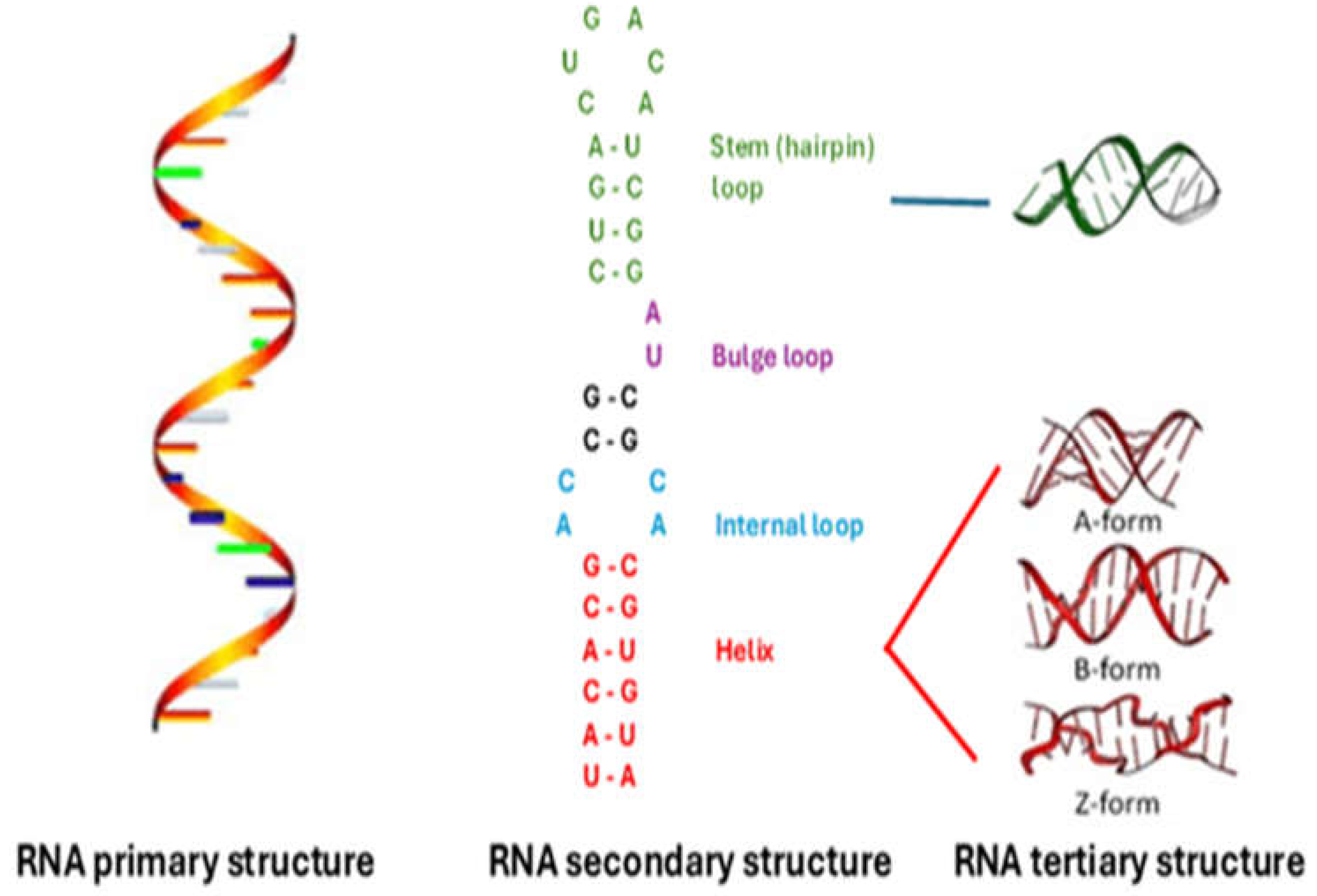
Figure 2.
H-type pseudoknot. Complementarity regions are shown as overlapping boxes.
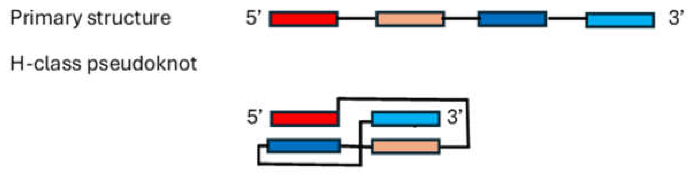
Figure 3.
RNA G-quadruplex. Guanine residues forming stacked tetrads are in red. Arrows follow the primary sequence.
Figure 3.
RNA G-quadruplex. Guanine residues forming stacked tetrads are in red. Arrows follow the primary sequence.
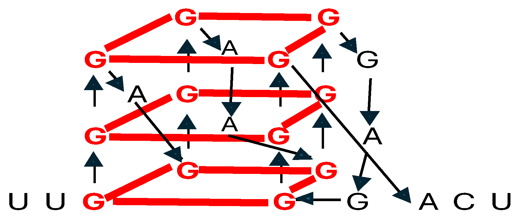
Figure 4.
RNA in gene therapy. RNA types used, challenges, and future perspectives.

Disclaimer/Publisher’s Note: The statements, opinions and data contained in all publications are solely those of the individual author(s) and contributor(s) and not of MDPI and/or the editor(s). MDPI and/or the editor(s) disclaim responsibility for any injury to people or property resulting from any ideas, methods, instructions or products referred to in the content. |
© 2024 by the authors. Licensee MDPI, Basel, Switzerland. This article is an open access article distributed under the terms and conditions of the Creative Commons Attribution (CC BY) license (http://creativecommons.org/licenses/by/4.0/).
Copyright: This open access article is published under a Creative Commons CC BY 4.0 license, which permit the free download, distribution, and reuse, provided that the author and preprint are cited in any reuse.


