Preprint
Review

EEG in Education: A Scoping Review of Hardware, Software, and Methodological Aspects

Altmetrics

Downloads

70

Views

34

Comments

0

Submitted:

06 December 2024

Posted:

06 December 2024

You are already at the latest version

Alerts
Abstract
Education is an activity that involves great cognitive load for learning, understanding, concentrating and other high level cognitive tasks. The use of the electroencephalogram (EEG) and other brain imaging techniques in education has opened the scientific field of neuroeducation. Insights about the brain mechanisms involved in learning and assistance in evaluation and optimization of education methodologies according to student brain responses is the main target of this field. Being a multidisciplinary field, neuroeducation requires expertise in various fields such as education, neuroinformatics, psychology, cognitive science and neuroscience. The need for a comprehensive guide where various important issues are presented and examples of their application in neuroeducation research projects are given is apparent. This paper presents an overview of the current hardware and software options, discusses methodological issues, and gives examples of best practices as found in the recent literature. These were selected applying the PRISMA statement on results returned by searching in PubMed, Scopus and Google Scholar with keywords “EEG and neuroeducation” for projects published on the last six years (2018-2024). Apart from the basic background knowledge, two research questions regarding methodological aspects (experimental settings, hardware and software used) and the subject of research and type of information used from the EEG signals are addressed and discussed.
Keywords: 
Subject: Computer Science and Mathematics  -   Mathematical and Computational Biology

1. Introduction

Almost a century after the first recording, EEG has provided precious information about the activity of cerebral neurons for scientific fields such as neuroscience and cognitive science. The technological developments in the area have provided portability and wireless connectivity to the EEG devices and they have facilitated the use of the necessary apparatus outside of medical laboratory settings. Thus, brain signals are being used for applications in human computer interaction (HCI) to control devices with brain computer interfaces (BCI) and examine the responses of the brain while performing various cognitive tasks.
Learning is a cognitive process which results in change of behavior and acquisition of new knowledge [1]. It is a lifelong process that helps organisms adapt to their environment. Education is an organized approach to learning where specific tasks, goals, methods, materials, and interactions are involved. Whether an educational method is successful or not it is a question of how well students perform in assessment procedures related to the taught materials. What happens within their minds though is a research question of psychology, cognitive science, educational science, and neuroscience.
The combination of the above fields have given rise to Neuroeducation, or educational neuroscience, or ‘mind, brain and education’ [2] for studying the activities occurring in the brain when individuals learn and applying this knowledge to improve teaching practices and optimize educational methods [3]. Although the gap between the neuronal functions and education might be too far to be the subject of an interdisciplinary field, as some early skepticism mentioned [4], there are quite a few examples where cognitive neuroscience has been successfully combined with behavioral data analysis in order to complement and extend existing knowledge of the relevant cognitive processes [2].
Brain imaging methods such as electroencephalogram (EEG), magnetoencephalogram (MEG), functional magnetic resonance imaging (fMRI) and functional near infrared spectroscopy (fNIR) have provided the necessary tools to study the operation of the brain while performing cognitive tasks. EEG is based on recording mainly the postsynaptic electrical activity of pyramidal cerebral neurons which are almost vertical to cerebral surface [5]. MEG records the related electromagnetic field alterations from the electrical activity of the brain [6]. The fMRI [7] and fNIR [8] are based on measuring the blood oxygenation levels alterations in the brain based on magnetic and optical absorption characteristics respectively. All the above methods are registering signals from the continuous operation of the brain. These signals convey information about the time and the place where action is taking place. However, apart from MEG which has both detailed time and space resolution, EEG is very precise about the time things are happening but lacks space resolution, while fMRI and fNIR have very high space resolution but lack specific time resolution since blood oxygen level variations do not happen concurrently with the corresponding neuronal activity. While spatial information is very important to determine functional and structural relations of brain areas, precise time details are also very important, especially when event related potentials (ERPs) are involved as explained later. As far as mobility is concerned, EEG and fNIR are the basic methods which can operate in out of laboratory settings while portable MEG systems based on optical pumped magnetometers (OPMs) are still in their early stages [9]. As far as cost is concerned, EEG is the most competitive method for limited budget projects.
The above characteristics of EEG have made this brain imaging method the main tool for research in cognitive science and related areas and scientific fields. As mentioned above, neuroeducation is such a field. Brain signals can be used to get insights of cognitive processes providing connections between their occurrences and the signal features appearing at the same time. Functional brain connectivity is a very important aspect to reveal coordinated brain activity, and different approaches, including deep learning, can be used to estimate coherence among neuronal areas [10]. Mental states during learning tasks, concentration, engagement, and arousal levels along with brain synchronization between the people involved are only some of the key performance indicators that can be used. This form of brain computer interfacing is defined as passive BCI in [10] and regards the analysis of brain activity of the user without any target monitoring. However, both the two other forms of BCI according to [10], active and reactive can be used. The former is related to the use of changes in the brain activity directed from the neurointerface operator for control commands and it is closer to the general idea of BCI. The latter detects and classifies brain responses to external stimulation. Passive and reactive BCIs seem to be closer to the needs of neuroeducation.
Regarding the use of biomedical information in education, a systematic review of Wearable Biosensor Technology in Education was recently made available [11]. A descriptive and bibliometric focused study in educational neuroscience is also available in [12]. As there is a large variety of research methodologies, metrics, scenarios and circumstances, and research targets that can be used in neuroeducation, a comprehensive guide describing basic issues and presenting current neuroeducation research practices is useful for entering this field from various disciplines. This is the motivation behind this scoping review which is focused on EEG and the research questions that are addressed in this paper are the following:
[Q1] What are the methodological aspects that are followed by the research projects in neuroeducation in terms of experimental settings such as sample sizes, hardware and software used?
[Q2] What is investigated in neuroeducation research projects? What is the subject of their research and what information is extracted and used from the EEG signals?
The rest of the paper is organized as follows: Section 2, Section 3 and Section 4 provide some necessary background knowledge before Q1 and Q2 can be faced. More specifically, Section 2 presents information regarding the information that can be extracted from EEG and the necessary steps. Section 3 outlines the hardware aspects of EEG devices and Section 4 discuss the software aspects and options that exist. Section 5 provides the methodology which was followed for the selection of the papers and the protocol for their review. The results are presented in section 6 to provide information for discussing about Q1 and Q2 in section seven. The paper concludes by summarizing the presented issues and discussing future trends in the field.

2. Information from EEG

Neurons are the cells of the neural system that are related to coding and transmitting information. There are about 1011 neurons in the human brain and each one of them can communicate with up to 10 thousand other neurons. In their generic form, neurons consist of (i) their cell body, (ii) the dendrites which receive chemical messages from other cells and (iii) an axon which transfers messages to other neurons, muscles, or glands. At the end of each neuron’s axon there can exist many endings which are connected to the dendrites of other neurons. These connections are called synapses, and they operate electrochemically using chemical substances which are called neurotransmitters. These can control the ion gates regulating input and output flow of charged ions thus creating electrical potentials. The action potential is created and propagates within the neuron from its dendrites to its axon endings and the postsynaptic potential (PSP) is the one which is created at the neurons inputs (dendrites). Although at a smaller scale from the action potential, PSP (~10mV) appears in a more synchronized basis with other neurons and has longer duration (~30ms). This is why PSP is considered the main source of the currents which are recorded from the EEG [5]. More specifically, the recordings of EEG come from the activity of the cerebral cortex neurons which have pyramidal form and are almost vertical to its surface. Since the human body is filled with electrically conductive material, these currents can be transferred to the outer surface of the scalp where they are recorded using the EEG electrodes [13].

2.1. Brain Waves

As mentioned, the signals which are recorded in EEG come from the PSPs of the neurons in the cerebral cortex. The amplitude and the frequency of these signals are related to the actions and the states of the brain (e.g. sleep, awake, active, concentrated, etc.). There are five basic bands of the human brain waves. In ascending frequency order, they are the following [13]: (i) δ (delta) from 0.5 to 4 Hz. These are usually related to deep sleep although they can be measured also in awaken subjects. (ii) θ (theta) from 4 to 7.5 Hz. These appear in relaxation states, drowsiness, and deep meditation. (iii) α (alpha) from 8 to 13 Hz. They are indication of relaxed state without focusing specific on something and they are usually recorded in the occipital lobe area (where the visual cortex lies) when subjects have their eyes closed. In this band, but mainly in frontal areas of the brain, in the motor cortex, the μ (mu) waves can be also recorded in relaxed states without movements. (iv) β (beta) from 14 to 26 Hz. These are related to concentration and focusing in solving some problem. (v) γ (gamma) from 30 to 45 Hz. These are not very frequent in normal conditions and they have low amplitudes.
The study of the characteristics of brain waves and their distribution on the scalp have created a link between them and specific states of mind. The degree of correlation or synchronization among signals from different electrodes can be a measurement of connectivity patterns [14]. Alpha band intensity ratios have been studied as indexes of mental workload where EEG features based on alpha-to-theta and theta-to-alpha ratios have provided high classification accuracy [15]. Engagement, relaxation, interest and focus are measured from EEG signals in [16], based in characteristic features extracted from the relations between the bands [17]. The use of high gamma band as a correlate for engagement is proposed in [18]. Frontal Alpha Asymmetry (FAA), defined as alpha activity difference between right and left hemispheres of the brain, serves as a biomarker for motivation and affect [19] and it is also linked to exaggerated physiological arousal [20] or avoidance and negation [21].

2.2. Event Related Potentials

One of the greatest advantages of EEG compared to other brain imaging methods is its very fine time resolution. Brain activity is recorded by its electrical representation at the very exact time instant that it happens [22]. Every time there is a stimulus that is perceived by the brain, a series of mental processes begin to analyze it and prepare a possible reaction. The corresponding electrical activity variations which are recorded in the EEG are called evoked potentials (EP). When the stimulus is related to higher cognitive functions that are taking place in the cerebral cortex and they are related to sensory, cognitive, affective or motor processes, these potentials are called event related potentials (ERP) and they can serve as a measure of the brain activity [23].
The basic characteristic of ERPs is the time dependance to the events they relate to and they are thus time locked with the events. The time in which they appear is called latency. Their intensity and their spatial distribution are also characterizing them although the spatial resolution of EEG is poor. That is, EEG can tell exactly when something happened in the brain, but an indication about the location must be derived after a series of data processing. ERPs are EEG fluctuations that are observed after an event and the polarity and latency of their signal variation can be used for their naming (e.g. a negative peak at 170 msec after the event is called N170). The time of the event is time 0. When latency is greater than 100, ERPs are related to the cognitive processing which is evoked and they are characterized as endogenous [13]. ERPs are considered the result of brain electrical activity in a collection of areas of the cerebral cortex where equivalent electrical dipoles correspond to electrical postsynaptic activity of sets of neurons [24]. That is, the ERPs which are recorded by the electrodes which are placed in specific locations of the scalp comes from the combination of currents created in various areas of the brain. An additional advantage of ERPs is that not only the time but also what is happening in the cerebral cortex in-between the event and the resulting action can be revealed [25].
As mentioned already, an initial naming convention for the ERPs was from the polarity of the greatest peak in the signal and its latency. Thus, there are ERPs with names such as P300, P3, N170, N400 etc. Some of the most well-known ERPs which are used in neurocognitive research are the following:
N170: it mainly relates to optical discrimination, face and familiar objects recognition [26]. N250 is also of a similar kind [27]. These ERPs are also called N1 and N2 and they also relate to inhibition in control tasks along with the P3 ERP that we present below.
N400: It relates to response to meaningful content in a flow of words that we read [28].
P3 (or P300): It relates to optical and audio oddballs and it is mainly traced in occipital lobe areas. The frequency of the oddball is in inverse analogy with the amplitude of the signal [29].
MMN (Mismatch Negativity): Similar to P300 but requiring no attention from the participant. It is associated with acoustic operation for oddball recognition and it is observed around 150-200 ms after the stimuli over fronto-central regions of the brain [30].
ERN (Error related negativity): It is related to acknowledgment of a wrong choice [31]. It is a negative variation of about 100 ms after the incorrect movement. It is mainly observed around the frontal lobe.
Reward Positivity (RewP): This is a positive waveform appearing in frontal to medial lobe areas in around 200-300 ms after the presentation of a positive feedback or outcome of an action [32]. It is related with ERN in the sense that it can be considered absent or suppressed following non reward [33].
The above ERPs along with many others (C1, N2pc, LRP, SSVEP etc.) are the basic elements in applications such as brain computer interfaces (BCI) and research in neuroeducation. The main problem in their use is the many preprocessing steps needed for their isolation as these EEG variations (1-30 μV) are observed together with the usual recorded brain activity (~100 μV). These issues are discussed in the next subsections.

2.3. EEG Analysis Steps

Due to the small scale of the currents that are being recorded and the various interferences that may exist, the raw EEG multichannel signal which is recorded by the electrodes may be contaminated with noise and artifacts of various frequencies and forms. Thus, a series of processing and analysis steps is necessary before any information can be extracted from the EEG.
High pass filtering with a cutoff frequency of 0.5 Hz and low pass filtering with cut off frequency in the range of about 50-70 Hz is the first step. The ubiquitous 50 Hz (up to 60Hz) signal due to the electrical power network can be eliminated with notch filters. Artifacts can be due to physiological or systemic factors. Physiological artifacts can be due to body movement and muscle activity, movements of the eyes or electrocardiogram (ECG) signal and sweating. Systemic artifacts are the power supply interference, cable defects, electrical noise and unbalanced impedances of the electrodes.
A recent presentation of the denoising methods and other processing steps in the pipeline of EEG signal analysis can be found in [34] and [35] and it is thoroughly presented in [36]. Linear regression, blind source separation (BSS), independent components analysis (ICA), principal components analysis (PCA), canonical correlation analysis (CCA), wavelet transform (WT), and empirical mode decomposition (EMD) are the main methods that are used [36]. The basic principle in most of them is the decomposition of the EEG signals, the detection and removal of contaminated parts and artifacts and the reconstruction of the signals.
Downsampling of the data to have files of smaller sizes which facilitate the computer analysis can be performed as well. Signal acquisition can take place at rates as high as 1024 Hz thus a conversion to 256 or 128 Hz would reduce sizes four or six times respectively.
After the above initial preprocessing steps, a feature extraction procedure needs to take place [14,37]. Joint time frequency analysis can be applied to data epochs to reveal the frequencies of the brain waves and their intensities in function with time. Segmentation in epochs is also very useful for extracting ERPs information. The triggering events are usually located at time 0 and a time window before and after this instant is included in the epoch. These epochs are then averaged together per channel to give a clear view of the ERP under examination.
Both classical signal analysis techniques and machine learning (ML) tools can be used for the classification and detection of patterns of neuronal activity during specific cognitive tasks. Representation learning using deep learning [38] can be used to transform raw data into representations of feature vectors needed for classification providing adequate levels of tolerance to accommodate variability among the users

3. Hardware Aspects

Down to its main components, a digital EEG device consists of the sensors, an amplifier and filtering unit, an analog to digital converter (ADC) and a communication unit. The EEG used in neuroeducation is non-invasive and electrodes are attached externally, on the surface of the skin. However, regarding other uses of EEG in brain machine interfaces (BMI) and especially for rehabilitation purposes, there is also the possibility for implanted electrodes, either to the surface of the brain (electrocorticography, EcoG) or even to the outer 1 to 2mm of the cortex (intracortical) [39]. Due to the external placement of the electrodes on the skin of the scalp, the voltages that are recorded are in the range of 1-100μV and this is a crucial factor in most applications that use EEG. This is because extra care must be taken in the correct placement of the electrodes.
Since the outer layer of the skin is a bad conductor of electricity, a gel might need to be used in order to lower the impedance between the skin and the electrode [40]. Although improving the signal to noise ratio (SNR), the use of gel is time consuming and not easy, especially when the EEG recordings should be in classroom conditions out of laboratory settings. The need to use the equipment in a more flexible and fast manner has made space to the development of portable, low-cost and consumer grade devices which can also communicate wirelessly with the signal acquisition computer. The existence of these devices has really made a revolution in the field of EEG research for non-medical purposes. A crucial factor for the existence of low-cost portable devices is the development of dry electrodes which allows faster and easier preparation. Beside the two options (gel or dry) there is also the use of wet electrodes which is a compromise between using gel and the related discomfort but better conduction, and the flexibility and fast placement of the dry ones. However, the use of dry electrodes seems to be achieving high standards obtaining comparable results to wet electrodes [41]. Another categorization of electrodes is whether they are active or passive. In the former case they coexist with a pre-amplifier so that the signal is directly amplified at the source before it is sent to the collecting module. The latter does not use preamplification at the electrode level. Although this can be important for the quality of the signal, the final performance depends also on other parameters [40].
EEG recording can be bipolar of unipolar. In the first case, electrodes are paired, and their potential differences are recorded. In the unipolar case, the recorded potential difference is between an electrode and a reference electrode which is common between more electrodes. Electrodes can be placed on the head either attached to an elastic cap following the standardized 10-20 set of positioning and naming [42] or they can be fixed on an elastic head strap, or they can be fixed on a special made spider-like plastic base. The first option is the standard way and have preset positions where 128 or more electrodes can be attached although usually much less are used. The second option is used by smaller scaled systems of 2 to 4 electrodes and the third option can be used with up to 14-20 electrodes.
Although there are on-going efforts to build open and do-it-yourself EEG devices, such as the openEEG project in [43], the more convenient case is to use portable, wireless, low cost consumer-grade devices. A very recent and complete survey of such EEG systems is presented by Niso et al in [40]. A previous similar review exists in [44]. As it will be observed from the presentation of some recent neuroeducation projects in the next section as well, the most common low-cost devices which are reportedly used in the literature are among the Emotiv (epoc, epoc-flex) openBCI (Ganglion and Cyton+Daisy) and InteraXon (muse 2 and muse S) companies. With a cost of either less than 1000 $ or up to 2000 $ they are an affordable and decent solution, especially in cases where many of them must be used concurrently to measure brain activity of groups of students in classroom settings. Ear-centered devices have also been presented and they appear to be an alternative to classical cap EEG recording for auditory attention monitoring [45].

4. Software Aspects

The role of the software used in EEGs is the management and analysis of the EEG data. All the digital equipment which can be used for EEG recording, from the least expensive till the highest quality medical devices, is accompanied by the relevant proprietary software which offers a specific range of processing, viewing, and storing options. However, the lack of compatibility in file formats and the limitation of data processing to the options provided by that software was an inhibitory factor in EEG research [46].
With the gradual emergence of open source EEG processing software and data standardization, the research community has been able to develop, modify, and use a variety of processing tools through these software packages [47]. Depending on processing requirements, user interface preferences, working environment and familiarity with programming, the options that exist nowadays are plenty. Some of these are presented below.
EEGLAB [48]. An integrated software package running on MATLAB (The Mathworks, Inc., Natick, MA, USA). It provides functionality both through command line and graphical user interface (GUI). It supports processing of raw EEG data and provides a set of ERP processing options. It can apply ICA analysis, create topographic maps of wave and energy distribution on the head following time and frequency analysis, artefacts detection, etc. The main EEGLAB reference article ([48]) has almost 22000 citations (Dec.2023) and maybe this fact by itself describes the great acceptance of this package. It provides levels of functionality, starting with command line functions and reaching GUI interaction for all operations. EEGLAB supports many datafile types (e.g. set, edf, edf+, bdf, gdf, mff, cnt, eeg) and its open architecture allows the creation of many plugins (~150) with many additional functionality and enhanced data handling. EEGLAB can also run on Octave [49] which is an open software alternative to MATLAB. Although EEGLAB can run on Octave, this compatibility is not yet guaranteed for its extensions, such as ERPLAB which is an EEGLAB plugin presented next. However, due to the grant privileged pricing options to academia [47], the use of MATLAB as a basis for software packages can be beneficial in academic research.
ERPLAB [50]. As mentioned above, this is an EEGLAB plugin. Its aim is to provide extra functionality for ERPs processing, visualization and analysis. It offers enhanced filtering capabilities, reference point alteration, data preprocessing, epoching and artifact detection.
Corrmap [51] is also a MATLAB toolbox for performing ICA and semi-automatic clustering of independent components to artifacts and brain-related signals from EEG recordings.
OpenViBE [52]. An open software platform that provides functionalities for designing brain-computer interfaces, interfacing them with virtual environments and enabling visual feedback of brain activity in real time. It has a set of modules for the acquisition, preprocessing, main processing, and visualization of EEG data that can be interconnected according to the user's requirements using a graphical interface. The user can also add new modules according to their requirements as a software development kit (SDK) in C++ is provided.
Fieldtrip [53]. A software toolkit that runs on top of MATLAB and can be used to analyze data derived from MEG, EEG and other biosignals. It does not use a GUI but provides a large set of high and low-level functions. The former are used by users while the latter are largely hidden from them.
BCIPy [54]. Python-based software focused on research in brain-computer interface systems using ERP interfacing for text dictation. It can also be of more general use and supports data acquisition, stimulus presentation, data search, signal processing and visualization, language modeling and uses a simple graphical user interface.
MNE [55]. Software package in Python that provides analysis and workflow tools for preprocessing, source localization, analysis in the time and frequency domains, statistical analysis, and methods for estimating functional connectivity between brain regions.
BIOSIG [46]. An open-source software library for processing a variety of biomedical signals such as EEG, electrocorticalogram (ECoG), electrocardiogram (ECG), electroophthalmogram (EOG), electromyogram (EMG), etc. Tools are provided for many applications, such as BCIs, neurophysiology, psychology, etc. Options are also given for data acquisition, artefact processing, quality control, feature extraction, categorization, modeling, visualization, etc. Originally for MATLAB and Octave, it also enables use with C++, Python, Java and R.
PsychoPy [56]. An application for creating experiments which offers ease of use due to its graphical interface and flexibility due to the Python scripts availability. In comparison with the previous mentioned software tools, PsychoPy is not concerned with EEG processing. Its main role is to conduct experiments in behavioral sciences and neuroimaging experiments. It can present stimuli on a computer screen and communicate with the EEG device which is being used, to send a “trigger”. This is especially useful in experiments involving ERPs where the exact time of the stimuli onsets must be recorded along with the corresponding EEG data. The trigger signals can also be sent by network connections allowing for remotely recording and experimentation.
Stimulus presentation can also be created and controlled by Psychophysics (Psychtoolbox-3) which is a MATLAB toolbox fully compatible with Octave [57,58].
The above were some of the software options which can be employed in experiments involving EEG such as the ones in Neuroeducation. These are mainly open software solutions while at the same time, the proprietary software which comes with the EEG devices can offer a range of processing and analysis tools.

5. Methodology

As mentioned in the introduction, the purpose of this paper is to provide a comprehensive guide and survey to the current state of the multidisciplinary field of neuroeducation by examining relevant projects based on using EEG. The kind of information that can be extracted from the brain signals and the relevant hardware and software options were discussed in the previous sections to provide an initial background knowledge so that projects related to neuroeducation can be presented next. Thus, recent neuroeducation research projects, as found in the literature searching in PubMed, Scopus and Google Scholar with keywords “EEG and neuroeducation” are presented in the following section focusing on their methodological aspects regarding Q1 and Q2. The Preferred Reporting Items for Scoping Reviews and Meta-Analyses (PRISMA) statement [10] was followed for the selection of the papers and the corresponding flow diagram is depicted in Figure 1.
The search was focused on neuroeducation projects published on the last six years (2018-2024). The initial screening of the papers was based on their keywords and abstracts. Focus on neuroeducation using EEG, clear presentation of research targets, methodological issues, and detailed explanation of the research procedures regarding hardware, software and samples used were the basic criteria for the inclusion of a paper in this review. Research projects also had to be published in peer reviewed journals or international conferences. PubMed returned 8 papers and 6 of them were included in the review. Scopus returned 20 papers and 13 of them were included in the review. Google scholar returned 485 results and 8 of them were included in the review as they were not in the results of the previous databases.

6. Results

This section briefly presents a collection of recent and characteristic neuroeducation projects focusing on their methodological characteristics as well as the hardware and software options that were used.
The efficacy of Just in Time Teaching in anatomy education, where preclass exercises are used from instructors to establish foundational knowledge in novice learners and focus on more complex concepts, was tracked using EEG in [59]. Two ERPs were used as metrics in that research. The first was the N250 and the second was the RewP (Reward Positivity). As presented earlier, N250 relates to object recognition and increases in amplitude with perceptual expertise [27]. RewP is associated with evaluation of performance feedback [32] and decreases with the enhancement of evaluating the correctness of a response as the subject learns. The above ERP behaviors were observed in the experiments with 24 (11 male + 23 female) participants of 20.74 years mean age (m.age), where computer-based learning modules were used. The Psychophysics Toolbox extensions [57,58] and a customized MATLAB script were used for questions presented on computer screen. The actiCAP XPress EEG with 16ch V-Amp system (Brain Products, GmbH, Munich, Germany) and Brain Vision Recorder Software (Version 1.20, Brain Products, GMbH, Munich, Germany) were employed for EEG acquisition.
ERPs were also used as metrics in [60] were the level of diagnostic accuracy in images of bovine pathology was under examination whether it was the result of image recognition or acquisition of diagnostic expertise. The ERPs that were used were RewP and the N170. As mentioned above, RewP is associated with evaluation of performance feedback. N170 is related to optical discrimination and recognition of faces, familiar objects or words [26]. According to the study in [60], while the amplitude of the RewP decreased with learning in novices it had remained significantly different from the corresponding ERP in experts where it did not change throughout the course of the experiment. Regarding N170, there was no change in the signals from the novices and they also remained significantly lower in amplitude comparing to those of the experts. MATLAB and the Psychophysics Toolbox extensions were again used for displaying the questions to the subjects as in [59] and the actiCAP slim with 16ch V-Amp EEG system (Brain Products, GmbH, Munich, Germany) and Brain Vision Recorder Software (Version 1.20, Brain Products, GMbH, Munich, Germany) were again employed for EEG acquisition. Twenty-six undergraduate students (23 f. +3 m.) were recruited as novices with a mean age of 19.8 years. Six female and three male experts with a mean age of 44.1 years were recruited as experts.
The negative value of the means of EEG, in addition to the time needed to perform programming tasks were used as metrics in a research about the brain activity of computer science students in visual and textual programming [61,62]. The BIOPAC MP150 data acquisition unit and AcqKnowledge 4.3 Software was used for data acquisition, analysis, storage, and retrieval. Eight volunteers, first year computer science students, were recruited (3 f. and 5 m.) and the research was carried out in university labs in a properly designed place. The negative value of the means of EEG is considered to indicate a higher cognitive response [63], and that is the reason that it is being used as a metric in this study. The need for differentiated education programs depending on learning skills and cognitive abilities of the students was stated in this study. Also, the invariance of the programming language used, was also in the results. The small number of students for the experiments and the necessity of special places to perform the EEG measurements were mentioned as limitations of this study.
Another research which is related to computer programming learning and knowledge transferring is presented in [64]. The relationship between cognitive load and prior knowledge as indicated by the working memory usage is investigated in a project with eight male (8 m.) participants, 18-21 years old which were about to learn the C programming language. Their prior programming knowledge varied from beginner to average creating two groups; sufficient previous knowledge (SPK) and insufficient previous knowledge (IPK). EEG data were collected using the EPOC-X device while the participants were completing 17 learning tasks related to computer programming and presented with the OpenSesame software. EEGLAB was used for filtering and ICA. The spatio-temporal brain data (SPBD) were provided to NeuCube software to create neural connections and connectivity patterns using computational models based on Spike Neural Networks (SNNs). Percentages of the brainwave bands theta and alpha during the learning tasks, related to cognitive load, were also considered. The research demonstrated that neuronal connectivity and spike activity patterns as visualized in NeuCube can be used to understand and interpret brain activity patterns during the transfer learning process
Brain synchronization and engagement was under investigation in [65] where the inter-subject correlation (ISC) [66] was measured in EEGs recorded by 42 female students of m.age=22.4 watching videos in a classroom. The feasibility of performing ISC using low cost comfortable and affordable equipment such as the Smartphone Brain Scanner [67] which uses a modified version of Emotiv EPOC was successfully demonstrated. EEG processing was performed with the Corrmap plug-in [51] for EEGLAB [48].
Spatial reasoning is the subject of the research in [68] where the learning of the law of reflection was examined using the N2 and P3 ERPs and joint time frequencies (JFTs) of alpha waves at sites F3, F4 and Pz while learning through video game play. These neural correlates together with behavioral measures of performance in learning the concepts were investigated in 21 children (11 boys and 10 girls) from 6 to 12 years old. EEG recording was performed using a NetAmps 300 system from Electrical Geodesics Inc. (EGI, Eugene, OR) with the help of Netstation 4.5 software of the same company. Suppression of alpha band during correct trials at site F3 was observed and it relates to learning. Additionally, children under seven had difficulties learning the concept of the reflection law.
The exploration of the effectiveness of an Augmented Reality (AR) application to assist mobile users learn mobile photography concepts is presented in [16]. Emotional state indexes for engagement (ENG), relaxation (MED), interest (INT), and focus (FOC) are measured from the EEG of the users of this app. Twenty-eight participants (7 m. and 21 f.) of m.age=20 were divided into two groups in half. An Emotiv EPOC+ is used for this study with 128Hz sampling frequency and EEG processing is performed by the EmotivPro software which also extract the emotional state indexes from the raw data. As it is mentioned in Emotiv knowledge base [17], ENG is characterized by increased physiological arousal and beta waves along with attenuated alpha waves. INT is the degree of attraction or aversion to the current stimuli; MED is a measurement index of the ability to switch off and recover from intense concentration and FOC is a measurement index of the fixed attention to one specific task. The effectiveness of the AR-based mobile photography application (ARMPA) compared to using a typical 2D mobile application is observed and verified by the results of the pre-post-tests and also by measures of cognitive load [69] and the emotional state indexes mentioned above. According to them, students expend the least mental effort when using the ARMPA and they are more engaged to the learning task.
The potentialities and the issues concerning the use of portable EEG devices in educational contexts is presented in [70] where a case study is performed with a group of third-grade primary school students. The disruption of everyday life in the classroom, the collaboration between educators and researchers and the balance between budget, ease of equipment preparation and usefulness of the collected data is discussed. Also providing an overview of recent studies using low-cost EEG devices, the paper describes a study conducted with a group of 17 third-grade primary school children aged between 8 and 9 years (10 girls and 7 boys) in a regular school with which the researchers collaborate. Familiarity between researchers and educators and participants is very important in these contexts. Two baseline recordings of EEG activity were made, two minutes with eyes closed and two minutes with eyes open. The brain activity of the students listening to different explanations about mathematical applications and performing some arithmetic tasks was collected. Four types of portable devices were used: i) Brainlink Pro, 512Hz, two dry sensors, Bluetooth connection to Lucid Scribe software, ii) Emotiv Epoc, 14 channel EEG semi-dry sensors, 128Hz. Data collected with Emotiv TestBench software, iii) Epoc Flex, 32 channels, 128Hz, passive Ag/AgCl gel sensors mounted on a neoprene cap. EmotivPro software for data collection and analysis, and iv) The Muse, 4 channels of dry contact electrodes from frontal and tempo-parietal lobes areas, 250Hz, Mind Monitor application for data collection. EEGLab was mainly used for the EEG data analysis. As the main purpose of this study was to examine the issues and the methodology for EEG analysis in classroom settings, the sensations the students had with the devices were also collected with questionnaires.
The effects of context and gender in physics problems was investigated in [71]. The study focused in triggered situational interest. Being closely related to engagement, situational interest has behavioral, cognitive, and affective dimensions which were measured using psychophysiological tools including pupillometry, electrodermal activity (EDA), facial expressions processing and electroencephalography. The results were obtained combining the information from each measurement, with pupillometry and EEG relating to cognitive engagement, EDA to arousal and emotional engagement and facial expressions to emotional valence respectively. Regarding EEG, the quotient of beta and the sum of alpha and theta power spectral density (PSD) in F3, F4, O1 and O2 scalp sites was employed for generating cognitive engagement indexes (CEI) every second averaging over Hanning sliding windows from the last 20 sec. Sixty participants (32 f. and 28 m. of m.age=23.7) were asked to read and solve 21 physics multiple choice questions of three different types depending on the kind of context they used: Seven with technical (T), seven with humanistic (H) and seven with no context (N). A BrainVision BrainAmp with 32 channels with ActiCap active electrodes and the BrainVision Recorder software was used. Experiments were carried out in lab environment and the surprising result (as the authors are mentioning) that decontextualized problems (N) produced significantly higher performance that the others kinds (H and N), was found.
The levels of attention measured when a binaural wave stimulus was being heard by the students were examined in [72]. The research had two participants, Science Department students aged in the range from 20 to 28 and having a similar degree of knowledge. The Neurosky Mindwave portable EEG device was used, and data processing and analysis was performed by the accompanying Effective Learner application. It was observed that a frequency induction of a binaural wave at 10Hz helped increasing the levels of attention during tasks such as viewing a video and reading of a text. The duration of the EEG recordings was varying from 2 min to 7 min.
The differences and similarities in the brain responses when performing readings and tasks in digital and paper textbooks were investigated in [73]. Apart from EEG they also collected eye tracking, galvanic skin response (GSR) and facial expression analysis (FEA) data. An emotive 14 channels portable device was used (probably EPOC-X although it is not mentioned specifically) and the authors used the final metrics provided by the dedicated software of the devices without further processing. The sample consisted of three participants, two girls and a boy, in the sixth grade of primary education (around 12 years old) from different schools. Although the results obtained were mostly similar for both types of textbooks, a preference of the paper textbook was noticed in one participant. This was indicated by the frontal alpha asymmetry (FAA) metric, which was positive and related with motivation of focus, liking and attaching towards the textbook.
The frontal ERP P300 assymetry (fP3As) was investigated as a possible biomarker of cognitive control abilities related to judging performance during school days (Mondays and Wednesdays) and times (morning and afternoon) in [74]. This was to reveal important aspects of students learning such as school starting times, testing times, sleep/nap and break duration guidelines to optimize student performance. The participants were 24 female high school students in SouthWestern Canada with m.age=16.9 years and they performed a Stroop-like word-colour judgement task randomly assigned to two groups (morning or afternoon test groups). The words were created with the Stim2 software and reaction times (RTs) and accuracy were recorded for each trial. The fP3A was determined via subtraction of the P300 amplitude in electrode F3 from the one in F4. EEG data were recorded with Neurosoft, Inc. EEG with nine electrodes, and they were amplified and digitized with a SynAmps2 EEG system (Compumedics Neuroscan) at 1000Hz using the SCAN 4.3 software. High pass filtering at 0.15 Hz and 100 Hz low pass were applied. In summary, behavioral, and neural evidence was found to relate congruence judgement especially in the mornings supporting a misalignment between adolescent circadian circle and school start time.
Inhibitory control in negative priming (NP) is the subject of the research in [75]. The concept in NP is that if a stimulus has been previously ignored on purpose, subsequent reaction to the same stimulus would be impaired. This can cause a slower response time (RT) or a higher error rate. In [75], the process under investigation is the use of heuristics to solve fraction comparison tasks where one needs to think that a larger denominator leads to a smaller fraction and needs to inhibit the “larger natural number -larger fraction” strategy. Twenty-eight undergraduate students participated in the project with m.age = 20.8 years and none of them were math majors. The metrics used were the N1, N2 and P3 ERPs which are related to inhibition in control. EEG data were recorded from 64-channels using Brain Products (Germany) apparatus. Six electrode sites (Fz, F3, F4, Cz, C3 and C4) were used for N1 and N2 and nine sites (Fz, F3, F4, Cz, C3, C4, Pz, P3, and P4) for P3. Analysis of the data was performed with Brain Vision Analyzer 2.0. The study revealed that adults still needed to inhibit misleading strategy when performing fraction comparisons.
Again the role of misconceptions, while studying biological phenomena this time, was examined in [76]. Inhibitory control plays an important part in this procedure where students must suppress tempting but inaccurate answers. The persistence of the “moving things are alive” heuristic into adulthood was the subject of the current research. Twenty-eight undergraduate students were recruited (13 f. and 15 m., m.age=23.7 years). Images of intuitive (I) and contra intuitive (CI) drawings where the participants had to select the ones depicting living beings were displayed to the participants. EEG was recorded with Brain Vision Acticap, 64ch. Stimuli were presented using the E-prime 2.0 software and data were analyzed using Brainvision analyzer v2.0. The N2 and LPP (Late Positive Potential) ERPs were used as metrics. The results indicated that identifying the living thing in the CI condition took significantly more time than in the I condition, and that the N2 and LPP components were greater in the CI conditions. This is an indication of the suppression of the heuristic that must be applied before the decision.
Intuitive thinking and analytical thinking were the subjects of the research in [77]. The former is mentioned as System 1 and the latter as System 2 in bibliography. A simple task as repeating four one-digit numbers adding either 0 or 1 to them was employed for the EEG and pupillometry measurements. Thirty undergraduate students (m.age=22.8, 22 f. and 8 m..) were recruited for the research. The Psychophysics MATLAB toolbox was used for the experimental task and EEG data were recorded with ActiCAP (Brain Products) 64ch layout with ActiCHamp using Brain Vision software (v.1.10). EEGLAB and R language were also used for statistical analysis and graphical representations. The metrics used were the alpha and theta activity in parietal and frontal areas respectively. It was demonstrated that cognitive control, working memory, attention and long-term memory are involved in System 1 and 2 thinking demonstrating an increased parietal alpha (at CPz) for the former and a decreased frontal theta (at Fz) for the latter. These results are in accordance with previous research findings.
An investigation related to the stress, attention, interest, and engagement levels in onsite and online higher education is presented in [78]. Eye tracking, galvanic skin response and EEG were used in a sample of 20 postgraduate students (ages 22-25, 50% male, 50% fem.). The level of the brain waves was used for the metrics with the variation of the β wave correlated with attention while α is strongly correlated with meditation. Commitment, which is an indicator of motivation, was reflected in the β/(α+θ) index. The Emotiv EPOC+ was used for the EEG measurements with EmotivPRO v.2.0 software. Statistical analysis was performed with R language (v.3.6.3). The results of the research demonstrated that onsite learning is related with more attention, interest, and emotional intensity levels although stress levels are higher.
The differences in brain activity whilst performing various types of throwing games in children was explored in [79]. The power spectral densities of the brain waves were compared in three types of games: (i) throwing to a goal, (ii) throwing simultaneously with another opponent to the same goal and (iii) just throwing. Eight children volunteers (4 boys and 4 girls, m.age=7.2) participated in the experiments. EEG recording was made using a portable Emotiv EPOC-X headset with 14 recording channels and 2 reference electrodes. Artifact cancelation and notch filter (at 50Hz) was applied by the device. The Emotiv Brain Activity Map and Emotive TestBench applications were employed for the spectral analysis. Further pre-processing and ICA decomposition were performed by EEGLAB. The most significant differences in brain activity among the three types of games were observed in beta oscillations. The goal and simultaneous, (i) and (ii), types of games presented significantly higher values in beta band power spectral densities compared to just throwing. At the same time, the goal case exhibited significant higher values than ‘simultaneous’ in the same band, indicating the association of beta waves with decision making processes.
An effort to monitor the cortical activities of children and adults while completing complex cognitive tasks in the form of Schulte tables is presented in [80]. In their research, 12 children (3 f. and 9 m, aged 7-8) and 10 adults (3 f. and 7 m., aged 18-20) participated in the experiments. The tasks were considered difficult for the first group and easier for the second group due to the age and the relevant skills difference. Visual search, working memory and mental arithmetic were employed for the tasks. The actiCHamp (Brain Products) EEG amplifier and ActiCap electrode sensors with 32 channels were used for EEG recording. The EEGLAB and Fieldtrip MATLAB toolboxes were used for preprocessing and wavelets analyses. Response times and brain bands (δ,θ,α,β,γ) power spectral densities were used as metrics for the performances. The use of different problem-solving strategies (e.g. procedural vs fact-retrieval) according to the age groups and experience gained was indicated by the results which are trying to shed light on brain development and age-related changes in cognitive processes.
The role of the reward positivity ERP (RewP) and whether this component reflects an underlying learning process is the aim of the research presented in [81]. Thirty undergraduate students (23 f. and 8m., mean age=20) participated in the research where they had to learn to predict a disease type based only on health indexes information and positive or negative feedback upon correct or incorrect predictions respectively. The experimental setup included the registration of responses on stimuli controlled by the Psychophysics toolbox. EEG was recorded using 64 channels ActiCAP and ActiCHamp amplifier (Brain Products) and analyzed with Brain Vision Analyzer software (version 7.6). According to the researchers, RewP reflects reward learning systems in the brain as its amplitude is diminished with learning. The same research question about the role of RewP is examined in a similar study by mainly the same team of researchers in [82]. In that research, a computational model and 30 human participants learn 60 words of a novel language.
Whether or not action is required to elicit a RewP and the impact of cue, choice and action on its amplitude is investigated in [83]. Twenty six students (13 m. and 13f., m. age=21,54) participated in the experiments where they completed four versions of a computer-based guessing game with different degrees of choice of the winning actions from them. EEG data were collected and amplified by ActiCHamp (Brain Products) and preprocessing was done in Brain Vision Analyzer (version 2.1.2). EEGLAB was also used for the analysis of the data. The results indicate that agency, which is defined as the sense of control over our actions and their outcomes, affects the generation of RewP supporting the existence of a reinforcement learning (RL) system in the human brain.
The cognitive activities and performance on complex tasks was investigated in [84]. Cognitive load metrics were acquired from twenty-three university students (10f. and 10m. aged from 18 to 23, m.age=19.25 and s.d=1.19) while performing listing and concept mapping tasks related to sustainability issues such as climate change, food systems, renewable energy or water availability. Subjective behavioral metrics based on the NASA Task Load Index (NASA-TLX) and the Systems Thinking Scale Revisited (STSR) questionnaires were collected along with EEG measurements from the B-Alert X10 EEG system by Advanced Brain Monitoring (ABM). The cognitive load was estimated using the alpha/theta waves energy ratios in a second-to-second basis.
A data set of parallel EEG recordings between subject pairs is presented in [85]. The public availability of synchronized EEG data is very important and can provide a platform for experimentation and benchmarking for hyperscanning and new processing algorithms exploring temporal, spatial, spectral and functional synchronization aspects [86]. The data set includes recordings of 16 subject pairs of one female and one male aged 18-24 and consists of four sections for each subject. Two sections of 1 min each correspond to the resting states of Eyes Open and Eyes Closed and two sections of 10 min each corresponding to the parallel performance of a collaboration (solving a 100-piece puzzle) and a competition task (playing a domino game). Half recordings have taken place face to face and half online and they include four channels with signals from A1, A2, C3 and C4 electrode sites and they were obtained using a pair of Enophones (Eno, Montral, CA) wearable EEG devices and EEGLAB was used for the preprocessing.
Another dataset of EEG and other physiological signals is published in [87]. It consists of 24 recordings of students (10 m. and 14 f., aged between 18 and 25 years old, m.age=21.33 and s.d=1.4) engaged in learning experiences in Humanities under two scenarios; One is traditional learning and the other is with partially immersive learning experiences. Along with the physiological data a set of psychometric data was collected from the participants through questionnaires. OpenBCI Ultracortex Mark IV headset was used for the EEG recordings at 250Hz using eight dry electrodes located in positions FP1, FP2, C3, C4, P7, P8, O1, and O2 based on the 10-20 system. The OpenBCI_GUI software and EEGLAB were used for processing the recordings. Four sessions lasting three minutes each were recorded from each participant preceded by two minutes of resting state (eyes closed and eyes open for one minute each).
Brain to brain synchronization, or hyperscanning, during remote learning sessions was the subject of investigation in [88]. The participants were 10 undergraduate information technology students from the United Arab Emirates University, aged from 18 to 28 years old and an instructor. EEG data were captured using a wireless Unicorn Hybrid system by g.Tec of 8 channels at 250Hz. The channels corresponded to positions Fz, C3, Cz, C4, Pz, PO7, Oz, and PO8, attaching the ground and reference electrodes to the participants’ mastoids. Cross correlation of ten student: instructor pairs time series EEG signals were produced and synchronization percentages were calculated. Correlation matrices in the frequency domain were also produced and patterns of similarity were detected using KNN classification.
An open-loop neuroadaptive system is proposed in [89]. The aim of this system is to assess the cognitive abilities (CA) of the students and then suggest ways for their enhancement. This process is performed sequentially in an open loop manner. The system operates in five stages: experimental session, data processing and formation, verification and implementation of recommendations for CA enhancement. The types of the assessed CAs include visual search, working memory, mental arithmetic and ability to combine the above. Experimental studies of the proposed system were conducted using the LiveAmp EEG device (Brain Products) to register the brain signals using 64 channels at 500Hz sampling rate and EEGLAB for their preprocessing. The sample consisted of 60 young students of two age groups, 9-10 years old and 11-12 years old and there were 36 boys and 24 girls. 15 participants were excluded for various reasons. Behavioral (correctness and time needed) and physiological characteristics (normalized alpha band power variance) were used for attention and fatigue. Electrooculogram (EOG) extracted from EEG were used for the latter.
The above project descriptions are summarized in Table 1.
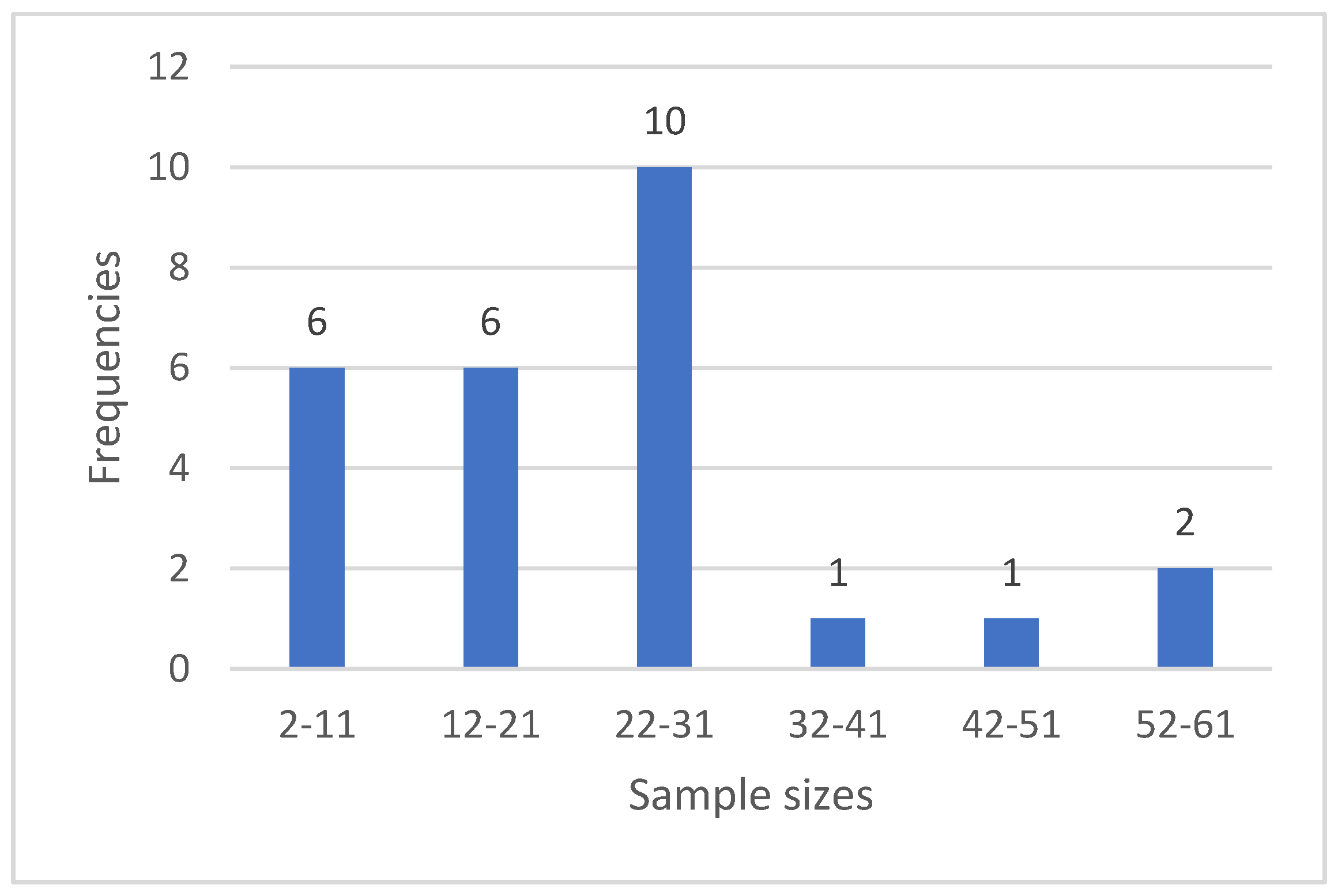
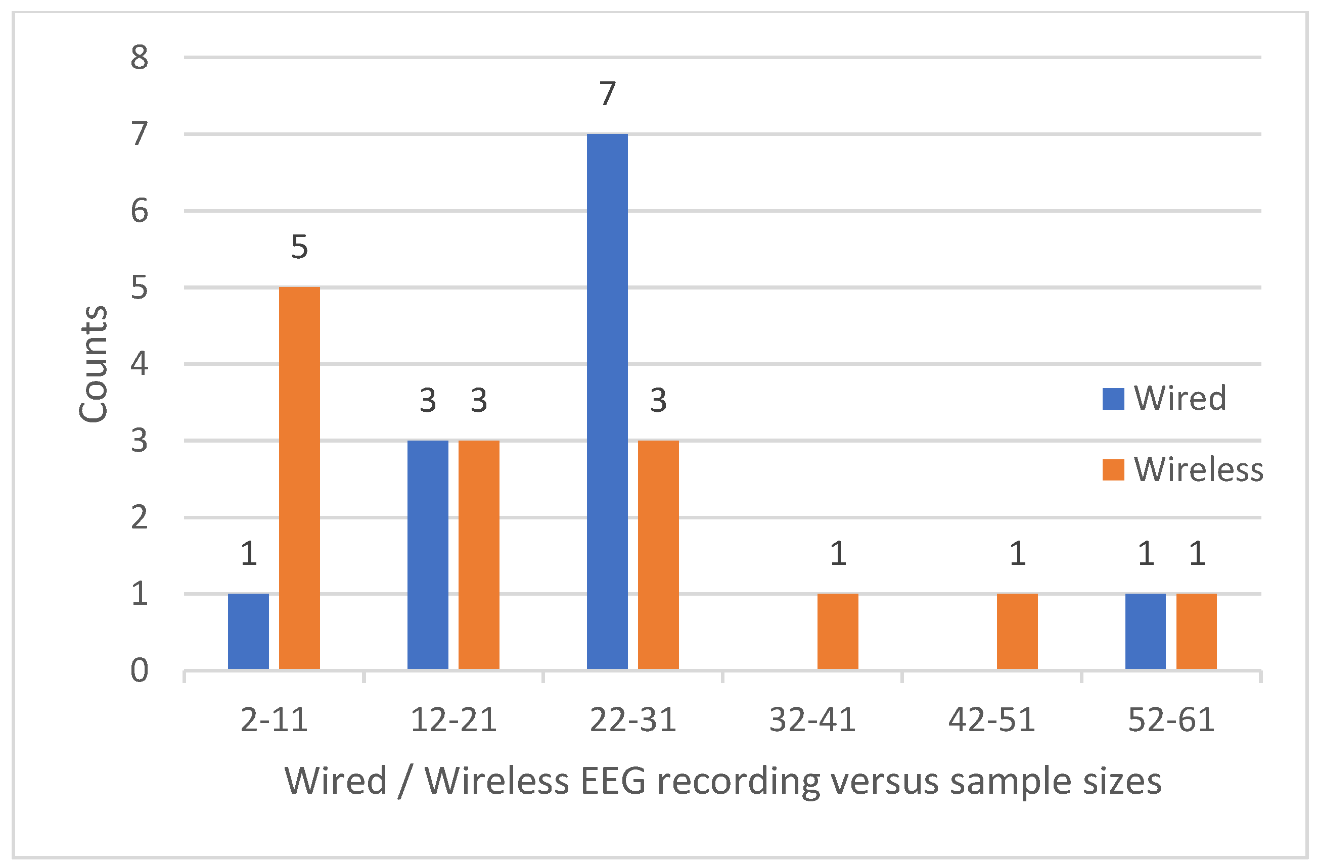
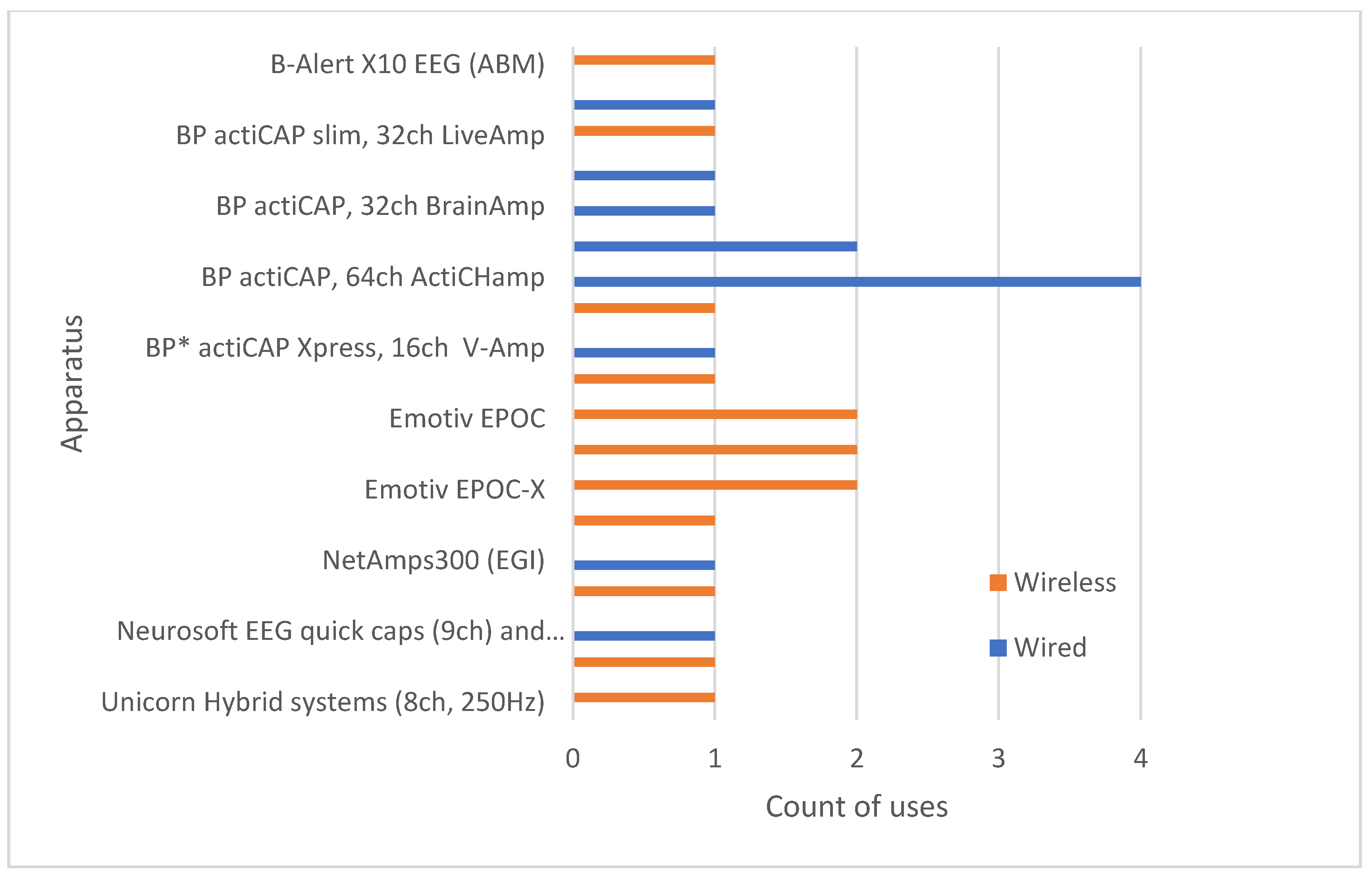
In the following figures we can see the frequencies of the sample sizes (Figure 2), the frequency of using wireless systems (Figure 3) and the frequencies of each EEG system (Figure 4).
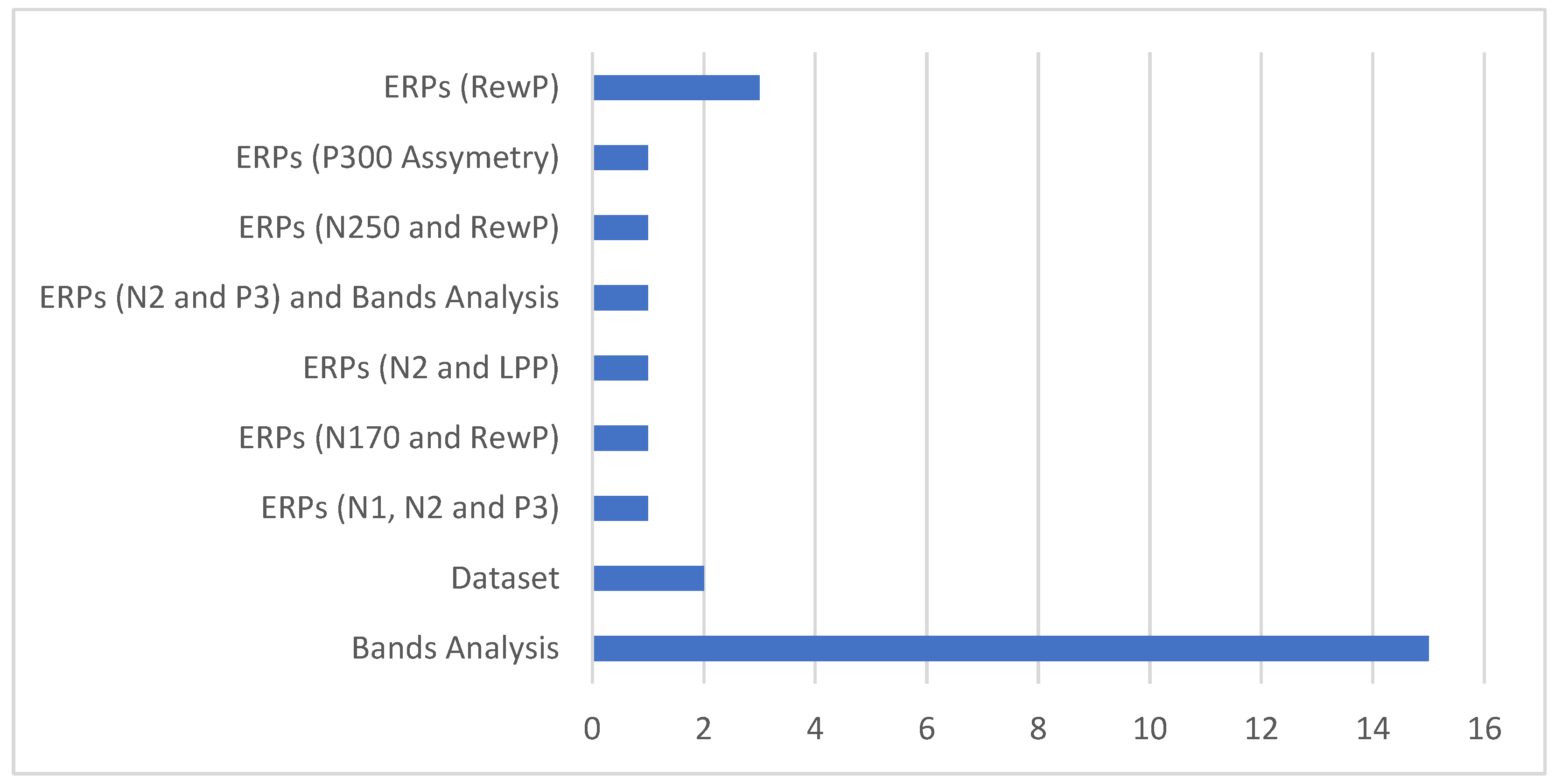
The ways in which the EEG signals were used can be seen in Figure 5.

7. Discussion

A variety of neuroeducation projects were presented in the previous section. The discussion in this section is organized according to the research questions set at the introduction.

7.1. Q1: Methodological Aspects, Experimental Settings, Sample Sizes, Hardware and Software Used

A challenge for neuroeducation as presented in [2] is the lack of understanding between neuroscientists and educational researchers. Additionally, the highly controlled experimental settings used in neuroscience are not the typical natural environments where education is taking place. The availability of EEG apparatus, the comprehension of the relevant research methodologies, and the effectiveness of their applications in experiments is an important factor towards the bridging of these two worlds.
As mentioned in [70], some of the issues in neuroeducation research are the dominance of studies with university students while at the same time studies with primary school students are not very often. This is mainly due to the organizational difficulties with studies involving pupils. However, learning at this period of life is much more vivid related to later stages of education [90] and it is worth arriving to a methodological framework allowing its study without disrupting everyday school life. This brings also the issue of “naturalism” [91], regarding the research environment; controlled laboratory, partial naturalistic laboratory and naturalistic research. The latter is an ideal setting to register brain function while learning in natural conditions. However, it is the most difficult as well due to noise and interference problems which are highly expected at these conditions. Low-cost devices can offer portability, wireless connectivity and ease of use. These characteristics make them ideal for long scale studies in naturalistic environments although the noise issues must be faced to a higher degree compared to more controlled environments. These issues exist in the projects that have been examined; The mean sample size is 23.32 ranging from 2 to 60. The frequencies of the sample sizes in groups of ten are depicted in Figure 2 where it is shown that the most frequent sample size belongs to the zone 22-31 with 10 occurrences. At the same time, smaller sample sizes are more frequently met (12 times) than higher sizes (4 times). Six papers describe projects with children and twenty with adult students. That is, almost 75% of the projects involve students and adults in general.
Portable low-cost devices are used in 11 of the cases examined in Section 6 (Emotiv EPOC and Flex, OpenBCI, Neurosky, Enophones, Brainlink Pro and Unicorn) while medium to high cost devices are used for the rest of the projects. This is depicted in Figure 4. In the same Figure it is shown whether the apparatus is wireless or not. Wireless equipment is being used in 14 cases and wired connections are used in 12 cases. From the 6 projects with children, wireless devices are used in 4 of them (Emotiv EPOC-X, Flex, Muse, Macrotellect Brainlink pro and BrainProducts LiveAmp). Half of the twenty projects with adults are using wireless devices and the other half are using wired ones. Wireless devices are being used in all sample sizes but mostly in the group 2-11 as it is shown in Figure 3.
Together with the variety of low to medium cost EEG apparatus to perform the experiments, there is also a variety of software performing the necessary signal preprocessing and analysis procedures. Availability of raw data can enhance these procedures with new processing methods and tools when open source and/or extensible software toolboxes and packages are used. It is therefore a very important characteristic when it is offered, as most of the low-cost devices prefer to pair with their proprietary software and provide this availability, of raw data, at an extra cost [44]. A combination of different software packages depending on the level of analysis which is required in each case was observed. Both open source and proprietary software is being used, the latter in combination with the corresponding EEG apparatus which is being employed in each case.

7.2. Q2: What Is Investigated, What Is the Subject of Research and what Information is Extracted from EEG Signals

Efficacy of learning methods [16,59,60], students’ performance [61,62], brain synchronization [65,88], creation of datasets [85,87], examination of differences among different groups and factors [71,72,73,74,78,80], reinforcement learning and reward processing [81,83], cognitive load and abilities measurements [84,89] and forms of thinking and reasoning [68,75,76,77] were among the subjects of investigation.
Attention and engagement as markers towards the “gestalt”, the feeling of a class as a distinct organization with its own “atmosphere” and mentality, can be derived using EEG technology [92]. Brain synchronization among the people involved in the teaching process (other students and instructor) is also very important. The “synergy zone” as is mentioned in [92], the ideal learning classroom environment, depends on factors such as engagement, attention, connection and enjoyment creating flow [93] and brain synchronization. The latter is investigated in [65] and [88] while a dataset of parallel EEG recordings is presented in [85].
Active learning, where students learn by doing, provides a special experimental ground from where important conclusions can be derived regarding educational procedures. The use of virtual and augmented reality (VR and AR) tools in the learning process can also provide a very interesting experimental ground [16,87]. Neurofeedback, the self-regulation of brain activity, is also a very promising approach, with important applications in facing learning disorders [94]. An example of a system for the assessment of cognitive abilities and providing feedback in the form of suggestions for their enchantment is presented in [89] as mentioned in the previous section.
Research in a multidisciplinary field such as the one of neuroeducation requires a team of specialists in the individual fields of education, neuroinformatics, psychology and cognitive science and neuroscience. The EEG signal creates a large quantity of data which must be checked for their validity and then undergoes a series of preprocessing and analysis steps before they arrive in a condition so that interpretation and conclusions can be derived. The experimentation protocols must be carefully designed so that the data which will be collected will be the ones needed for the subject of investigation. To derive reliable conclusions from the EEG data, a proper combination of biomarkers should be used. Either based on band characteristics, on ERPs or in connectivity features, machine learning can provide a set of tools to classify signals in proper states. However, behavioral information might also need to be combined with EEG data in neuroeducation. ERPs are being used in 9 of the projects presented in the previous section while 15 are based on EEG bands processing to extract the features they need. Two projects create datasets for further processing. This is presented in Figure 5 which shows the way in which EEG signals were used.
As it can be pointed out, neuroeducation can offer the tools for better understanding the learning processes, the effects of using new educational methods and offers the required expertise to optimize the learning conditions as it happens with neurofeedback. New technological advances in the required hardware and software resources and the relevant methodological enhancements are opening a whole new horizon in this interdisciplinary field.

8. Conclusions

A scoping review of the use of EEG in neuroeducation was presented in this paper. Some basic elements of what EEG is measuring and what can be done with these signals were presented. Issues concerning the hardware which is required, focusing on portable, wireless and low cost solutions, were discussed. Some of the most common software solutions for preprocessing and further analysis of the recorded signals were mentioned. A brief review of 26 recent neuroeducation projects, focusing and discussing methodological aspects was provided as well. These projects were selected applying the PRISMA statement on results returned by searching in PubMed, Scopus and Google Scholar.
The analysis of the signals originating from the operation of the brain during educational tasks can offer invaluable insights and information regarding both neurophysiological, cognitive, and educational aspects. The emerging of new technologies to facilitate signal collection through low-cost, wireless, and portable devices and the variety of open software solutions to support the analysis of the results, can leverage research and new methodological approaches in neuroeducation and support the interdisciplinary cooperation of experts needed to accomplish research projects in this area.

Author Contributions

Conceptualization, C.O.; T.S.; C.V. and E.K, methodology, C.O.; T.S.; C.V. and E.K, investigation, C.O.; T.S.; C.V. and E.K, resources, C.O.; T.S.; C.V. and E.K, data curation, C.O.; T.S.; C.V. and E.K, writing—original draft preparation, C.O.; T.S. and C.V., writing—review and editing, C.O.; T.S.; C.V. and E.K, visualization, C.O.; T.S. and C.V, supervision and project administration, C.O. and E.K. All authors have read and agreed to the published version of the manuscript.

Funding

This research received no external funding.

Conflicts of Interest

The authors declare no conflict of interest.

References

  1. Stangor, C.; Walinga, J. Introduction to Psychology; 1st Canadian edition.; BCcampus, BC Open Textbook Project: Victoria, 2014; ISBN 978-1-77420-005-6. [Google Scholar]
  2. Ansari, D.; De Smedt, B.; Grabner, R.H. Neuroeducation – A Critical Overview of An Emerging Field. Neuroethics 2012, 5, 105–117. [Google Scholar] [CrossRef]
  3. APA APA Dictionary of Psychology. Available online: https://dictionary.apa.org/ (accessed on 7 March 2023).
  4. Bruer, J.T. Education and the Brain: A Bridge Too Far. Educ. Res. 1997, 26, 4–16. [Google Scholar] [CrossRef]
  5. Im, C.-H. Basics of EEG: Generation, Acquisition, and Applications of EEG. In Computational EEG Analysis: Methods and Applications; Springer Singapore: Singapore, 2018 ISBN 9789811309076.
  6. Singh, S.P. Magnetoencephalography: Basic Principles. Ann. Indian Acad. Neurol. 2014, 17, S107–S112. [Google Scholar] [CrossRef] [PubMed]
  7. Glover, G.H. Overview of Functional Magnetic Resonance Imaging. Neurosurg. Clin. N. Am. 2011, 22, 133–139. [Google Scholar] [CrossRef] [PubMed]
  8. Chen, W.-L.; Wagner, J.; Heugel, N.; Sugar, J.; Lee, Y.-W.; Conant, L.; Malloy, M.; Heffernan, J.; Quirk, B.; Zinos, A.; et al. Functional Near-Infrared Spectroscopy and Its Clinical Application in the Field of Neuroscience: Advances and Future Directions. Front. Neurosci. 2020, 14. [Google Scholar] [CrossRef] [PubMed]
  9. Schofield, H.; Hill, R.M.; Feys, O.; Holmes, N.; Osborne, J.; Doyle, C.; Bobela, D.; Corvilian, P.; Wens, V.; Rier, L.; et al. A Novel, Robust, and Portable Platform for Magnetoencephalography Using Optically Pumped Magnetometers 2024, 2024.03.06.583313.
  10. Hramov, A.E.; Maksimenko, V.A.; Pisarchik, A.N. Physical Principles of Brain–Computer Interfaces and Their Applications for Rehabilitation, Robotics and Control of Human Brain States. Phys. Rep. 2021, 918, 1–133. [Google Scholar] [CrossRef]
  11. Hernández-Mustieles, M.A.; Lima-Carmona, Y.E.; Pacheco-Ramírez, M.A.; Mendoza-Armenta, A.A.; Romero-Gómez, J.E.; Cruz-Gómez, C.F.; Rodríguez-Alvarado, D.C.; Cruz-Garza, J.G.; Arceo, A.; Ramírez-Moreno, M.A. Wearable Biosensor Technology in Education: A Systematic Review. 2024.
  12. SAYGINER, Ş.; BALAMAN, F.; TİRYAKİ, S.H. The Current Trend in Educational Neuroscience Research: A Descriptive and Bibliometric Study. J. Comput. Educ. Res. 2022, 10, 184–201. [Google Scholar] [CrossRef]
  13. Sanei, S.; Chambers, J. EEG Signal Processing; John Wiley & Sons: Chichester, England; Hoboken, NJ, 2007; ISBN 978-0-470-02581-9. [Google Scholar]
  14. Lotte, F.; Bougrain, L.; Cichocki, A.; Clerc, M.; Congedo, M.; Rakotomamonjy, A.; Yger, F. A Review of Classification Algorithms for EEG-Based Brain–Computer Interfaces: A 10 Year Update. J. Neural Eng. 2018, 15, 031005. [Google Scholar] [CrossRef]
  15. Raufi, B.; Longo, L. An Evaluation of the EEG Alpha-to-Theta and Theta-to-Alpha Band Ratios as Indexes of Mental Workload. Front. Neuroinformatics 2022, 16. [Google Scholar] [CrossRef]
  16. Zhao, G.; Zhang, L.; Chu, J.; Zhu, W.; Hu, B.; He, H.; Yang, L. An Augmented Reality Based Mobile Photography Application to Improve Learning Gain, Decrease Cognitive Load, and Achieve Better Emotional State. Int. J. Human–Computer Interact. 2023, 39, 643–658. [Google Scholar] [CrossRef]
  17. Emy Nguyen What Are the Performance Metrics Detection Suite? EMOTIV 2019.
  18. Halderman, L.K.; Finn, B.; Lockwood, J. r.; Long, N.M.; Kahana, M.J. EEG Correlates of Engagement During Assessment. ETS Res. Rep. Ser. 2021, 2021, 1–17. [Google Scholar] [CrossRef]
  19. Gollan, J.K.; Hoxha, D.; Chihade, D.; Pflieger, M.E.; Rosebrock, L.; Cacioppo, J. Frontal Alpha EEG Asymmetry before and after Behavioral Activation Treatment for Depression. Biol. Psychol. 2014, 99, 198–208. [Google Scholar] [CrossRef]
  20. Butt, M.; Espinal, E.; Aupperle, R.L.; Nikulina, V.; Stewart, J.L. The Electrical Aftermath: Brain Signals of Posttraumatic Stress Disorder Filtered Through a Clinical Lens. Front. Psychiatry 2019, 10, 368. [Google Scholar] [CrossRef]
  21. Lacey, M.F.; Gable, P.A. Frontal Asymmetry in an Approach–Avoidance Conflict Paradigm. Psychophysiology 2021, 58, e13780. [Google Scholar] [CrossRef]
  22. Jackson, A.F.; Bolger, D.J. The Neurophysiological Bases of EEG and EEG Measurement: A Review for the Rest of Us: Neurophysiological Bases of EEG. Psychophysiology 2014, 51, 1061–1071. [Google Scholar] [CrossRef]
  23. Kappenman, E.S.; Farrens, J.L.; Zhang, W.; Stewart, A.X.; Luck, S.J. ERP CORE: An Open Resource for Human Event-Related Potential Research. NeuroImage 2021, 225, 117465. [Google Scholar] [CrossRef]
  24. The Oxford Handbook of Event-Related Potential Components; Kappenman, E.S., Luck, S.J., Eds.; 1st ed.; Oxford University Press, 2011; ISBN 978-0-19-537414-8.
  25. Luck, S.J. An Introduction to the Event-Related Potential Technique; Second edition.; The MIT Press: Cambridge, Massachusetts, 2014; ISBN 978-0-262-52585-5. [Google Scholar]
  26. Bentin, S.; Allison, T.; Puce, A.; Perez, E.; McCarthy, G. Electrophysiological Studies of Face Perception in Humans. J. Cogn. Neurosci. 1996, 8, 551–565. [Google Scholar] [CrossRef]
  27. Kaufmann, J.M.; Schweinberger, S.R.; Burton, A.M. N250 ERP Correlates of the Acquisition of Face Representations across Different Images. J. Cogn. Neurosci. 2009, 21, 625–641. [Google Scholar] [CrossRef]
  28. Kutas, M.; Hillyard, S.A. Reading Senseless Sentences: Brain Potentials Reflect Semantic Incongruity. Science 1980, 207, 203–205. [Google Scholar] [CrossRef] [PubMed]
  29. Comerchero, M.D.; Polich, J. P3a and P3b from Typical Auditory and Visual Stimuli. Clin. Neurophysiol. 1999, 110, 24–30. [Google Scholar] [CrossRef] [PubMed]
  30. Näätänen, R.; Paavilainen, P.; Rinne, T.; Alho, K. The Mismatch Negativity (MMN) in Basic Research of Central Auditory Processing: A Review. Clin. Neurophysiol. Off. J. Int. Fed. Clin. Neurophysiol. 2007, 118, 2544–2590. [Google Scholar] [CrossRef] [PubMed]
  31. Gehring, W.J.; Goss, B.; Coles, M.G.H.; Meyer, D.E.; Donchin, E. The Error-Related Negativity. Perspect. Psychol. Sci. 2018, 13, 200–204. [Google Scholar] [CrossRef]
  32. Muir, A.M.; Eberhard, A.C.; Walker, M.S.; Bennion, A.; South, M.; Larson, M.J. Dissociating the Effect of Reward Uncertainty and Timing Uncertainty on Neural Indices of Reward Prediction Errors: A Reward Positivity (RewP) Event-Related Potential (ERP) Study. Biol. Psychol. 2021, 163, 108121. [Google Scholar] [CrossRef]
  33. Proudfit, G.H. The Reward Positivity: From Basic Research on Reward to a Biomarker for Depression. Psychophysiology 2015, 52, 449–459. [Google Scholar] [CrossRef]
  34. Chaddad, A.; Wu, Y.; Kateb, R.; Bouridane, A. Electroencephalography Signal Processing: A Comprehensive Review and Analysis of Methods and Techniques. Sensors 2023, 23, 6434. [Google Scholar] [CrossRef]
  35. Kim, S.-P. Preprocessing of EEG. In Computational EEG Analysis: Methods and Applications; Springer Singapore: Singapore, 2018 ISBN 9789811309076.
  36. Cohen, M.X. Analyzing Neural Time Series Data: Theory and Practice; The MIT Press, 2014; ISBN 978-0-262-31955-3.
  37. Lotte, F.; Congedo, M.; Lécuyer, A.; Lamarche, F.; Arnaldi, B. A Review of Classification Algorithms for EEG-Based Brain-Computer Interfaces. J. Neural Eng. 2007, 4, R1–R13. [Google Scholar] [CrossRef]
  38. LeCun, Y.; Bengio, Y.; Hinton, G. Deep Learning. Nature 2015, 521, 436–444. [Google Scholar] [CrossRef]
  39. Kandel, E.R.; Koester, J.; Mack, S.H.; Siegelbaum, S.A. Principles of Neural Science, Sixth Edition; McGraw Hill, 2021; ISBN 978-1-259-64224-1.
  40. Niso, G.; Romero, E.; Moreau, J.T.; Araujo, A.; Krol, L.R. Wireless EEG: A Survey of Systems and Studies. NeuroImage 2023, 269, 119774. [Google Scholar] [CrossRef]
  41. Di Flumeri, G.; Aricò, P.; Borghini, G.; Sciaraffa, N.; Di Florio, A.; Babiloni, F. The Dry Revolution: Evaluation of Three Different EEG Dry Electrode Types in Terms of Signal Spectral Features, Mental States Classification and Usability. Sensors 2019, 19, 1365. [Google Scholar] [CrossRef]
  42. eeg10-20 10–20 System (EEG). Wikipedia 2023.
  43. openEEG Welcome to the OpenEEG Project. Available online: https://openeeg.sourceforge.net/doc/ (accessed on 14 December 2023).
  44. Sawangjai, P.; Hompoonsup, S.; Leelaarporn, P.; Kongwudhikunakorn, S.; Wilaiprasitporn, T. Consumer Grade EEG Measuring Sensors as Research Tools: A Review. IEEE Sens. J. 2020, 20, 3996–4024. [Google Scholar] [CrossRef]
  45. Bleichner, M.G.; Mirkovic, B.; Debener, S. Identifying Auditory Attention with Ear-EEG: cEEGrid versus High-Density Cap-EEG Comparison. J. Neural Eng. 2016, 13, 066004. [Google Scholar] [CrossRef] [PubMed]
  46. Schlogl, A.; Brunner, C. BioSig: A Free and Open Source Software Library for BCI Research. Computer 2008, 41, 44–50. [Google Scholar] [CrossRef]
  47. Baillet, S.; Friston, K.; Oostenveld, R. Academic Software Applications for Electromagnetic Brain Mapping Using MEG and EEG. Comput. Intell. Neurosci. 2011, 2011, 1–4. [Google Scholar] [CrossRef]
  48. Delorme, A.; Makeig, S. EEGLAB: An Open Source Toolbox for Analysis of Single-Trial EEG Dynamics Including Independent Component Analysis. J. Neurosci. Methods 2004, 134, 9–21. [Google Scholar] [CrossRef]
  49. Eaton, J.W.; Bateman, D.; Hauberg, S.; Wehbring, R. GNU Octave Version 7.2.0 Manual: A High-Level Interactive Language for Numerical Computations. 2022.
  50. Lopez-Calderon, J.; Luck, S.J. ERPLAB: An Open-Source Toolbox for the Analysis of Event-Related Potentials. Front. Hum. Neurosci. 2014, 8. [Google Scholar] [CrossRef]
  51. Viola, F.C.; Thorne, J.; Edmonds, B.; Schneider, T.; Eichele, T.; Debener, S. Semi-Automatic Identification of Independent Components Representing EEG Artifact. Clin. Neurophysiol. Off. J. Int. Fed. Clin. Neurophysiol. 2009, 120, 868–877. [Google Scholar] [CrossRef]
  52. Renard, Y.; Lotte, F.; Gibert, G.; Congedo, M.; Maby, E.; Delannoy, V.; Bertrand, O.; Lécuyer, A. OpenViBE: An Open-Source Software Platform to Design, Test, and Use Brain–Computer Interfaces in Real and Virtual Environments. Presence Teleoperators Virtual Environ. 2010, 19, 35–53. [Google Scholar] [CrossRef]
  53. Oostenveld, R.; Fries, P.; Maris, E.; Schoffelen, J.-M. FieldTrip: Open Source Software for Advanced Analysis of MEG, EEG, and Invasive Electrophysiological Data. Comput. Intell. Neurosci. 2011, 2011, 1–9. [Google Scholar] [CrossRef] [PubMed]
  54. Memmott, T.; Koçanaoğulları, A.; Lawhead, M.; Klee, D.; Dudy, S.; Fried-Oken, M.; Oken, B. BciPy: Brain–Computer Interface Software in Python. Brain-Comput. Interfaces 2021, 8, 137–153. [Google Scholar] [CrossRef]
  55. Gramfort, A.; Luessi, M.; Larson, E.; Engemann, D.A.; Strohmeier, D.; Brodbeck, C.; Parkkonen, L.; Hämäläinen, M.S. MNE Software for Processing MEG and EEG Data. NeuroImage 2014, 86, 446–460. [Google Scholar] [CrossRef] [PubMed]
  56. Peirce, J.; Gray, J.R.; Simpson, S.; MacAskill, M.; Höchenberger, R.; Sogo, H.; Kastman, E.; Lindeløv, J.K. PsychoPy2: Experiments in Behavior Made Easy. Behav. Res. Methods 2019, 51, 195–203. [Google Scholar] [CrossRef] [PubMed]
  57. Kleiner, M.; Brainard, D.; Pelli, D.; Ingling, A.; Murray, R.; Broussard, C. What’s New in Psychtoolbox-3. Perception 2007, 36, 1–16. [Google Scholar]
  58. Brainard, D.H. The Psychophysics Toolbox. Spat. Vis. 1997, 10, 433–436. [Google Scholar] [CrossRef]
  59. Anderson, S.J.; Hecker, K.G.; Krigolson, O.E.; Jamniczky, H.A. A Reinforcement-Based Learning Paradigm Increases Anatomical Learning and Retention-A Neuroeducation Study. Front. Hum. Neurosci. 2018, 12, 38. [Google Scholar] [CrossRef]
  60. Anderson, S.J.; Warren, A.L.; Abdullayeva, N.; Krigolson, O.; Hecker, K.G. Pathologists Aren’t Pigeons: Exploring the Neural Basis of Visual Recognition and Perceptual Expertise in Pathology. Adv. Health Sci. Educ. Theory Pract. 2023. [Google Scholar] [CrossRef]
  61. Doukakis, S. Exploring Brain Activity and Transforming Knowledge in Visual and Textual Programming Using Neuroeducation Approaches. AIMS Neurosci. 2019, 6, 175–190. [Google Scholar] [CrossRef]
  62. Doukakis, S.; Papalaskari, M.-A.; Vlamos, P.; Plerou, A.; Giannopoulou, P. Undergraduate Students’ Brain Activity in Visual and Textual Programming. Adv. Exp. Med. Biol. 2020, 1194, 425–435. [Google Scholar] [CrossRef]
  63. Read, G.L.; Innis, I.J. Electroencephalography ( Eeg ). In The International Encyclopedia of Communication Research Methods; Matthes, J., Davis, C.S., Potter, R.F., Eds.; Wiley, 2017; pp. 1–18 ISBN 978-1-118-90176-2.
  64. Fard, M.H.; Petrova, K.; Doborjeh, M.; Kasabov, N. Using EEG Data and NeuCube for the Study of Transfer of Learning. In Proceedings of the 2020 International Conference on Computational Science and Computational Intelligence (CSCI), IEEE; 2020; pp. 443–449. [Google Scholar]
  65. Poulsen, A.T.; Kamronn, S.; Dmochowski, J.; Parra, L.C.; Hansen, L.K. EEG in the Classroom: Synchronised Neural Recordings during Video Presentation. Sci. Rep. 2017, 7, 43916. [Google Scholar] [CrossRef] [PubMed]
  66. Dmochowski, J.; Sajda, P.; Dias, J.; Parra, L. Correlated Components of Ongoing EEG Point to Emotionally Laden Attention – A Possible Marker of Engagement? Front. Hum. Neurosci. 2012, 6. [Google Scholar] [CrossRef] [PubMed]
  67. Stopczynski, A.; Stahlhut, C.; Larsen, J.E.; Petersen, M.K.; Hansen, L.K. The Smartphone Brain Scanner: A Portable Real-Time Neuroimaging System. PLOS ONE 2014, 9, e86733. [Google Scholar] [CrossRef] [PubMed]
  68. Schroer, J.E.; Thomas, R.D. Learning the Law of Reflection: Event-Related Potentials of Children Aged 6–12 during Educational Video Game Play. Mind Brain Educ. 2020, 14, 361–375. [Google Scholar] [CrossRef]
  69. Kutafina, E.; Heiligers, A.; Popovic, R.; Brenner, A.; Hankammer, B.; Jonas, S.M.; Mathiak, K.; Zweerings, J. Tracking of Mental Workload with a Mobile EEG Sensor. Sensors 2021, 21, 5205. [Google Scholar] [CrossRef]
  70. García-Monge, A.; Rodríguez-Navarro, H.; Marbán, J.-M. Potentialities and Limitations of the Use of EEG Devices in Educational Contexts. Comun. Media Educ. Res. J. 2023, 31, 47–57. [Google Scholar] [CrossRef]
  71. Bouhdana, I.; P, C.; Lb, F.; Hg, L.; Pm, L.; G, A.-D.; P, P.; S, M.; M, R.; Ma, M. Effects of Reading Contextualized Physics Problems among Men and Women: A Psychophysiological Approach. Trends Neurosci. Educ. 2023, 30. [Google Scholar] [CrossRef]
  72. Bos, A.S.; Donato, L.G.; Vettori, M.; Zaro, M.A. Effects of the Binaural Wave as a Stimulus for Student Hyperattention: Brain Frequency Records without Interactive Media Context. Int. J. Adv. Eng. Res. Sci. 2020, 7, 208–213. [Google Scholar] [CrossRef]
  73. Onieva López, J.L.; Maqueda Cuenca, E.; Felipe Morales, A.; García Ruiz, M.A. E-Textbooks vs. Print Textbooks: A Neuroscientific Study on Reading and Completing Exercises in Primary School Students. 2021.
  74. Byczynski, G.E.; D’Angiulli, A. Frontal P300 Asymmetry and Congruence Judgment: Retroactive Switching Is Impaired during School Day Mornings in Female Adolescents. Curr. Res. Neurobiol. 2024, 100128. [Google Scholar] [CrossRef]
  75. Fu, X.; Li, X.; Xu, P.; Zeng, J. Inhibiting the Whole Number Bias in a Fraction Comparison Task: An Event-Related Potential Study. Psychol. Res. Behav. Manag. 2020, Volume 13, 245–255. [Google Scholar] [CrossRef]
  76. Skelling-Desmeules, Y.; Brault Foisy, L.-M.; Potvin, P.; Lapierre, H.G.; Ahr, E.; Léger, P.-M.; Masson, S.; Charland, P. Persistence of the “Moving Things Are Alive” Heuristic into Adulthood: Evidence from EEG. CBE Life Sci. Educ. 2021, 20, ar45. [Google Scholar] [CrossRef] [PubMed]
  77. Williams, C.C.; Kappen, M.; Hassall, C.D.; Wright, B.; Krigolson, O.E. Thinking Theta and Alpha: Mechanisms of Intuitive and Analytical Reasoning. NeuroImage 2019, 189, 574–580. [Google Scholar] [CrossRef] [PubMed]
  78. Juárez Varón, D.; Bellido-García, I.; Gupta, B.B. Analysis of Stress, Attention, Interest, and Engagement in Onsite and Online Higher Education: A Neurotechnological Study. Comunicar 2023, 31, 21–34. [Google Scholar] [CrossRef]
  79. García-Monge, A.; Rodríguez-Navarro, H.; González-Calvo, G.; Bores-García, D. Brain Activity during Different Throwing Games: EEG Exploratory Study. Int. J. Environ. Res. Public. Health 2020, 17, 6796. [Google Scholar] [CrossRef]
  80. Khramova, M.V.; Kuc, A.K.; Maksimenko, V.A.; Frolov, N.S.; Grubov, V.V.; Kurkin, S.A.; Pisarchik, A.N.; Shusharina, N.N.; Fedorov, A.A.; Hramov, A.E. Monitoring the Cortical Activity of Children and Adults during Cognitive Task Completion. Sensors 2021, 21, 6021. [Google Scholar] [CrossRef]
  81. Williams, C.C.; Hecker, K.G.; Paget, M.K.; Coderre, S.P.; Burak, K.W.; Wright, B.; Krigolson, O.E. The Application of Reward Learning in the Real World: Changes in the Reward Positivity Amplitude Reflect Learning in a Medical Education Context. Int. J. Psychophysiol. 2018, 132, 236–242. [Google Scholar] [CrossRef]
  82. Williams, C.C.; Hassall, C.D.; Lindenbach, T.; Krigolson, O.E. Reward Prediction Errors Reflect an Underlying Learning Process That Parallels Behavioural Adaptations: A Trial-to-Trial Analysis. Comput. Brain Behav. 2020, 3, 189–199. [Google Scholar] [CrossRef]
  83. Hassall, C.D.; Hajcak, G.; Krigolson, O.E. The Importance of Agency in Human Reward Processing. Cogn. Affect. Behav. Neurosci. 2019, 19, 1458–1466. [Google Scholar] [CrossRef]
  84. Barrella, E.M.; Cowan, C.; Girdner, J.; Watson, M.K.; Anderson, R. Measuring Connections: Engineering Students’ Cognitive Activities and Performance on Complex Tasks. In Proceedings of the 2019 IEEE Frontiers in Education Conference (FIE); IEEE; 2019; pp. 1–8. [Google Scholar]
  85. Hernández-Mustieles, M.A.; Lima-Carmona, Y.E.; Mendoza-Armenta, A.A.; Hernandez-Machain, X.; Garza-Vélez, D.A.; Carrillo-Márquez, A.; Rodríguez-Alvarado, D.C.; Lozoya-Santos, J. de J.; Ramírez-Moreno, M.A. An EEG Dataset of Subject Pairs during Collaboration and Competition Tasks in Face-to-Face and Online Modalities. Data 2024, 9, 47. [Google Scholar] [CrossRef]
  86. Mendoza-Armenta, A.A.; Blanco-Téllez, P.; García-Alcántar, A.G.; Ceballos-González, I.; Hernández-Mustieles, M.A.; Ramírez-Mendoza, R.A.; Lozoya-Santos, J. de J.; Ramírez-Moreno, M.A. Implementation of a Real-Time Brain-to-Brain Synchrony Estimation Algorithm for Neuroeducation Applications. Sensors 2024, 24, 1776. [Google Scholar] [CrossRef]
  87. Romo-De León, R.; Cham-Pérez, M.L.L.; Elizondo-Villegas, V.A.; Villarreal-Villarreal, A.; Ortiz-Espinoza, A.A.; Vélez-Saboyá, C.S.; Lozoya-Santos, J.D.J.; Cebral-Loureda, M.; Ramírez-Moreno, M.A. EEG and Physiological Signals Dataset from Participants during Traditional and Partially Immersive Learning Experiences in Humanities. Data 2024, 9. [Google Scholar] [CrossRef]
  88. Jamil, N.; Belkacem, A.N. Investigating the Phenomenon of Brain-to-Brain Synchronization and Cognitive Dynamics in Remote Learning. IEEE Access 2024, 12, 80086–80098. [Google Scholar] [CrossRef]
  89. Grubov, V.V.; Khramova, M.V.; Goman, S.; Badarin, A.A.; Kurkin, S.A.; Andrikov, D.A.; Pitsik, E.; Antipov, V.; Petushok, E.; Brusinskii, N.; et al. Open-Loop Neuroadaptive System for Enhancing Student’s Cognitive Abilities in Learning. IEEE Access 2024, 12, 49034–49049. [Google Scholar] [CrossRef]
  90. Guerrero, J.A.A.; Moreira, J.A.M.; Zambrano, M.J.Z.; Rivas, F.E.C.; Pilligua, M.L.B. Applied Neuroscience in Early Childhood and High School Education. Int. J. Soc. Sci. Humanit. 2021, 5, 223–231. [Google Scholar] [CrossRef]
  91. Matusz, P.J.; Dikker, S.; Huth, A.G.; Perrodin, C. Are We Ready for Real-World Neuroscience? J. Cogn. Neurosci. 2019, 31, 327–338. [Google Scholar] [CrossRef]
  92. Zadina, J.N. The Synergy Zone: Connecting the Mind, Brain, and Heart for the Ideal Classroom Learning Environment. Brain Sci. 2023, 13. [Google Scholar] [CrossRef]
  93. Csikszentmihalyi, M. Flow: The Psychology of Optimal Experience; 1st ed.; Harper & Row: New York, 1990; ISBN 978-0-06-016253-5. [Google Scholar]
  94. Patil, A.U.; Madathil, D.; Fan, Y.-T.; Tzeng, O.J.L.; Huang, C.-M.; Huang, H.-W. Neurofeedback for the Education of Children with ADHD and Specific Learning Disorders: A Review. Brain Sci. 2022, 12. [Google Scholar] [CrossRef]
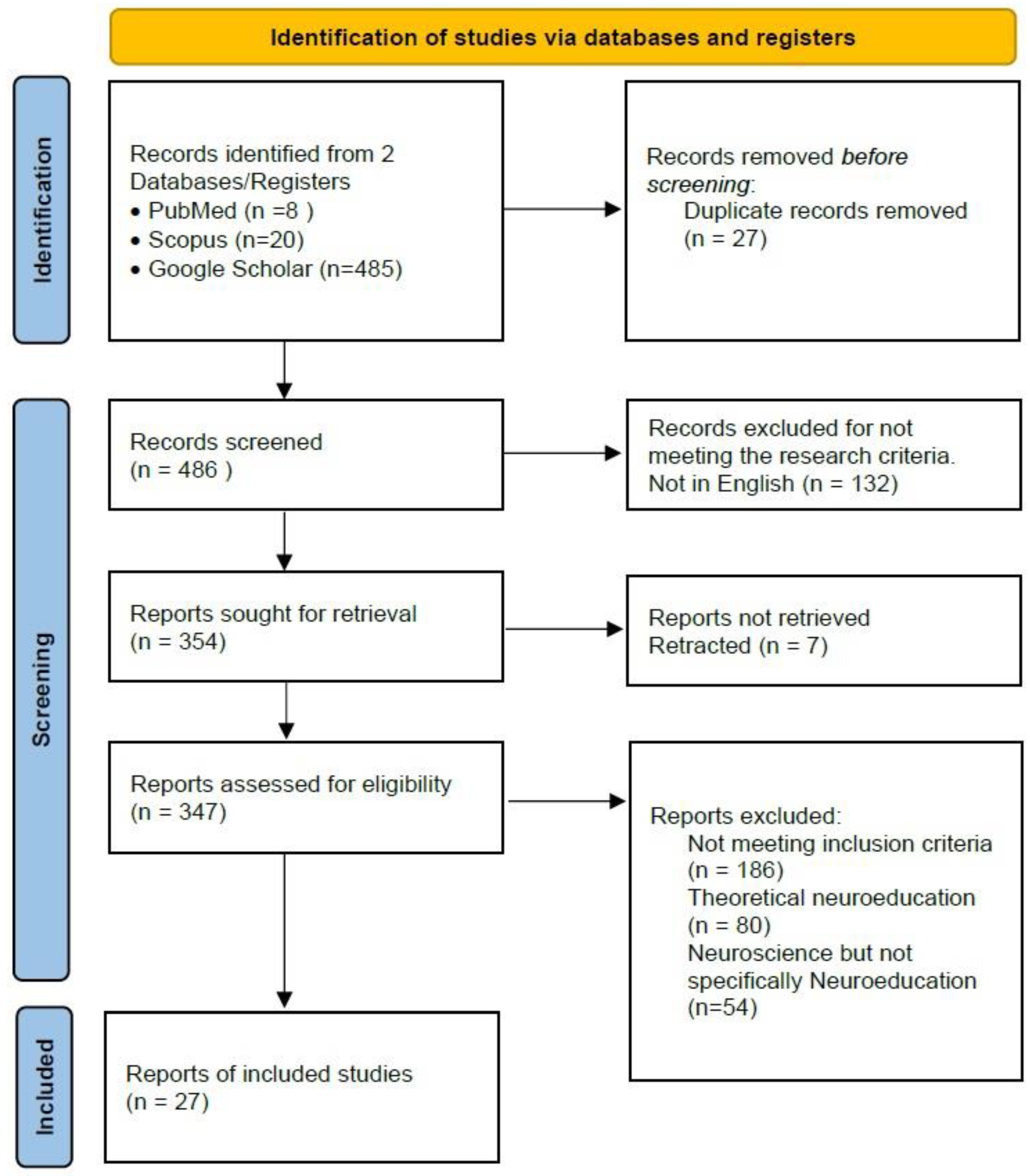
Figure 1. The PRISMA flow diagram.
Preprints 142071 g001
Figure 2. Frequencies of the sample sizes in groups of ten.
Preprints 142071 g002
Figure 3. Frequencies of wired and wireless EEG recordings.
Preprints 142071 g003
Figure 4. Frequency of use of EEG devices and configurations (amplifier and caps).
Preprints 142071 g004
Figure 5. The way in which EEG signals were used in the presented projects.
Preprints 142071 g005
Table 1. Neuroeducation projects details.
Project Subject Sample EEG metrics H/W used S/W used
Anderson et al (2018), [59] Efficacy of Just in Time teaching in anatomy education. 24 students (11m.+23 f.), m.age=20.74 N250 and RewP ERPs BP* actiCAP Xpress + 16ch V-Amp Psychophysics Matlab Toolbox, Brain Vision Recorder
Anderson et al (2023), [60] Diagnostic accuracy factors examination 26 students(23f.+3m.), m.age=19.8 and 9 experts (6f+3m), m.age=44.1 RewP and N170 ERPs BP 32ch actiCAP slim + LiveAmp Psychophysics Matlab Toolbox, Brain Vision Recorder
Doukakis et al (2019-20), [61,62] Students’ performance in programming tasks 8 first year students (3f. + 5m.) EEG negative means value and time. BIOPAC MP150 AckKnowledge 4.3
Fard et al (2020), [64] Transfer learning during computer programming tasks 8 male students (18-21 y.o) EEG alpha and theta bands power spectrums and models of neuronal connectivity Emotiv EPOC-X OpenSesame, EEGLAB, NeuCube with Spike Neural Networks (SNNs) models.
Poulsen et al (2017), [65] Brain synchronization and engagement while watching videos 42 f. (m.age=22.4) Intersubject correlation (ISC) Emotiv EPOC Corrmap Matlab Toolbox
Schroer et al (2020), [68] Spatial reasoning 21 children (11m.+10f.), 6-12 y.old N2 and P3 ERPs, Joint time frequencies of alpha NetAmps300 (EGI) Nestation 4.5 (EGI)
Zhao et al (2023), [16] Effectiveness of AR app to assist learning 28 persons (7m.+21f.) m.age=20 Emotional state indexes from Brain waves Emotiv EPOC+ EmotivPro
García-Monge et al, (2023), [70] Exploration of potentialities and issues using portable EEG in classrooms 17 primary sch. Children, (10f.+7m.), 8-9 y.o 2’ EEG recording with eyes closed and 2’ with eyes open. Frequency domain analyses. Brainlink Pro, Emotiv Epoc, Epoc Flex, Muse EEGLAB, Lucid Scribe, Emotiv TestBench, EmotivPro, Mind monitor
Bouhdana et al (2023), [71] Effects of context and gender in physics problems 60 participants, (32f.+28m.), m.age=23.7 Cognitive engagement indexes β/(α+θ) BP ActiCAP + 32ch BrainAmp BrainVision Analyzer (v2.0), Matlab
Bos et al (2020), [72] Effects of low frequency binaural waves to attention levels 2 students with ages from 20 to 28 y.o Frequencies analysis performed by the proprietary software Neurosky Mindwave Effective Learner app. (Neurosky)
Onieva et al (2021), [73] Brain responses differences and similarities while reading digital or paper textbooks 3 pupils (1m. +2f.), 12 y.o Emotional state indexes from Brain waves and FAA Emotiv EPOC Emotiv software
Byczynski and Angiulli (2024), [74] Congruence judgement in relation to school days and starting times 24 female students, m.age=16.9 Frontal P300 assymentry Neurosoft EEG quick caps (9ch) and SynAmps2 (Compumedics Neuroscan) SCAN 4.3 (Compumedics Neuroscan) and Stim2 for stimuli presentation
Fu et al (2020), [75] Inhibitory control performing fraction comparisons 28 students, m.age=20.8 N1, N2 and P3 ERPs BP ActiCAP, 64ch BrainVision Analyzer (v2.0)
Skelling-Desmeules et al (2021), [76] Persistence of minconceptions in biological studies 28 students, (13f.+15m.),m.age= 23.7 N2 and LPP ERPs BP ActiCAP, 64ch BrainVision Analyzer (v2.0), E-prime
Williams et al (2019), [77] Intuitive and analytical thinking 30 undergrad students (22f.+8m.), (m.age=22.8) Alpha and theta activity in parietal and frontal areas. BP ActiCAP, 64ch + ActiCHamp BrainVision (v.1.10), EEGLAB and R language
Juarez-Varon et al (2023), [78] Analysis of stress, attention, interest, and engagement in onsite and online learning 20 pg students (22-25 y.o, 10m.+10f.) Alpha, beta, and theta activity Emotiv EPOC + EmotivPRO (v2.0) and R language
Garcia-Monge et al (2020), [79] Brain activity differences in various types of throwing games 8 children (m.age=7.2) Brain bands (mostly beta) activity Emotiv EPOC-X Emotiv Brain Activity Map and Emotiv TestBench, EEGLAB
Khramova et al (2021), [80] Differences in cortical activities of adults and children 12 children (3f.+9m.) 7-8 y.o. and 10 adults (3f+7m) (18-20 y.o.) Brain bands (mostly alpha and beta) activity BP ActiCAP, 32ch + ActiCHamp EEGLAB and FieldTrip
Williams et al (2018), [81] Reflection of reinforcement learning in RewP ERP 30 students (23f.+7m.), m.age=20 RewP ERP BP ActiCAP, 64ch ActiCHamp BrainVision Analyzer
Williams et al (2020), [82] Reward Predictions errors as indications of learning processes 30 students (19f.+11m.), m.age=20 RewP ERP BP ActiCAP, 64ch ActiCHamp Brain Vision Analyzer and Psychophysics Toolbox for stimuli presentation
Hassal et al (2019), [83] The role of control over actions in reward processing 26 students (13m.+13f.), m.age=21.54) RewP ERP BP ActiCAP, 64ch ActiCHamp BrainVision (v.2.1.2) and EEGLAB
Barrella et al (2019), [84] Measurement of cognitive load in concept handling tasks 23 students (13m.+10f.), m.age=19.25) Alpha over theta waves ratio B-Alert X10 EEG (ABM) ABM’s B-Alert Live software
Hernandez-Mustieles et al (2024), [85] Public Dataset of parallel EEG recordings during Collaboration and Competition Tasks 16 subject pairs by 1 m. and 1 f. (aged 18-24) Enophones EEGLAB
Rome-De Leon et al (2024), [87] Public dataset of EEG and other physiological signals recordings during two teaching scenarios for humanities studies 24 students (10 m. and 14 f.., aged 18-25, mean=21.33, sd=1.4) OpenBCI Ultracortex Mark IV OpenBCI GUI and
EEGLAB
Jamil et al (2024), [88] Brain to brain synchronization during remote learing. 10 students aged 18-28 and one instructor Time series and frequency domain correlations. Unicorn Hybrid systems (8ch, 250Hz)
Grubov et al (2024), [89] Cognitive abilities assessment and feedback in form of recommendations 60 pupils in two age groups, 9-10 and 11-12 years old, 36 boys and 24 girls. Alpha band power variance. BP LiveAmp (64ch, 500Hz) EEGLAB
* BP: Brain Products GmbH (also as Brain Vision LLC in the U.S).
Disclaimer/Publisher’s Note: The statements, opinions and data contained in all publications are solely those of the individual author(s) and contributor(s) and not of MDPI and/or the editor(s). MDPI and/or the editor(s) disclaim responsibility for any injury to people or property resulting from any ideas, methods, instructions or products referred to in the content.
Copyright: This open access article is published under a Creative Commons CC BY 4.0 license, which permit the free download, distribution, and reuse, provided that the author and preprint are cited in any reuse.
Prerpints.org logo

Preprints.org is a free preprint server supported by MDPI in Basel, Switzerland.

Subscribe

© 2024 MDPI (Basel, Switzerland) unless otherwise stated