Preprint
Article

This version is not peer-reviewed.

Outcomes of Broader Genomic Profiling in Metastatic Colorectal Cancer: a Portuguese Cohort Study

A peer-reviewed article of this preprint also exists.

Submitted:

13 December 2024

Posted:

16 December 2024

Read the latest preprint version here

Abstract

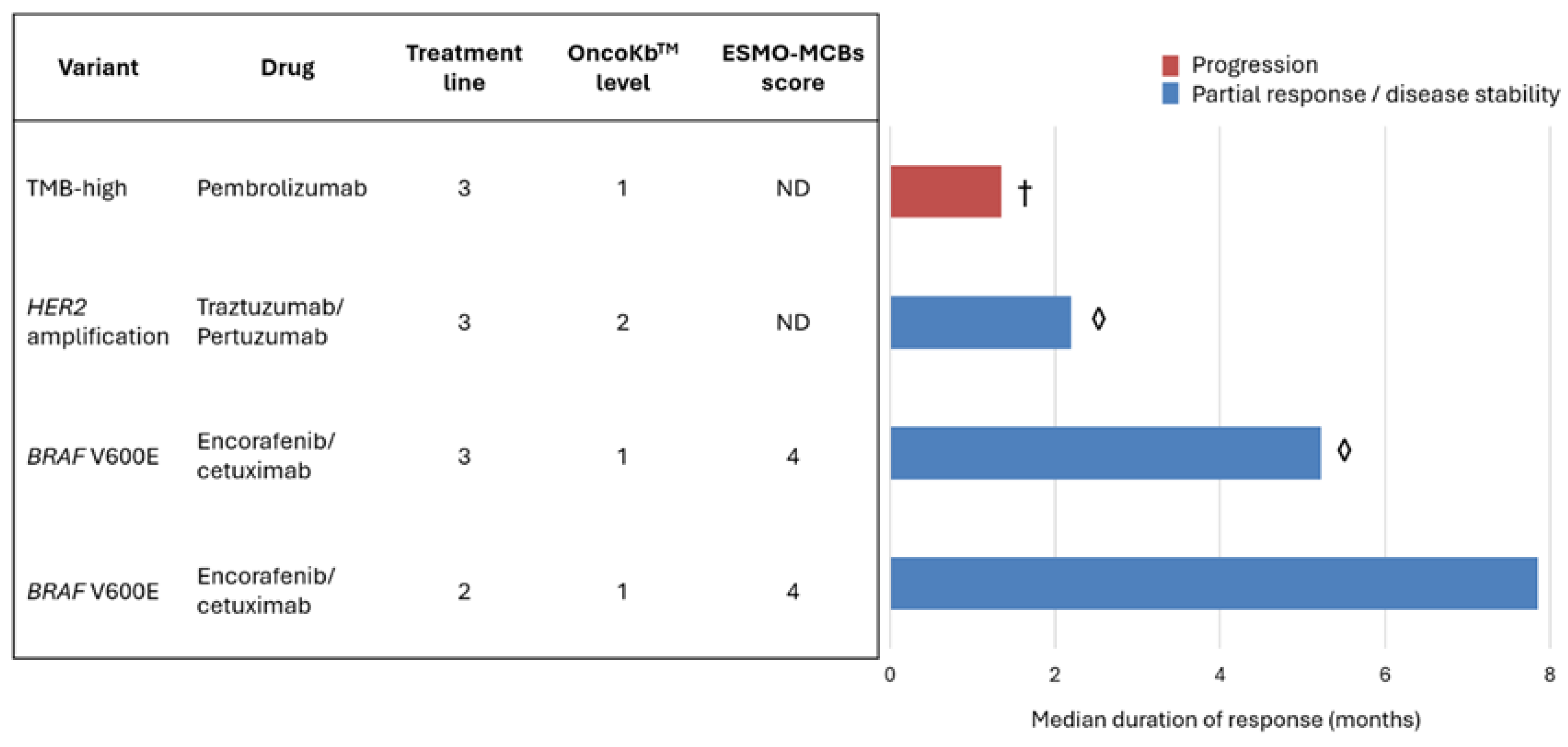
Background: Colorectal cancer (CRC) is the third most diagnosed cancer globally and the second leading cause of cancer-related deaths. Despite advancements, metastatic CRC (mCRC) has a five-year survival rate below 20%. Next-generation sequencing (NGS) can identify rare actionable mutations and assess tumour mutational burden (TMB), but its clinical utility in mCRC is debated due to limited survival improvement and cost-effectiveness concerns. Methods: This retrospective study included mCRC patients (≥18 years) treated at a single oncology center who underwent NGS during treatment planning. Tumour samples were analyzed using either a 52-gene Oncomine™ Focus Assay or a 500+ gene Oncomine™ Comprehensive Assay Plus. Variants were classified by clinical significance (ESMO ESCAT) and potential benefit (ESMO-MCBS and OncoKBTM). Kaplan-Meier and Cox regression analyses evaluated survival outcomes, with significance at p<0.005. Results: Eighty-six metastatic colorectal cancer (mCRC) patients were analysed, all MMR proficient. Most cases (73.3%) underwent sequencing at metastatic diagnosis, using primary tumour samples (74.4%) and a focused NGS assay (75.6%). A total of 206 somatic variants were detected in 86.0% of patients, 31.1% of which were classified as clinically significant, predominantly KRAS mutations (76.6%), with G12D and G12V variants as the most frequent. Median overall survival (OS) was 39.5 months, with no single mutation predictive of OS. Among 33.7% RAS/BRAF wild-type patients, 65.5% received anti-EGFR therapies. Eleven patients (12.8%) had other actionable variants ESCAT level I-II, including four identified as TMB-high, four KRAS G12C, two BRAF V600E and one HER2 amplification. Four received OncoKbTM level 1-2 and ESMO-MCBS score 4, leading to disease control in three cases. Conclusions: NGS enables the detection of rare variants, supports personalized treatments, and expands therapeutic options. As new drugs emerge and genomic data integration improves, NGS is poised to enhance real-world mCRC management.

Keywords: 
;  ;  ;  ;  

1. Introduction

Colorectal cancer ranks as the third most diagnosed cancer globally and stands as the second leading cause of cancer-related mortality [1] . Despite significant advancements in treatment over the past decade, 20% of patients are diagnosed with upfront metastatic colorectal cancer (mCRC) and up to 40% of patients initially treated for localised cancer will eventually develop metastatic disease. Despite recent advances in systemic treatment of mCRC, five-year survival rate remains below 20% [2,3]. Prognosis and treatment approaches for mCRC are strongly influenced by clinical and pathological factors, including tumour location (right or left colon) and the presence of RAS or BRAF variants [3].
To date, regarding molecular profiling, to define the first-line treatment for mCRC patients it is only required to know the presence or absence of RAS/BRAF variants and the microsatellite instability (MSI) status [4]. Searching for KRAS and NRAS variants at codons 12, 13, 61,117,146 and for BRAF at codon 600 is considered standard of care[5]. However, most experts and societies recommend multigene next-generation sequencing testing (NGS) at diagnosis of mCRC, particularly when there are no extra costs when compared with more traditional gene-directed polymerase chain reaction (PCR) [6,7]. Larger NGS panels not only allow for the calculation of tumoral mutational burden (TMB), determination of MSI status, and detection of HER2 amplifications but also potentially rare actionable gene variants [5,8]. Because such alterations tend to be truncal, one-time testing in the primary tumour or metastasis tissue is sufficient [5,9].
The Princess Margaret IMPACT/COMPACT multitumour trial showed that patients submitted to NGS and enrolled in genotype-matched trials had higher response rates to treatment than those included in genotype-unmatched trials [10]. A few years later, the OCTANE study enrolled over 4500 patients and found that NGS results changed the drug treatment in 15.7% of patients, however, overall survival was not different for patients receiving genotype-matched therapies [11]. Specifically for colorectal cancer, patients undergoing NGS testing in OCTANE had higher general health-associated costs and were more exposed to supportive care rather than clinical trial involvement [12].
While wide genome analyses are unveiling the genetic profile of colorectal cancer and the potential impact of different gene signatures on patient’s prognosis and clinical outcomes, the methodologies used in such studies are dissociated from clinical practice [13,14]. Some cohorts have used more targeted sequencing to analyse real-world patients, but usually in a retrospective manner or using surgical samples, failing to show the impact of NGS in selecting patients’ treatment [15,16].
Up to 53% of patients with colorectal cancer have KRAS/NRAS variants. BRAF V600E variants can be found in 8,5% of patients but their hotspot variants are detectable through more traditional techniques [6]. A high level of MSI (MSI-high) can be found in 8,5% of patients and can be detected by an immunohistochemical surrogate biomarker, mismatch repair (MMR) proteins expression [4,6]. Therefore, beyond the RAS/BRAF status and MSI-high, other actionable variants are rare, but detectable by NGS [6]. With unknown cost-benefit or real-world implications of NGS in mCRC, we aim to explore de clinical impact of the sequencing in a real-world cohort of mCRC, regarding the actionability of the encountered gene variants and its influence on disease management and prognosis.

2. Materials and Methods

2.1. Patient Selection

Metastatic CRC patients, followed in the same oncological centre, aged 18 years-old or more, were selected between 2022 and 2023, to perform NGS. Patients with newly diagnosed mCRC were included (before palliative treatment), as well as previously treated mCRC patients in need for further treatment strategies, accordingly to physician evaluation. Only patients eligible for treatment were included (systemic or locoregional). Patients with multiple active cancers, without clinical information regarding previous lines of treatment or accurate diagnostic dates were excluded.

2.2. Molecular Studies

Sequencing was performed using the most recently available biological tumour sample (surgical sample or biopsy of primary tumour or metastasis). Two different sequencing platforms were used, according to physician choice upon patient evaluation: 1) A focused assay, using the OncomineTM Focus Assay (Thermo Fisher Scientific), that allows the study of DNA and RNA across 52 genes; 2) a comprehensive assay, using OncomineTM Comprehensive Assay Plus (Thermo Fisher Scientific) for DNA analysis across more than 500 genes, and OncomineTM Focus Assay (Thermo Fisher Scientific) for RNA analysis. Both tests can detect SNVs, indels, CNVs and gene fusions and the last allows for tumoral mutational burden (TMB) determination.
MSI status was determined by the surrogate marker of MMR proteins expression. It was preformed using a four-antibody immunohistochemical panel including MLH1, MLH2, PMS2 and MSH6.

2.3. Variant and Targeted Treatment Classification

Variants were classified according to the Consensus Recommendation of the Association for Molecular Pathology, American Society of Clinical Oncology, and College of American Pathologists in variants with clinical significance (CSV) or variants with uncertain clinical significance (VUS) [17]. Bening variants were not reported. TMB was considered high if above 10 mutation/mb [18].
The clinical actionability of the variants was classified according to the European Society for Medical Oncology (ESMO) Scale for Clinical Actionability of Molecular Targets (ESCAT) in tiers. The ESCAT scale defines six levels of clinical evidence for molecular targets: tiers I to V and X (lack of evidence). Tier I corresponds to the highest level of clinical evidence [19]. In this study, we only considered ESCAT tier I and II molecular alterations.
The potential clinical benefit of targeted treatments offered was classified according to the ESMO Magnitude of Clinical Benefit Scale (ESMO-MCBS) in the non-curative setting, from level 1 to 5, and to OncoKBTM, from level 1 to 4 and level R (resistance). Level 5 corresponds to the highest level of clinical benefit for ESMO-MCBS, while level 1, in the therapeutic setting of OncoKBTM, corresponds to the highest evidence [20,21,22].

2.4. Patient Characterization

All clinical, histological and radiological data were collected retrospectively from electronic entries. Clinical data include the patient’s age and gender, all systemic and locoregional cancer treatments received, and time of death. Histological data was retrieved from pathological reports, including tumour stage and anatomical location and identification of the origin of the sample used for sequencing (primary tumour specimen/biopsy or metastasis specimen/biopsy). Radiological information was used to determine the pattern of metastatic spread at diagnosis of mCRC, response to treatments and time of disease progression.

2.5. Statistical Considerations

The sample was described with descriptive statistics. The chi-square test was used to study the influence of clinical factors on the capability of NGS to detect genetic variants. Stage, primary tumour location, previous treatments, the biological sample used for sequencing, the sequencing panel used, and genetic variants were used as stratification factors.
Overall survival (OS) was defined as the time between diagnosis of mCRC and the time of death. Median duration of response (DOR) was defined as the time between the first cycle of treatment and the time of clinical or radiological progression. The impact of the stratification factors on OS and DOR was analysed with Kaplan Meier curves and univariate Cox regression. Results were considered significant for p<.0053.

3. Results

This section may be divided by subheadings. It should provide a concise and precise description of the experimental results, their interpretation, as well as the experimental conclusions that can be drawn.

3.1. Sample Characteristics

Eighty-six patients were included, 66.3% of male sex, 57% with colon and 43% with rectal cancer, predominantly left-side disease (83.7%). Sequencing was offered to most patients at diagnosis of metastatic disease (73.3%), using biological material from the primary tumour (74.4%) and a focused NGS assay (75.6%). Regarding the molecular profile, no patient had MMR deficiency. Most included tumours were RAS/BRAF mutant (65.1%). The sample baseline characteristics are presented in Table 1.

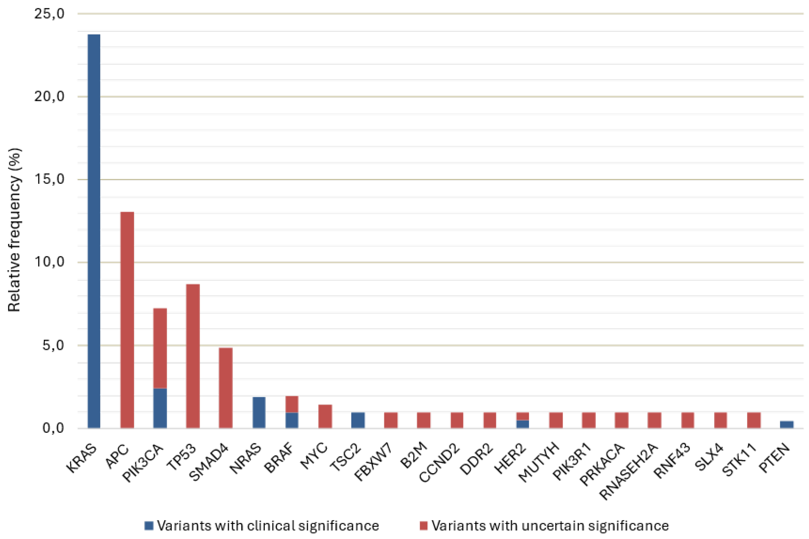
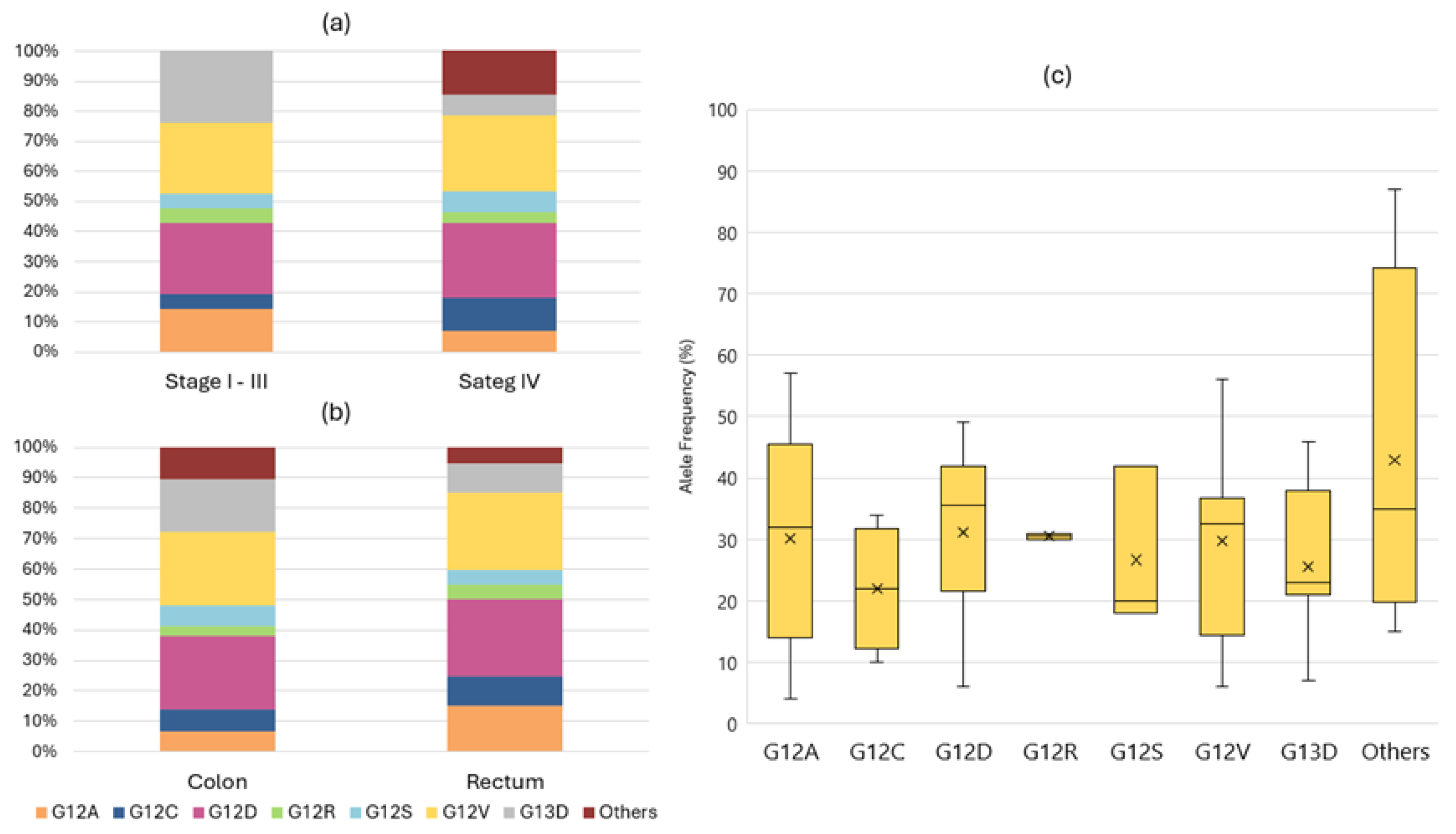
3.2. Detected Variants

A total of 206 somatic variants were detected in 74 patients (86.0%). Sixty-four variants (31.1%) were classified as clinically significant (CSV). Of these, 49 occurred in KRAS (76.6%), 4 in NRAS (6.3%) and 2 in BRAF (3.1%). Other clinically significant variants were also found in PIK3CA, PTEN, HER2 and TSC2. All CSV found were single nucleotide variants, except for one HER2 amplification and the two TSC2 deletions. The frequency of encountered variants can be found in Figure 1.
Of the 49 KRAS variants, all were classified as CSV and occurred in exons 2,3 and 4. Most were G12D and G12V (24,5% each). Detailed information regarding KRAS variants found is presented in Figure 2. The median allele frequency (MAF) for KRAS variants was 31% (IQR 17). The detection of RAS variants was independent of primary tumour location (p=.592), stage at diagnosis (p=.852), origin of biological material (p=.516) or technique used for its collection (p=.209).
Regarding actionable variants, KRAS G12C represented 8.2% of the KRAS CSV and was found in 4.7% of patients. The BRAF variants classified as CSV were two BRAF V600E and were found in 2.3% of patients. Median TMB of the patients that performed de comprehensive assay was 5.7 mut/mb (IQR 4.73).

3.3. Actionability and Therapeutic Implications

Table 2 shows the scoring of actionable variants in the sample according to ESCAT. Twenty-nine patients were RAS/BRAF wild type (33.7%). Nineteen received anti-EGFR therapies (65.5%), 17 of which in first-line treatment (89.5%). Eight patients already progressed, with a median duration of response for anti-EGFR therapies in combination with fluoropyrimidine-based chemotherapy in first-line of 20.9 months (CI95% 9.6 – 32.1). This combination is therapeutic level 1 in OncoKbTM and ESMO-MCBs score 4 in the non-curative setting.
In subsequent lines of treatment, two RAS/BRAF wild-type patients received irinotecan/cetuximab in third-line and achieved partial response, despite the absence of ESCAT or ESMO-MCBs scoring for this strategy. Another patient received panitumumab monotherapy beyond the third-line (ESMO-MCBS score 3) with progression at first radiological evaluation. All subsequent lines of treatment containing anti-EGFR were guided by liquid biopsy for confirmation of RAS/BRAF status.
Besides RAS/BRAF wild-type patients, there are 11 patients (12.8%) with other actionable variants, including four patients that were identified as being TMB-high. Of these eleven patients, four (36.4%) already received targeted treatment in subsequent lines of mCRC. Figure 3 shows in more detail the therapies received, treatment outcomes, and potential clinical benefit scoring for those 4 patients.

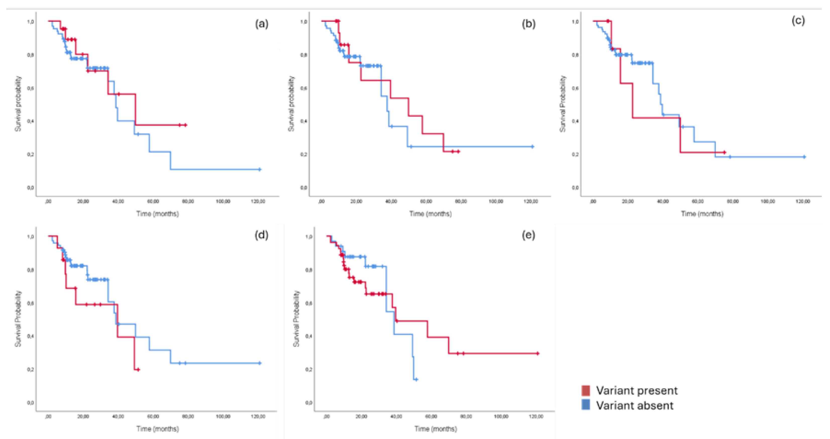
3.4. Prognostic Significance of NGS-Detected Variants

With a median follow-up of 22.8 months (IQR 18.4), 29 patients died (33.7%). The median overall survival was 39.5 months (CI95% 25.5-53.6). None of the most frequently mutated genes in the sample (KRAS/NRAS, APC, PIK3CA, TP53 or SMAD4) showed to be predictive of OS as shown in Figure 4. Whitin KRAS mutant tumours, no variant showed to be predictive of a worse prognosis. Median OS for patients that were treated beyond first-line with NGS-guided treatments, for other than BRAF/BRAF wild-type variants, was not met.

3.5. Factors Impacting NGS Results

The origin of biological material (p=.556) or the technique used for its collection (p=.699) did not influence the capability of the NGS test to detect variants of clinical significance. The capability of detecting any variant was influenced by the NGS platform used (p=.034) but not the detection of pathogenic variants (p=.100). Considering other potential clinically relevant factors like location of primary tumour (p=.300), sidedness (p=.537), stage at diagnosis (p=.475) and use of chemotherapy in the curative setting (p.921), none affected the detection of pathogenic variants.

4. Discussion

Beyond the lack of real-world data on the clinical utility of NGS for mCRC, there is also the need to explore the clinical impact of these molecular platforms in different populations, since each country regulations may impact the possibility of offering NGS-directed cancer treatments [23]. In our Portuguese cohort, beyond RAS/BRAF wild-type profile, NGS was able to detect actionable variants or molecular characteristics in 11 out of 86 patients (12.8%), classified as ESCAT tier 1 and 2 [19]. Of these, to date, four already accessed targeted therapy, with a high potential clinical benefit of OncoKbTM level 1 or 2, three of each with disease control [22]. Our sample shows a slightly lower detection of targetable alterations classified by ESCAT when compared with a recent larger real-world cohort. However, there was no representativity for MMR deficient cancers in our sample and we only considered ESCAT tier I and II genomic alterations, excluding hypothetical targets (tier III and beyond) for which would be difficult to obtain matched treatments outside clinical trials, like PIK3CA and HER2 activating mutations [24].
NGS helps to better characterize the tumour genome, particularly when a comprehensive assay is used. A total of 206 somatic variants were identified in our sample, with 64 CSV. Fifty-eight percent of patients were RAS mutant, with RAS variants occurring in exons 2, 3 and 4, as expected from the preexisting bibliography [6]. Despite lack of futile parallel application of traditional techniques in our study, NGS already showed to detect rare RAS mutations, but also other gene variants related to anti-EGFR resistance, providing a more complete platform when choosing the first-line treatment in mCRC [23]. This is the case of HER2 amplifications, which we could find in one patient (1.2%). For this, it was possible to not only personalize treatment choices but also treat the patient with a NGS-directed therapy in a subsequent line [25,26].
Beyond the detection of rare variants, NGS provides new biomarkers with potential prognostic and therapeutic value, like MAF and TMB. While the prognostic role of MAF seems to be restricted to tumour-circulating DNA and not tissue samples, the possibility of NGS to detect variants with low MAF improves treatment selection, particularly for RAS mutant mCRC [27,28,29]. TMB is a known drug target that predicts response to immunotherapy [6], however, TMB calculation requires more comprehensive and expensive NGS panels [18]. In our sample, only one-quarter of patients received a comprehensive assay, which yielded the detection of 4 TMB-high cases (19%), one treated with pembrolizumab to date [30].
A retrospective study showed a prognostic impact of the number of detected variants in a Chinese cohort of colorectal cancer, but this finding may be highly dependent on the restricted panel used and the genes included [23,31]. Differently, previous studies demonstrated the potential impact of independent variants, like APC, on the prognosis of colorectal cancer, which we could not find in our sample [32]. Recent evidence confirms that there may not be a prognostic role of individual gene variants in mCRC [33,34], however codon-specific variants or expression profiles, may be used, in the future, as predictive biomarkers [35]. A large integrated genome and transcriptome analysis of colorectal cancer showed that the prognostic impact of variants should be integrated into the mutational profile of the tumour and disease stage. For example, APC mutations seem to predict better OS, but only for non-hypermutated colorectal cancers [13].
Our study has several limitations, namely the short follow-up and limited sample size. Mainly, the use of two different NGS platforms, causes an imbalance between the available genomic information between patients and a selection bias regarding the timing and the NGS panel offered. This is supported by the observed interference of the NGS panel and the capability of detecting any variants, but not pathogenic ones, since the most common pathogenic variants in mCRC are included in both panels. This selection bias could explain the absence of MMR-deficient cancers in this sample for whom immunotherapy is already approved in first line, without the need for a comprehensive molecular study before the beginning of treatment [4]. Despite the use of different tests, as expected, the use of primary cancer or metastatic tissue didn’t affect the detection of variants [9,36].
In advanced cancers, previous studies had discouraging results, with a low number of patients benefiting from comprehensive gene sequencing and lack of clinical benefit for genome-matched therapies [5,37,38]. More recently, a comprehensive review shows that NGS-informed treatments may be beneficial across all tumour types, with no exception for mCRC [39]. However, the evidence remains restricted to small cohorts [40,41]. In the future, a longer follow-up of this sample and more sequenced patients will allow for a more extended contribution of this work to understanding the real-world benefit of routine NGS sequencing and NGS-matched therapies in advanced mCRC.

5. Conclusions

Our study shows that standard NGS testing is feasible in the real-world setting. While some targetable genomic alterations may be detectable by PCR, rare and low allele frequency variants, amplifications, rearrangements and other biomarkers, such as tumour mutational burden, allow for more personalized treatment and increase access to further lines of treatment. The importance of sequencing as a biomarker and its clinical benefit will possibly increase in the future, as more drugs are developed and more genomic information is collected and discussed in molecular tumour boards, to generate real-world evidence on NGS-directed treatment.

Author Contributions

Conceptualization, R.R. and N.B.; methodology, R.R., R.S. and L.G.S.; formal analysis, R.R., R.C., I.F., G.C. and M.G; writing—original draft preparation, R.R., R.S. and L.G.S.; writing—review and editing, R.R., T.F. and J.P.; visualization, R.R., T.F., J.P. and N.B.; supervision, N.B.; project administration, R.R.; funding acquisition, Y.Y. All authors have read and agreed to the published version of the manuscript.

Funding

This research received no external funding.

Institutional Review Board Statement

The study was conducted in accordance with the Declaration of Helsinki, and approved by the Institutional Ethics Committee of Coimbra Portuguese Institute of Oncology.

Informed Consent Statement

Patient consent was waived due to anonymized retrospective collection of data, high number of included subjects and high mortality rate. .

Data Availability Statement

Anonymized data will be available upon request to the corresponding author.

Conflicts of Interest

The authors declare no conflicts of interest.

References

  1. Bray, F.; Laversanne, M.; Sung, H.; Ferlay, J.; Siegel, R.L.; Soerjomataram, I.; Jemal, A. Global Cancer Statistics 2022: GLOBOCAN Estimates of Incidence and Mortality Worldwide for 36 Cancers in 185 Countries. CA Cancer J Clin 2024, 74, 229–263. [Google Scholar] [CrossRef]
  2. Biller, L.H.; Schrag, D. Diagnosis and Treatment of Metastatic Colorectal Cancer. JAMA 2021, 325, 669. [Google Scholar] [CrossRef]
  3. Doleschal, B.; Petzer, A.; Rumpold, H. Current Concepts of Anti-EGFR Targeting in Metastatic Colorectal Cancer. Front Oncol 2022, 12. [Google Scholar] [CrossRef]
  4. Cervantes, A.; Adam, R.; Roselló, S.; Arnold, D.; Normanno, N.; Taïeb, J.; Seligmann, J.; De Baere, T.; Osterlund, P.; Yoshino, T.; et al. Metastatic Colorectal Cancer: ESMO Clinical Practice Guideline for Diagnosis, Treatment and Follow-Up. Annals of Oncology 2023, 34, 10–32. [Google Scholar] [CrossRef]
  5. Johnson, D.; Chee, C.E.; Wong, W.; Lam, R.C.T.; Tan, I.B.H.; Ma, B.B.Y. Current Advances in Targeted Therapy for Metastatic Colorectal Cancer – Clinical Translation and Future Directions. Cancer Treat Rev 2024, 125, 102700. [Google Scholar] [CrossRef]
  6. Mosele, M.F.; Westphalen, C.B.; Stenzinger, A.; Barlesi, F.; Bayle, A.; Bièche, I.; Bonastre, J.; Castro, E.; Dienstmann, R.; Krämer, A.; et al. Recommendations for the Use of Next-Generation Sequencing (NGS) for Patients with Advanced Cancer in 2024: A Report from the ESMO Precision Medicine Working Group. Annals of Oncology 2024, 35, 588–606. [Google Scholar] [CrossRef]
  7. Cornish, A.J.; Gruber, A.J.; Kinnersley, B.; Chubb, D.; Frangou, A.; Caravagna, G.; Noyvert, B.; Lakatos, E.; Wood, H.M.; Thorn, S.; et al. The Genomic Landscape of 2,023 Colorectal Cancers. Nature 2024, 633, 127–136. [Google Scholar] [CrossRef]
  8. National Comprehensive Cancer Network Colon Cancer (Version 5.2024).
  9. Lee, S.E.; Park, H.Y.; Hwang, D.-Y.; Han, H.S. High Concordance of Genomic Profiles between Primary and Metastatic Colorectal Cancer. Int J Mol Sci 2021, 22, 5561. [Google Scholar] [CrossRef]
  10. 1Stockley, T.L.; Oza, A.M.; Berman, H.K.; Leighl, N.B.; Knox, J.J.; Shepherd, F.A.; Chen, E.X.; Krzyzanowska, M.K.; Dhani, N.; oshua, A.M.; et al. Molecular Profiling of Advanced Solid Tumors and Patient Outcomes with Genotype-Matched Clinical Trials: The Princess Margaret IMPACT/COMPACT Trial. Genome Med 2016, 8, 109. [Google Scholar] [CrossRef]
  11. Bogdan, L.; Saleh, R.R.; Avery, L.; Del Rossi, S.; Yu, C.; Bedard, P.L. Clinical Utility of Tumor Next-Generation Sequencing Panel Testing to Inform Treatment Decisions for Patients With Advanced Solid Tumors in a Tertiary Care Center. JCO Precis Oncol 2024. [CrossRef]
  12. Hernando-Calvo, A.; Nguyen, P.; Bedard, P.L.; Chan, K.K.W.; Saleh, R.R.; Weymann, D.; Yu, C.; Amir, E.; Regier, D.A.; Gyawali, B.; et al. Impact on Costs and Outcomes of Multi-Gene Panel Testing for Advanced Solid Malignancies: A Cost-Consequence Analysis Using Linked Administrative Data. EClinicalMedicine 2024, 69, 102443. [Google Scholar] [CrossRef]
  13. Nunes, L.; Li, F.; Wu, M.; Luo, T.; Hammarström, K.; Torell, E.; Ljuslinder, I.; Mezheyeuski, A.; Edqvist, P.-H.; Löfgren-Burström, A.; et al. Prognostic Genome and Transcriptome Signatures in Colorectal Cancers. Nature 2024, 633, 137–146. [Google Scholar] [CrossRef]
  14. Cornish, A.J.; Gruber, A.J.; Kinnersley, B.; Chubb, D.; Frangou, A.; Caravagna, G.; Noyvert, B.; Lakatos, E.; Wood, H.M.; Thorn, S.; et al. The Genomic Landscape of 2,023 Colorectal Cancers. Nature 2024, 633, 127–136. [Google Scholar] [CrossRef]
  15. Gao, X.H.; Yu, G.Y.; Hong, Y.G.; Lian, W.; Chouhan, H.; Xu, Y.; Liu, L.J.; Bai, C.G.; Zhang, W. Clinical Significance of Multiple Gene Detection with a 22-Gene Panel in Formalin-Fixed Paraffin-Embedded Specimens of 207 Colorectal Cancer Patients. Int J Clin Oncol 2019, 24, 141–152. [Google Scholar] [CrossRef]
  16. Lee, C.S.; Song, I.H.; Lee, A.; Kang, J.; Lee, Y.S.; Lee, I.K.; Song, Y.S.; Lee, S.H. Enhancing the Landscape of Colorectal Cancer Using Targeted Deep Sequencing. Sci Rep 2021, 11, 8154. [Google Scholar] [CrossRef]
  17. Li, M.M.; Datto, M.; Duncavage, E.J.; Kulkarni, S.; Lindeman, N.I.; Roy, S.; Tsimberidou, A.M.; Vnencak-Jones, C.L.; Wolff, D.J.; Younes, A.; et al. Standards and Guidelines for the Interpretation and Reporting of Sequence Variants in Cancer. The Journal of Molecular Diagnostics 2017, 19, 4–23. [Google Scholar] [CrossRef]
  18. Mo, S.-F.; Cai, Z.-Z.; Kuai, W.-H.; Li, X.; Chen, Y.-T. Universal Cutoff for Tumor Mutational Burden in Predicting the Efficacy of Anti-PD-(L)1 Therapy for Advanced Cancers. Front Cell Dev Biol 2023, 11. [Google Scholar] [CrossRef]
  19. Mateo, J.; Chakravarty, D.; Dienstmann, R.; Jezdic, S.; Gonzalez-Perez, A.; Lopez-Bigas, N.; Ng, C.K.Y.; Bedard, P.L.; Tortora, G.; Douillard, J.-Y.; et al. A Framework to Rank Genomic Alterations as Targets for Cancer Precision Medicine: The ESMO Scale for Clinical Actionability of Molecular Targets (ESCAT). Annals of Oncology 2018, 29, 1895–1902. [Google Scholar] [CrossRef]
  20. Suehnholz, S.P.; Nissan, M.H.; Zhang, H.; Kundra, R.; Nandakumar, S.; Lu, C.; Carrero, S.; Dhaneshwar, A.; Fernandez, N.; Xu, B.W.; et al. Quantifying the Expanding Landscape of Clinical Actionability for Patients with Cancer. Cancer Discov 2024, 14, 49–65. [Google Scholar] [CrossRef]
  21. Cherny, N.I.; Dafni, U.; Bogaerts, J.; Latino, N.J.; Pentheroudakis, G.; Douillard, J.-Y.; Tabernero, J.; Zielinski, C.; Piccart, M.J.; de Vries, E.G.E. ESMO-Magnitude of Clinical Benefit Scale Version 1. 1. Annals of Oncology 2017, 28, 2340–2366. [Google Scholar] [CrossRef]
  22. Chakravarty, D.; Gao, J.; Phillips, S.; Kundra, R.; Zhang, H.; Wang, J.; Rudolph, J.E.; Yaeger, R.; Soumerai, T.; Nissan, M.H.; et al. OncoKB: A Precision Oncology Knowledge Base. JCO Precis Oncol 2017, 1–16. [Google Scholar] [CrossRef] [PubMed]
  23. Bayle, A.; Basile, D.; Garinet, S.; Rance, B.; Laurent-Puig, P.; Blons, H.; Taieb, J.; Perkins, G. Next-Generation Sequencing Targeted Panel in Routine Care for Metastatic Colon Cancers. Cancers (Basel) 2021, 13, 5750. [Google Scholar] [CrossRef]
  24. Mulet Margalef, N.; Castillo, C.; Mosteiro, M.; Pérez, X.; Aguilar, S.; Ruíz-Pace, F.; Gil, M.; Cuadra, C.; Ruffinelli, J.C.; Martínez, M.; et al. Genomically Matched Therapy in Refractory Colorectal Cancer According to ESMO Scale for Clinical Actionability of Molecular Targets: Experience of a Comprehensive Cancer Centre Network. Mol Oncol 2023, 17, 1908–1916. [Google Scholar] [CrossRef]
  25. Spiekman, I.A.C.; Zeverijn, L.J.; Geurts, B.S.; Verkerk, K.; Haj Mohammad, S.F.; van der Noort, V.; Roepman, P.; de Leng, W.W.J.; Jansen, A.M.L.; Gootjes, E.C.; et al. Trastuzumab plus Pertuzumab for HER2-Amplified Advanced Colorectal Cancer: Results from the Drug Rediscovery Protocol (DRUP). Eur J Cancer 2024, 202, 113988. [Google Scholar] [CrossRef]
  26. Meric-Bernstam, F.; Hurwitz, H.; Raghav, K.P.S.; McWilliams, R.R.; Fakih, M.; VanderWalde, A.; Swanton, C.; Kurzrock, R.; Burris, H.; Sweeney, C.; et al. Pertuzumab plus Trastuzumab for HER2-Amplified Metastatic Colorectal Cancer (MyPathway): An Updated Report from a Multicentre, Open-Label, Phase 2a, Multiple Basket Study. Lancet Oncol 2019, 20, 518–530. [Google Scholar] [CrossRef]
  27. Manca, P.; Corallo, S.; Lonardi, S.; Fucà, G.; Busico, A.; Leone, A.G.; Corti, F.; Antoniotti, C.; Procaccio, L.; Smiroldo, V.; et al. Variant Allele Frequency in Baseline Circulating Tumour DNA to Measure Tumour Burden and to Stratify Outcomes in Patients with RAS Wild-Type Metastatic Colorectal Cancer: A Translational Objective of the Valentino Study. Br J Cancer 2022, 126, 449–455. [Google Scholar] [CrossRef]
  28. Lin, C.-Y.; Shen, M.-Y.; Chen, W.T.-L.; Yang, C.-A. Evaluation of the Prognostic Value of Low-Frequency KRAS Mutation Detection in Circulating Tumor DNA of Patients with Metastatic Colorectal Cancer. J Pers Med 2023, 13. [Google Scholar] [CrossRef]
  29. Jin, K.; Bao, Q.; Zhao, T.; Wang, H.; Huang, L.; Wang, K.; Xing, B. Comparing Baseline VAF in Circulating Tumor DNA and Tumor Tissues Predicting Prognosis of Patients with Colorectal Cancer Liver Metastases after Curative Resection. Clin Res Hepatol Gastroenterol 2024, 48, 102464. [Google Scholar] [CrossRef]
  30. Duvivier, H.L.; Rothe, M.; Mangat, P.K.; Garrett-Mayer, E.; Ahn, E.R.; Al Baghdadi, T.; Alva, A.S.; Dublis, S.A.; Cannon, T.L.; Calfa, C.J.; et al. Pembrolizumab in Patients With Tumors With High Tumor Mutational Burden: Results From the Targeted Agent and Profiling Utilization Registry Study. Journal of Clinical Oncology 2023, 41, 5140–5150. [Google Scholar] [CrossRef]
  31. Gao, X.H.; Yu, G.Y.; Hong, Y.G.; Lian, W.; Chouhan, H.; Xu, Y.; Liu, L.J.; Bai, C.G.; Zhang, W. Clinical Significance of Multiple Gene Detection with a 22-Gene Panel in Formalin-Fixed Paraffin-Embedded Specimens of 207 Colorectal Cancer Patients. Int J Clin Oncol 2019, 24, 141–152. [Google Scholar] [CrossRef]
  32. Schell, M.J.; Yang, M.; Teer, J.K.; Lo, F.Y.; Madan, A.; Coppola, D.; Monteiro, A.N.A.; Nebozhyn, M. V.; Yue, B.; Loboda, A.; et al. A Multigene Mutation Classification of 468 Colorectal Cancers Reveals a Prognostic Role for APC. Nat Commun 2016, 7, 11743. [Google Scholar] [CrossRef]
  33. Ottaiano, A.; Santorsola, M.; Capuozzo, M.; Perri, F.; Circelli, L.; Cascella, M.; Ianniello, M.; Sabbatino, F.; Granata, V.; Izzo, F.; et al. The Prognostic Role of P53 Mutations in Metastatic Colorectal Cancer: A Systematic Review and Meta-Analysis. Crit Rev Oncol Hematol 2023, 186, 104018. [Google Scholar] [CrossRef]
  34. Mei, Z.B.; Duan, C.Y.; Li, C.B.; Cui, L.; Ogino, S. Prognostic Role of Tumor PIK3CA Mutation in Colorectal Cancer: A Systematic Review and Meta-Analysis. Ann Oncol 2016, 27, 1836–1848. [Google Scholar] [CrossRef]
  35. Hsieh, M.-C.; Rau, K.-M.; Liu, K.-W.; Chiu, C.-C.; Chen, C.-I.; Song, L.-C.; Chen, H.-P. Prognostic Impact of Codon-Specific KRAS Mutational Status on Survival in Patients with Metastatic Colorectal Cancer Treated with TAS-102 or Regorafenib. Journal of Clinical Oncology 2024, 42, 75–75. [Google Scholar] [CrossRef]
  36. Bhullar, D.S.; Barriuso, J.; Mullamitha, S.; Saunders, M.P.; O’Dwyer, S.T.; Aziz, O. Biomarker Concordance between Primary Colorectal Cancer and Its Metastases. EBioMedicine 2019, 40, 363–374. [Google Scholar] [CrossRef]
  37. Massard, C.; Michiels, S.; Ferté, C.; Le Deley, M.-C.; Lacroix, L.; Hollebecque, A.; Verlingue, L.; Ileana, E.; Rosellini, S.; Ammari, S.; et al. High-Throughput Genomics and Clinical Outcome in Hard-to-Treat Advanced Cancers: Results of the MOSCATO 01 Trial. Cancer Discov 2017, 7, 586–595. [Google Scholar] [CrossRef]
  38. Le Tourneau, C.; Delord, J.-P.; Gonçalves, A.; Gavoille, C.; Dubot, C.; Isambert, N.; Campone, M.; Trédan, O.; Massiani, M.-A.; Mauborgne, C.; et al. Molecularly Targeted Therapy Based on Tumour Molecular Profiling versus Conventional Therapy for Advanced Cancer (SHIVA): A Multicentre, Open-Label, Proof-of-Concept, Randomised, Controlled Phase 2 Trial. Lancet Oncol 2015, 16, 1324–1334. [Google Scholar] [CrossRef]
  39. Gibbs, S.N.; Peneva, D.; Cuyun Carter, G.; Palomares, M.R.; Thakkar, S.; Hall, D.W.; Dalglish, H.; Campos, C.; Yermilov, I. Comprehensive Review on the Clinical Impact of Next-Generation Sequencing Tests for the Management of Advanced Cancer. JCO Precis Oncol 2023. [CrossRef]
  40. Kato, S.; Schwaederlé, M.C.; Fanta, P.T.; Okamura, R.; Leichman, L.; Lippman, S.M.; Lanman, R.B.; Raymond, V.M.; Talasaz, A.; Kurzrock, R. Genomic Assessment of Blood-Derived Circulating Tumor DNA in Patients With Colorectal Cancers: Correlation With Tissue Sequencing, Therapeutic Response, and Survival. JCO Precis Oncol 2019, 1–16. [Google Scholar] [CrossRef]
  41. Carter, P.; Alifrangis, C.; Chandrasinghe, P.; Cereser, B.; Del Bel Belluz, L.; Leo, C.A.; Moderau, N.; Tabassum, N.; Warusavitarne, J.; Krell, J.; et al. The Benefit of Tumor Molecular Profiling on Predicting Treatments for Colorectal Adenocarcinomas. Oncotarget 2018, 9, 11371–11376. [Google Scholar] [CrossRef] [PubMed]
Figure 1. Relative frequency of variants classified as clinically significant, and variants classified as of uncertain significance. The relative frequency was calculated based on the total amount of identified variables (n=206), however there are other 49 VUS not represented in the graphic. From those 49 VUS, each occurred only once in the sample and all in different genes. The total amount of CSV is represented in the graphic.
Preprints 142877 g001
Figure 2. Relative frequencies of each KRAS variant according to stage at diagnosis (a) or location of primary tumour (b). Graphic (c) shows a box plot for the distribution of median allele frequency of each KRAS variant.
Preprints 142877 g002
Figure 3. The table (left) presents four patients treated with targeted therapies in subsequent lines of systemic treatment for mCRC. The variant matched treatment is scored according to its potential clinical benefit with ESMO Magnitude of Clinical Benefit Scale (ESMO-MCBS) in the non-curative setting and to OncoKBTM therapeutic level. The bars (right) show the median duration of treatment (months) and best response to treatment: progression (red) or partial response/disease stability (in blue). One patient as died (†) and two are still on treatment (◊). ND – not defined.
Preprints 142877 g003
Figure 4. Impact of different variants on overall survival. Individually, there is not a predictive role for variants on APC (a), TP53 (b), SMAD4 (c), PIK3CA (d) or KRAS/NRAS (e).Cox regression showed an hazard-ratio of 0.65 (CI95% 0.26-1.61) for APC (p=.332); 0.75 (CI95% 0.31-1.84) for TP53 (p=.524); 1.13 (CI95% 0.39-3.30) for SMAD4 (p=.831); 1.70 (CI95% 0.71-4.04) for PIK3CA (p=.254); and 1.11 (CI95% 0.51-2.42) for KRAS/NRAS (p=.800).
Preprints 142877 g004
Table 1. Patients and disease characterization.
Characteristics n =86 (%)
Sex
Male
Female

57 (66.3)
29 (33.7)
Age (years)
Median (amplitude)

64.5 (27-80)
Stage at diagnosis
II
III
IV

9 (10.5)
28 (32.6)
49 (56.9)
Location of primary
Colon
Rectum

49 (57.0)
37 (43.0)
Sidedness
Left
Right

72 (83.7)
14 (16.3)
MMRp 86 (100)
RAS/BRAF mutant 56 (65.1)
Surgery (primary tumour) 53 (61.6)
Chemotherapy in curative setting 39 (45.3)
Number of metastatic locations
Median (amplitude)

1,62 (1-4)
Metastatic locations
Liver
Lung
Peritoneal
Lymph nodes
Local recurrence
Others

58 (67.4)
33 (38.4)
20 (23.3)
16 (18.6)
6 (7.0)
6 (7.0)
NGS setting
Before palliative treatment
Previously treated mCRC

63 (73.3)
23 (26.7)
NGS panel
Focused assay
Comprehensive assay

65 (75.6)
21 (24.4)
Origin of biological material
Primary tumour
Metastasis

59 (74.4)
27 (25.6)
Collection of biological material
Surgical sample
Biopsy

49 (57.0)
37 (43.0)
ECOG PS: Eastern Cooperative Oncology Group performance status;
MMRp: mismatch repair proteins proficiency (tissue).
Table 2. clinical actionability of different tier I and II variants and molecular profiles according to the Scale for Clinical Actionability of Molecular Targets (ESCAT).
Variant/profile N=86 (%) ESCAT tier
RAS/BRAF wild-type 29 (33.7) ND
KRAS G12C 4 (4.7) IA
TMB high (>10 mut/mb) 4 (4.7) ICa
BRAF V600E 2 (2.3) IA
HER2 amplification 1 (1.2) IIB
a – ESCAT scoring for tumour-agnostic genomic alteration; ND – not defined.
Disclaimer/Publisher’s Note: The statements, opinions and data contained in all publications are solely those of the individual author(s) and contributor(s) and not of MDPI and/or the editor(s). MDPI and/or the editor(s) disclaim responsibility for any injury to people or property resulting from any ideas, methods, instructions or products referred to in the content.
Copyright: This open access article is published under a Creative Commons CC BY 4.0 license, which permit the free download, distribution, and reuse, provided that the author and preprint are cited in any reuse.
Prerpints.org logo

Preprints.org is a free preprint server supported by MDPI in Basel, Switzerland.

Subscribe

Disclaimer

Terms of Use

Privacy Policy

Privacy Settings

© 2025 MDPI (Basel, Switzerland) unless otherwise stated