
Submitted:
25 March 2025
Posted:
26 March 2025
You are already at the latest version
Abstract
Inland recirculating aquaculture is a thriving food industry providing sustainable and locally sourced high-quality protein. However, its expansion is accompanied by emerging challenges regarding the spread of pathogens and diseases. Detection and management of pathogens in aquaculture remain underdeveloped compared to other animal farming sectors due to the vast diversity of species involved, the non-domesticated species, and limited knowledge regarding pathogens, host responses, and disease mechanisms. Furthermore, recirculating aquaculture systems are heavily dependent on beneficial bacterial communities for waste product removal and water quality maintenance, with opportunistic pathogens constituting an inherent component of these microbial communities. To enhance the potential of inland aquaculture as a sustainable source of protein, it is imperative to adopt advanced tools for pathogen detection and monitoring and for assessing the overall health of the microbial ecosystem. This paper presents an overview of promising current molecular and technological advancements that offer solutions for pathogen detection and system monitoring in aquaculture. We focus on recently developed point-of-care and on-site detection methods using miniaturized laboratory equipment and robust workflows that operate independently of cold chain logistics. We explore current methodologies for monitoring pathogens in the environment rather than through fish health assessments. Lastly, we discuss techniques from other scientific disciplines in aquaculture, including CRISPR-Cas protocols for pathogen detection and the implementation of "omics" approaches for comprehensive characterization of microbial states. These methods demonstrate considerable potential for pathogen surveillance and, subsequently, early responses in the dynamic aquaculture field. Through a better understanding of available options aquaculture managers and molecular scientist can collaborate and optimize systems. This paper aims to facilitate communication between molecular scientists and aquaculture managers, equipping the aquaculture industry with knowledge to enhance pathogen management techniques in their facilities.
Keywords:
pathogen surveillance
; detection
; monitoring
; molecular tools
; CRISPR‐CAS
; metagenomics
; metatranscriptomics
Introduction
Aquaculture is a rapidly developing sector, experiencing significant annual growth rates. Currently, global inland aquaculture produces 50 million tons of food annually (FAO, 2022). This growth has been fueled by declining natural fisheries, increasing per capita incomes, and a growing demand for aquatic animal protein (FAO, 2020). Additionally, the sector’s expansion is closely tied to ongoing advancements in technology and methodology, particularly with recirculating systems (Naylor et al., 2021).
Pathogen spread and disease emergence are growing global concerns for aquaculture managers, both in open and recirculating aquaculture settings. Disease outbreaks in aquaculture are responsible for an estimated annual loss of 6 billion USD globally (World Bank, 2014). Similar to other animal farming industries, these outbreaks are influenced by various factors, including operational practices, rearing densities (Saraiva et al., 2022), Click or tap here to enter text.biosecurity measures, environmental parameters (e.g., water quality), and stock properties (e.g., limited genetic diversity) (Wright et al., 2023).
Predicting, detecting, and treating diseases in aquaculture presents unique challenges, compared to other animal-farming industries. First, aquaculture involves over 400 evolutionarily and behaviorally distinct species (Stentiford et al., 2022), which may respond differently to similar conditions. For example, Atlantic salmon (Salmo salar) reared at high densities exhibit increased stress and susceptibility to disease (Ellison et al., 2020), while the opposite is observed in the territorial Nile tilapia (Oreochromis niloticus) (Ellison et al., 2018). Also, the underwater environment and the limited behavioral responses of fish to stress, pain, and disease complicate detection through visual and behavioral assessments. Although research on fish behavior in response to stress (Sharma, 2019; Fu et al., 2022), pain (Sneddon, 2009), and disease (Huntingford et al., 2020) has been expanding, it remains largely under represented within aquaculture species and has primarily focused on zebrafish. The structural design of aquaculture enclosures may hinder visual inspections compared to controlled laboratory experiments in small glass tanks, potentially limiting behavioral assessments necessary for the early detection of behavioral changes. As a result, disease detection often occurs after the opportunity for rapid treatment has passed (Rupp et al., 2019), potentially leading to significant stock losses. Additionally, many aquatic pathogens lack specific, consumer-safe treatments (Cain, 2022). Therefore, prevention and early detection methods are crucial lines of defense, making tools that support early detection essential for the sector’s continued and sustainable growth.
Disease management should extend beyond individual pathogenic species to encompass the study of entire microbial communities, especially in recirculating facilities. Analyzing the complete microbial community, including its overall composition, interactions between or within microbial species, and their competition for space and resources, is crucial for the system (Lee et al., 2023) and animal health (Almeida et al., 2021). Understanding the structure and function of these microbial communities opens up opportunities for implementing specific microbial amendments and probiotics (Bentzon-Tilia et al., 2016; Verschuere et al., 2000; El-Saadony et al., 2021), enhancing immune system responses (De Schryver & Vadstein, 2014; Yang et al., 2020), fostering biocontrol mechanisms (Khan et al., 2019), and creating conditions that are unfavorable for pathogens (Yang et al., 2020).
Current challenges in early pathogen detection and rapid response in aquaculture stem from three primary obstacles: (1) reliance on off-site diagnostic laboratories, requiring the transport of samples and coordination with external facilities; (2) the need for controlled conditions and specialized infrastructure, including expensive equipment, cold chains, and temperature-sensitive reagents; and (3) the necessity of capturing and handling farmed animals for sampling, which is labor-intensive and raises animal welfare concerns. These challenges are particularly pronounced in settings where diagnostic laboratories are scarce, time is limited, and resources are constrained.
To address these limitations, three key advancements have emerged: (1) point-of-care technology, which enables on-site microbial detection, effectively “bringing the lab to the farm”, (2) field-compatible molecular methods, which reduce reliance on controlled environmental conditions, allowing for “independence from lab infrastructure”, and (3) the use of environmental samples, minimizing the need for direct animal handling and shifting detection efforts to “move away from animal handling.” Together, these innovations may streamline pathogen detection, improve access to molecular diagnostics, and facilitate rapid, precise management responses. They empower farms to perform certain diagnostic tests independently, enable veterinarians to conduct advanced on-site analyses, and could be integrated into mobile labs that travel between farms.
Furthermore, we explore the potential applications of two emerging research developments in aquaculture: CRISPR-Cas technologies for precise pathogen detection and sequence-based community analyses for comprehensive microbial monitoring. These advancements hold promises for transforming disease surveillance and management into aquaculture systems.
Our aim is not to instruct aquaculture managers in these methods, but to summarize current possibilities, spark ideas, and foster collaboration between trained molecular scientists and aquaculture managers. This collaboration seeks to bridge the gap between fundamental and applied aquaculture research. Ultimately, integrating advanced methods for rapid pathogen monitoring and comprehensive community analysis will enhance disease prevention and improve the health of both farmed animals and the environment.
Figure 1.
Overview of current detection options. Managers can select the most suitable approach based on their specific needs, considering factors such as the goal of detecting individual pathogen species or characterizing the entire microbial community, the availability of a fully equipped laboratory versus field-compatible on-site methods, and the level of bioinformatic expertise accessible.
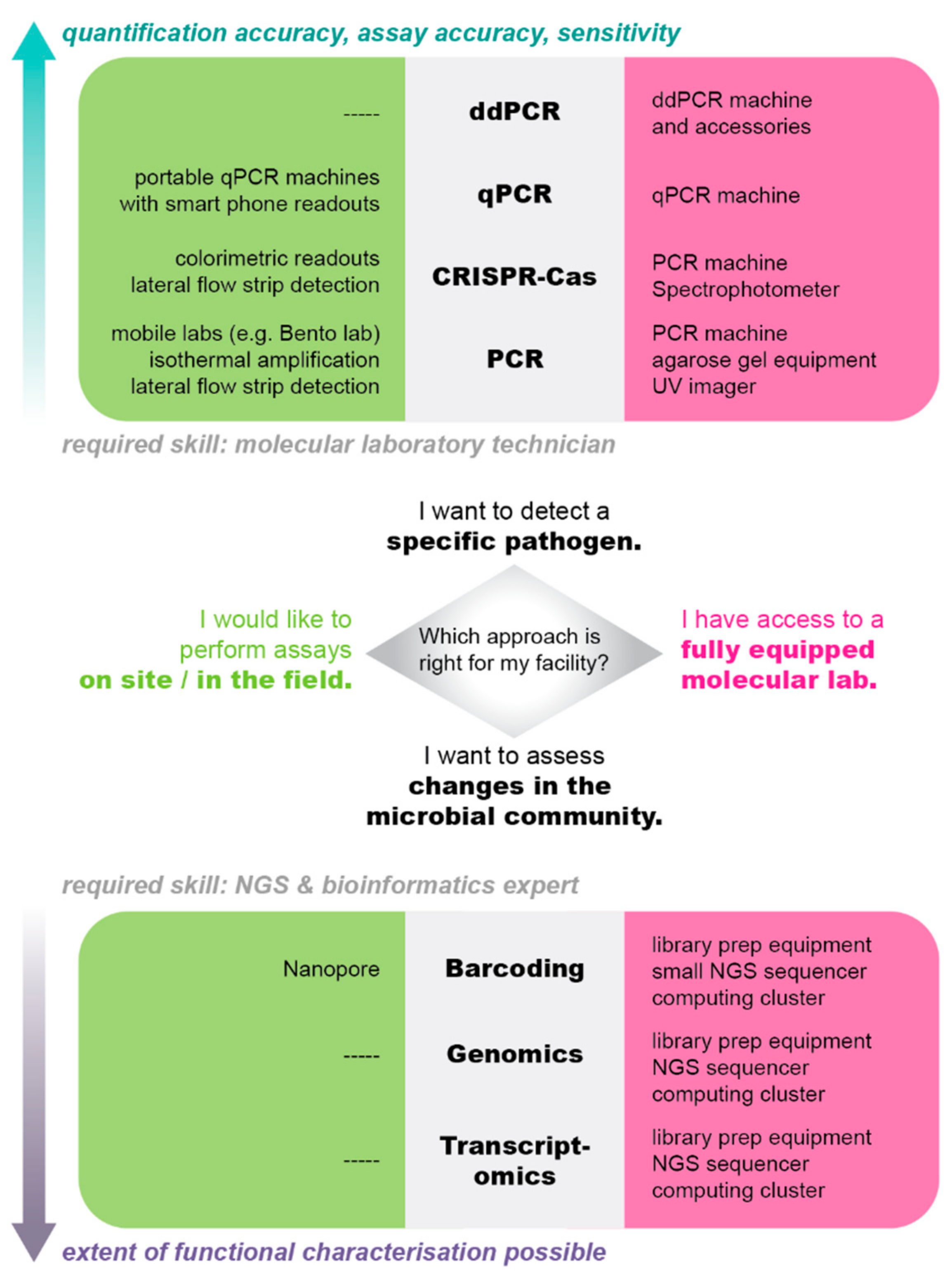

Table 1.
Examples of molecular technologies that are currently being applied for microbial detection in aquaculture. Bringing the lab to the farm.
Table 1.
Examples of molecular technologies that are currently being applied for microbial detection in aquaculture. Bringing the lab to the farm.
| Molecular technology | Group or disease of interest | Sample type/info | References |
|---|---|---|---|
| LAMP | Streptococcus agalactiae, koi herpes virus, Iridovirus, and Aeromonas hydrophila | Cultured ornamental fishes | (Chang et al., 2013) |
| Vibrio parahaemolyticus | Seafood samples | (Anupama et al., 2020) | |
| Vibrio vulnificus | Various sample types | (Z. Tian et al., 2022) | |
| Decapod iridescent virus 1 (DIV1), White spot syndrome virus (WSSV), and Enterocytozoon hepatopenaei (EHP) | Clinical infection experiment using shrimp | (Hu et al., 2023) | |
| Lyophilized qPCR | Gyrodactylus salaris and Aphanomyces astaci | Environmental water samples | (Rieder et al., 2022) |
| Metabarcoding and Metagenomics | Microbial communities | Recirculating aquaculture systems | (Rieder et al., 2023) |
| Metabarcoding | Nitrifying biofilms | Freshwater, brackish, and marine biofilters | (Hüpeden et al., 2020) |
| Microbial communities | Sole (Solea senegalensis) | (Almeida et al., 2021) | |
| Skin and gill microbiomes | Atlantic salmon (Salmo salar) | (Lorgen-Ritchie et al., 2022) | |
| Gut, skin, and microbiomes | Sea bass (Dicentrachus labrax) | (Nikouli et al., 2021; Meziti et al., 2024) | |
| Gut, skin, and microbiomes | Sea bream (Sparus aurata) | (Nikouli et al., 2018, 2021) | |
| Metatranscriptomics | Viruses | Multiple sources | (Mordecai et al., 2020) |
| Microbial communities | Mussels | (Rey-Campos et al., 2022) | |
| CRISPR-Cas | See Table 2 for an exhaustive list of CRISPR-Cas studies | ||
Table 2.
Overview of CRISPR-based technologies, highlighting the benefits and flexibility of various methods and associated proteins. The table also details the amplification methods used, including Recombinase Polymerase Amplification (RPA), Recombinase Assisted Amplification (RAA), Dualplex RAA (dRAA), Chemically Enhanced RAA (CE-RAA), and Reverse Transcription RAA or RPA (RT-RAA or RT-RPA), as well as Loop-Mediated Isothermal Amplification (LAMP). Additionally, the readout methods—fluorescence (F), lateral flow strips (LF), and colorimetry (C)—are specified. Abbreviations for Limit of Detection (LoD) (i.e., the lowest copies required for detection) measurements include Colony Forming Units (CFU), nanograms (ng), and femtograms (fg).
| CAS Protein | Amplification method | Detection Method | Target | Limit of Detection | Time, min | References |
|---|---|---|---|---|---|---|
| Cas12a | RPA | F | Salmo salar, Salmo trutta, Salvenius alpinus | 10−5 ng/µL | <150 | Williams et al., 2019, 2021, 2022 |
| F, C | Vibrio spp. | 20 copies/μL | 120 | C. Li et al., 2022 | ||
| F, LF | Enterocytozoon hepatopenaei | 50 copies/reaction | 60 | Kanitchinda et al., 2020 | ||
| F | Vibrio spp. | 100 copies/reaction | 90 | P. Wang, Guo et al., 2023 | ||
| F, LF | Clonorchis sinensis | 1 copy/μL | 60 | Huang et al., 2023 | ||
| F | Vibrio vulnificus | 4 copies/reaction | 40-60 | X. Zhang et al., 2024 | ||
| F | Enterocytozoon hepatopenaei | 10 copies/reaction | 40 | Wang et al., 2022 | ||
| F | Renibacterium salmoninarum | 10 copies/µL | 60 | D’Agnese et al., 2023 | ||
| F, LF | Megalocytivirus | 40 copies/reaction | 60 | Sukonta et al., 2022 | ||
| F | White Spot Syndrome Virus | 200 copies/reaction | <60 | Chaijarasphong et al., 2019 | ||
| F | WSSV | 10 copies/reaction | 60 | P. Wang, Yang et al., 2023 | ||
| F | Mandarin fish ranavirus (MFR), Infectious spleen and kidney necrosis virus (ISKNV) | 1 copy/μL (MFR), 0.1 copy/μL (ISKNV) |
45-60 | Lu et al., 2024 | ||
| RAA | F | Vibrio vulnificus | 2 copies/reaction | 40 | Xiao et al., 2021 | |
| F | Cyprinid herpesvirus 2 | 10 copies/reaction | 60 | Hou et al., 2025 | ||
| dRAA | F | Aeromonas hydrophila | 2 copies/reaction | 45 | Lin et al., 2022 | |
| CE-RAA | F | Vibrio parahemolyticus | 67 CFU/ml in pure cultures, 73 CFU/g in shrimp tissues |
N/A | Lv et al., 2022 | |
| RT-RAA | F | Nervous necrosis virus | 0.5 copies/μL | 30 | Gao et al., 2018 | |
| RT-RPA | F, LF | Infectious hematopoietic necrosis virus | 9.5 copies/μL | 60 | Rong et al., 2024 | |
| F, LF | Hirame novirhabdovirus | 8.7 copies/μL | 60 | Tang et al., 2024 | ||
| F, LF | Tilapia tilapinevirus | 200 copies/reaction | <60 | Sukonta et al., 2022 | ||
| F, LF | Yellow head virus | 100 fg RNA | 90 | Aiamsa-At et al., 2023 | ||
| Cas12b | LAMP (with/without reverse transcriptase) |
F | White Spot Syndrome Virus, Taura Syndrome Virus (TSV) |
100 copies/reaction (WSSV), 200 copies/reaction (TSV) |
30 | Major et al., 2023 |
| Cas13a | RPA | F, LF | Delta smelt (Hypomesus transpacificus), Longfin smelt (Spirinchus thaleichthys), Wakasagi (Hypomesus nipponensis) | 300 copies/reaction | <60 | Baerwald et al., 2020 |
| LF | Vibrio alginolyticus | 10 copies/μL | 50 | Y. Wang et al., 2023 | ||
| F, LF | Yersinia ruckeri | RNA: 2 aM in bacterial lysates of planktonic and biofilm cultures DNA: 1 ng/reaction in biofilm samples |
70 | Calderon et al., 2024 | ||
| F, C | White Spot Syndrome Virus | 1.06 copies/reaction | 60 | Sullivan et al., 2019 | ||
| RAA | F, LF | Largemouth bass ranavirus | 31 copies/ul | 60 | Guang et al., 2024 | |
| Cas12a and Cas13a | RPA | F | White Spot Syndrome Virus, Enterocytozoon hepatopenaei | 200 copies/reaction (WSSV), 20 copies/reaction (EHP) |
<60 | Kanitchinda et al., 2024 |
In recent years, there has been a significant development of automated, miniaturized, downscaled, and affordable laboratory equipment by start-up companies. This innovation has primarily been driven by needs in three research areas: human epidemiology in countries of the global south, the environmental DNA field, and molecular ecology. These demands have spearheaded the development of microfluidic devices (Kulkarni & Goel, 2020), portable quantitative PCR (qPCR) machines (e.g., Bento Lab https://bento.bio/), and handheld DNA sequencing devices (e.g., Nanopore MinION), as well as complete field laboratories, including “lab in a bag” options or automobiles converted to fully functional labs (e.g., EMBL Mobile Service (https://www.embl.org/about/info/trec/mobile-labs/). The implementation of these tools directly within aquaculture facilities has the potential to significantly reduce turnaround times, minimize the risk of sample loss or degradation during transport, and enable molecular diagnostics in regions with limited access to centralized laboratory infrastructure.
Devices and solutions capable of rapidly identifying or ruling out specific pathogens are of particular interest in aquaculture, as they facilitate timely and disease-appropriate responses, such as treatment or quarantine of affected units. Devices capable of portable qPCR functionality hold significant potential in this regard. These devices use the same reagents as standard laboratory qPCR machines, allowing for straightforward adaptation of well-established assays. The primary trade-off associated with miniaturization is reduced sample throughput. In contrast to traditional 96-well formats, portable machines accommodate fewer simultaneous reactions; for instance, the Bio Molecular Systems (Australia) Mic can process 48 samples, the Biomeme Franklin (USA) handles 9 samples, and the Aqua Kit processes 16 samples.
Fortunately, throughput is typically not a limiting factor in point-of-care testing, as a small number of samples is generally sufficient for effective pathogen detection (Nguyen et al. 2018). Portable qPCR machines are characterized by their lightweight, compact design, and often reduced cost. For example, the footprint dimensions of the Biomeme Franklin and the Bio Molecular Systems Mic are 101.3 mm × 182 mm × 89.8 mm and 130 mm × 150 mm × 150 mm, respectively, with weights of 1.2 kg and 2 kg. Additionally, these portable systems offer rapid turnaround times (< 60 minutes per run), and certain models may not require regular calibration services or access to plug-in electricity, particularly battery-operated variants. An example of a portable amplification device is the Bio-Key qPCR 1. Currently, Bio-Key qPCR kits are tailored to detect well-known aquaculture pathogens, including Shrimp Hemocyte Iridescent Virus (SHIV), Vibrio spp., and Infectious Hypodermal Hematopoietic Necrosis virus 2.
Digital droplet PCR (ddPCR) is an advanced molecular diagnostic technology that represents the latest evolution in PCR methodologies. There are currently more than six commercialized ddPCR platforms available, such as the Biomark HD system, Bio-Rad's QX100 and QX200 systems, and RainDrop from RainDance (Dong et al., 2015). Unlike qPCR, ddPCR partitions a sample into thousands of nanoliter-scale reactions, generating a vast number of individual data points from a single sample. This approach enables robust statistical analysis without the necessity of standard curves, which are required for qPCR assay (see H. Zhang et al. (2024) for a detailed explanation of digital PCR mechanisms).
A key advantage of ddPCR is its exceptional sensitivity and ability to provide absolute quantification (Pavšič et al., 2016), making it particularly suitable for applications requiring the detection of rare target molecules, such as in the early stages of a disease outbreak. Additionally, ddPCR can detect small fold changes in target nucleic acids (Quan et al., 2018), allowing for the identification of subtle variations that may facilitate early and rapid intervention by resource managers. However, ddPCR also has certain 2limitations. Compared to qPCR, it has a lower throughput, typically processing 96 samples per run compared to the 384-well plates used in qPCR, and the initial setup costs in terms of machinery and training are higher (H. Zhang et al., 2024). These challenges may present a barrier to widespread adoption in some laboratories.
When animals exhibit signs of illness, but the causative agent remains unidentified, there is a need for systems that can detect novel or unknown pathogens. In this context, portable sequencers, particularly the Nanopore MinION, present significant potential (Urban et al., 2021). The MinION is a cost-effective portable sequencing device, with a Nanopore Flongle flow cell run priced at approximately USD 90 for viral and bacterial sequencing. However, successful sequencing requires relatively high-quality DNA, and data interpretation may necessitate a certain level of expertise (Bloemen et al., 2023), thus collaboration with a trained molecular ecologist is recommended. An application example for portable sequencing in the aquaculture sector is the rapid and precise sequencing of salmonid alphavirus and infectious salmon anemia virus - two viruses with significant implications for global salmonid aquaculture - as demonstrated by Gallagher et al. (2018). A more general overview of this technology's application is provided by Delamare-Deboutteville (2021).
Both specific detection and sequencing processes can be accomplished by microfluidic devices. These miniaturized devices facilitate the precise manipulation and analysis of small fluid volumes, allowing for the sequential combination of multiple steps in molecular protocols - such as sample preparation, nucleic acid amplification, and detection - within a single chip or cartridge. See review by Gorgannezhad et al. (2019) for a more in-depth discussion of designs and options. Application examples for microfluidics-based pathogen detection in aquaculture encompass a variety organisms, including decapod iridescent virus 1 (DIV1), white spot syndrome virus (WSSV), infectious hypodermal and hematopoietic necrosis virus, infectious spleen kidney necrosis virus, koi herpesvirus, and Iridovirus, as well as the Enterocytozoon hepatopenaei parasite and various bacteria, such as Aeromonas hydrophila, Edwardsiella tarda, Vibrio harveyi, V. alginolyticus, V. anguillarum, V. parahaemolyticus, V. vulnificus, and Pseudomonas aeruginosa (Chang et al., 2013; Xiao et al., 2021; Guptha Yedire et al., 2023; P. Wang, Guo et al., 2023).
Independence from Controlled Environments
In recent years, protocols for molecular analyses under field conditions have been developed, some of which we will highlight. Traditional molecular assays are often sensitive to environmental parameters, as the enzymes involved typically require stringent cooling chains to maintain activity or rely on precisely controlled temperature sequences for effective DNA amplification. Additionally, the results usually necessitate sophisticated machinery for analysis. The increasing need for on-site pathogen surveillance in human health programs in remote areas (Song et al., 2022) have driven the advancement in the development of enzymes capable of surviving lyophilization, allowing for transportation and storage at room temperature or refrigeration. Furthermore, there is a growing focus on assays that operate at constant (“isothermal”) temperatures that are easily achievable, as well as those that can be assessed visually through color changes.
Lyophilization
Lyophilization, or freeze-drying, enhances independence from stringent cooling chains. Traditional PCR and qPCR reagents lose activity at temperatures above -20°C. However, when lyophilized, PCR assays containing all necessary reagents and enzymes can be transported and stored at room temperature, requiring only reconstitution with water on-site prior to use (Rieder et al., 2022). An additional advantage of lyophilization is that central laboratories can prepare quality-controlled reaction batches, which can then be shipped to the end-users and stored on-site for extended periods (Rieder et al., 2022). Consequently, lyophilization is particularly advantageous in resource-limited or remote settings that lack continuous access to freezer capacity.
Isothermal Amplification
Isothermal amplification eliminates the need for precise cycling and high temperatures required in traditional PCR and qPCR assays. Isothermal polymerases function at constant, lower temperatures that can be achieved using incubators, water baths, or even body heat. Two isothermal protocols are currently available, loop-mediated isothermal amplification (LAMP) and recombinase polymerase amplification (RPA). RPA functions at 37°C (Williams et al., 2022), while LAMP operates at 65°C (Notomi et al., 2000). Both methods are time-efficient (< 60 min) (Sullivan et al., 2019; Notomi et al., 2000) and sensitive enough to detect low copy numbers of DNA (e.g., ~1.06 copies for white spot syndrome virus) (Sullivan et al., 2019). Furthermore, these techniques can be integrated with microfluidic devices (Giuffrida & Spoto, 2017; Kant et al., 2018). The successful application of this in aquaculture, specifically LAMP, has already been demonstrated to detect Vibrio parahaemolyticus (Anupama et al., 2020) and V. vulnificus (Z. Tian et al., 2022), both known fish pathogens (Novoslavskij et al., 2016). A review by Das et al. (Das et al., 2022) provides an overview of LAMP, highlighting its efficiency and widespread use as a rapid diagnostic tool for pathogen detection. The authors emphasize LAMP’s ability to generate a high copy number (~109) within a short timeframe (<1 h), making it a valuable method for rapid and sensitive pathogen detection.
RPA
RPA has been used to detect various viruses, including penaeus stylirostris densovirus (Jaroenram & Owens, 2014), infectious hypodermal and hematopoietic necrosis virus (Xia et al., 2015), cyprinid herpesvirus 3 (Prescott et al., 2016), abalone herpes-like virus, red-spotted grouper nervous necrosis virus (Gao et al., 2018), tilapia lake virus (Sukonta, Senapin, Taengphu, et al., 2022), megalocytivirus, the causative agent of scale drop disease (Sukonta, Senapin, Meemetta, et al., 2022), and Vibrio parahaemolyticus, the causative agent of acute hepatopancreatic necrosis disease (C. Li et al., 2022). In addition to viral detection, RPA has been used to detect opportunistic bacterial pathogens such as Flavobacterium columnare (Mabrok et al., 2021), and Edwardsiella ictaluri (H. Li et al., 2022), V. parahaemolyticus (Geng et al., 2019), Francisella noatunensis subsp. orientalis (Shahin et al., 2018), as well as the parasite Tetracapsuloides bryosalmonae (Soliman et al., 2018). During lab testing there have been occurrences of false positives with RPA (personnel experience); however, this likely depends on the species and specific assay conditions.
Colorimetric Readouts
Colorimetric readouts facilitate independence from machine-dependent result interpretation by providing direct, visually perceptible outputs. Currently, two options are available: lateral flow strips, which exhibit a color change in a specific section (typically a narrow band) when a target is detected and liquid colorimetric assays, where the entire reaction volume changes color with detection. These methods provide a human readable output, thereby facilitating important decision making in real time without the need for sensitive spectrophotometry or fluorometers typically used for standard laboratory assays, although there is still room for improvement and expansion. In the aquaculture sector, colorimetric readouts have been explored in various studies. For example, Morsy et al. (2016) developed a colorimetric assay for monitoring food spoilage during fish and meat processing. Additionally, colorimetric assays have been applied for pathogen detection, such as largemouth bass ranavirus (Jin et al., 2020) and Flavobacterium columnare (Suebsing et al., 2015). Despite these promising applications, further successful case studies are required to achieve widespread adoption and maximize the benefits of these methods in aquaculture.
Moving away from Fish Handling
Monitoring fish health in aquaculture traditionally requires the capture, examination, and often sacrifice of healthy animals for routine checks. This practice is time-consuming, necessitating significant personnel resources, and conflicts with animal welfare principles. In contrast, the assessment of pathogen presence and in abundance from environmental samples (such as water or swabs) is non-invasive, equally sensitive, and aligns with the United Nations Sustainable Development Goals (United Nations, 2016) as well as the 3Rs objectives (Replace, Reduce, and Refine) for animal welfare (Hubrecht & Carter, 2019). While there are presently proof-of-concept diagnostic tools for specific pathogens, substantial research and development are necessary to fully realize their capabilities and practical applications, but they represent the potential future of aquaculture testing.
Environmental DNA and RNA
Environmental DNA (eDNA) or RNA (eRNA) is organismal DNA or RNA that can be found in the environment. Both eDNA and eRNA originate from cellular material shed by organisms (via skin, excrement, etc.) and can be collected from a variety of environmental samples, including water, soil, plants, and air, rather than directly sampling an individual organism. Both these tools are currently revolutionizing both ecological research and pathogen surveillance (Bass et al., 2023). In the aquaculture sector, eDNA methodologies could enhance biosecurity measures (Bohara et al., 2022) by targeting potential contamination sources, such as transport water. For instance, newly imported fish could be placed under quarantine until the screening of transport water yields negative results for specific pathogens.
Applications of eDNA in aquaculture include single-target assays for detecting ectoparasitic flukes such as Gyrodactylus salaris (Fossøy et al., 2020), Tetracapsuloides bryosalmonae (Sieber et al., 2020), and the bacterial pathogen Flavobacterium psychrophilum (Nguyen et al., 2018). Additionally, Peters et al. (2018) conducted a metabarcoding study demonstrating the potential of this approach for detecting eukaryotic aquaculture pathogens and harmful algae species in a salmon farm, highlighting its broader applicability in aquaculture healthy monitoring. While the detection of pathogens represents a major advancement for the sector, it is equally important to assess whether the abundances of target pathogenic organisms exceed established thresholds for food safety or water quality. Species quantification remains a major challenge in eDNA studies, limiting the ability to translate detection/non-detection data into actionable risk assessments.
Addressing this challenge, recent approaches detecting eRNA can distinguish between the mere detection of a pathogen’s RNA and an active, pathogenic state. For instance, eRNA has been used to detect Bonamia ostreae, a protozoan parasite responsible for significant mortality in flat oyster (Ostrea edulis) populations (Mérou et al., 2020). Importantly, eDNA and eRNA approaches stand to benefit from advancements in miniaturization, automation, and integration with colorimetric readouts, isothermal amplification, and field compatibility, like those applied in PCR and qPCR methodologies.
Transfer of CRISPR Technology to Aquaculture
CRISPR is widely recognized for its role in genome editing, but it has quickly evolved into a powerful, highly specific, and sensitive diagnostic tool. As of recent, it has also been adapted for the development of highly specific diagnostic assays. CRISPR-based detection is a valid, target-specific alternative to PCR and qPCR-based amplification techniques (Phelps, 2019). Briefly, the mRNA guide, also known as the guide RNA, is a short synthetic RNA sequence that directs the Cas protein to a specific nucleotide target by complementary base pairing. Once the guide RNA binds to the target nucleotide sequence, the Cas protein acts as a molecular “scissors,” creating a break at the designated site. This action also results in the Cas protein cleaving the report, thus releasing the fluorophore, which emits a signal that can be detected via fluorescence, colorimetry, or lateral flow assays. One caveat is that the Cas system is not suited for detecting low levels of nucleic acids. However, when combined with nucleic acid amplification technologies such as LAMP, RPA, or RAA, it achieves sensitivity comparable to other pathogen detection methods (Figure 2; RPA-CRISPR-Cas). A comprehensive overview of the fundamental principles and relevant considerations for pathogen detection is provided by Y. Tian et al. (2022).
Initially, diagnostic assays were limited to using the Cas9 protein (Pardee et al., 2016; X. Zhang et al., 2018), which lacks collateral cleavage activity. However, the discovery of Cas12 and Cas13 proteins revolutionized the diagnostic field, resulting in the development of methods such as SHERLOCK and DETECTR. These methods were successfully applied for detection of various viruses including Zika (Gootenberg et al., 2017) and SARS-COV-2 in humans (Broughton et al., 2020). Given the success of CRISPR in human medicine, CRISPR-based diagnostics has since been adapted for aquaculture applications. An overview of the CRISPR-based methods established in the aquaculture sector is provided in Table 2.
Currently, CRISPR-based diagnostic assays in aquaculture demonstrate exceptional versatility, with applications ranging from disease monitoring to ecological studies that enable precise aquatic species identification (Williams et al., 2019, Baerwald et al., 2020). The types of samples used for analysis are diverse and include animal-derived tissue (Guang, 2024; Sukonta, 2022; Wang, 2022), mucus swabs (“fish in hand” method) (Baerwald, 2020), biofilm samples (Calderon et al., 2024) and even eDNA samples (Williams, 2019, D’Agnese, 2023). These assays have been developed to detect either DNA or RNA, with recent advancements demonstrating the potential to target small non-coding RNAs (Calderon et al., 2024). CRISPR assays can also be designed for the simultaneous detection of multiple genes or the DNA/RNA of target species/strains, enabling the rapid identification of multiple targets at once (Lin et al., 2022; Gao et al., 2024; Kanitchinda et al., 2024).
Several advancements have facilitated the transition of CRISPR-based methods from the laboratory to field applications. These methods can be performed at room temperature, typically without the need for expensive specialized equipment, and have been further optimized to eliminate thermocycling (Major et al., 2023; Sukonta et al., 2022; Gao et al., 2024). Additionally, they bypass labor-intensive nucleic acid isolations (Baerwald et al., 2020), and certain protocols allow for one-tube reactions, minimizing procedural steps (i.e., time) (Major et al., 2023; P. Wang, Yang et al., 2023; P. Wang, Guo et al, 2023; Wang et al., 2022).
To enhance the applicability of CRISPR-based assay in field settings, various read-out methods have been developed. Some of these read-out methods include fluorescence readout via mobile phones, microplate readers, or direct visualization with UV illumination (Lin et al., 2022; Lu, 2024; Kanitchinda et al., 2024). Additionally, many assays have been adapted for compatibility with lateral flow strips, which eliminate the need for trained personnel or specialized equipment, facilitating rapid and accessible deployment in field conditions (Huang et al., 2023; Rong et al., 2024; Aiamsa-At et al., 2023).
However, further advancements and innovations for field applications are needed regarding quantitative assessments of pathogen abundance rather than binary detection/non-detection results. Additionally, the development of field-compatible nucleic acid isolation protocols remains a critical area for improvement. The recently discovered small Cas14 enzyme, which does not require a protospacer adjacent motif (PAM) sequence for target recognition, holds promise as a tool for ssDNA diagnostics (Aquino-Jarquin, 2019). Moreover, technical innovations from human diagnostics, such as smartphone-based fluorescence readers for CRISPR detection (Samacoits et al., 2021), could further enhance the feasibility of on-site implementation, increasing accessibility and ease of use.
In summary, CRISPR-based diagnostics offer a highly specific, sensitive, rapid, and potentially cost-effective approach for detecting the DNA or RNA of targeted pathogens. In aquaculture, CRISPR has the potential to facilitate rapid and cost-effective (Huang et al., 2023) pathogen detection, potentially aiding in the reduction mortalities and economic losses. Beyond disease monitoring, CRISPR is emerging as a powerful tool in ecological and conservation biology, facilitating non-invasive diagnostics and enabling almost real-time decision making (Baerwald et al., 2020; Phelps, 2019).
Transfer of Microbial Community Ecology to Aquaculture
Aquaculture, particularly when introducing novel fish species or interacting with external water bodies, often deals with symptoms caused by unknown disease agents. Additionally, recirculating aquaculture relies on a diverse microbial community for optimal functioning, and imbalance within these microbial communities has been implicated in poor fish health and suboptimal productivity (Rey-Campos et al., 2022; Sutherland et al., 2022; Rieder et al., 2023). The previously discussed approaches typically target just one or a few candidate pathogen species and depend on the availability of species-specific reagents, such as primers and probes, and are therefore less suited to detect novel pathogens or to answer questions related to community composition and function.
To analyze microbial communities comprehensively, metabarcoding (Almeida et al., 2021; Rieder et al., 2023), metagenomics (Rieder et al., 2023), and metatranscriptomics (Hook et al., 2021; Rey-Campos et al., 2022) are valuable tools. These methods enable the identification of microbial species, including unculturable taxa, while also providing insights into the overall composition, diversity, dynamics, and functional contributions of the aquaculture microbiome. However, the effectiveness of these methods in species identification and functional analysis is contingent upon the availability of reference genomes and gene annotations. While these techniques may not be the primary choice for rapid pathogen diagnostics and on-site monitoring in typical aquaculture settings, they offer significant potential for evaluating management strategies, assessing the impacts of feed changes (Rojas-Tirado et al., 2018), and monitoring microbial recolonization following treatments or disease outbreaks (Xue et al., 2017).
Metabarcoding approaches can effectively classify which taxonomic units are present, and thus assesses overall community composition (Bakke et al., 2017; Almeida et al., 2021; Rieder et al., 2023). This technique can be used to detect pathogens associated with fish, such as those present on gills or lesions, as well as to characterize bacteria or fungi within the system (Rieder et al., 2023). Metabarcoding is relatively straightforward to implement and can accurately classify species or at least group pathogens when applied correctly, but its efficacy is constrained by the organisms for which marker gene sequences are already documented. If a species has not been previously cataloged in a database (e.g., reference library), it cannot be assigned a taxonomic identity, as identification relies solely on known species within the available reference library. In the absence of a taxonomic assignment, it is likely that the species is not a recognized pathogen, since these are typically well-studied, sequenced (e.g., within reference libraries), and documented in scientific literature. Additionally, the species group of interest needs to be defined upfront, since metabarcoding methods for bacteria, fungi, and eukaryotic families rely on different, group-specific marker genes and cannot be analyzed together. Viruses, due to their extensive diversity, cannot be effectively sequenced as a group using metabarcoding approaches. Examples of metabarcoding application in aquaculture include the detection of spatial-temporal patterns in recirculating freshwater aquaculture systems (Rieder et al., 2023) or tracking of microbial communities changes in response to environmental changes that could lead to a disease outbreak (Bastos Gomes et al., 2017).
In contrast, metagenomics enables the simultaneous detection of all taxonomic groups present in a sample based on the presence of their DNA, but without relying on marker genes. This approach provides a comprehensive overview of all organisms present, including viruses, phages, bacteria, plants, fish, as well as organisms found in food or the surrounding environment. When interpreted by skilled researchers, metagenomics data can elucidate microbial community structure, roles in nutrient cycling, pathogen dynamics, and overall ecological interactions within a system (Martínez-Porchas & Vargas-Albores, 2017; Nogueira & Botelho, 2021). The primary limitation of metagenomics lies in the substantial bioinformatics expertise and time required to analyze and interpret the data. As metagenomics focuses on DNA, it cannot differentiate between sequences derived from live organisms (e.g., proliferating pathogens) and those from dead organisms (e.g., plant material used in the food pellets).
An extension of the metagenomics approach, metatranscriptomics, detects not only the presence of an (alive or dead) organism, but also assesses its activity. This technique can elucidate processes such as nutrient uptake, metabolism, stress responses, host-microbe interactions, and the expression of antimicrobial resistance genes. Recent applications of metatranscriptomics in the aquatic sector include investigations of seasonal activity changes (Sutherland et al., 2022) and studies of pathogen-host dynamics, particularly concerning pathogen load (Rey-Campos et al., 2022). Furthermore, metatranscriptomics serves as a powerful approach for discovering novel viruses within aquaculture (Hierweger et al., 2017; Seuberlich et al., 2023). Mordecai et al. (2020) applied a combination of molecular techniques to detect the Atlantic salmon calicivirus (ASCV), a new variant of Cutthroat trout virus (CTV-2), and three previously unidentified RNA viruses, which have been shown to have a high prevalence in farmed Atlantic salmon in British Columbia and in Chinook salmon. As for metagenomics, challenges usually occur during the data analysis step, particularly because very few organisms’ genomes are known to the level of detail required for transcriptomics and expression analyses.
Although molecular technologies have greatly advanced in the last decades, translating data into practical applications remains complex. One key challenge is the absence of a universal baseline or definition of a “healthy” microbiome, as microbial communities differ across farmed species, environments, and production systems (Hoque et al., 2023; Bhat et al., 2023). Microbial taxonomy alone does not fully capture ecosystem functionality, since species exhibit functional redundancy and their functions are sometimes unknown (Ramond et al., 2025). Consequently, rather than focusing solely on the presence or absence of specific taxon, an approach that examines microbial metabolic capabilities may provide more meaningful insights. While -omic methodologies can detect microbial community shifts in response to environmental changes, establishing definitive causal relationships between microbiome composition and host health remains an ongoing research challenge.
Overall, the adoption of omics approaches can be regarded as a long-term strategic tool for the aquaculture sector. These methods necessitate substantial financial investment into consumables, NGS-trained laboratory personnel, and bioinformaticians with specific expertise, and often require considerable time for implementation and analysis (depending on question, level of analysis, and experience, many months can pass between sampling and actionable conclusions). Once established, however, -omics approaches offer considerable potential for sustainable management of biofilter communities and the discovery of novel biocontrol agents, such as bacteriophages (Liu et al., 2022). The application of these approaches is thus most effective when conducted in long-term collaboration between the aquaculture industry and academic research institutions.
Conclusion
Field-compatible methodologies, techniques from the eDNA field, and community-targeting approaches exhibit significant potential for pathogen surveillance in aquaculture. The scientific field is undergoing a revolution in point-of-care diagnostics and on-site pathogen monitoring, driven by innovations that enable detection, identification, and characterization of pathogens without the need for laboratories, live samples, or refrigeration. Furthermore, community sequencing techniques offer unprecedented insights into how management interventions, cleaning protocols, stocking cycles, and other operational practices influence the microbial community of tanks and biofilters. Familiarity with these methods and their potential applications will equip the next generation of aquaculture managers to implement timely and targeted interventions and enhance their pathogen management strategies.
Author Contributions
JR conceptualized and drafted the manuscript, AB and AM contributed to literature search and specific sections, all authors contributed to review and editing, JR and IAK performed visualization, JR and IAK contributed to funding acquisition.
Ethics and Integrity Statement
The authors declare that there are no conflicts of interest relating to this manuscript. No animals were used to produce the manuscript. As a perspective piece, it does not contain any data. The work was funded by SNF grant 204838 “MiCo4Sys – Microbial Community Composition and Colonization in Compartmentalized Aquaculture Systems”.
Acknowledgments
We are grateful to Heike Schmidt-Posthaus and all members of her team for insightful discussions on aquaculture pathogens and practices, and to many aquaculture farms in Switzerland who provided insights into their operating practices.
References
- Aiamsa-At, P., Nonthakaew, N., Phiwsaiya, K., Senapin, S., & Chaijarasphong, T. (2023). CRISPR-based, genotype-specific detection of yellow head virus genotype 1 with fluorescent, lateral flow and DNAzyme-assisted colorimetric readouts. Aquaculture, 574, 739696. [CrossRef]
- Almeida, D. B., Magalhães, C., Sousa, Z., Borges, M. T., Silva, E., Blanquet, I., & Mucha, A. P. (2021). Microbial community dynamics in a hatchery recirculating aquaculture system (RAS) of sole (Solea senegalensis). Aquaculture, 539. [CrossRef]
- Anupama, K. P., Nayak, A., Karunasagar, I., & Maiti, B. (2020). Rapid visual detection of Vibrio parahaemolyticus in seafood samples by loop-mediated isothermal amplification with hydroxynaphthol blue dye. World Journal of Microbiology and Biotechnology, 36(5), Article 5. [CrossRef]
- Bakke, I., Åm, A. L., Kolarevic, J., Ytrestøyl, T., Vadstein, O., Attramadal, K. J. K., & Terjesen, B. F. (2017). Microbial community dynamics in semi-commercial RAS for production of Atlantic salmon post-smolts at different salinities. Aquacultural Engineering, 78, 42–49. [CrossRef]
- Bass, D., Christison, K. W., Stentiford, G. D., Cook, L. S. J., & Hartikainen, H. (2023). Environmental DNA/RNA for pathogen and parasite detection, surveillance, and ecology. Trends in Parasitology, 39(4), Article 4. [CrossRef]
- Bastos Gomes, G., Hutson, K. S., Domingos, J. A., Chung, C., Hayward, S., Miller, T. L., & Jerry, D. R. (2017). Use of environmental DNA (eDNA) and water quality data to predict protozoan parasites outbreaks in fish farms. Aquaculture, 479, 467–473. [CrossRef]
- Bentzon-Tilia, M., Sonnenschein, E. C., & Gram, L. (2016). Monitoring and managing microbes in aquaculture – Towards a sustainable industry. Microbial Biotechnology, 9(5), Article 5. [CrossRef]
- Bloemen, B., Gand, M., Vanneste, K., Marchal, K., Roosens, N. H. C., & De Keersmaecker, S. C. J. (2023). Development of a portable on-site applicable metagenomic data generation workflow for enhanced pathogen and antimicrobial resistance surveillance. Scientific Reports, 13(1), Article 1. [CrossRef]
- Bohara, K., Yadav, A. K., & Joshi, P. (2022). Detection of Fish Pathogens in Freshwater Aquaculture Using eDNA Methods. Diversity, 14(12), Article 12. [CrossRef]
- Cain, K. (2022). The many challenges of disease management in aquaculture. Journal of the World Aquaculture Society, 53(6), 1080–1083. [CrossRef]
- Chang, W.-H., Yang, S.-Y., Wang, C.-H., Tsai, M.-A., Wang, P.-C., Chen, T.-Y., Chen, S.-C., & Lee, G.-B. (2013). Rapid isolation and detection of aquaculture pathogens in an integrated microfluidic system using loop-mediated isothermal amplification. Sensors and Actuators B: Chemical, 180, 96–106. [CrossRef]
- Das, D., Lin, C.-W., & Chuang, H.-S. (2022). LAMP-Based Point-of-Care Biosensors for Rapid Pathogen Detection. Biosensors, 12(12), 1068. [CrossRef]
- De Schryver, P., & Vadstein, O. (2014). Ecological theory as a foundation to control pathogenic invasion in aquaculture. The ISME Journal, 8(12), Article 12. [CrossRef]
- Delamare-Deboutteville, J. (2021). Uncover aquaculture pathogens identity using Nanopore MinION.pdf. Available online: https://digitalarchive.worldfishcenter.org/bitstream/handle/20.500.12348/4931/eb0411d2c9e634acfae22ee2db9e7391.pdf?sequence2=.
- Dong, L., Meng, Y., Sui, Z., Wang, J., Wu, L., & Fu, B. (2015). Comparison of four digital PCR platforms for accurate quantification of DNA copy number of a certified plasmid DNA reference material. Scientific Reports, 5(1), 13174. 5, 1, 13174. [CrossRef]
- Ellison, A. R., Uren Webster, T. M., Rey, O., Garcia de Leaniz, C., Consuegra, S., Orozco-terWengel, P., & Cable, J. (2018). Transcriptomic response to parasite infection in Nile tilapia (Oreochromis niloticus) depends on rearing density. BMC Genomics, 19(1), Article 1. [CrossRef]
- Ellison, A. R., Uren Webster, T. M., Rodriguez-Barreto, D., de Leaniz, C. G., Consuegra, S., Orozco-terWengel, P., & Cable, J. (2020). Comparative transcriptomics reveal conserved impacts of rearing density on immune response of two important aquaculture species. Fish & Shellfish Immunology, 104, 192–201. [CrossRef]
- El-Saadony, M. T., Alagawany, M., Patra, A. K., Kar, I., Tiwari, R., Dawood, M. A. O., Dhama, K., & Abdel-Latif, H. M. R. (2021). The functionality of probiotics in aquaculture: An overview. Fish & Shellfish Immunology, 117, 36–52. [CrossRef]
- FAO. (2020). The State of World Fisheries and Aquaculture 2020. In brief. FAO. [CrossRef]
- FAO. (2022). The State of World Fisheries and Aquaculture 2022. FAO. [CrossRef]
- Fossøy, F., Brandsegg, H., Sivertsgård, R., Pettersen, O., Sandercock, B. K., Solem, Ø., Hindar, K., & Mo, T. A. (2020). Monitoring presence and abundance of two gyrodactylid ectoparasites and their salmonid hosts using environmental DNA. Environmental DNA, 2(1), 53–62. [CrossRef]
- Fu, C.-W., Horng, J.-L., & Chou, M.-Y. (2022). Fish Behavior as a Neural Proxy to Reveal Physiological States. Frontiers in Physiology, 13, 937432. [CrossRef]
- Gallagher, M. D., Matejusova, I., Nguyen, L., Ruane, N. M., Falk, K., & Macqueen, D. J. (2018). Nanopore sequencing for rapid diagnostics of salmonid RNA viruses. Scientific Reports, 8(1), Article 1. [CrossRef]
- Gao, F., Jiang, J.-Z., Wang, J.-Y., & Wei, H.-Y. (2018). Real-time isothermal detection of Abalone herpes-like virus and red-spotted grouper nervous necrosis virus using recombinase polymerase amplification. Journal of Virological Methods, 251, 92–98. [CrossRef]
- Geng, Y., Tan, K., Liu, L., Sun, X. X., Zhao, B., & Wang, J. (2019). Development and evaluation of a rapid and sensitive RPA assay for specific detection of Vibrio parahaemolyticus in seafood. BMC Microbiology, 19(1), Article 1. [CrossRef]
- Giuffrida, M. C., & Spoto, G. (2017). Integration of isothermal amplification methods in microfluidic devices: Recent advances. Biosensors and Bioelectronics, 90, 174–186. [CrossRef]
- Gorgannezhad, L., Stratton, H., & Nguyen, N.-T. (2019). Microfluidic-Based Nucleic Acid Amplification Systems in Microbiology. Micromachines, 10(6), Article 6. [CrossRef]
- Guptha Yedire, S., Khan, H., AbdelFatah, T., Moakhar, R. S., & Mahshid, S. (2023). Microfluidic-based colorimetric nucleic acid detection of pathogens. Sensors & Diagnostics. [CrossRef]
- Hierweger, M. M., Koch, M. C., Rupp, M., Maes, P., Paola, N. D., Bruggmann, R., Kuhn, J. H., Schmidt-Posthaus, H., & Seuberlich, T. (2017). Novel Filoviruses, Hantavirus, and Rhabdovirus in Freshwater Fish, Switzerland, 2017. Emerging Infectious Diseases. [CrossRef]
- Hook, S. E., White, C., & Ross, D. J. (2021). A metatranscriptomic analysis of changing dynamics in the plankton communities adjacent to aquaculture leases in southern Tasmania, Australia. Marine Genomics, 59, 100858. [CrossRef]
- Hu, K., Li, Y., Wang, F., Liu, J., Li, Y., Zhao, Q., Zheng, X., Zhu, N., Yu, X., Fang, S., & Huang, J. (2023). A loop-mediated isothermal amplification-based microfluidic chip for triplex detection of shrimp pathogens. Journal of Fish Diseases, 46(2), 137–146. [CrossRef]
- Hubrecht, R. C., & Carter, E. (2019). The 3Rs and Humane Experimental Technique: Implementing Change. Animals : An Open Access Journal from MDPI, 9(10), 754. [CrossRef]
- Huntingford, F., Rey, S., & Quaggiotto, M.-M. (2020). Behavioural fever, fish welfare and what farmers and fishers know. Applied Animal Behaviour Science, 231, 105090. [CrossRef]
- Hüpeden, J., Wemheuer, B., Indenbirken, D., Schulz, C., & Spieck, E. (2020). Taxonomic and functional profiling of nitrifying biofilms in freshwater, brackish and marine RAS biofilters. Aquacultural Engineering, 90, 102094. [CrossRef]
- Jaroenram, W., & Owens, L. (2014). Recombinase polymerase amplification combined with a lateral flow dipstick for discriminating between infectious Penaeus stylirostris densovirus and virus-related sequences in shrimp genome. Journal of Virological Methods, 208, 144–151. [CrossRef]
- Jin, R., Zhai, L., Zhu, Q., Feng, J., & Pan, X. (2020). Naked-eyes detection of Largemouth bass ranavirus in clinical fish samples using gold nanoparticles as colorimetric sensor. Aquaculture, 528, 735554. [CrossRef]
- Kanitchinda, S., Srisala, J., Suebsing, R., Prachumwat, A., & Chaijarasphong, T. (2020). CRISPR-Cas fluorescent cleavage assay coupled with recombinase polymerase amplification for sensitive and specific detection of Enterocytozoon hepatopenaei. Biotechnology Reports, 27, e00485. [CrossRef]
- Kant, K., Shahbazi, M.-A., Dave, V. P., Ngo, T. A., Chidambara, V. A., Than, L. Q., Bang, D. D., & Wolff, A. (2018). Microfluidic devices for sample preparation and rapid detection of foodborne pathogens. Biotechnology Advances, 36(4), Article 4. [CrossRef]
- Khan, R., Petersen, F. C., & Shekhar, S. (2019). Commensal Bacteria: An Emerging Player in Defense Against Respiratory Pathogens. Frontiers in Immunology, 10. Available online: https://www.frontiersin.org/articles/10.3389/fimmu.2019.01203.
- Kulkarni, M. B., & Goel, S. (2020). Advances in continuous-flow based microfluidic PCR devices—A review. Engineering Research Express, 2(4), Article 4. [CrossRef]
- Lee, M. D., Pedroso, A. A., & Maurer, J. J. (2023). Bacterial composition of a competitive exclusion product and its correlation with product efficacy at reducing Salmonella in poultry. Frontiers in Physiology, 13. Available online: https://www.frontiersin.org/articles/10.3389/fphys.2022.1043383.
- Li, C., Lin, N., Feng, Z., Lin, M., Guan, B., Chen, K., Liang, W., Wang, Q., Li, M., You, Y., & Chen, Q. (2022). CRISPR/Cas12a Based Rapid Molecular Detection of Acute Hepatopancreatic Necrosis Disease in Shrimp. Frontiers in Veterinary Science, 8. Available online: https://www.frontiersin.org/articles/10.3389/fvets.2021.819681.
- Li, H., Zhang, L., Yu, Y., Ai, T., Zhang, Y., & Su, J. (2022). Rapid detection of Edwardsiella ictaluri in yellow catfish (Pelteobagrus fulvidraco) by real-time RPA and RPA-LFD. Aquaculture, 552, 737976. [CrossRef]
- Liu, R., Han, G., Li, Z., Cun, S., Hao, B., Zhang, J., & Liu, X. (2022). Bacteriophage therapy in aquaculture: Current status and future challenges. Folia Microbiologica, 67(4), 573–590. [CrossRef]
- Lorgen-Ritchie, M., Clarkson, M., Chalmers, L., Taylor, J. F., Migaud, H., & Martin, S. A. M. (2022). Temporal changes in skin and gill microbiomes of Atlantic salmon in a recirculating aquaculture system – Why do they matter? Aquaculture, 558, 738352. [CrossRef]
- Mabrok, M., Elayaraja, S., Chokmangmeepisarn, P., Jaroenram, W., Arunrut, N., Kiatpathomchai, W., Debnath, P. P., Delamare-Deboutteville, J., Mohan, C. V., Fawzy, A., & Rodkhum, C. (2021). Rapid visualization in the specific detection of Flavobacterium columnare, a causative agent of freshwater columnaris using a novel recombinase polymerase amplification (RPA) combined with lateral flow dipstick (LFD) assay. Aquaculture, 531, 735780. [CrossRef]
- Martínez-Porchas, M., & Vargas-Albores, F. (2017). Microbial metagenomics in aquaculture: A potential tool for a deeper insight into the activity. Reviews in Aquaculture, 9(1), 42–56. [CrossRef]
- Mérou, N., Lecadet, C., Pouvreau, S., & Arzul, I. (2020). An eDNA/eRNA-based approach to investigate the life cycle of non-cultivable shellfish micro-parasites: The case of Bonamia ostreae, a parasite of the European flat oyster Ostrea edulis. Microbial Biotechnology, 13(6), 1807–1818. [CrossRef]
- Meziti, A., Nikouli, E., Papaharisis, L., Ar. Kormas, K., & Mente, E. (2024). The response of gut and fecal bacterial communities of the European sea bass (Dicentrachus labrax) fed a low fish-plant meal and yeast protein supplementation diet. Sustainable Microbiology, 1(1), qvae005. [CrossRef]
- Mordecai, G. J., Di Cicco, E., Günther, O. P., Schulze, A. D., Kaukinen, K. H., Li, S., Tabata, A., Ming, T. J., Ferguson, H. W., Suttle, C. A., & Miller, K. M. (2020). Discovery and surveillance of viruses from salmon in British Columbia using viral immune-response biomarkers, metatranscriptomics, and high-throughput RT-PCR. Virus Evolution, 7(1), veaa069. [CrossRef]
- Morsy, M. K., Zór, K., Kostesha, N., Alstrøm, T. S., Heiskanen, A., El-Tanahi, H., Sharoba, A., Papkovsky, D., Larsen, J., Khalaf, H., Jakobsen, M. H., & Emnéus, J. (2016). Development and validation of a colorimetric sensor array for fish spoilage monitoring. Food Control, 60, 346–352. [CrossRef]
- Naylor, R. L., Hardy, R. W., Buschmann, A. H., Bush, S. R., Cao, L., Klinger, D. H., Little, D. C., Lubchenco, J., Shumway, S. E., & Troell, M. (2021). A 20-year retrospective review of global aquaculture. Nature, 591(7851), Article 7851. [CrossRef]
- Nguyen, P. L., Sudheesh, P. S., Thomas, A. C., Sinnesael, M., Haman, K., & Cain, K. D. (2018). Rapid Detection and Monitoring of Flavobacterium psychrophilum in Water by Using a Handheld, Field-Portable Quantitative PCR System. Journal of Aquatic Animal Health, 30(4), 302–311. [CrossRef]
- Nikouli, E., Meziti, A., Antonopoulou, E., Mente, E., & Kormas, K. A. (2018). Gut Bacterial Communities in Geographically Distant Populations of Farmed Sea Bream (Sparus aurata) and Sea Bass (Dicentrarchus labrax). Microorganisms, 6(3), 92. [CrossRef]
- Nikouli, E., Meziti, A., Smeti, E., Antonopoulou, E., Mente, E., & Kormas, K. A. (2021). Gut Microbiota of Five Sympatrically Farmed Marine Fish Species in the Aegean Sea. Microbial Ecology, 81(2), 460–470. [CrossRef]
- Nogueira, T., & Botelho, A. (2021). Metagenomics and other omics approaches to bacterial communities and antimicrobial resistance assessment in aquacultures. Antibiotics, 10(7). [CrossRef]
- Notomi, T., Okayama, H., Masubuchi, H., Yonekawa, T., Watanabe, K., Amino, N., & Hase, T. (2000). Loop-mediated isothermal amplification of DNA. Nucleic Acids Research, 28(12), Article 12. [CrossRef]
- Novoslavskij, A., Terentjeva, M., Eizenberga, I., Valciņa, O., Bartkevičs, V., & Bērziņš, A. (2016). Major foodborne pathogens in fish and fish products: A review. Annals of Microbiology, 66(1), Article 1. [CrossRef]
- Pavšič, J., Žel, J., & Milavec, M. (2016). Assessment of the real-time PCR and different digital PCR platforms for DNA quantification. Analytical and Bioanalytical Chemistry, 408, 107–121. [CrossRef]
- Peters, L., Spatharis, S., Dario, M. A., Dwyer, T., Roca, I. J. T., Kintner, A., Kanstad-Hanssen, Ø., Llewellyn, M. S., & Praebel, K. (2018). Environmental DNA: A new low-cost monitoring tool for pathogens in salmonid aquaculture. Frontiers in Microbiology, 9(DEC), 3009. [CrossRef]
- Phelps, M. (2019). Increasing eDNA capabilities with CRISPR technology for real-time monitoring of ecosystem biodiversity. Molecular Ecology Resources, 19(5), 1103–1105. [CrossRef]
- Prescott, M. A., Reed, A. N., Jin, L., & Pastey, M. K. (2016). Rapid Detection of Cyprinid Herpesvirus 3 in Latently Infected Koi by Recombinase Polymerase Amplification. Journal of Aquatic Animal Health, 28(3), Article 3. [CrossRef]
- Quan, P.-L., Sauzade, M., & Brouzes, E. (2018). dPCR: A Technology Review. Sensors (Basel, Switzerland), 18(4), 1271. [CrossRef]
- Ramond, P., Galand, P. E., & Logares, R. (2025). Microbial functional diversity and redundancy: Moving forward. FEMS Microbiology Reviews, 49, fuae031. [CrossRef]
- Rey-Campos, M., Ríos-Castro, R., Gallardo-Escárate, C., Novoa, B., & Figueras, A. (2022). Exploring the Potential of Metatranscriptomics to Describe Microbial Communities and Their Effects in Molluscs. International Journal of Molecular Sciences, 23(24), 16029. [CrossRef]
- Rieder, J., Kapopoulou, A., Bank, C., & Adrian-Kalchhauser, I. (2023). Metagenomics and metabarcoding experimental choices and their impact on microbial community characterization in freshwater recirculating aquaculture systems. Environmental Microbiome, 18(1), 8. [CrossRef]
- Rieder, J., Martin-Sanchez, P. M., Osman, O. A., Adrian-Kalchhauser, I., & Eiler, A. (2022). Detecting aquatic pathogens with field-compatible dried qPCR assays. Journal of Microbiological Methods, 202, 106594. [CrossRef]
- Rojas-Tirado, P., Pedersen, P. B., Vadstein, O., & Pedersen, L.-F. (2018). Changes in microbial water quality in RAS following altered feed loading. Aquacultural Engineering, 81, 80–88. [CrossRef]
- Rupp, M., Pilo, P., Müller, B., Knüsel, R., von Siebenthal, B., Frey, J., Sindilariu, P.-D., & Schmidt-Posthaus, H. (2019). Systemic infection in European perch with thermoadapted virulent Aeromonas salmonicida (Perca fluviatilis). Journal of Fish Diseases, 42(5), Article 5. [CrossRef]
- Samacoits, A., Nimsamer, P., Mayuramart, O., Chantaravisoot, N., Sitthi-amorn, P., Nakhakes, C., Luangkamchorn, L., Tongcham, P., Zahm, U., Suphanpayak, S., Padungwattanachoke, N., Leelarthaphin, N., Huayhongthong, H., Pisitkun, T., Payungporn, S., & Hannanta-anan, P. (2021). Machine Learning-Driven and Smartphone-Based Fluorescence Detection for CRISPR Diagnostic of SARS-CoV-2. ACS Omega, 6(4), 2727–2733. [CrossRef]
- Saraiva, J. L., Rachinas-Lopes, P., & Arechavala-Lopez, P. (2022). Finding the “golden stocking density”: A balance between fish welfare and farmers’ perspectives. Frontiers in Veterinary Science, 9. Available online: https://www.frontiersin.org/articles/10.3389/fvets.2022.930221.
- Seuberlich, T., Kuhn, J. H., & Schmidt-Posthaus, H. (2023). Near-Complete Genome Sequence of Lötschberg Virus (Mononegavirales: Filoviridae) Identified in European Perch (Perca fluviatilis Linnaeus, 1758). Microbiology Resource Announcements, 12(4), e00028-23. [CrossRef]
- Shahin, K., Gustavo Ramirez-Paredes, J., Harold, G., Lopez-Jimena, B., Adams, A., & Weidmann, M. (2018). Development of a recombinase polymerase amplification assay for rapid detection of Francisella noatunensis subsp. Orientalis. PloS One, 13(2), Article 2. [CrossRef]
- Sharma, M. (2019). Behavioural responses in effect to chemical stress in fish: A review. International Journal of Fisheries and Aquatic Studies, 7(1), 1–5.
- Sieber, N., Hartikainen, H., & Vorburger, C. (2020). Validation of an eDNA-based method for the detection of wildlife pathogens in water. Diseases of Aquatic Organisms, 141, 171–184. [CrossRef]
- Sneddon, L. U. (2009). Pain perception in fish: Indicators and endpoints. ILAR Journal, 50(4), 338–342. [CrossRef]
- Soliman, H., Kumar, G., & El-Matbouli, M. (2018). Recombinase polymerase amplification assay combined with a lateral flow dipstick for rapid detection of Tetracapsuloides bryosalmonae, the causative agent of proliferative kidney disease in salmonids. Parasites & Vectors, 11(1), Article 1. [CrossRef]
- Song, X., Coulter, F. J., Yang, M., Smith, J. L., Tafesse, F. G., Messer, W. B., & Reif, J. H. (2022). A lyophilized colorimetric RT-LAMP test kit for rapid, low-cost, at-home molecular testing of SARS-CoV-2 and other pathogens. Scientific Reports, 12(1), Article 1. [CrossRef]
- Stentiford, G. D., Peeler, E. J., Tyler, C. R., Bickley, L. K., Holt, C. C., Bass, D., Turner, A. D., Baker-Austin, C., Ellis, T., Lowther, J. A., Posen, P. E., Bateman, K. S., Verner-Jeffreys, D. W., van Aerle, R., Stone, D. M., Paley, R., Trent, A., Katsiadaki, I., Higman, W. A., … Hartnell, R. E. (2022). A seafood risk tool for assessing and mitigating chemical and pathogen hazards in the aquaculture supply chain. Nature Food, 3(2), Article 2. [CrossRef]
- Suebsing, R., Kampeera, J., Sirithammajak, S., Withyachumnarnkul, B., Turner, W., & Kiatpathomchai, W. (2015). Colorimetric Method of Loop-Mediated Isothermal Amplification with the Pre-Addition of Calcein for Detecting Flavobacterium columnare and its Assessment in Tilapia Farms. Journal of Aquatic Animal Health, 27(1), Article 1. [CrossRef]
- Sukonta, T., Senapin, S., Meemetta, W., & Chaijarasphong, T. (2022). CRISPR-based platform for rapid, sensitive and field-deployable detection of scale drop disease virus in Asian sea bass (Lates calcarifer). Journal of Fish Diseases, 45(1), 107–120. [CrossRef]
- Sukonta, T., Senapin, S., Taengphu, S., Hannanta-anan, P., Kitthamarat, M., Aiamsa-at, P., & Chaijarasphong, T. (2022). An RT-RPA-Cas12a platform for rapid and sensitive detection of tilapia lake virus. Aquaculture, 560, 738538. [CrossRef]
- Sullivan, T. J., Dhar, A. K., Cruz-Flores, R., & Bodnar, A. G. (2019). Rapid, CRISPR-Based, Field-Deployable Detection Of White Spot Syndrome Virus In Shrimp. Scientific Reports, 9(1), Article 1. [CrossRef]
- Sutherland, B. J. G., Finke, J. F., Saunders, R., Warne, S., Schulze, A. D., Strohm, J. H. T., Chan, A. M., Suttle, C. A., & Miller, K. M. (2022). Metatranscriptomics reveals a shift in microbial community composition and function during summer months in a coastal marine environment. Environmental DNA. [CrossRef]
- Tian, Y., Liu, T., Liu, C., Xu, Q., & Liu, Q. (2022). Pathogen detection strategy based on CRISPR. Microchemical Journal, 174, 107036. [CrossRef]
- Tian, Z., Yang, L., Qi, X., Zheng, Q., Shang, D., & Cao, J. (2022). Visual LAMP method for the detection of Vibrio vulnificus in aquatic products and environmental water. BMC Microbiology, 22(1), Article 1. [CrossRef]
- United Nations. (2016). The 2030 Agenda for Sustainable Development.
- Urban, L., Holzer, A., Baronas, J. J., Hall, M. B., Braeuninger-Weimer, P., Scherm, M. J., Kunz, D. J., Perera, S. N., Martin-Herranz, D. E., Tipper, E. T., Salter, S. J., & Stammnitz, M. R. (2021). Freshwater monitoring by nanopore sequencing. eLife, 10, e61504. [CrossRef]
- Verschuere, L., Rombaut, G., Sorgeloos, P., & Verstraete, W. (2000). Probiotic Bacteria as Biological Control Agents in Aquaculture. Microbiology and Molecular Biology Reviews, 64(4), Article 4.
- Wang, P., Guo, B., Zhang, X., Wang, Y., Yang, G., Shen, H., Gao, S., & Zhang, L. (2023). One-Pot Molecular Diagnosis of Acute Hepatopancreatic Necrosis Disease by Recombinase Polymerase Amplification and CRISPR/Cas12a with Specially Designed crRNA. Journal of Agricultural and Food Chemistry, 71(16), 6490–6498. [CrossRef]
- Williams, M. A., de Eyto, E., Caestecker, S., Regan, F., & Parle-McDermott, A. (2022). Development and field validation of RPA-CRISPR-Cas environmental DNA assays for the detection of brown trout (Salmo trutta) and Arctic char (Salvelinus alpinus). Environmental DNA. [CrossRef]
- World Bank. (2014). Reducing Disease Risk in Aquaculture. Available online: http://hdl.handle.net/10986/18936.
- Wright, A., Li, X., Yang, X., Soto, E., & Gross, J. (2023). Disease prevention and mitigation in US finfish aquaculture: A review of current approaches and new strategies. Reviews in Aquaculture, n/a(n/a), Article n/a. [CrossRef]
- Xia, X., Yu, Y., Hu, L., Weidmann, M., Pan, Y., Yan, S., & Wang, Y. (2015). Rapid detection of infectious hypodermal and hematopoietic necrosis virus (IHHNV) by real-time, isothermal recombinase polymerase amplification assay. Archives of Virology, 160(4), Article 4. [CrossRef]
- Xiao, X., Lin, Z., Huang, X., Lu, J., Zhou, Y., Zheng, L., & Lou, Y. (2021). Rapid and Sensitive Detection of Vibrio vulnificus Using CRISPR/Cas12a Combined With a Recombinase-Aided Amplification Assay. Frontiers in Microbiology, 12. [CrossRef]
- Xue, S., Xu, W., Wei, J., & Sun, J. (2017). Impact of environmental bacterial communities on fish health in marine recirculating aquaculture systems. Veterinary Microbiology, 203, 34–39. [CrossRef]
- Yang, W., Zheng, Z., Lu, K., Zheng, C., Du, Y., Wang, J., & Zhu, J. (2020). Manipulating the phytoplankton community has the potential to create a stable bacterioplankton community in a shrimp rearing environment. Aquaculture, 520, 734789. [CrossRef]
- Zhang, H., Cao, L., Brodsky, J., Gablech, I., Xu, F., Li, Z., Korabecna, M., & Neuzil, P. (2024). Quantitative or digital PCR? A comparative analysis for choosing the optimal one for biosensing applications. TrAC Trends in Analytical Chemistry, 174, 117676. [CrossRef]
Figure 2.
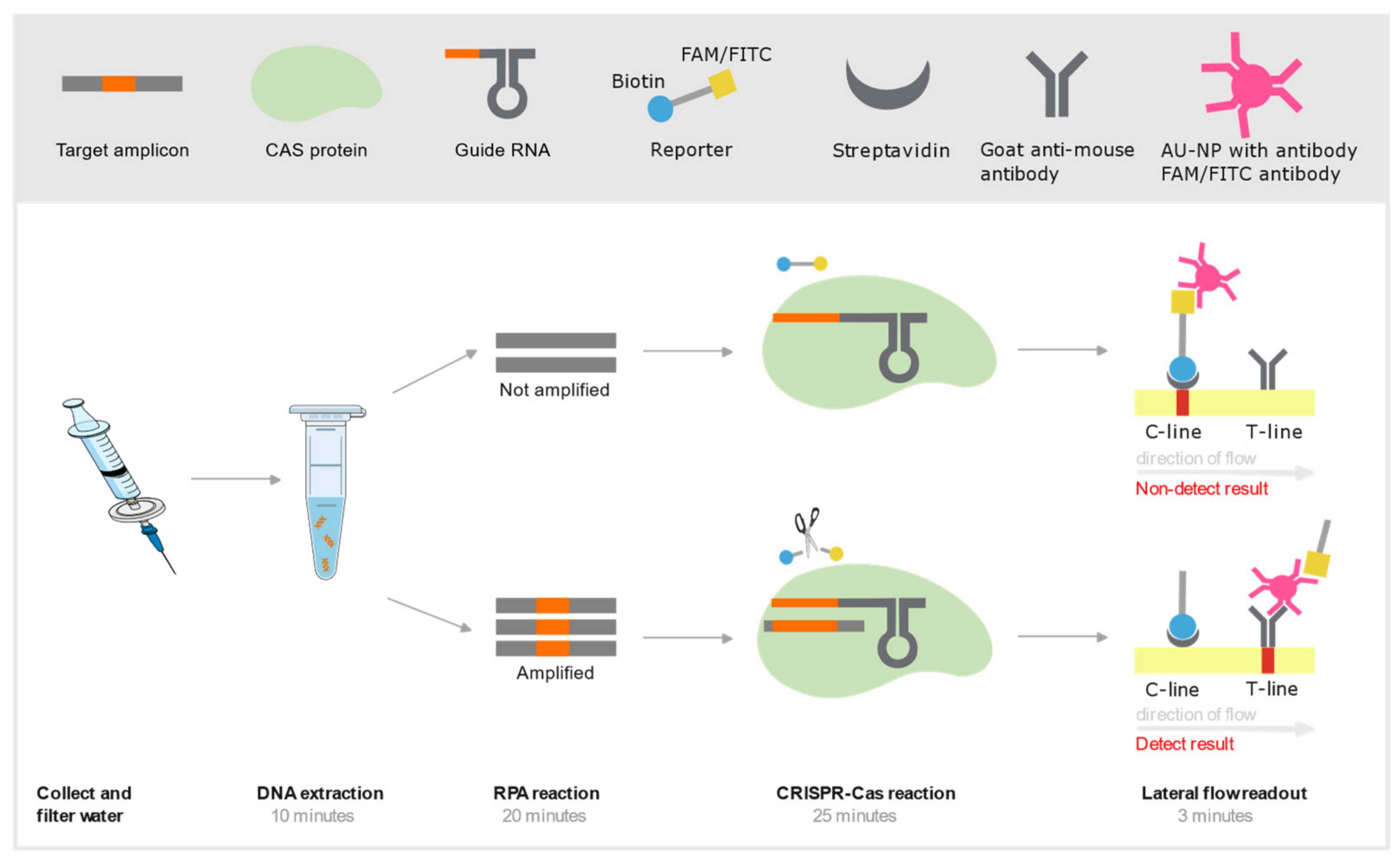
Schematic representation of an RPA-CRISPR-Cas assay integrated with a lateral flow strip for rapid detection (< 1hr). The process begins with the collection and filtration of a water sample, followed by crude DNA extraction. The target DNA is then amplified using a recombinase polymerase amplification (RPA) reaction at 37°C for 20 minutes. The resulting amplicon is subsequently introduced into a CRISPR-Cas reaction mixture, where the presence of the target sequence activates a trans-cleavage reaction. Finally, the mixture is applied to a lateral flow strip, enabling visual detection of the target species. Streptavidin is a biotin-binding protein and AU-NP is a gold conjugated nanoparticle.
Figure 2.
Schematic representation of an RPA-CRISPR-Cas assay integrated with a lateral flow strip for rapid detection (< 1hr). The process begins with the collection and filtration of a water sample, followed by crude DNA extraction. The target DNA is then amplified using a recombinase polymerase amplification (RPA) reaction at 37°C for 20 minutes. The resulting amplicon is subsequently introduced into a CRISPR-Cas reaction mixture, where the presence of the target sequence activates a trans-cleavage reaction. Finally, the mixture is applied to a lateral flow strip, enabling visual detection of the target species. Streptavidin is a biotin-binding protein and AU-NP is a gold conjugated nanoparticle.
| 1 | |
| 2 |
Disclaimer/Publisher’s Note: The statements, opinions and data contained in all publications are solely those of the individual author(s) and contributor(s) and not of MDPI and/or the editor(s). MDPI and/or the editor(s) disclaim responsibility for any injury to people or property resulting from any ideas, methods, instructions or products referred to in the content. |
© 2025 by the authors. Licensee MDPI, Basel, Switzerland. This article is an open access article distributed under the terms and conditions of the Creative Commons Attribution (CC BY) license (http://creativecommons.org/licenses/by/4.0/).
Copyright: This open access article is published under a Creative Commons CC BY 4.0 license, which permit the free download, distribution, and reuse, provided that the author and preprint are cited in any reuse.


