Preprint
Article

This version is not peer-reviewed.

Spore-Derived Isolates from a Single Basidiocarp of Bioluminescent Omphalotus olivascens Reveal Multifaceted Phenotypic and Physiological Variations

A peer-reviewed article of this preprint also exists.

Submitted:

21 December 2024

Posted:

24 December 2024

You are already at the latest version

Abstract

The fungal genus Omphalotus is noted for its bioluminescence and the production of biologically active secondary metabolites. We isolated 47 fungal strains of Omphalotus olivascens germinated from spores of a single mushroom. We first noted a high degree of variation in the outward appearances in radial growth and pigmentation among the cultures. Radial growth rates fell into at least five distinct categories, with only slower growing isolates obtained compared with the parental dikaryon. Scanning UV-vis spectroscopy of liquid-grown cultures showed variation in pigmentation in both the absorption intensity and peak absorption wavelengths, indicating that some isolates vary from the parental strain in both pigment concentration and composition. Bioluminescence intensity was observed to have isolates with both greater and lesser intensities, while the increased emission in response to caffeic acid was inversely proportional to the unstimulated output. Under UV illumination the media of parental strain was observed to be brightly fluorescent, which was not due to the pigment, while the isolates also varied from greater to lesser intensity and in their peak emission. At least three separate fluorescent bands were observed by gel electrophoresis from one of the cultures, while only one was observed in others. Fluorescence intensity varied significantly in response to casamino acids. None of a subset of the cultures produced an antibiotic effective against Staphylococcus aureus, and only the haploids, but not the parental heterokaryon, produced an antibiotic consistent with illudin M effective against Mycobacterium smegmatis. This same subset all produced an anticancer agent that was highly potent against MDA-MB-468 breast cancer tumor cells. We interpret these variations in haploids as significant in altering Omphalotus physiology and its production of secondary metabolites, which may in turn alter their ecology and life cycle, and could be further applied to studying fungal physiologies and facilitate linking them to their genetic underpinnings.

Keywords: 
;  ;  ;  ;  ;  ;  

1. Introduction

The fungal genus Omphalotus (Fayod) has long been known as a source of bioactive secondary metabolites and is one of the limited numbers of bioluminescent fungi, thus this genus is of widespread interest. The species Omphalotus olivascens H.E. Bigelow, O.K. Mill. & Thiers is largely endemic to California and has not been extensively examined for its physiological and genetic potential.
Bioluminescence is a property shared by all the members of the genus Omphalotus [1], and its genes bear a unique genetic signature [2]. Fungal luminescence remained an enigma until the luciferin, hispidin [3,4,5], and then its luciferase [6] were revealed.
Earlier, other fluorescent compounds were thought to potentially be related to fungal bioluminescence. Ichihara et al. [7] isolated dihydroilludin S, and suggested a possible connection to bioluminescence. Endo et al. [8] determined the fluorescence of two compounds from Omphalotus (Clitocybe) illudins, illudin S and ergosta-4,6,8(14),22-en-3-one. It was noted that the fluorescence emission peak of the ergosta compound at 530 nm was very similar to the light emission peak of fungal bioluminescence. Additional fluorescent molecules were also found in Omphalotus mushrooms, and were considered as candidates for its luciferin. Isobe et al. isolated flavin [9], and then lampteroflavin [10], both with fluorescent emission of 524 nm, which was also similar to their measurements of mushroom bioluminescence emission. The possible relationship of flavin mononucleotide (FMN) to fungal bioluminescence seemed at the time to be supported by the earlier discovery of FMN is the luciferin in bacterial bioluminescence [11]. Later, the sesquiterpene panal was also proposed as the mushroom luciferin [12]. Hispidin was only much later definitely determined to be the true luciferin.
Early in the history of antibiotic discovery, mushrooms in Basidiomycota were among the sources investigated for their antibiotic potential [13,14,15,16,17,18,19,20,21,22,23]. Among these, Robbins et al. [13] began investigation of Omphalotus illudens (synonym Clitocybe illudens); work which continues today. Anchel et al. [20] found that extracts of these mushrooms showed activity against Staphylococcus aureus and Mycobacterium smegmatis. Chloroform extracts contained the anti-mycobacterial component, which concomitantly decreased in the aqueous phase, and was designated illudin M. The substance remaining in the aqueous fraction was more strongly active against S. aureus, and was further extractable with ethyl acetate and was designated illudin S.
Nakanishi et al. demonstrated antitumor activity of the compound lampterol isolated from Omphalotus guepiniiformis (synonym Lampteromyces japonicus) against Ehrlich murine ascites tumors [24]. It was subsequently recognized that lampterol was identical with illudin S [20] when its chemical structure was determined [25,26,27,28]. Derivatives of illudins were later investigated for improved anticancer properties. It was shown that the acylfulvene illudin derivative hydroxymethylacylfulvene (irofulven) was selectively toxic against myelocytic leukemia and epidermoid, lung, ovarian, and breast carcinoma cells [29,30]. Irofulven was subsequently shown to antitumor activity against the human xenograft lung carcinoma model MV522 [31,32]. More recently, conjugates of illudin M have been shown to have potent antitumor activity [33], and improved production of illudin M has been investigated in order to supply material for larger studies [34,35,36].
Fungi in the phylum Basidiomycota typically have a life cycle in which sexual reproduction occurs with the formation of a basidiocarp (mushroom) that is haploid and dikaryotic, produced from a haploid dikaryotic mycelium, and then undergoes karyogamy only in its basidia in order to produce haploid basidiospores through meiosis [37]. These haploid spores are dispersed, often by the wind, and are capable of germinating to give rise to a haploid mycelium. In most Basidiomycetes, they must undergo fusion with a compatible haploid mycelium in order to be capable of completing their sexual life cycle. This sexual compatibility is mostly regulated by heterozygosity of two mating type loci which are linked in some systems and results in two mating types (bipolarity) and segregate independently in other systems which results in four mating types (tetrapolarity)[38,39]. In the genus Omphalotus, tetrapolar sexuality has been described, with compatible monokaryons forming clamp connections [40]. Sub-variations in mating types can result in a greater range of incompatibilities, resulting in many more mating types [41,42]. Once established, a mycelium such as that of Armillaria gallica may remain in the haploid dikaryotic state for millennia [43], although there may also be many different genetic events that can occur among monokaryons and dikaryons to generate a variety of mosaic individuals [44,45,46,47,48]. There are also notable exceptions where a mycelium that produces a basidiocarp is not haploid dikaryotic, including homokaryotic fruiting in Agrocybe aegerita [49] and Amanita phalloides [50].
Much of what is known about the Basidiomycetes originally became evident through study of their sexually reproductive structures — the mushrooms they produce. Geographic distribution, species and/or biotype differences, associations with plants, toxicity, food and medicinal values, bioluminescence and decomposition, all had contributions to our knowledge derived from study of the mushrooms alone. Knowledge of the mycelial basis of Basidiomycetes came first through their cultivation, eventually leading to the general understanding of their mode of reproduction.
Based on the prominence of Basidiomycete dikaryons and their fruiting bodies, there has been overall less study of their monokaryons. Of the areas of interest in monokaryons, mating type studies initially focused on Schizophyllum commune as an informative model [51], and haploids of other mushroom-forming species have been studied physiologically for edible mushroom production [52], and plant biomass degradation [53]. Peabody et al. [54] studied phenotypic differences in natural haploid isolates of Armillaria gallica. They found variation in rates of growth and in genetic plasticity in response to water potential, and A. gallica was revealed to generate a diploid state within the vegetative mycelium. Variation in bioluminescence intensity has been shown in spore-derived isolates of Armillaria mellea [55], and similar findings were earlier reported for Panellus stipticus (orth. var. P. stypticus) [56]. More recently, monokaryons of S. commune were studied and found to exhibit differences in lignocellulose degradation, culture morphology, fruiting body morphology, strain-specific carbon source profiles and other physiological differences [57]. Genomic analysis of the haploid strains revealed unexpectedly large variances, with monokaryons exhibiting genetic variation of 11.2% across strains, with the genes for mushroom formation and lignocellulose being largely conserved.
Here, we describe phenotypic variations found in basidiospore-derived isolates of Omphalotus olivascens. The survey included typical presumptive monokaryons that lacked clamp connections and a few heterokaryons that were obtained, as well as atypical highly branched isolates that lack clamp connections and grow very slowly. We show that basidiospore-derived isolates are a rich source of phenotypic variation in physiology and in production of secondary metabolites, and suggest they could help link such traits to their genetic underpinnings.

2. Materials and Methods

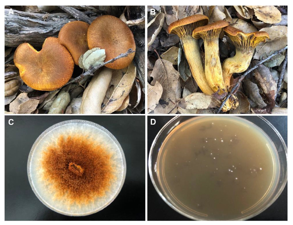
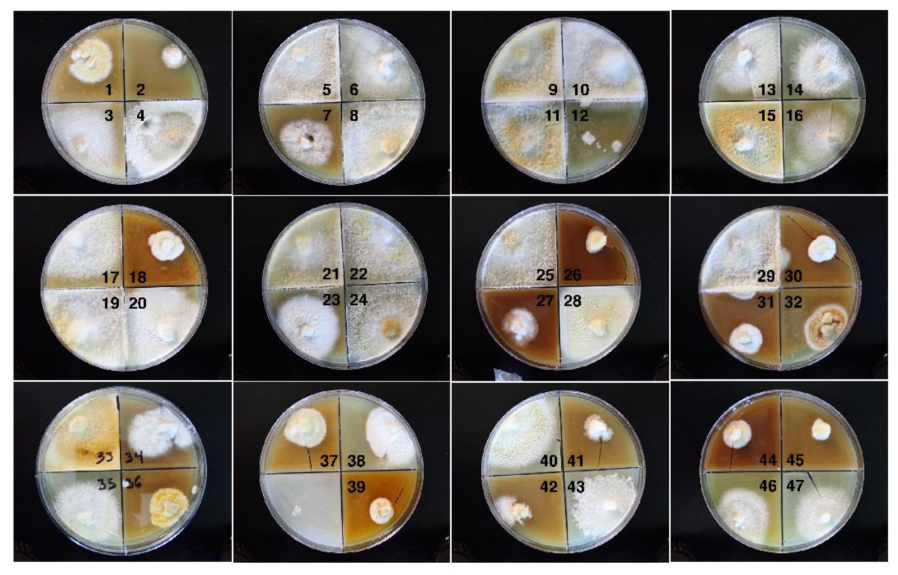
2.1. Fungal Cultures

O. olivascens mushrooms were collected under permission from the TreePeople Land Trust (TPLT). A spore print was collected from an individual fruiting body and a tissue sample of the parental heterokaryon was obtained from the stipe trama. A voucher specimen was deposited in the University of California, Los Angeles Herbarium (LA; fungarium col. #: DB.24.001). The spores were resuspended in sterile distilled water, vortexed vigorously, counted with a hemocytometer and examined for clumping. The spores were subjected to a dilution series in sterile distilled H2O and then plated to malt extract agar (MEA; 2% malt extract with 2% agar and 30 mg/ml chloramphenicol (CAM30)[40] and maintained under ambient laboratory conditions. Germinated isolates were collected between 5 and 17 days. Differences in the size of the cultures was noted and initially thought to represent possible early and late germination times, although we subsequently found there were genuine differences in the growth rates as described in our results. The isolates were transferred to bread crumb yeast agar (BCY; 2% white breadcrumbs, 0.5% yeast extract, and 1.5% agar on quadrant plates with 30 mg/ml chloramphenicol; BCYCAM30). After adherence of the growing culture to the agar was observed (approx. 3-10 days), the plates were transferred to 4° C for 2 months and subsequently photographed and subcultured. This and subsequent transferred culture materials were used for qualitative studies. For quantitative studies, the cultures were transferred to MEA and allowed to grow under ambient conditions whereby the cultures were actively growing and had not reached the periphery of the petri plate. Slower growing cultures necessitated staggered starts of the culture inoculum. Uniform transfers were made all on the same day by utilizing the back end of a sterilized glass Pasteur pipette (approx. 4.5 mm diameter) to obtain a punch taken from the periphery of the actively growing cultures. Other fungal cultures used in the study were Omphalotus olearius Harold H Burdsall (HHB) 7441, Omphalotus guepiniiformis (Lampteromyces japonicus) Ronald H Petersen (RHP), 2305, Panellus stipticus (P. stypticus) American Type Culture Collection (ATCC; Manassas, VA, USA) 66462, Armillaria mellea (155798, Carolina Biological, Burlington, NC, USA) and Pleurotus ostreatus (Mueller’s Mushrooms, Alpine, CA, USA).

2.2. Growth Assessment

2.2.1. Radial Growth

Radial growth was first surveyed among the isolates, and given semiquantitative rankings of + (slowest growing), ++, +++, ++++ and +++++ (fastest growing). Growth range representative isolates were quantitatively assessed by transferring four back-end Pasteur pipette plugs from the margin of the same actively-growing cultures to 100 mm petri dishes with MEA. Putative monokaryotic isolates representing five pre-observed growth rates were compared with the parental isolate, as well as spore-derived heterokaryons. The cultures were grown at 25°C in darkened conditions for 12 days, then photographed and measured. Diameter of an individual culture was calculated by taking the mean of the longest axis and the axis orthogonal. Difference in radial growth between strains was then analyzed with one-way analysis of variance (ANOVA), followed by post hoc comparisons with Tukey’s HSD test.

2.2.2. Mating Type Determinations

Mating trials were conducted for the first 12 isolates as described by Petersen and Hughes [40] using several different pairing approaches. Initially, four different isolates were separated by a sterilized coverslip “windows” and a central plastic barrier (mahjong tiles) to reduce the number of MEA plates needed (Figure S1). Faster growing cultures with slow growing cultures on the same plate sometimes resulted in overgrowth from neighboring cultures, and necessitated further isolation of each pair of isolates per plate. The macro-morphology of confrontations was scored as robust (compatible), barrage (same B mating type), flat (same A mating type), or overlap (same A and B; no apparent reaction). Confronted hyphae were then observed with light microscopy (AmScope T340B compound microscope with MU500-HS digital camera and AmLite software) directly through the coverslip on the media and scored for the presence or absence of clamp connections and the degree of branching.

2.3. Pigmentation

Variations in pigmentation of cultures grown on BCY agar were visually notable. Pigmentation was further assessed in clear M9 minimal media pH 6.8 (Difco, Becton Dickinson, Franklin Lakes, NJ, USA) with 0.4% glucose (Na+ 103.7, mM; phosphate, 69.54 mM; chloride, 27.3 mM; f mM; K, 21.9 mM; ammonium, 18.7 mM; Mg2+, 2 mM; sulfate, 2 mM), with and without 50 mM FeCl3. Cultures of varying growth rates were allowed to grow from 2-4 weeks until pigmentation was visibly detectable. UV-Vis spectroscopy was performed on the culture supernatants that were spun at 17,000 x g for 2 min. Separate scans were performed from 225 to 350 and 350 to 600 nm with a SpectraMax M3 (Molecular Devices, San Jose, CA, USA).

2.4. Bioluminescence

Luminescence was measured by luminometry as relative light units (REU) with an Invitrogen iBright 1500 and using iBright Analysis software (Invitrogen/ThermoFisher Scientific, Waltham, MA, USA). The isolates were cultured in BCYCAM30 within black 24-well plates, with the cultures transferred as 1-2 mm pieces obtained by cutting actively growing mycelia with a scalpel. The non-luminescent Pleurotus ostreatus was used as a control. Cultures were then grown for 7 days with a diurnal cycle in ambient conditions (approx. 25°C). Phenotypic variation in the response to caffeic acid was determined by its addition to cultures on the 7th day, applied with 100 μl of 1 mM caffeic acid (Pure Health Solutions, eBay) and 0.1% DMSO diluted from a 10x stock that was filter-sterilized (Puradisc 25 mm PES Syringe Filter, 0.2 µm).

2.5. Fluorescence

2.5.1. Fluorescence in Minimal Media

Fluorescence of the culture media of the parental heterokaryon and other isolates growing on BCY media was noted using a hand-held 365 nm UV lamp. Qualitative comparison of the strains was assessed in 24 well black tissue culture plates with clear bottoms containing translucent M9-glucose agar. The plates were subjected to UV transillumination at 365 nm and photographed. The fluorescence excitation and emission of culture supernatants grown in clear M9 liquid media with glucose were determined from culture supernatants comparing the parental heterokaryon with notably fluorescent isolates that gave slightly different colored fluorescence. Scanning fluorimetry was used to determine the excitation and emission maxima with a SpectraMax M3. The M9 supernatants were also concentrated 10-fold by subjecting them to evaporation at low pressure, and then separated in a 2% agarose gel with 1X M9 salts as the buffer with 200 mA of current and photographed using 365 nm transillumination.
The fluorescent supernatants were subjected to organic solvent extraction by addition of an equal volume of either chloroform or ethyl acetate, followed by vortexing and centrifugation. The supernatants were also heated to 90° C in a heat block.

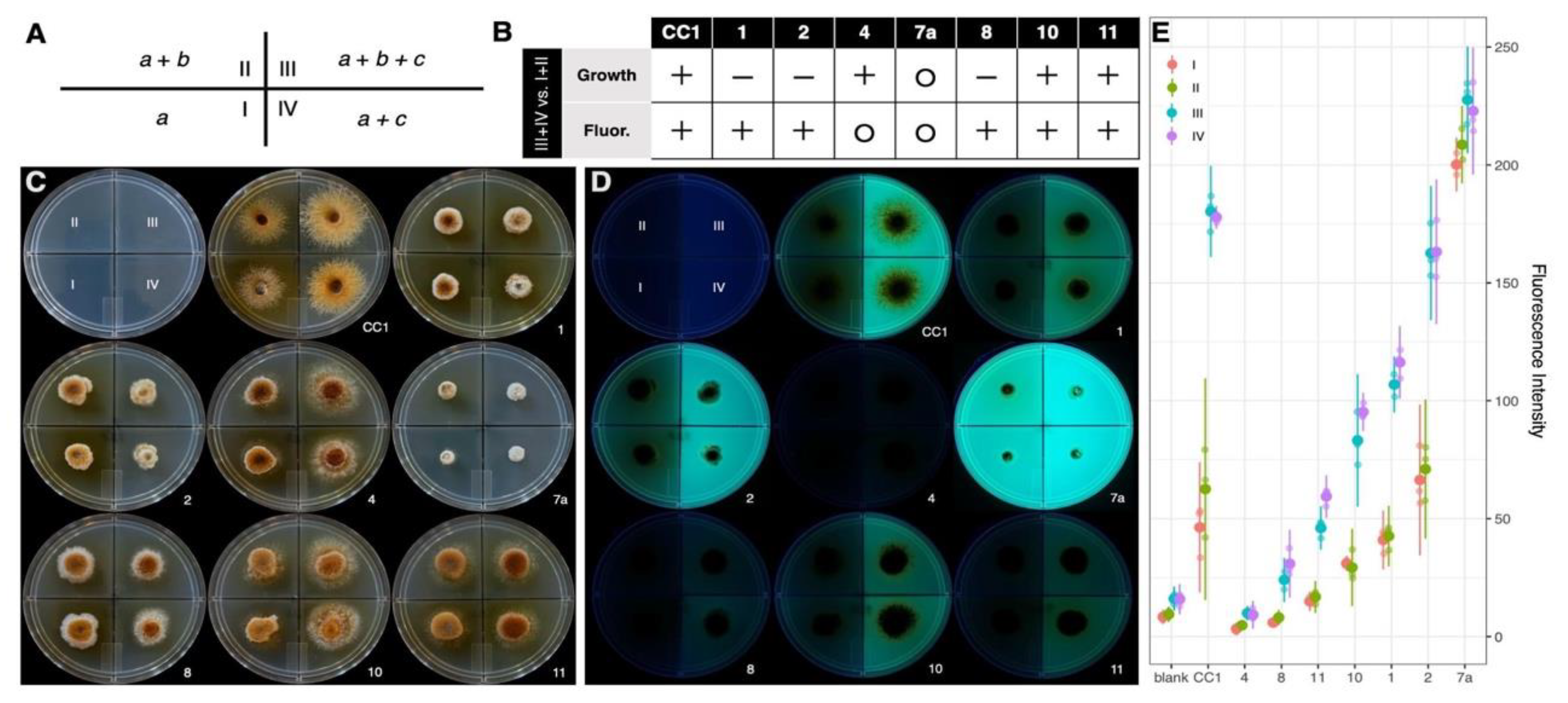
2.5.2. Growth and Fluorescence with Casamino Acids

The parental strain and non-clamped isolates pre-screened on 0.1% peptone agar were assayed for relative growth and fluorescence on four preparations of minimal media with 1.5% agar varying in composition of I, 0.2% glucose; II, 0.2% glucose, 0.1% KH2PO4, and 0.1% MgSO4 (from an autoclaved 10% stock); III, 0.2% glucose, 0.1% KH2PO4, 0.1% MgSO4, and 0.1% casamino acids; and IV, 0.2% glucose and 0.1% casamino acids only. X-plates with quadrants containing each of the four media were inoculated with plugs from the margins of the same inoculum grown on MEA. These cultures were then grown at 25°C in darkened conditions for 10 days. They were then viewed with 365 nm transillumination and photographed. Relative growth between treatments was scored as more robust (+), less robust (–), or no contrast (o). Relative fluorescence intensity was analyzed quantitatively with ImageJ (NIH) by first splitting the UV images into RGB channels, then with the green channel, selecting three regions (50-pixel diameter) from uncolonized corners within each quadrant of the X-plate. Intensity in each region was measured as mean gray value (sum of values divided by number of pixels). Correcting for autofluorescence of blank media, differences in fluorescence intensity were analyzed with one-way ANOVA and Tukey contrasts.

2.6. Antibiological Activity

2.6.1. Antibacterial Activity

Antibacterial disc diffusion assays were performed using fungal M9-glucose culture supernatants. 10 μl of filter-sterilized (0.2 μm Whatman (ThermoFisher, Waltham, MA, USA) Puradisc PES membrane) culture supernatants were absorbed into sterile 6 mm blank filter paper discs (Difco, Becton Dickinson, Franklin Lakes, NJ, USA). Confluent bacterial cultures of Staphylococcus aureus (ATCC 29523) and Mycobacterium smegmatis (ATCC 14468) were inoculated onto tryptic soy agar (Difco), the culture supernatant discs applied on top, and then incubated at 37 °C overnight. The plates were photographed and the diameter of the zones of clearing were determined.

2.6.2. Antitumor Cell Activity

MDA-MB-468 human breast adenocarcinoma was obtained from the ATCC (MDA-MB- 468 was kindly provided by Dr. Jonathan Kelber, Baylor University), and was authenticated at the University of Arizona Genetics Core, and determined to be mycoplasma-free (IDEXX Laboratories, Westbrook, ME, USA). Fungal culture supernatants were used to assess cytotoxicity toward MDA-MB-468. MDA-MB-468 cells were cultured in Dulbecco’s Modified Eagle Medium (DMEM), supplemented with heat-inactivated fetal bovine serum (10% vol/vol; Gibco/Life Technologies, Grand Island, NY, USA) and 1% of penicillin-streptomycin (10,000 units penicillin and 10mg/mL streptomycin; Sigma- Aldrich, St. Louis, MO, USA). Cells were trypsinized (0.5% trypsin 0.2% EDTA, Sigma–Aldrich) for use in passages and seeding cell proliferation assays. Cancer cell survival was determined using Alamar Blue (R&D Systems, Minneapolis, MN, USA) with relative fluorescence 544/590 nm, using a SpectraMax M3 (Molecular Devices, Sunnydale, CA, USA) running SoftmaxPro 7.0.

3. Results and Discussion

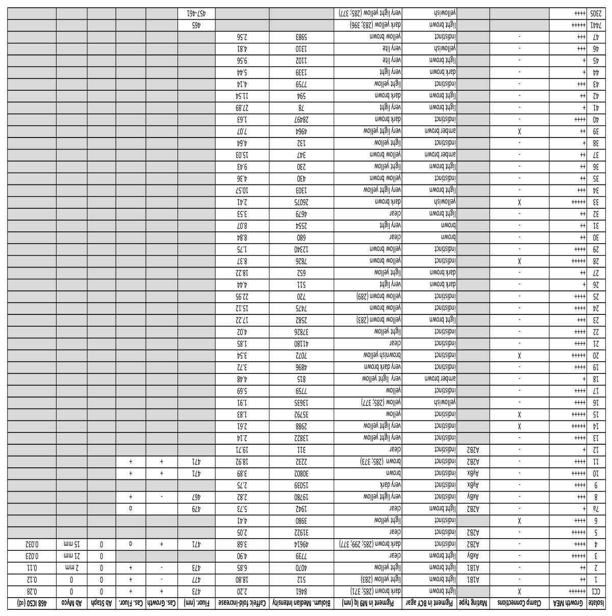
3.1. Fungal Cultures

The mushroom that was collected and its tissue sampled successfully grew on MEA CAM30 and the spores were germinated (Figure 1).
Along with this parental tissue isolate that had a rusty brown color somewhat similar to the mushrooms themselves, 47 basidiospore-derived isolates were included in this study (Figure 2; Table 1). Clone CC1-7, which initially grew slowly, exhibited a faster growing sector, and the slower growing component most similar to the initial strain was reisolated and designated CC1-7a. Clamp connections were observed in hyphae of the parental isolate as well as seven spore-derived isolates (CC1-6, 14, 15, 20, 28, 33, 39), suggesting heterokaryotic status. All other isolates did not possess clamp connections.

3.2. Growth

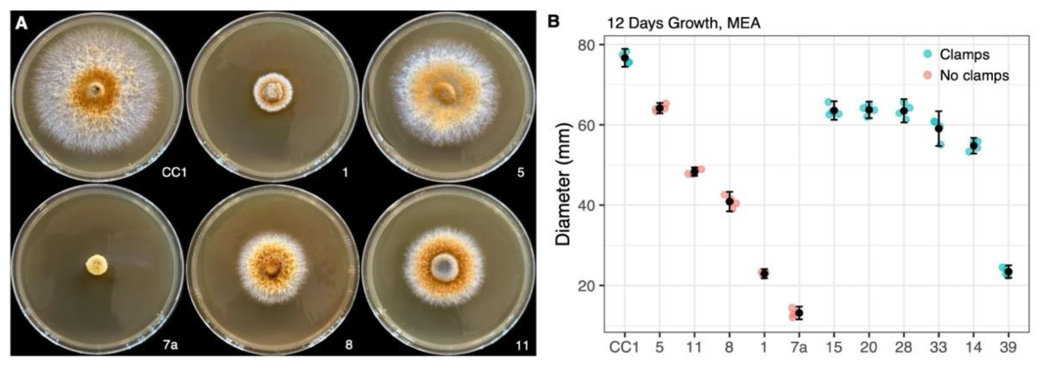
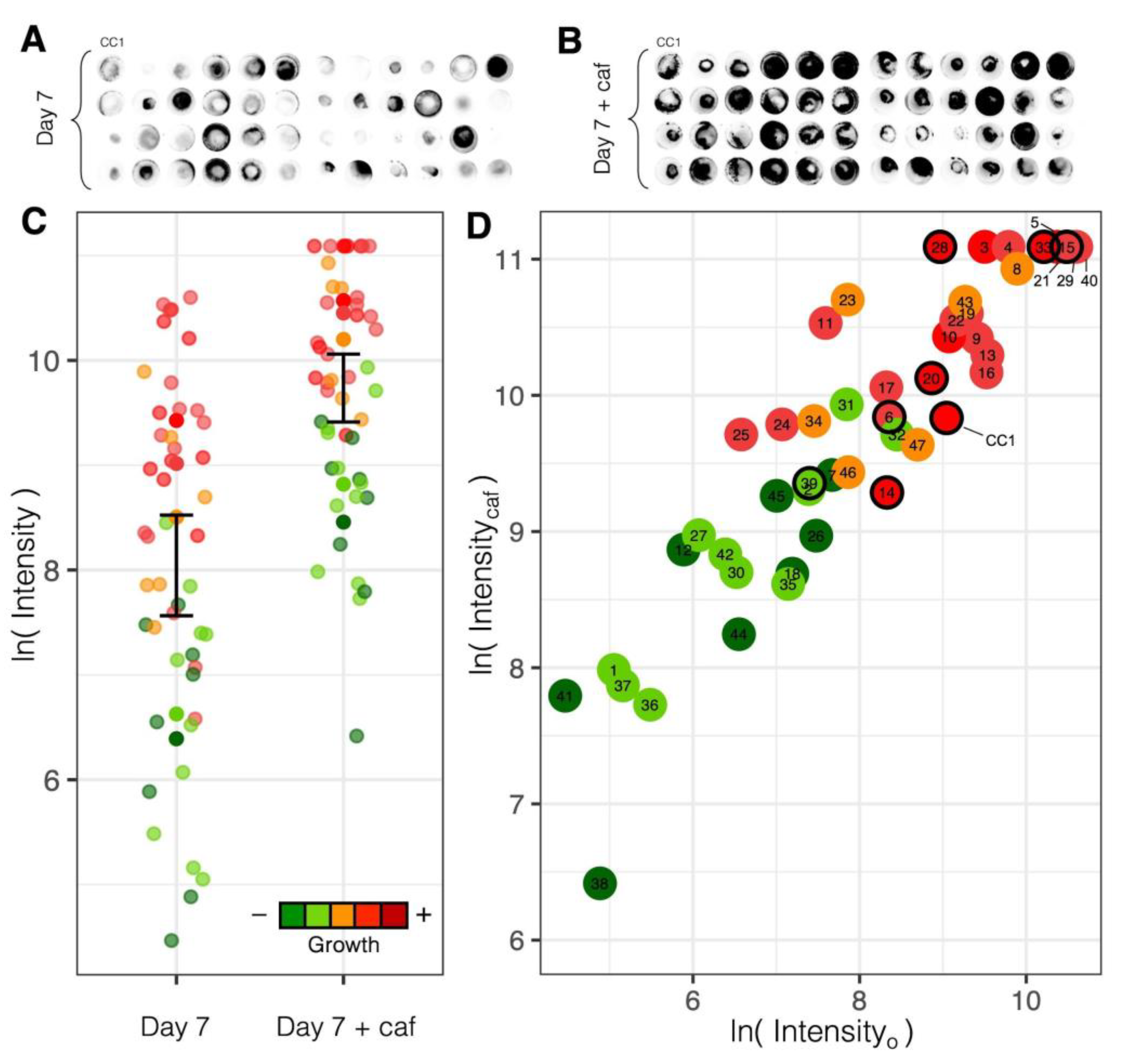
3.2.1. Variation in Radial Growth
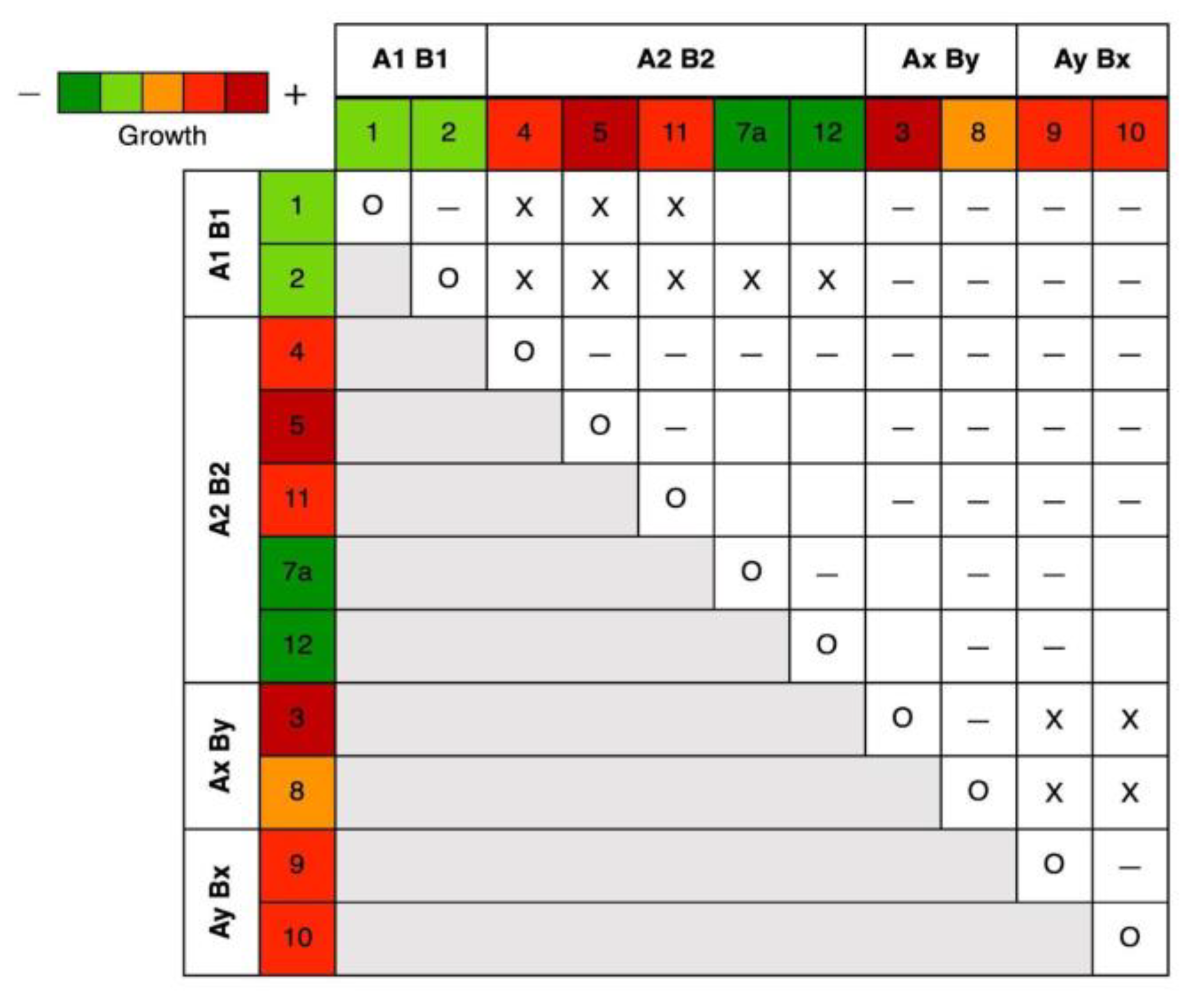
Radial growth on MEA was highly consistent for replicates of the parental strain and non-clamped, range-representative isolates, which showed significant difference after 12 days (one-way ANOVA, F(5, 17) = 1932.34, p < .001) and varied in mean diameter from 76 mm in the parental strain to 13 mm in CC1-7a (Figure 3; four replicates were measured for all isolates except CC1-1, which lost one due to contamination). Compared to the parental heterokaryotic strain, all spore-derived clamped isolates measured (N = 6) showed significantly less radial growth (Tukey’s test, p < 0.001), ranging from 54–63 mm in five of these isolates (CC1-15, 20, 28, 33, and 14), to 23 mm in CC1-39 (Figure 3B). CC1-6, another clamped isolate, was omitted from radial growth assessment due to reproducibly switching its growth from slow-growing white mycelia to fast-growing rusty brown mycelia of various intensities. Pigmentation of mycelia varied with other factors based on age, room temperature vs. cold, but was not comparatively assessed.
The observation that all spore-derived isolates, including heterokaryons, achieve only as much as 82% of the radial growth of the parental isolate could be consistent with physiology under selection for outcrossing; though, differences in nutrient composition of media can yield different inferences about natural physiology in Agaricomycetes [58].

3.2.2. Mating Types

A heterothallic tetrapolar system was observed on MEA, consistent with past studies of Omphalotus [40]. All four mating types were identified from non-clamped individuals among the first 12 isolates (Figure 4), with each mating type represented by at least two isolates. Examples of pairings with 4 strains per plate using barrier (mahjong tiles) with coverslip windows and with only 2 strains per plate with only a single coverslip is shown in Figure S1. Between isolates, heterozygosity at both mating loci was determined from compatible matings, which resulted in the formation of clamp connections in confronted hyphae for each of the studied individuals. Two isolates were determined homozygous at both loci if they showed compatible matings when paired with the same partner. After arbitrarily designating the first two mating types A1B1 and A2B2, isolates which did not show compatible matings with either of these first two were inferred to have mating types A1B2 or A2B1; however, morphological variability between confrontations of these non-mating isolates left us unable to resolve their status as either A or B locus incompatibilities. As such, the second set of complementary mating types are reported here as AxBy and AyBx. Compatible matings in the slowest-growing isolates (CC1-7a and CC1-12) were observed with pairings against an isolate from each of the four determined mating types.

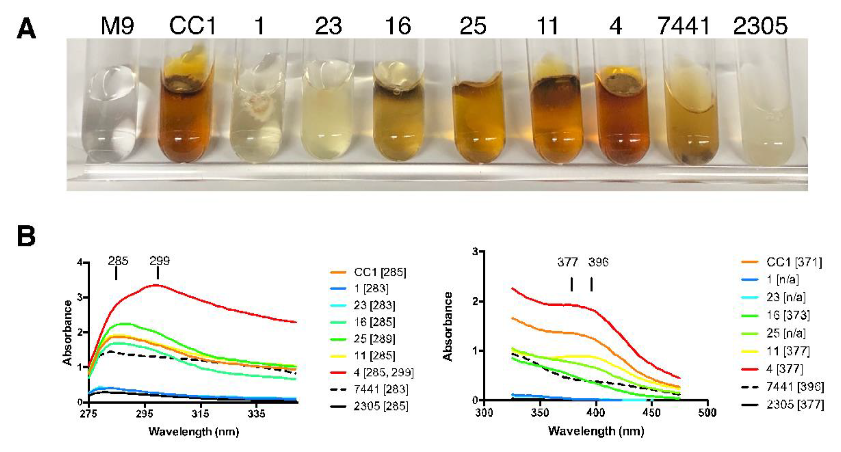
3.3. Variation in Pigmentation

Pigmentation varied in liquid media, and did not follow the same pattern on BCY agar as shown in M9-glucose (Figure 5; Table 1). This is exemplified by isolate #4, which was nearly white on BCY agar, but was the darkest grown in M9-glucose broth. We also noted other variations in pigmentation based on age, room temperature vs. cold, but did not comparatively assess them. The ability to separate the culture from the initially clear supernatant made use of this media preferable, and it seems likely that it would vary in yet other media and/or other carbon sources. The M9-glucose control and the parental CC1 are shown next to a gradation of pigment intensity (CC1-1, 23, 16, 25, 11, 4) as well as Omphalotus olearius (HHB 7441) and Omphalotus guepiniiformis (RHP 2305)(Figure 5A). In the shorter wavelength scan (Figure 5B, left side), there was a predominant peak at 285, with only CC1-4 showing a stronger additional peak at 299, indicating at least one additional pigment in high concentration. In the longer wavelength scan (Figure 5B, right side), a peak at 377 was the most prominent amongst the isolates, but with the parent having a peak at 371. Omphalotus olearius (HHB 7441) was the most different, with a peak at 396.
Based on these observations, pigmentation intensity is highly variable among isolates, and is differentially expressed when grown in different culture media. Although purified pigments would be required to precisely describe these differences, it is clear that CC1-4 makes an additional prominent pigment with an absorption peak at 299 and that minor variation occurred among the other isolates. These data also show that O. olivascens pigmentation is similar to O. guepiniiformis and that O. olearius is not similar to these other two species when assessed in M9-glucose. Numerous pigments in fungi and bacteria have been studied (see Gill [59] and Ramesh et al. [60] for reviews), and several pigments in Omphalotus have been identified [61]. Pigments from O. olivascens are commonly used for dyeing fabrics, and differences in pigment production across haploids from another dye mushroom, Phaeolus schweinitzii, have also recently been noted (Sidnee Ober-Singleton, personal communication 2024).

3.4. Bioluminescence

3.4.1. Variation in Luminosity

All O. olivascens isolates (N = 48) exhibited luminosity on BCY greater than background noise measured in the non-luminescent control species Pleurotus ostreatus. These luminous isolates varied from 460- to over 800-fold between the brightest and dimmest within three replicate sets started on different days, with several isolates showing consistently brighter or dimmer luminosity than the parental isolate (Figure 6A). The top five isolates ranked across replicates by mean luminescence intensity (CC1-21, 4, 22, 5, and 10) were among the top one third brightest isolates in each replicate, and were brighter than the parent culture by as much as 8-fold. All five of these isolates lacked clamp connections and were scored a priori as belonging to the two fastest growth classes. The five isolates ranked last by mean luminescence (CC1-41, 36, 12, 35, and 1) were in the bottom two thirds in each replicate, and showed an average luminosity dimmer than the parental isolate by 30- to 600-fold. All of the five dimmest isolates also lacked clamp connections, and belonged to the two slowest growth classes.
It was generally observed that mycelia of fast-growing isolates had more volume and were more rusty brown than mycelia of slow-growing isolates, which were more white. Overall, brighter strains were among the faster-growing isolates, though it was not strictly observed that luminosity corresponded to growth rate; several spore-derived isolates reproducibly showed greater luminescence than the parental strain, which was the isolate that exhibited the fastest growth on MEA.

3.4.2. Response to Caffeic Acid

The functional response of the fungal bioluminescence pathway [6] was assessed across isolates through stimulation with caffeic acid, which is a biosynthetic component for the luciferin precursor hispidin [5]. The addition of caffeic acid enhanced the luminescence intensity of mycelia in all O. olivascens isolates (Figure 6B,C; Table 1), collectively showing a significantly greater mean luminescence intensity immediately after the treatment (Welch’s t test: t = -5.8807, df = 82.383, p < 1E-07). A negative control of 100 μl of 0.1% DMSO without caffeic acid yielded an insignificant difference in measured luminescence after application to six replicate cultures of the parental strain (Welch’s t test: t = 1.1253, df = 9.8644, p > 0.2). The magnitude of the luminescence response to caffeic acid ranged from a 1.6- to 27-fold increase in luminosity, with dimmer isolates showing a greater increase in luminosity than brighter isolates. A regression analysis revealed a strong linear relationship in log-transformed luminescence intensity before and after treatment (Figure 6D; adjusted R2 = 0.823, p < 1E-18), indicating that initial luminescence intensity is a strong predictor of the intensity post-treatment.
Altogether, the parental strain and derived isolates studied here show luminosity that is predictably stimulated through the application of caffeic acid, consistent with known functions underlying fungal bioluminescence [5], and with isolates of higher baseline luminosity showing diminishing returns suggestive of saturation. The consistent functional response observed across O. olivascens isolates is in line with the view that fungal bioluminescence may serve in one or more adaptive roles that are not well defined which likely varies across species [1]. In Omphalotus, environmental stresses were found to induce differential transcriptional regulation of bioluminescence in O. guepiniiformis [62], though biological interpretation for such responses remains unclear.

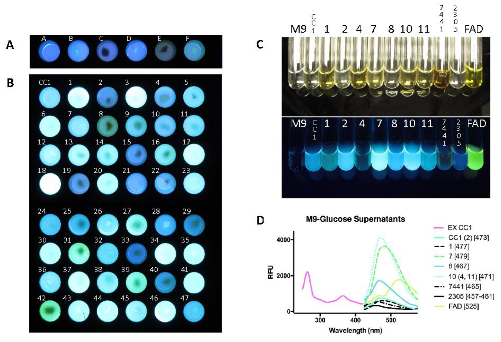
3.5. Variation in Fluorescence

3.5.1. Fluorescence Variation in Minimal Media

Variation in fluorescence was notable among petri plates using a hand-held UV light, and growth in 24 well plates with M9-glucose agar allowed all the cultures to be observed simultaneously (Figure 7A and B). The empty well (Figure 7A, well A) exhibited the color of the 365 transillumination passing through the translucent M9-glucose agar. Among the comparator fungal species in Figure 7A, only Armillaria mellea (well C) showed any observable variation from the media only well, which may have also had a contribution from non-fluorescent pigmentation. Variation between the parental strain (CC1) and isolates occurred in both fluorescence intensity and in minor color variations (Figure 7B). These fluorescent color variations were further apparent in M9-glucose culture supernatants of representative strains (Figure 7C). The fluorescence excitation and emission peaks were determined for these strains and showed variations consistent with their apparent minor color shifts, e.g., with culture CC1-8 appearing bluish (Em: 467 nm) and culture CC1-7a appearing more greenish (Em: 479 nm). The other two Omphalotus species (7441 and 2305) were bluer isolates than the parental strain or any of the isolates.
We further separated the fluorescent supernatants by agarose gel electrophoresis (Figure S2). A major bright band that was similar in migration rate was observed in all but CC1-4 and O. olearius and O. guepiniiformis (Figure S2A). Clone CC1-7a was the brightest following electrophoresis, and was further separated and showed at least three distinct bands (Figure S2B) that were not as readily apparent in the other strains. Because their observation may be due to concentration rather than their complete presence or absence, we only conclude that variation in the observable band composition occurred, and may account for the differences in peak fluorescence emissions.
Fluorescent compounds have previously been isolated from Omphalotus spp. including illudin S an ergosta [8], flavin [9], lampteroflavin [10], and panal [12] . The fluorescence substances in the O. olivascens culture supernatant were observed not to be diminished by 10 min at 90 °C (Figure S3), which was confirmed by fluorimetry (data not shown). The substances in our study were not extracted with chloroform or ethyl acetate (Figure S3). These results establish the lack of overall heat sensitivity of the fluorescent components, lack of being readily volatile, and insolubility in organic solvents. These features are not consistent with the known fluorescent compounds of bioluminescent fungi described by Endo et al. [8], or Nakamura et al. [12]. The absence of the characteristic flavin peak at 525 also indicates the O. olivascens fluorescent compounds are not the flavins FMN or lampteroflavin previously found in Omphalotus guepiniiformis [9,10]. Other known fluorescent compounds from mushrooms such as aurantricholides A and B isolated from Tricholoma aurantium are also extractable in organic solvents, and are unstable [63], so are not consistent with the fluorescent compounds in O. olivascens culture supernatants. The compounds we observed may represent new fluorescent substances, or known substances for which fluorescence has not been described. Their secretion into the culture supernatant which separates them from the mycelium, their water solubility and ability to be separated by electrophoresis should facilitate their purification and the elucidation of their chemical structures.

3.5.2. Fluorescence and Growth Variation in Response to Casamino Acids

Following the preliminary observation of differential stimulation with peptone, phenotypes for the parental strain and non-clamped isolates with representative variation (N = 8) were scored in four treatments of minimal media with and without casamino acids and inorganic salts (Figure 8A). After 10 days of growth, discrete contrasts were apparent between treatments with and without casamino acids for phenotypes including mycelial growth and fluorescence of media (Figure 8B), which were consistent in a biological replicate started on a separate day. The parental heterokaryon and three derived isolates (CC1-4, 10, 11) showed more robust growth with casamino acids, while three other isolates (CC1-1, 2, 8) showed less robust growth (Figure 8C); none of these showed contrasts in growth with inorganic salts. The isolate belonging to the slowest a priori growth category (CC1-7a) showed the least growth in each treatment for all isolates, and showed no apparent growth contrast between treatments after 10 days.
Relative fluorescence intensity for media of a given treatment varied widely across strains (Figure 8D–E), and differed as much as 60-fold between the most and least intense. Relative fluorescence intensity also differed significantly between treatments within all strains (one-way ANOVA, p < .001), except for CC1-4 and CC1-7a (p > .05). For the strains with significant fluorescence contrast, treatments with casamino acids (III and IV) showed greater intensity than treatments without casamino acids (I and II; Tukey’s test, p < 0.05). Notably, CC1-7a had greater relative fluorescence in its treatments without casamino acids than all other isolates showed in treatments with casamino acids. CC1-4 was the only isolate to measure lower fluorescence intensity than the blank media.
On the whole, growth and fluorescence are differentially inducible through the addition of a complex organic nitrogen source, suggesting differences in nitrogen metabolism. This result is consistent with the finding in another mushroom-forming species, the ectomycorrhizal Tricholoma matsutake, that nitrogen metabolism varies considerably across basidiospore-derived isolates generated from a single basidiocarp, suggesting variation in ecophysiology [52]. In an ecological context, the response to a complex organic nitrogen source could lead to selective pressure between individuals if their substrate is homogeneous in nitrogen composition, or foster complementary interactions between individuals if the substrate is heterogeneous [64].

3.6. Antibiological Activity

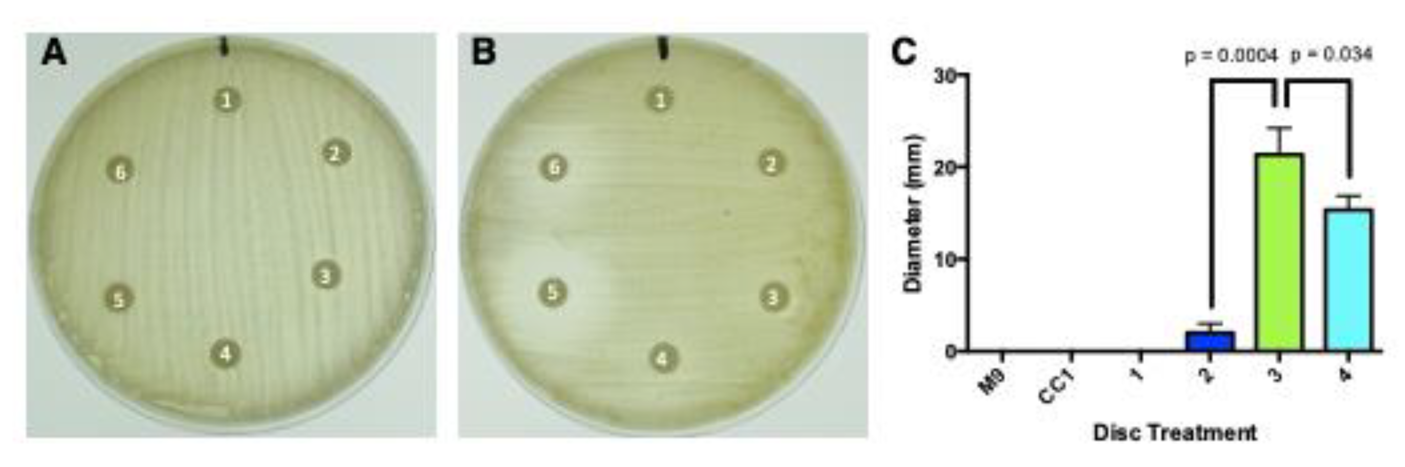
3.6.1. Antibacterial Activity

Of the 5 culture supernatants compared with the M9-glucose media control, none were antibiotically active against S. aureus (Figure 9A). However, isolates CC1-3 and CC1-4 consistently had observable zones of inhibition against M. smegmatis, with only very minor inhibition from CC1-2, and none from the parental CC1 or CC1-1 (Figure 9B). Inhibition zones of those that were measurable were also statistically different from each other (Figure 9C). Based upon the bacterial sensitivity pattern established by Anchel et al. [20], these results indicate O. olivascens makes detectable illudin M, but not illudin S, which was apparent only from the monokaryons and not the parental heterokaryon.

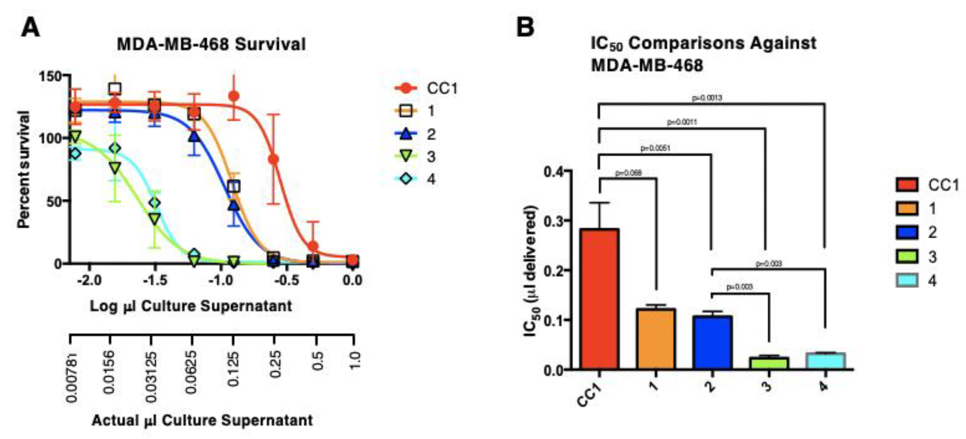
3.6.2. Antitumor Cell Activity

Variation occurred in the μL IC50 values of the parental heterokaryon (CC1) and the first 4 monokaryons (Figure 10). CC1 had the highest (least potent) IC50 (0.28 μL +/- 0.031), followed by CC1-1 and CC1-2 with similar IC50 values (O.12 μL +/- 0.0051 and 0.11 μL +/- 0.0063) and then CC1-4 and CC1-3 with IC50 values of 0.032 μL (+/- 0.0018) and 0.023 μL (+/- 0.0032) respectively. The CC1 heterokaryon was significantly less toxic than all the other isolates, while isolates CC1-1 and CC1-2 grouped together and were not significantly different from each other but were significantly different from isolates CC1-3 and CC1-4 that also did not differ significantly from each other and were the most potent.
As with other behaviors of the isolates, cytotoxicity showed strong physiological variation. While only four of the isolates were compared with the parental heterokaryon, the most potent of these was approximately 10-fold greater than the parental strain. Based upon the antibacterial activity, the active component is consistent with illudin M. Enhanced production of illudin M has recently been a focus of interest [34,35,36], and thus this approach offers the potential for additional improvements.
Although promising, the anticancer candidate irofulven did not progress past phase II clinical studies in humans [65]. However, Kim et al. [2] noted that there is renewed interest in illudins because of the enhanced sensitivity in cancers with mutations in the transcription-coupled nucleotide-excision repair (TC-NER) pathway. They also noted that genetic modifications to increase production of the illudin precursor of irofulven by Omphalotus have not been attempted because of the lack of a genetic system. Our work suggests examining monokaryons for enhanced illudin production could reveal isolates with substantially increased production. Such clones could further be increased using the enhanced production methods recently developed [34,35,36].
While Omphalotus has not been genetically manipulated, genomic analysis of Omphalotus spp. has provided the basis of terpene production [2,66,67]. Wawrzyn et al. [66] were successful in first identifying the genes from Omphalotus olearius and then expressing them in Escherichia coli. They were able to produce twelve different products including protoilludine. Kim et al. [2], further examined O. guepiniiformis and identified homologous genes and performed a synteny analysis, which is likely to further the understanding of the genes involved in terpene biosynthesis, including illudins. The genes Omp4, Omp6 and Omp7, for example, are components of the pathway that produces protoilludene, the precursor to illudin S, and were further studied by Yang et al. [67]. As heterologous gene expression in tumor-targeted Salmonella and E. coli strains have been used to enhance antitumor activity [68], expression of therapeutic illudins by these strains offers a potential therapeutic delivery system, as illustrated by heterologous expression of cytosine deaminase in Salmonella, which successfully converts 5-fluorocytosine to the anticancer agent 5-fluoruricil [69]. Our work shows that the cytotoxic product of Omphalotus olivascens that is consistent with illudin M is variably produced by monokaryons, which could offer the opportunity to genetically dissect its production by comparative genomic analysis.

4. Conclusions

All of the physiological/phenotypic characteristics we assessed varied substantially (Table 1). Only in one case, radial growth rate, was only a reduction in that quantitative characteristic found. And in only two cases was only greater activity found, antibacterial and anticancer agent production. In all other cases, pigmentation, bioluminescence, response to caffeic acid, fluorescence, and responses to casamino acids, significantly greater and lesser quantitative properties were obtained. Such variations, alone and in combination, can be considered emergent properties relative to the parental strain.
These observations are consistent with both known classical genetics, and the emerging feature(s) of fungal genomics whereby the genetic content of haploids varies significantly [57]. The processes of crossing over, random assortment, and dominance and/or recessive traits being pronounced in haploids is consistent with our findings. Indeed, genetic loci associated with agronomic traits have been identified through segregation analysis in other mushroom-forming fungi [70,71,72,73]. However, with the backdrop of larger than expected heterozygosity becoming apparent in fungal genomes, we suspect that genomic analysis of Omphalotus haploids may reveal significant variations in DNA content.
The significance of these findings are multifold. First, the relationship of such significant physiological variation to the ecology and life cycle of Basidiomycetes must be considered as the haploid stage constitutes its initial stage in the environment. The roles of each of these physiological features outside the laboratory environment could play a role in survival of haploids in different environmental conditions and at different times, as well as in sexual competition across haploids for limited mating partners [74]. The features we measured are of general significance in fungal biology. Only recently has terpene biosynthesis and the basis for fungal bioluminescence been elucidated, but still retains areas of uncertainty. By comparison among haploids, increases in bioluminescence or other characteristics could reveal the existence of suppressive regulatory elements, inhibitors or competing biosynthetic pathways, while decreases in bioluminescence could represent increased regulatory suppression, lack of activators, promoter elements or cofactors not yet known. Thus, this approach provides an opportunity to link physiological functions to their corresponding genetic components. The use of fungi in biosynthesis is also directly addressed by this approach. Illudins have both antibiotic and anticancer properties; current approaches to increasing their production in O. nidiformis have included process development [35], improving downstream processing [36] and improving production by the producer strain [34]. Our work on O. olivascens haploids demonstrates an alternative and potentially complementary approach.
Overall, these data support physiological evaluation of haploids as a broad approach to studying fungal physiologies with the potential to link them to their genetic components consistent with recent reports [57,75].

Supplementary Materials

The following supporting information can be downloaded at the website of this paper posted on Preprints.org, Figure S1: Plating setup for mating type pairing experiments.; Figure S2, Visualization of fluorescence components separated by agarose gel electrophoresis; Figure S3, Effect of heat and organic solvent extraction on fluorescence.

Author Contributions

Conceptualization, D.B.; formal analysis, R.D. and D.B.; investigation, R.D. and D.B.; resources, D.B.; data curation, R.D. and D.B.; writing—original draft preparation, D.B. and R.D.; writing—review and editing, R.D. and D.B.; visualization, R.D. and D.B.; supervision, D.B.; project administration, D.B.; funding acquisition, D.B. All authors have read and agreed to the published version of the manuscript.

Funding

This work was supported by the National Institutes of Health (NIH) Grant SC3GM098207 to DB.

Data Availability Statement

The data is available from the Investigators.

Acknowledgments

We thank TreePeople Land Trust for permission to collect fungal specimens. We thank Rachael Mackelprang for helpful discussions.

Conflicts of Interest

R.D. declares no conflicts of interest. D.B. has a financial interest in Theobald Therapeutics LLC, which owns patent rights that include subject materials discussed in this report. The funders had no role in the design of the study, in the collection, analyses, or interpretation of data; in the writing of the manuscript; or in the decision to publish the results.

References

  1. Ke, H.-M.; Lu, M.R.; Chang, C.-C.; Hsiao, C.; Ou, J.-H.; Taneyama, Y.; Tsai, I.J. Evolution and Diversity of Bioluminescent Fungi. In The Mycota; Springer International Publishing: Cham, 2023; pp. 275–294. ISBN 9783031291982. [Google Scholar] [CrossRef]
  2. Kim, J.; Park, M.J.; Shim, D.; Ryoo, R. De Novo Genome Assembly of the Bioluminescent Mushroom Omphalotus Guepiniiformis Reveals an Omphalotus-Specific Lineage of the Luciferase Gene Block. Genomics 2022, 114, 110514. [Google Scholar] [CrossRef] [PubMed]
  3. Purtov, K.V.; Petunin, A.I.; Rodicheva, E.K.; Bondar, V.S.; Gitelson, J.I. Components of the Luminescent System of the Luminous Fungus Neonothopanus Nambi. Dokl. Biochem. Biophys. 2015, 461, 65–68. [Google Scholar] [CrossRef] [PubMed]
  4. Purtov, K.V.; Petushkov, V.N.; Baranov, M.S.; Mineev, K.S.; Rodionova, N.S.; Kaskova, Z.M.; Tsarkova, A.S.; Petunin, A.I.; Bondar, V.S.; Rodicheva, E.K.; et al. The Chemical Basis of Fungal Bioluminescence. Angew. Chem. Int. Ed Engl. 2015, 54, 8124–8128. [Google Scholar] [CrossRef] [PubMed]
  5. Oba, Y.; Suzuki, Y.; Martins, G.N.R.; Carvalho, R.P.; Pereira, T.A.; Waldenmaier, H.E.; Kanie, S.; Naito, M.; Oliveira, A.G.; Dörr, F.A.; et al. Identification of Hispidin as a Bioluminescent Active Compound and Its Recycling Biosynthesis in the Luminous Fungal Fruiting Body. Photochem. Photobiol. Sci. 2017, 16, 1435–1440. [Google Scholar] [CrossRef]
  6. Kotlobay, A.A.; Sarkisyan, K.S.; Mokrushina, Y.A.; Marcet-Houben, M.; Serebrovskaya, E.O.; Markina, N.M.; Gonzalez Somermeyer, L.; Gorokhovatsky, A.Y.; Vvedensky, A.; Purtov, K.V.; et al. Genetically Encodable Bioluminescent System from Fungi. Proc. Natl. Acad. Sci. U. S. A. 2018, 115, 12728–12732. [Google Scholar] [CrossRef]
  7. A, Ichihara; H, Shirahama; T, Matsumoto Dihydroiludin S, A New Constitutent from Lampteromyes Japonicus. Tetrahedron Lett. 1969, 45, 3965–3968.
  8. Endo, M.; Kajiwara, M.; Nakanishi, K. Fluorescent Constituents and Cultivation of Lampteromyces Japonicus. Journal of the Chemical Society D: Chemical Communications 1970, 0, 309–310. [Google Scholar] [CrossRef]
  9. Isobe, M.; Uyakul, D.; Goto, T. Lampteromyces Bioluminescence--1. Identification of Riboflavin as the Light Emitter in the Mushroom L. Japonicus. J. Biolumin. Chemilumin. 1987, 1, 181–188. [Google Scholar] [CrossRef]
  10. M, Isobe; D, Uyakul; T, Goto. Lampteromyces Bioluminescence - 2 Lampteroflavin, A Light Emitter in the Luminous Mushroom L. Japonicus. Tetrahedron Lett. 1988, 29, 1169–1172. [Google Scholar] [CrossRef]
  11. McElroy, W.D.; Hastings, J.W.; Sonnenfeld, V.; Coulombre, J. The Requirement of Riboflavin Phosphate for Bacterial Luminescence. Science 1953, 118, 385–386. [Google Scholar] [CrossRef]
  12. Nakamura, H.; Kishi, Y.; Shimomura, O. Panal: A Possible Precursor of Fungal Luciferin. Tetrahedron 1988, 44, 1597–1602. [Google Scholar] [CrossRef]
  13. Robbins, W.J.; Hervey, A.; Davidson, R.W.; Ma, R.; Robbins, W.C. A Survey of Some Wood-Destroying and Other Fungi for Antibacterial Activity. Bull. Torrey Bot. Club 1945, 72, 165. [Google Scholar] [CrossRef]
  14. Robbins, W.J.; Kavanagh, F.; Hervey, A. Antibiotic Substances from Basidiomycetes: I. Pleurotus Griseus. Proc. Natl. Acad. Sci. U. S. A. 1947, 33, 171–176. [Google Scholar] [CrossRef] [PubMed]
  15. Robbins, W.J.; Kavanagh, F.; Hervey, A. Antibiotic Substances from Basidiomycetes: II. Polyporus Biformis. Proc. Natl. Acad. Sci. U. S. A. 1947, 33, 176–182. [Google Scholar] [CrossRef]
  16. Anchel, M.; Hervey, A.; Kavanagh, F.; Polatnick, J.; Robbins, W.J. Antibiotic Substances from Basidiomycetes: III. Coprinus Similis and Lentinus Degener: III. Coprinus Similis and Lentinus Degener. Proc. Natl. Acad. Sci. U. S. A. 1948, 34, 498–502. [Google Scholar] [CrossRef]
  17. Kavanagh, F.; Hervey, A.; Robbins, W.J. Antibiotic Substances from Basidiomycetes: IV. Marasmius Conigenus. Proc. Natl. Acad. Sci. U. S. A. 1949, 35, 343–349. [Google Scholar] [CrossRef]
  18. Kavanagh, F.; Hervey, A.; Robbins, W.J. Antibiotic Substances from Basidiomycetes: V. Poria Corticola, Poria Tenuis and an Unidentified Basidiomycete. Proc. Natl. Acad. Sci. U. S. A. 1950, 36, 1–7. [Google Scholar] [CrossRef]
  19. Kavanagh, F.; Hervey, A.; Robbins, W.J. Antibiotic Substances from Basidiomycetes: VI. Agrocybe Dura. Proc. Natl. Acad. Sci. U. S. A. 1950, 36, 102–106. [Google Scholar] [CrossRef]
  20. Anchel, M.; Hervey, A.; Robbins, W.J. Antibiotic Substances from Basidiomycetes. VII. Clitocybe Illudens. Proc. Natl. Acad. Sci. U. S. A. 1950, 36, 300–305. [Google Scholar] [CrossRef]
  21. Kavanagh, F.; Hervey, A.; Robbins, W.J. Antibiotic Substances from Basidiomycetes: VIII. Pleurotus Multilus (Fr.) Sacc. and Pleurotus Passeckerianus Pilat. Proc. Natl. Acad. Sci. U. S. A. 1951, 37, 570–574. [Google Scholar] [CrossRef]
  22. Kavanagh, F.; Hervey, A.; Robbins, W.J. Antibiotic Substances from Basidiomycetes: IX. Drosophila Subtarata. (Batsch Ex Fr.) Quel. Proc. Natl. Acad. Sci. U. S. A. 1952, 38, 555–560. [Google Scholar] [CrossRef] [PubMed]
  23. Anchel, M.; Hervey, A.; Robbins, W.J. Antibiotic Substances from Basidiomycetes: X. Fomes Juniperinus Schrenk. Proc. Natl. Acad. Sci. U. S. A. 1952, 38, 655–659. [Google Scholar] [CrossRef]
  24. Nakanishi, K.; Tada, M.; Yamada, Y.; Ohashi, M.; Komatsu, N.; Terakawa, H. Isolation of Lampterol, an Antiumour Substance from Lampteromyces Japonicus. Nature 1963, 197, 292. [Google Scholar] [CrossRef] [PubMed]
  25. McMorris, T.C.; Anchel, M. The Structures of the Basidiomycete Metabolites Illudin S and Illudin M. J. Am. Chem. Soc. 1963, 85, 831–832. [Google Scholar] [CrossRef]
  26. Tada, M.; Yamada, Y.; Bhacca, N.S.; Nakanishi, K.; Ohashi, M. STRUCTURE AND REACTIONS OF ILLUDIN-S (LAMPTEROL). Chem. Pharm. Bull. (Tokyo) 1964, 12, 853–855. [Google Scholar] [CrossRef]
  27. Matsumoto, T.; Shirahama, H.; Ichihara, A.; Fukuoka, Y.; Takahashi, Y.; Mori, Y.; Watanabe, M. Structure of Lampterol (Illudin S). Tetrahedron 1965, 21, 2671–2676. [Google Scholar] [CrossRef]
  28. Nakanishi, K.; Ohashi, M.; Tada, M.; Yamada, Y. Illudin S (Lampterol). Tetrahedron 1965, 21, 1231–1246. [Google Scholar] [CrossRef]
  29. Kelner, M.J.; McMorris, T.C.; Beck, W.T.; Zamora, J.M.; Taetle, R. Preclinical Evaluation of Illudins as Anticancer Agents. Cancer Res. 1987, 47, 3186–3189. [Google Scholar]
  30. Kelner, M.J.; McMorris, T.C.; Taetle, R. Preclinical Evaluation of Illudins as Anticancer Agents: Basis for Selective Cytotoxicity. J. Natl. Cancer Inst. 1990, 82, 1562–1565. [Google Scholar] [CrossRef]
  31. Kelner, M.J.; McMorris, T.C.; Estes, L.; Starr, R.J.; Rutherford, M.; Montoya, M.; Samson, K.M.; Taetle, R. Efficacy of Acylfulvene Illudin Analogues against a Metastatic Lung Carcinoma MV522 Xenograft Nonresponsive to Traditional Anticancer Agents: Retention of Activity against Various Mdr Phenotypes and Unusual Cytotoxicity against ERCC2 and ERCC3 DNA Helicase-Deficient Cells. Cancer Res. 1995, 55, 4936–4940. [Google Scholar]
  32. Kelner, M.J.; McMorris, T.C.; Montoya, M.A.; Estes, L.; Uglik, S.F.; Rutherford, M.; Samson, K.M.; Bagnell, R.D.; Taetle, R. Characterization of Acylfulvene Histiospecific Toxicity in Human Tumor Cell Lines. Cancer Chemother. Pharmacol. 1998, 41, 237–242. [Google Scholar] [CrossRef] [PubMed]
  33. Schobert, R.; Biersack, B.; Knauer, S.; Ocker, M. Conjugates of the Fungal Cytotoxin Illudin M with Improved Tumour Specificity. Bioorg. Med. Chem. 2008, 16, 8592–8597. [Google Scholar] [CrossRef] [PubMed]
  34. Chaverra-Muñoz, L.; Briem, T.; Hüttel, S. Optimization of the Production Process for the Anticancer Lead Compound Illudin M: Improving Titers in Shake-Flasks. Microb. Cell Fact. 2022, 21, 98. [Google Scholar] [CrossRef] [PubMed]
  35. Chaverra-Muñoz, L.; Hüttel, S. Optimization of the Production Process for the Anticancer Lead Compound Illudin M: Process Development in Stirred Tank Bioreactors. Microb. Cell Fact. 2022, 21, 145. [Google Scholar] [CrossRef]
  36. Chaverra-Muñoz, L.; Briem, T.; Hüttel, S. Optimization of the Production Process for the Anticancer Lead Compound Illudin M: Downstream Processing. Microb. Cell Fact. 2022, 21, 165. [Google Scholar] [CrossRef]
  37. Coelho, M.A.; Bakkeren, G.; Sun, S.; Hood, M.E.; Giraud, T. Fungal Sex: The Basidiomycota. In The Fungal Kingdom; ASM Press: Washington, DC, USA, 2017; pp. 147–175. ISBN 9781683670827. [Google Scholar]
  38. Fincham, J.R.S. Fungal Genetics. Encyclopedia of Life Sciences 2001.
  39. Idnurm, A.; Hood, M.E.; Johannesson, H.; Giraud, T. Contrasted Patterns in Mating-Type Chromosomes in Fungi: Hotspots versus Coldspots of Recombination. Fungal Biol. Rev. 2015, 29, 220–229. [Google Scholar] [CrossRef]
  40. Petersen, R.H.; Hughes, K.W. Mating Systems in Omphalotus (Paxillaceae, Agaricales). Osterr. Bot. Z. 1998, 211, 217–229. [Google Scholar] [CrossRef]
  41. Kothe, E. Tetrapolar Fungal Mating Types: Sexes by the Thousands. FEMS Microbiol. Rev. 1996, 18, 65–87. [Google Scholar] [CrossRef]
  42. Kües, U. From Two to Many: Multiple Mating Types in Basidiomycetes. Fungal Biol. Rev. 2015, 29, 126–166. [Google Scholar] [CrossRef]
  43. Smith, M.L.; Bruhn, J.N.; Anderson, J.B. The Fungus Armillaria Bulbosa Is among the Largest and Oldest Living Organisms. Nature 1992, 356, 428–431. [Google Scholar] [CrossRef]
  44. Peabody, R.B.; Peabody, D.C.; Sicard, K.M. A Genetic Mosaic in the Fruiting Stage of Armillaria Gallica. Fungal Genet. Biol. 2000, 29, 72–80. [Google Scholar] [CrossRef] [PubMed]
  45. Selosse, M.A. Adding Pieces to Fungal Mosaics. New Phytol. 2001, 149, 159–162. [Google Scholar] [CrossRef] [PubMed]
  46. Hughes, K.W.; Petersen, R.H.; Lodge, D.J.; Bergemann, S.E.; Baumgartner, K.; Tulloss, R.E.; Lickey, E.; Cifuentes, J. Evolutionary Consequences of Putative Intra-and Interspecific Hybridization in Agaric Fungi. Mycologia 2013, 105, 1577–1594. [Google Scholar] [CrossRef]
  47. Vreeburg, S.; Nygren, K.; Aanen, D.K. Unholy Marriages and Eternal Triangles: How Competition in the Mushroom Life Cycle Can Lead to Genomic Conflict. Philos. Trans. R. Soc. Lond. B Biol. Sci. 2016, 371. [Google Scholar] [CrossRef]
  48. Tyrrell, M.G.; Peabody, D.C.; Peabody, R.B.; James-Pederson, M.; Hirst, R.G.; Allan-Perkins, E.; Bickford, H.; Shafrir, A.; Doiron, R.J.; Churchill, A.C.; et al. Mosaic Fungal Individuals Have the Potential to Evolve within a Single Generation. Sci. Rep. 2020, 10, 17625. [Google Scholar] [CrossRef]
  49. Herzog, R.; Solovyeva, I.; Rühl, M.; Thines, M.; Hennicke, F. Dikaryotic Fruiting Body Development in a Single Dikaryon of Agrocybe Aegerita and the Spectrum of Monokaryotic Fruiting Types in Its Monokaryotic Progeny. Mycol. Prog. 2016, 15, 947–957. [Google Scholar] [CrossRef]
  50. Wang, Y.-W.; McKeon, M.C.; Elmore, H.; Hess, J.; Golan, J.; Gage, H.; Mao, W.; Harrow, L.; Gonçalves, S.C.; Hull, C.M.; et al. Invasive Californian Death Caps Develop Mushrooms Unisexually and Bisexually. Nat. Commun. 2023, 14, 6560. [Google Scholar] [CrossRef]
  51. Raper, J.R. Heterokaryosis and Sexuality in Fungi. Trans. N. Y. Acad. Sci. 1955, 17, 627–635. [Google Scholar] [CrossRef]
  52. Yamada, A.; Hayakawa, N.; Saito, C.; Horimai, Y.; Misawa, H.; Yamanaka, T.; Fukuda, M. Physiological Variation among Tricholoma Matsutake Isolates Generated from Basidiospores Obtained from One Basidioma. Mycoscience 2019, 60, 102–109. [Google Scholar] [CrossRef]
  53. Casado López, S.; Theelen, B.; Manserra, S.; Issak, T.Y.; Rytioja, J.; Mäkelä, M.R.; de Vries, R.P. Functional Diversity in Dichomitus Squalens Monokaryons. IMA Fungus 2017, 8, 17–25. [Google Scholar] [CrossRef] [PubMed]
  54. Peabody, R.B.; Peabody, D.C.; Tyrrell, M.G.; Edenburn-MacQueen, E.; Howdy, R.P.; Semelrath, K.M. Haploid Vegetative Mycelia of Armillaria Gallica Show Among-Cell-Line Variation for Growth and Phenotypic Plasticity. Mycologia 2005, 97, 777–787. [Google Scholar] [CrossRef] [PubMed]
  55. Poole, V.A. … Analysis of Luminescence Intensity of Single Spore Isolates from the Naturally Bioluminescent Fungus Armillaria Mellea (Agaricales, Physalacriaceae). 2018.
  56. Petersen, R.H.; Bermudes, D. Panellus Stypticus: Geographically Separated Interbreeding Populations. Mycologia 1992, 84, 209–213. [Google Scholar] [CrossRef]
  57. Marian, I.M.; Valdes, I.D.; Hayes, R.D.; LaButti, K.; Duffy, K.; Chovatia, M.; Johnson, J.; Ng, V.; Lugones, L.G.; Wösten, H.A.B.; et al. High Phenotypic and Genotypic Plasticity among Strains of the Mushroom-Forming Fungus Schizophyllum Commune. Fungal Genet. Biol. 2024, 173, 103913. [Google Scholar] [CrossRef]
  58. Kauserud, H.; Schumacher, T. Outcrossing or Inbreeding: DNA Markers Provide Evidence for Type of Reproductive Mode in Phellinus Nigrolimitatus (Basidiomycota). Mycol. Res. 2001, 105, 676–683. [Google Scholar] [CrossRef]
  59. Gill, M. Pigments of Fungi (Macromycetes). Natural product reports 2003, 20, 615–639. [Google Scholar] [CrossRef]
  60. Ramesh, C.; Vinithkumar, N.V.; Kirubagaran, R.; Venil, C.K.; Dufossé, L. Multifaceted Applications of Microbial Pigments: Current Knowledge, Challenges and Future Directions for Public Health Implications. Microorganisms 2019, 7, 186. [Google Scholar] [CrossRef]
  61. Kirchmair, M.; Pöder, R.; Huber, C.; Miller, O.K. Chemotaxonomical and Morphological Observations in the Genus Omphalotus (Omphalotaceae). Persoonia 2002, 17, 583–600. [Google Scholar]
  62. Park, M.-J.; Kim, E.; Kim, M.-J.; Jang, Y.; Ryoo, R.; Ka, K.-H. Cloning and Expression Analysis of Bioluminescence Genes in Omphalotus Guepiniiformis Reveal Stress-Dependent Regulation of Bioluminescence. Mycobiology 2024, 52, 42–50. [Google Scholar] [CrossRef]
  63. Klostermeyer, D.; Knops, L.; Sindlinger, T.; Polborn, K.; Steglich, W. Novel Benzotropolone and 2H-Furo[3,2-b]Benzopyran-2-One Pigments FromTricholoma Aurantium (Agaricales). European J. Org. Chem. 2000, 2000, 603–609. [Google Scholar] [CrossRef]
  64. Loreau, M.; Hector, A. Partitioning Selection and Complementarity in Biodiversity Experiments. Nature 2001, 412, 72–76. [Google Scholar] [CrossRef] [PubMed]
  65. Staake, M.D.; Kashinatham, A.; McMorris, T.C.; Estes, L.A.; Kelner, M.J. Hydroxyurea Derivatives of Irofulven with Improved Antitumor Efficacy. Bioorg. Med. Chem. Lett. 2016, 26, 1836–1838. [Google Scholar] [CrossRef] [PubMed]
  66. Wawrzyn, G.T.; Quin, M.B.; Choudhary, S.; López-Gallego, F.; Schmidt-Dannert, C. Draft Genome of Omphalotus Olearius Provides a Predictive Framework for Sesquiterpenoid Natural Product Biosynthesis in Basidiomycota. Chem. Biol. 2012, 19, 772–783. [Google Scholar] [CrossRef]
  67. Yang, L.; Wang, C.; Zhou, J.; Kim, S.W. Combinatorial Engineering of Hybrid Mevalonate Pathways in Escherichia Coli for Protoilludene Production. Microb. Cell Fact. 2016, 15, 14. [Google Scholar] [CrossRef] [PubMed]
  68. Zhou, S.; Gravekamp, C.; Bermudes, D.; Liu, K. Tumour-Targeting Bacteria Engineered to Fight Cancer. Nat. Rev. Cancer 2018, 18, 727–743. [Google Scholar] [CrossRef]
  69. King, I.; Itterson, M.; Bermudes, D. Tumor-Targeted Salmonella Typhimurium Overexpressing Cytosine Deaminase: A Novel, Tumor-Selective Therapy. Methods Mol. Biol. 2009, 542, 649–659. [Google Scholar] [CrossRef]
  70. Gong, W.-B.; Liu, W.; Lu, Y.-Y.; Bian, Y.-B.; Zhou, Y.; Kwan, H.S.; Cheung, M.K.; Xiao, Y. Constructing a New Integrated Genetic Linkage Map and Mapping Quantitative Trait Loci for Vegetative Mycelium Growth Rate in Lentinula Edodes. Fungal Biol. 2014, 118, 295–308. [Google Scholar] [CrossRef]
  71. Gong, W.; Xie, C.; Zhou, Y.; Zhu, Z.; Wang, Y.; Peng, Y. A Resequencing-Based Ultradense Genetic Map of Hericium Erinaceus for Anchoring Genome Sequences and Identifying Genetic Loci Associated with Monokaryon Growth. Front. Microbiol. 2019, 10, 3129. [Google Scholar] [CrossRef]
  72. Yoneyama, S.; Maeda, K.; Sadamori, A.; Saitoh, S.; Tsuda, M.; Azuma, T.; Nagano, A.; Tomiyama, T.; Matsumoto, T. Construction of a Genetic Linkage Map and Detection of Quantitative Trait Locus for the Ergothioneine Content in Tamogitake Mushroom (Pleurotus Cornucopiae Var. Citrinopileatus). Mycoscience 2021, 62, 71–80. [Google Scholar] [CrossRef]
  73. Ryu, J.-S.; Park, B.; van Peer, A.F.; Na, K.S.; Lee, S.H. Quantitative Trait Loci Analysis for Molecular Markers Linked to Agricultural Traits of Pleurotus Ostreatus. PLoS One 2024, 19, e0308832. [Google Scholar] [CrossRef]
  74. Nieuwenhuis, B.P.S.; Aanen, D.K. Nuclear Arms Races: Experimental Evolution for Mating Success in the Mushroom-Forming Fungus Schizophyllum Commune. PLoS One 2018, 13, e0209671. [Google Scholar] [CrossRef] [PubMed]
  75. Gong, W.; Song, X.; Xie, C.; Zhou, Y.; Zhu, Z.; Xu, C.; Peng, Y. Landscape of Meiotic Crossovers in Hericium Erinaceus. Microbiological Research 2021, 245, 126692. [Google Scholar] [CrossRef] [PubMed]
Figure 1. Omphalotus olivascens basidiocarps, the heterokaryotic culture obtained from mushroom tissue, and germinants. A & B) O. olivascens was collected and a spore print obtained. C) The mushroom was dissected aseptically and trama from the stipe grown on BCYCAM30 (strain CC1). D) Spores were then subjected to limiting dilution and germinated on MEACAM30.
Preprints 143709 g001
Figure 2. Spore-derived isolates 1-47. The isolates were transferred to quadrant plates containing BCYCAM30.
Preprints 143709 g002
Figure 3. Variation in mycelial growth on MEACAM30. A) Growth across the parental strain (CC1) and five representative non-clamped isolates derived from basidiospores (CC1-1, 5, 7a, 8 and 11). B) Difference in mean growth across the clamped (blue) and non-clamped isolates (red). 95% confidence intervals are shown based on four replicates measured for all isolates except CC1-1, which had three.
Preprints 143709 g003
Figure 4. Mating trials for the first 12 isolates, with colors indicating pre-observed growth rates. Pairings were scored as self (O), mating compatible (X), not compatible (–), or not observed (blank).
Preprints 143709 g004
Figure 5. UV-Vis spectrum of isolates producing pigment in M9-glucoseCAM30 liquid minimal media. A) The M9-glucose control and the parental CC1 are shown next to a gradation of pigment intensity (CC1-1, 23, 16, 25, 11, 4) and Omphalotus olearius (HHB 7441) and Omphalotus guepiniiformis (RHP 2305). B) Representative scans of the pigmentation visible when growing in liquid. Absorption peaks are given in brackets.
Preprints 143709 g005
Figure 6. Bioluminescence across O. olivascens isolates. A) The parental strain (CC1) and all 47 basidiospore-derived isolates, imaged collectively after 7 days of growth on BCY in two 24-well plates. B) The same cultures imaged immediately after the addition of caffeic acid. C) Difference in luminescence intensity (natural log-transformed) before and after treatment with caffeic acid, with 95% confidence intervals for the collective mean. D) Enhancement in luminescence intensity (natural log-transformed) in each isolate after treatment with caffeic acid; black circles indicate isolates with clamp connections. Colors in C and D reflect pre-observed growth rate categories.
Preprints 143709 g006
Figure 7. Fluorescence of fungal cultures. Cultures were transferred to 24 well plates with clear glass bottoms containing translucent M9-glucose agar and grown for 7 days. Panel A: A, empty well; B, Pleurotus ostreatus; C, Armillaria mellea; D, Panellus stipticus; E, Omphalotus olearius; and F, Omphalotus guepiniiformis; Panel B: O. olivascens isolates CC1 (parental heterokaryon) and the spore-derived isolates 1-47. A) The cultures photographed with UV 365 nm transillumination. Panel C: Representative cultures supernatants (CC1, 1, 2, 4, 7a, 8, 10, 11) and Omphalotus olearius (7441), Omphalotus guepiniiformis (2305) and flavin adenine dinucleotide (FAD) in glass tubes photographed in light (top) and with UV 365 nm transillumination; the M9-glucose media was used as a control. Panel D: The excitation spectrum of the CC1 supernatant and the emission spectra of the same representative cultures. A representative culture number given first, with the other cultures having the same peaks in parentheses, and the emission peaks given in brackets.
Preprints 143709 g007
Figure 8. Phenotypes in minimal media with varied nitrogen. A) Schematic of experimental design, with media composition based on (a) glucose, (b) inorganic salts, and (c) casamino acids. B) Qualitative phenotypic contrasts between treatments with and without casamino acids; isolates are scored more (+), less (-), or no contrast (o) in mycelial growth and relative fluorescence of media with UV light (365 nm). C) Cultures after 10 days of growth. D) Fluorescence with UV transillumination. E) Fluorescence intensity between treatments and across isolates, with 95% confidence intervals for three sampled regions in each treatment.
Preprints 143709 g008
Figure 9. Disc diffusion assay of O. olivascens M9-glucose culture supernatants. A) Staphylococcus aureus exposed to 1 (M9); 2 (CC1 heterokaryon); 3 (CC1-1); 4 (CC1-2), 5 (CC1-3) and 6 (CC1-4). B) Mycobacterium smegmatis exposed to 1 (M9); 2 (CC1 heterokaryon); 3 (CC1-1); 4 (CC1-2), 5 (CC1-3) and 6 (CC1-4). C) Graphical representation of the diameters measured for M. smegmatis.
Preprints 143709 g009
Figure 10. A) Dose response curves of Omphalotus isolate culture supernatants with error expressed as the standard error of the mean (SEM). Dose responses were determined as relative percent survival of MDA-MB-468 triple negative breast cancer cells following exposure to two-fold dilutions initiated from a 1:10 dilution of the respective Omphalotus cultures. B) Comparison of the IC50 values and their significance.
Preprints 143709 g010
Table 1. Summary of the observations and measurements of the fungal strains made in this study.
Preprints 143709 i001
Disclaimer/Publisher’s Note: The statements, opinions and data contained in all publications are solely those of the individual author(s) and contributor(s) and not of MDPI and/or the editor(s). MDPI and/or the editor(s) disclaim responsibility for any injury to people or property resulting from any ideas, methods, instructions or products referred to in the content.
Copyright: This open access article is published under a Creative Commons CC BY 4.0 license, which permit the free download, distribution, and reuse, provided that the author and preprint are cited in any reuse.
Prerpints.org logo

Preprints.org is a free preprint server supported by MDPI in Basel, Switzerland.

Subscribe

Disclaimer

Terms of Use

Privacy Policy

Privacy Settings

© 2025 MDPI (Basel, Switzerland) unless otherwise stated