Preprint
Article

This version is not peer-reviewed.

Comparable Immune Escape Capacity Between KP.2 and Other SARS-CoV-2 Variants in the Central Chinese Population After the First COVID-19 Booster

Submitted:

21 January 2025

Posted:

22 January 2025

You are already at the latest version

Abstract

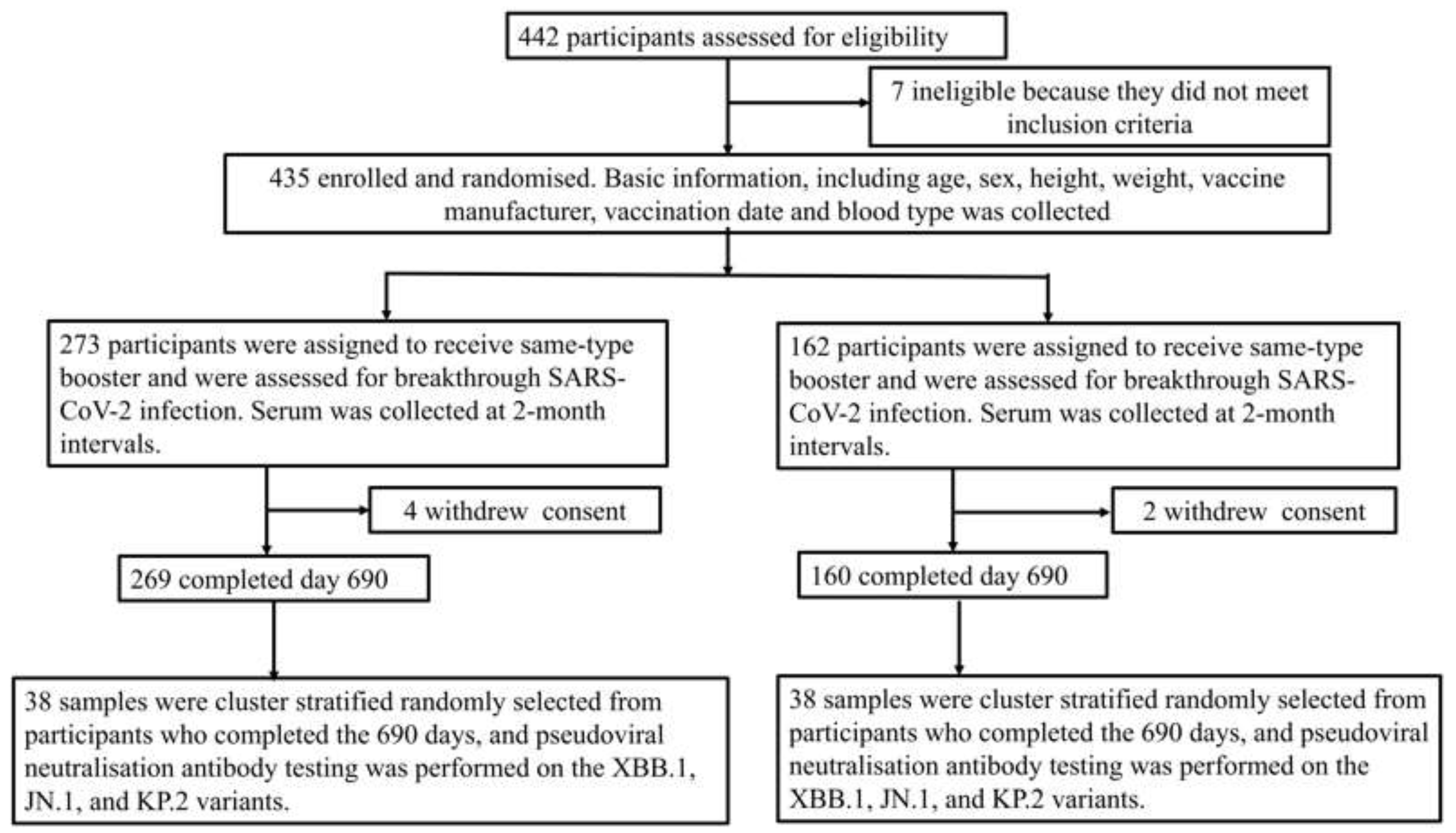
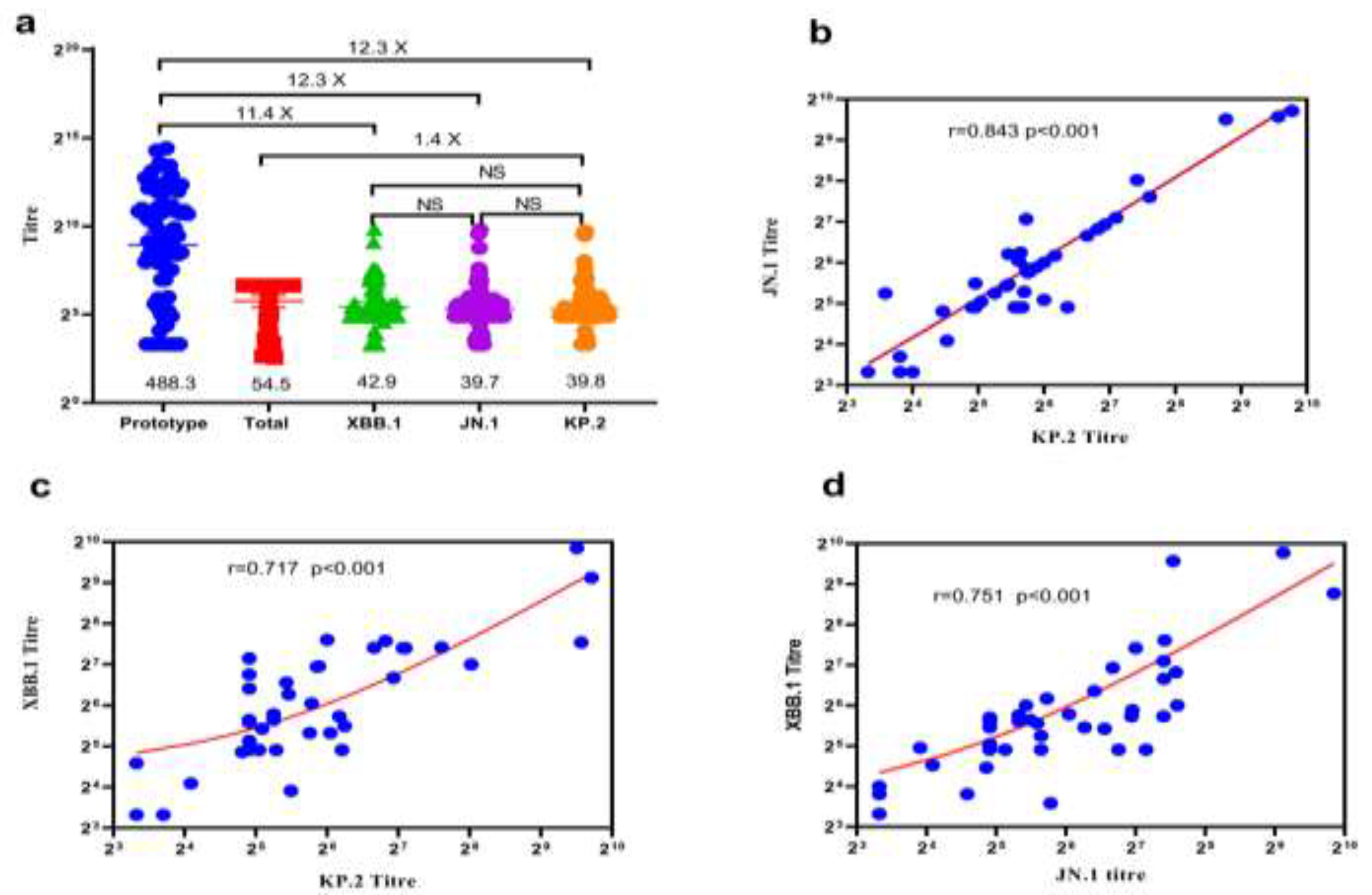
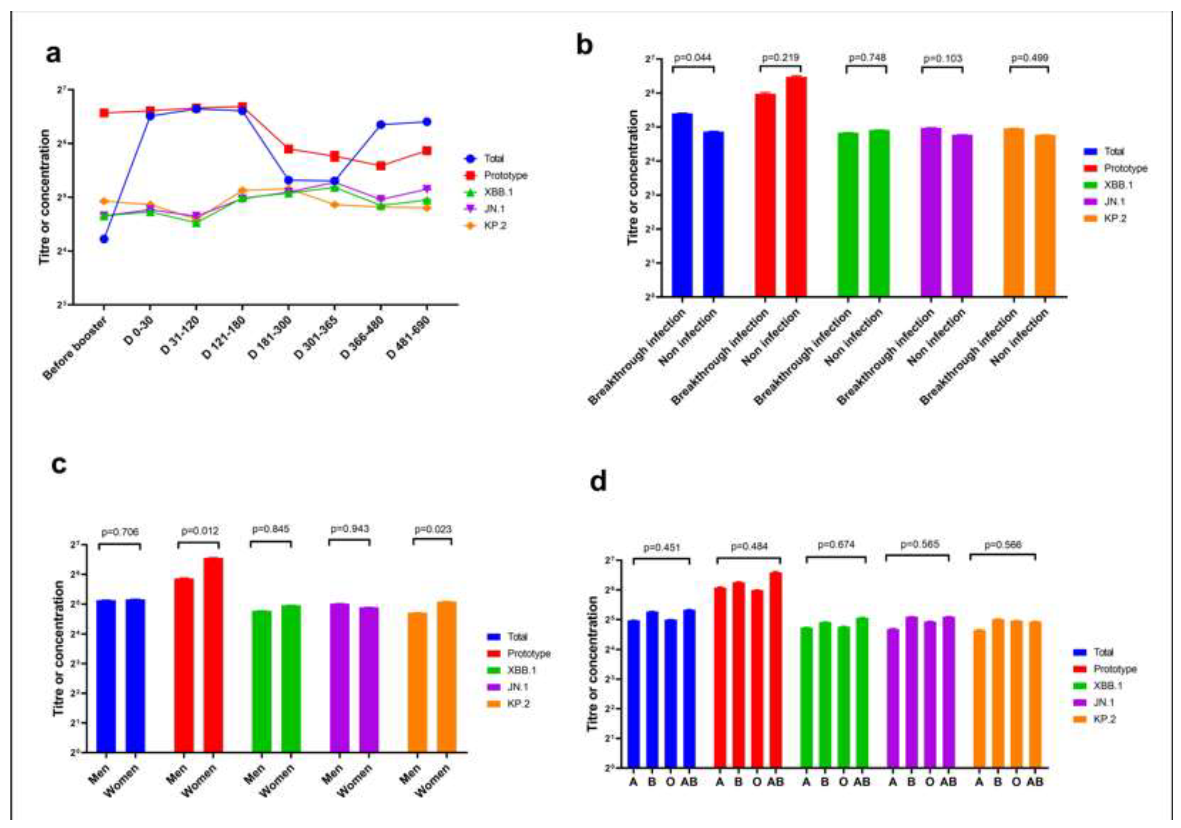
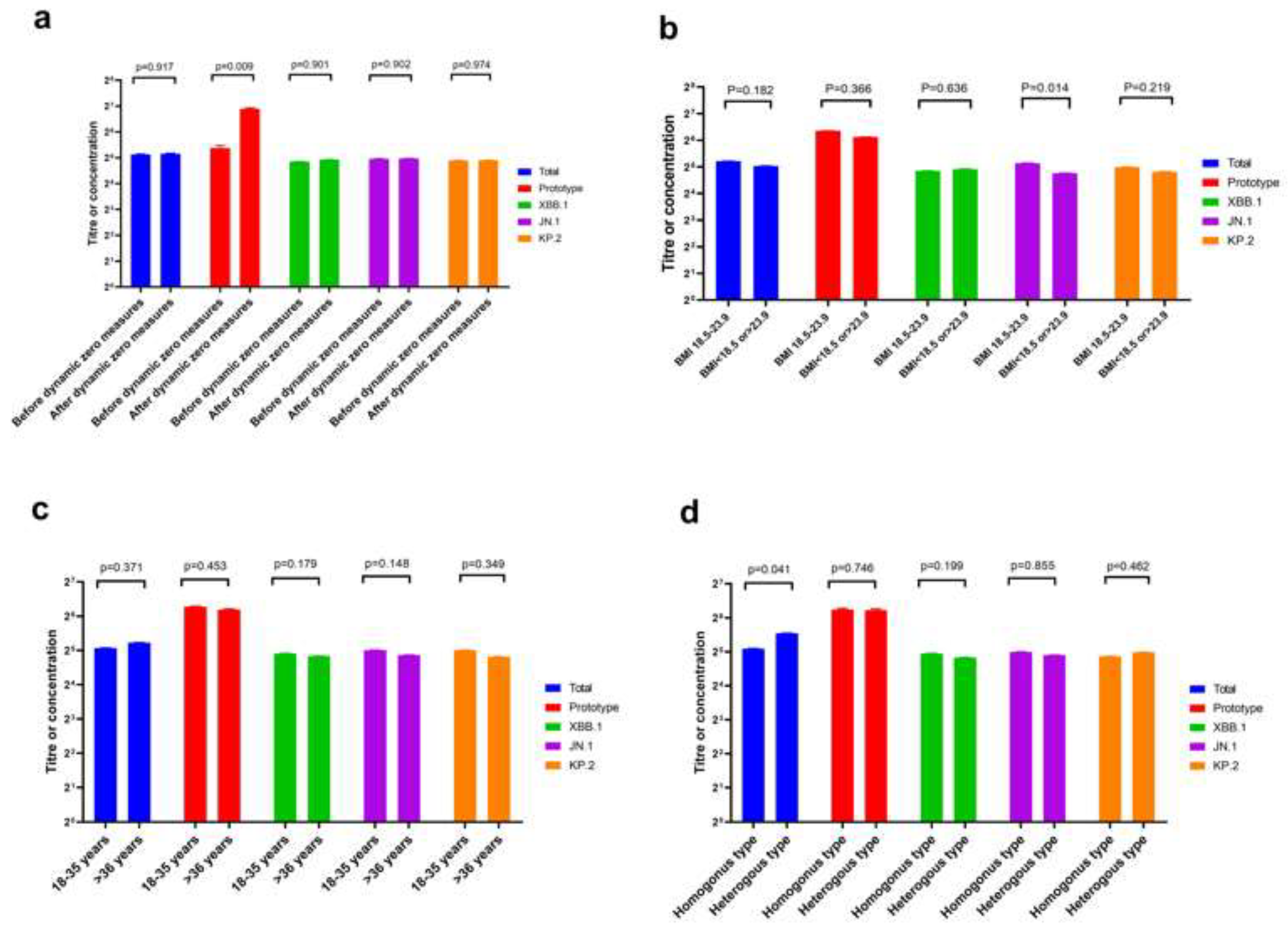
The neutralisation ability of homologous and heterologous booster vaccinations against the KP.2 variant of severe acute respiratory syndrome coronavirus 2 (SARS-CoV-2) is unknown. We evaluated Omicron variants (XBB.1, JN.1, and KP.2) neutralisation in participants vaccinated with heterologous versus homologous boosters. In 38 participants each from homologous and heter-ologous booster groups over 690 days, serum pseudovirus neutralisation was tested against the prototype, XBB.1, JN.1, and KP.2 variants to detect neutralisation titres. Total concentration of neutralising antibodies against SARS-CoV-2 receptor-binding domain was measured by en-zyme-linked immunosorbent assay. On throat swab samples, reverse transcription polymerase chain reaction was used to verify breakthrough SARS-CoV-2 infections in participants. Geometric mean neutralising titres against the prototype, total, XBB.1, JN.1, and KP.2 variants were 488.3, 54.5, 42.9, 39.7, and 39.8, respectively. Neutralisation assays revealed 12.3-, 12.3-, and 11.4-fold reduc-tions against JN.1, KP.2, and XBB.1 variants, respectively, compared with the prototype. No sig-nificant difference occurred in neutralising antibody titres among JN.1, KP.2, and XBB.1 Omicron variants. Homologous booster group and males produced fewer neutralising antibodies than heterologous booster group and females, respectively. KP.2 Omicron variant exhibited compa-rable immune evasion properties with other variants. A second different-type or broad-spectrum booster may improve neutralisation against Omicron variants KP.2.

Keywords: 
;  ;  ;  ;  ;  

1. Introduction

The KP.2 variant, a third-generation subbranch of the Omicron JN.1 variant of severe acute respiratory syndrome coronavirus 2 (SARS-CoV-2), was first detected in samples collected in India on 2 January 2024 [1, 2]. KP.2, known for its high transmissibility, is sometimes referred to as the ‘FLiRT’ variant due to its mutations in the spike protein, which increase its infectivity [1]. Following the rapid global spread of KP.2 strain since February 2024, the World Health Organisation listed KP.2 as a ‘variant to be monitored’ on 3 May, 2024 [3]. KP.2 began to spread globally between 7 and 13 May 2024 [4]. In April 2024, medical experts warned that FLiRT had a significant transmission advantage and could lead to a new wave of coronavirus disease 2019 (COVID-19) [5]. FLiRT was initially discovered in the United States’ sewer system; however, its exact source remains unknown [3]. Two new mutation sites in KP.2 are located in the S protein, suggesting enhanced transmission capabilities. Infectious disease experts have also reported that the latest mutation may be better at evading host immunity, predicting an imminent surge in COVID-19 cases [6]. The proportion of KP.2 subbranches in global circulating strains has gradually increased from 0.16% in early January 2024 to approximately 14% in early May 2024. In some countries, the prevalence of KP.2 subbranches is relatively high, accounting for 10–30% of infections [3]. As of 28 April 2024, Canadian national data showed that KP.2 was the most prevalent subvariant of JN.1, accounting for 26.6% of all COVID-19 cases in Canada (https://gisaid.org/). In the United States, as of May 2024, although other FLiRT variants, including KP.1.1, were also detected, they were not as prevalent as KP.2 [5]. Notably, data from the Centre for Disease Control and Prevention showed that KP.2 accounted for an estimated 28.2% of cases in the United States in the 2 weeks ending 11 May 2024, increasing from approximately 6% in mid-April and 1% in mid-March [7]. On 14 May 2024, the Chinese Centre for Disease Control and Prevention reported that, as of 12 May 2024, 25 KP.2 sequences had been detected in local cases in China. The proportion of KP.2 in sequences reported weekly was between 0.05% and 0.3%, indicating an extremely low prevalence (https://www.chinacdc.cn).
According to a population survey conducted by the Chinese Centre for Disease Control and Prevention in January 2023, the COVID-19 infection rate among the Chinese population reached 87.54% between 9 December 2022 and 30 January 2023 [8], with most individuals infected by the XBB Omicron variant despite receiving the first booster shot of the COVID-19 vaccine [5]. Conversely, based on the latest epidemic data from the Chinese Centre for Disease Control and Prevention in August 2024, the detection rate of COVID-19 infection in fever clinics in China was approximately 5%, with the primary circulating mutant strain being JN.1 and XDV.1 (https://www.chinacdc.cn); XDV.1 variant accounted for 43.4% prevalence in August 2024 (https://www.chinacdc.cn). However, the mechanism of breakthrough infection among individuals receiving a booster shot of the COVID-19 vaccine remains unknown. Moreover, there are no systematic descriptions of the neutralisation capacity of breakthrough infections in booster sera samples after different homologous and heterologous booster vaccinations against the Omicron KP.2 variant. Therefore, we aimed to evaluate the neutralisation of the Omicron variants XBB.1, JN.1, and KP.2 by comparing serum samples of Chinese individuals who received heterologous boosters (from different manufacturers) with those from Chinese individuals who received homologous boosters (from the same manufacturer) against SARS-CoV-2.

2. Materials and Methods

2.1. Participants and Flow of Study

This cross-sectional study enrolled 442 participants between 24 June 2021 and 16 September 2023 from Henan Provincial People’s Hospital, Zhengzhou Municipal Chinese Medicine Hospital, and Henan Electric Power Survey and Design Institute in central China through announcements and WeChat groups. The inclusion criteria were healthy or clinically stable adults (aged 18–80 years) who received the first dose of the booster COVID-19 vaccine between 6 and 8 months (180–240 days) prior to the study. Participants vaccinated with other vaccines, such as influenza or booster, were excluded. Additionally, participants with a clinically notable acute illness or a body temperature ≥38 °C within 24 h before receiving the planned booster dose of the study vaccine were excluded. Seven participants were excluded because they did not meet the inclusion criteria. Finally, 435 participants were enrolled and divided into a homologous-type group (n=273), consisting of participants who received a booster from the homologous manufacturer as their previous booster, and a heterologous group (n=162), together with those who received a booster from a different manufacturer. Participants were monitored for breakthrough infection for 690 days. Sera were collected every 2 months to detect the titre and total concentration of neutralising antibodies. Additionally, throat swab samples were collected to verify whether the participants were infected with SARS-CoV-2 using a specific reverse transcription polymerase chain reaction (RT-PCR). Notably, 269 and 160 of the 429 participants completed the 690-day period in the homologous and heterologous groups, respectively, with four and two participants in each group withdrawing consent. To ensure a better overall representation of our samples, we used the cluster-stratified random sampling method [9]. We classified 429 participants into groups of 11 average sections at every two monthly intervals, based on the time between booster and primary vaccinations. Since the longest interval between booster and primary vaccinations for some participants was 690 days, duration was divided into a total of 11 periods. Furthermore, seven study participants were randomly selected from every period group, and a similar number was required by sex and age to minimise control bias. Finally, 76 samples were randomly selected to detect titres against prototype, XBB.1, JN.1, and KP.2 variants and the total concentration of neutralising antibodies against the receptor binding domain (RBD) of SARS-CoV-2 (Figure 1). These 76 samples included 36 males and 40 females, with 38 samples each from the homologous and heterologous groups.

2.2. Assessment of Samples

Seventy-six samples, 38 and 38 from the homologous and heterologous groups, respectively, collected between 20 October 2021 and 16 September 2023, were selected for further analysis. The sample collection dates coincided with a change in epidemic measures from dynamic zero clearance to full liberalisation before and after 13 December 2022. Under the dynamic zero clearance policy, when a resident tested positive for COVID-19 in a residential community, they were immediately sent to a centralised isolation hospital for free isolation, treatment, and observation. Other residents in the community were required to undergo throat swab nucleic acid testing and isolation at home for seven consecutive days. Residents who tested positive during the next 7 days were also sent to a government isolation hospital. The isolation measures were only lifted if all community residents had negative throat swabs for seven consecutive days. Basic participant information was collected, including sex, age, vaccination date, vaccine type, blood type, body mass index (BMI), vaccination interval between primary and booster doses, and duration after booster dose calculated from the booster date to the sample collection date. Enzyme-linked immunosorbent assay (ELISA) was used to detect total neutralising antibodies against the RBD of SARS-CoV-2 in December 2023. The 76 samples also underwent simultaneous pseudovirus-neutralising antibody testing against the prototype, XBB.1, JN.1, and KP.2 variants in June 2024.

2.3. Serum Pseudovirus Neutralisation Test

Pseudotyped viruses were produced using 293T cells, which were transfected with S protein expression plasmid (prototype virus, XBB.1, JN.1, and KP.2 variants) and infected with vesicular stomatitis virus glycoprotein (VSV-G) pseudotyped virus (G*ΔG-VSV) [10]. Subsequently, a serum pseudovirus neutralisation test (pVNT) was performed to detect neutralisation titres. The pseudotyped viruses used for neutralisation titre detection and the mutation sites of the S genes are listed in Table S1. The initial dilution was 1:30 or 1:10, followed by three-fold serial dilutions. The final dilution of the sample was 1:7290. The 50% neutralisation dilution (ND50) was calculated using the Reed–Muench method, and the limit of detection (LOD) was 1:10. Results below the LOD were set to 0.5 times that of the LOD, and an ND50 titre > 1:30 was considered positive.

2.4. Measurement of Total Neutralising Antibodies and ABO Blood Typing

Total neutralising antibodies were detected using a commercial ELISA kit [11] (anti-SARS-CoV-2 S Kit; Shanghai GeneoDx Biotechnology Co., Ltd., Shanghai, China), which detects neutralising immunoglobulin antibodies against the SARS-CoV-2 spike protein RBD, using a universal microplate reader (DNM-9602; Beijing Pulong Co., Ltd., Beijing, China). A value > 6.5 IU/mL was considered positive. Following the manufacturer’s protocols and instructions (Chengdu United Co., Ltd., Chengdu, China), values > 100 IU/mL were capped at 100 IU/mL. The ABO blood group was determined using the test tube method based on the manufacturer’s instructions.

2.5. Throat Swab Samples Test

Throat swab samples collected for SARS-CoV-2 analysis were analysed using RT-PCR (Shanghai Zhejiang Biotechnology Ltd., Shanghai, China). Cycle threshold values of ≤ 44 on RT-PCR were considered positive.

2.6. Ethics

The study was approved by the Institutional Review Board of the Ethics Committee of Henan Provincial People’s Hospital (approval number: 20210051, approval date: 24 May 2021) and was conducted in compliance with the principles of the Declaration of Helsinki. All participants enrolled in this study provided written informed consent to participate.

2.7. Statistics

Summary statistics for the geometric means with 95% confidence intervals (CIs) are presented. Statistical significance between groups and subgroups was assessed using the Mann–Whitney U test, Student’s t-test, and one-way analysis of variance for continuous variables; however, Pearson’s χ² or Fisher’s exact test was used for categorical variables. Spearman’s rank correlation and r values were used to evaluate the correlation between two continuous variables. As the total neutralizing antibodies and four different neutralizing antibodies against the prototype strain, XBB.1, JN.1, KP.2 were respectively measured for each specimen, our data were repeated measures. Therefore, a mixed linear model was used to identify the factors affecting neutralising antibodies after booster vaccination [9]. The log2-transformed neutralising antibody titre or total antibody concentration was considered the dependent variable in this model. Age, sex, BMI, ABO blood type, vaccination mode, the interval between primary and booster vaccination doses, SARS-CoV-2 breakthrough infection, and epidemic measures were considered independent variables. Hypothesis testing was two-sided, with statistical significance at P< 0.05. SPSS (version 25.0; IBM Corp., Armonk, NY, USA) and GraphPad Prism (version 8.0; La Jolla, CA, USA) were used for statistical analyses and plotting.

3. Results

Thirty-six male and forty female participants were enrolled in this study. Of these, 37 (48.7%, 37/76) had breakthrough SARS-CoV-2 infections, with no difference in the rate of breakthrough infection between the heterologous-type (22/38, 57.9%) and homologous-type (15/38, 39.5%) booster groups (p=0.108) (Table 1). However, a significant difference (p < 0.001) was found in the rate of breakthrough infection before 13 December 2022 (0/30, 0%) when dynamic zero measures were strictly implemented, compared with that after 13 December 2022 (37/46, 80.4%), when dynamic zero measures were replaced by routine epidemic control measures (Table 1). The median (quartile) age of the participants was 34.0 (23.3–53.0) years in the total group and specifically, 27.0 (21–57.0) years and 37.5 (31–50) years in the heterologous-type and homologous-type booster groups, respectively (Mann–Whitney U test, p=0.084). Four samples (7/38, 18.4%) in the heterologous-type booster group and 23 samples (23/38, 60.5%) in the homologous-type booster group were collected before 13 December 2022 (p=0.001). Additionally, 18 (18/38, 47.4%) and 22 (22/38, 52.6%) participants in the heterologous-type and homologous-type booster groups, respectively, were men (p=0.358). BMI and blood type were similar between the two groups (p=0.702 and p=0.719, respectively), and no significant difference was observed in the duration of booster vaccination between the two groups (p=0.054). The interval between the primary and booster vaccinations was also similar between the two groups (p=0.238). Among the homologous group, 35 participants were vaccinated using Chinese inactive COVID-19 vaccine (produced by eight manufacturers including Beijing Kexing Zhongwei, Beijing Biologics, Changchun Biologics, Beijing Kexing, Wuhan Biologics, Lanzhou Biologics) whereas three participants were vaccinated Chinese by protein vaccine (produced by Anhui Zhifei Biologics) as primary and booster vaccinations. Additionally, among the heterologous group, 38 participants used inactive vaccine as the primary vaccination. However, for booster vaccination,31 participants used attenuated live vaccine (produced by Tianjin CanSino Biologics) and 7 participants used protein vaccine (the product manufacturers were the same as above). Additional basic characteristics are listed in Table 1.
We evaluated the neutralisation activity of sera from 76 booster vaccinations against the prototype, total, XBB.1, JN.1, and KP.2 Omicron variants, which showed the same reduced neutralisation as the prototype. The geometric mean neutralising titres (GMTs) against the prototype, total, XBB.1, JN.1, and KP.2 Omicron variants were 488.3 (95% CI: 293.31–812.9), 54.5 (42.9–69.3), 42.9 (35.0–52.4), 39.7 (32.9–47.9), and 39.8 (32.6–48.7), respectively (Figure 2a). Therefore, neutralisation assays against JN.1, KP.2, and XBB.1 indicated 11.4-, 12.3-, and 12.3-fold reductions in neutralising antibody titres, respectively, compared with the prototype. Notably, no significant difference was observed in the neutralising antibody titres among the JN.1, KP.2, and XBB.1 Omicron variants (adjusted p=0.97; Figure 2a, Table S2). Additionally, a high correlation was observed between the GMT of JN.1 and KP.2 (r=0.843, p < 0.001) (Figure 2c) and among XBB.1, JN.1, and KP.2 variants (r=0.717, r=0.751, and p < 0.001, respectively) (Figure 2c,d, Table S2). These results further confirmed that the immune escape capacity of the JN.1 and KP.2 variants did not increase compared with that of the previous XBB.1 variants (Table S2). However, no correlation was found between the total neutralising antibodies and the prototype, XBB.1, JN.1, and KP.2 variants (p=0.240 to 0.932, r=-0.01 to 0.136, respectively) (Table S2).
Subsequently, we analysed the dynamics of antibody neutralisation titres of sera against the three SARS-CoV-2 variants, XBB.1, JN.1, and KP.2, and the concentration of the total neutralising antibodies. We also analysed the influencing factors at different durations after the first booster dose of the COVID-19 vaccine within 690 days (Figure 3 and Figure 4). Sex, age, blood type, breakthrough infection, epidemic prevention and control measures, vaccination type, and booster vaccination interval affected the antibody titre and total antibody concentration of participants after the first booster vaccination (Figure 3 and Figure 4). Initially, the total antibody serum concentration was highest between 4 and 6 months after the first booster vaccine and gradually decreased with time (days 121–180 vs before booster, p=0.001) (Figure 3a and Table S3). However, antibody titres against the XBB.1, JN.1, and KP.2 variants were very low after the first booster vaccine and remained unchanged during the 690-day period (Figure 3a and Tables S3-S7). Moreover, the population with breakthrough infection after booster vaccination had higher total antibody concentration in the serum than those without breakthrough infection (p=0.044). Conversely, the antibody titres against the XBB.1, JN.1 and KP.2 variants did not differ between populations with and without breakthrough infection (p=0.748, 0.103 and 0.499, respectively) (Figure 3b). Women had a higher antibody titre against the prototype and KP.2 variant after vaccination compared with men (p=0.012, p=0.023, respectively), and there was no difference in antibody titres against the XBB.1 and JN.1 variants (p=0.845, p=0.943, respectively) (Figure 3c). Moreover, there was no difference in antibody titres against the XBB.1, JN.1 and KP.2 variants among different blood type booster vaccinators (p=0.674, p=0.565, P=0.556, respectively) (Figure 3d, Tables S3 and S7).
The serum collected after the liberation of the dynamic zero measures (after 13 December 2022) produced higher antibody titres against the prototype than those collected before 13 December 2022 (p=0.009) (Figure 4a). This trend was consistent, as shown in Figure 3c, where participants with breakthrough infection had a higher titre than those without infection. After the liberation of the dynamic zero measures (after 13 December 2022), more participants (37/46, 80.4%) had breakthrough SARS-CoV-2 Omicron variant XBB.1 infections compared with the period before 13 December 2022 (0/30, 0%) (Table 1). In addition, booster vaccine recipients' BMI was between 18.5 and 23.9 and they had a higher antibody titre against JN.1 than those with BMI >23.9 or <18.5 (p=0.014, Figure 4c). Booster vaccine recipients with age >35 years produced similar total antibody concentrations or titres to those with age <35 years (Figure 4c). Notably, participants who received heterologous booster vaccines produced an increased amount of total neutralising antibodies than those who received the homologous booster type vaccine (p=0.041, Table S4). However, no differences were observed in neutralising antibody titres against XBB.1, JN.1, and KP.2 between the heterologous- and homologous-type booster groups (p=0.199, p=0.855, and p=0.224, respectively) (Figure 4d, Tables S5–S7).

4. Discussion

This study evaluated the neutralisation of the Omicron variants XBB.1, JN.1, and KP.2 by analysing serum from individuals who had received the Chinese booster vaccine to determine if these variants would evade vaccine-elicited immunity previously established by the initial dose of the booster vaccine. To the best of our knowledge, this is the first report on the neutralisation serum capacity following the first booster vaccination against the newly circulating SARS-CoV-2 variants JN.1 and KP.2 in the Chinese population.
Our findings elucidated the mechanism of breakthrough infections among the Chinese population vaccinated with the first booster COVID-19 vaccine. Notably, most of the Chinese population received the first booster COVID-19 vaccine (inactivated vaccine, attenuated live vaccine, or protein subunit vaccine) before 13 December 2022 and before the complete liberation of epidemic control measures; however, the vaccine only contained the SARS-CoV-2 prototype strain and not the XBB.1 variant. Consequently, following the implementation of routine control measures in China, after 13 December 2022, the circulating SARS-CoV-2 strain mutated into Omicron variants, including the XBB series [12]. This implies that the neutralising antibodies generated by the COVID-19 vaccines, including the messenger RNA vaccine or inactivated vaccine, attenuated live vaccine, or protein subunit vaccine, were designed to contain only the prototype strain of SARS-CoV-2; they would have no cross-protection ability against other variant strains, including XBB.1, JN.1, KP.2, and XDV.1 [4].
Furthermore, our results showed that women who received the booster dose produced higher total antibodies and neutralizing antibodies against KP.2 variants than men. As documented in prior research [6, 7], this finding partially explained the pneumonia contributing to the susceptibility of men to SARS-CoV-2 variants. Notably, this finding has not yet been reported.
Several recent publications from the USA and other regions have reported that the latest Omicron variants, JN.1 and KP.2, exhibit a markedly reduced neutralisation capacity compared with the previous Omicron variant, XBB.1, which diminishes the binding affinity of most antibody drugs [8]. Conversely, our results showed that the neutralisation capacity against JN.1 and KP.2 variants in serum from Chinese booster vaccinees did not significantly decrease compared with the XBB.1 variant. The observed difference may primarily be ascribed to the different types of booster vaccinations administered to the study participants. In other regions, antibodies produced by boosters on the BA.4/5 spike protein or XBB vaccine have shown a diminished capacity to neutralise against the new JN.1 and KP.2 strains [13-17].
Consistent with previous studies, we found that the serum of individuals who received the Chinese booster vaccine exhibited higher immune escape capacity for the XBB.1 variant than those who received the prototype strain [16]. Furthermore, our results showed that the neutralising antibodies from the Chinese COVID-19 vaccine booster population against the prototype strain lasted only 4–6 months. However, the sera could not neutralise the emerging JN.1 and KP.2 variants within 690 days after booster vaccination. Since KP.2 variant has only one additional V1104L S gene mutation compared with XDV.1 variant (https://gisaid.org/lineage-comparison/), we speculated that the immune escape capacity of XDV.1 should be similar to that of KP.2. Notably, the sera from the population that received the booster could not neutralise the latest emerging XDV.1 variant in China. This significant difference suggests that, in the future, a novel vaccine incorporating JN.1, KP.2, and XDV.1 variants will be required to improve population immunity and prevent breakthrough infections with emerging SARS-CoV-2 variants [17].
Another notable finding of this study was that participants who received the booster vaccine and developed breakthrough infections exhibited higher total neutralising antibody than those without breakthrough infections. However, no difference was found between booster participants who produced antibody titre against XBB.1, JN.1, and KP.2 variants and those with and without breakthrough infection. This further suggests that the JN.1 and KP.2 variants have similar immune escape capacities with XBB.1. This is because the samples from individuals with breakthrough infection were collected before October 2023 when only the XBB.1 variant was circulating in China [12]. The JN.1, KP.2, and XDV.1 variants only began to emerge in China in January, May, and June 2024, respectively.
Furthermore, no difference was found in neutralising antibody titres against the prototype strains, XBB.1, JN.1, and KP.2 variants, between populations vaccinated with the heterologous-type booster and homologous-type booster. Conversely, individuals vaccinated with the heterologous-type booster had a higher total antibody concentration than those vaccinated with the homologous-type booster (Figure 4d). As these results suggest, there was a difference between total antibody concentration and pseudovirus neutralising antibody titres against different SARS-CoV-2 variants among booster populations. This finding is consistent with the finding that no correlation exists between ELISA and pseudovirus neutralisation test results. This may be due to differences in the detection methods for the serum-neutralising antibodies. Total neutralising antibodies against the RBD protein are detected using the reaction principle between antigen and antibody in ELISA [18]. Conversely, the pseudovirus neutralisation test employs the principle of cell transfection to identify the 50% neutralisation dilution titres for different SARS-CoV-2 variants in the serum [17]. Additionally, the heterologous booster group showed higher total antibody levels than the homologous group. This may be due to differences between booster and primary vaccine types, such as inactive, attenuated live, protein, or mRNA vaccines, since their mechanisms for producing antibodies in vaccinators differ [16, 19]. This finding is similar to those of studies in other countries on heterologous booster vaccines such as mRNA vaccines, protein vaccines, or vaccines containing JN.1 variants, which showed better neutralizing SARS-CoV-2 activity [13, 20-23].
This study has some limitations. First, the live virus neutralisation tests were inaccessible; however, we adopted the classic pseudovirus neutralisation test, commonly used in most published COVID-19 vaccination studies, to evaluate the immunogenicity of vaccination [14]. Additionally, the sample size of our study was limited due to the costs of the pseudoviral neutralisation test. Moreover, some participants may have had biased recollections of whether they had been ever infected with SARS-CoV-2, although throat swabs were used to confirm SARS-CoV-2 infection. Therefore, larger-scale studies are required to evaluate the immunogenicity and effectiveness of different vaccines against Omicron variants, including KP.3, LB.1, and XDV.1 [1, 24].

5. Conclusions

Conclusively, our study demonstrates that vaccine-induced immune protection may be more likely to be evaded by the Omicron variants, JN.1 and KP.2, compared with the prototypes and XBB.1 variants in Chinese individuals vaccinated with a first booster COVID-19 vaccine. Therefore, following the first booster, whether homologous or heterologous, a subsequent JN.1, KP.2, or XDV.1 and a broad-spectrum COVID-19 vaccine booster are recommended to improve neutralisation against these new SARS-CoV-2 variants. Future research should focus on increasing the sample size to determine whether new vaccines neutralise emerging COVID-19 variants, including KP.3, LB.1, and XDV.1, after booster vaccination.

Supplementary Materials

The following supporting information can be downloaded at: www.mdpi.com/xxx/s1, Table S1. Pseudotyped viruses used for neutralisation titre detection and mutation sites of S genes; Table S2. Correlation analysis of neutralising antibody titres among SARS-CoV-2 variants and total neutralising antibodies against the RBD; Table S3. Factors associated with total neutralising antibody concentration after Chinese COVID-19 booster vaccine administration; Table S4. Factors associated with neutralising antibody concentration against XBB.1 variant after Chinese COVID-19 booster vaccine administration; Table S5. Factors associated with neutralising antibody concentration against JN.1 variant after Chinese COVID-19 booster vaccine administration; Table S6. Factors associated with neutralising antibody concentration against KP.2 variant after Chinese COVID-19 booster vaccine administration; Table S7. Factors associated with neutralising antibody concentration against Prototype strain after Chinese COVID-19 booster vaccine administration

Author Contributions

YY, JX, GC, and LO conceptualised and designed the study and drafted the manuscript; BW, LO, WY, YG, and QM performed statistical analyses. GC, XM, XZ, and BM collected serum samples from volunteers; YL, PZ, QZ, and JZ helped conduct the clinical study and acquire, analyse, and interpret the data; Yan L performed the pseudovirus neutralisation tests; RW, XZ, Yi L, XM, PZ, and HW provided material support and supervised the study. All authors commented on previous versions of the manuscript. All authors read and approved the final manuscript

Funding

This research was funded by the Natural Science Foundation of China (recipient: R. Wang, grant number: 82002210); the Henan Provincial Key Programs in Science and Technology (recipient: B. Wang, grant number: 242102310116); the Joint Program of Medical Science and Technology Research of Henan Province (recipient: Y. Li, grant number: LHGJ 20230021); the Henan Natural Science Foundation (grant numbers: 222300420355 and 242300421502) from the Henan Provincial Department of Education; and the Henan Province Science and Technology Research Program Project from the Henan Provincial Health Commission (recipients: H. Wang and X. Zhang, grant number: SBGJ202103016).

Institutional Review Board Statement

The study was approved by the Institutional Review Board of the Ethics Committee of Henan Provincial People’s Hospital (approval number: 20210051, approval date: 24 May 2021) and was conducted in compliance with the principles of the Declaration of Helsinki.

Informed Consent Statement

All participants enrolled in this study provided written informed consent to participate. Written informed consent has been obtained from the patient(s) to publish this paper.

Data Availability Statement

The original contributions presented in the study are included in the article and Supplementary Files. Further inquiries can be directed to the corresponding author.

Acknowledgments

In this section, you can acknowledge any support given which is not covered by the author contribution or funding sections. This may include administrative and technical support, or donations in kind (e.g., materials used for experiments).

Conflicts of Interest

The authors declare no conflict of interest. The funders had no role in the design of the study; in the collection, analysis, or interpretation of data; in the writing of this manuscript; or in the decision to publish the results.

References

  1. Kaku, Y.; Yo, M. S.; Tolentino, J. E.; Uriu, K.; Okumura, K. Genotype to Phenotype Japan (G2P-Japan) Consortium, Ito, J.; Sato, K. Virological characteristics of the SARS-CoV-2 KP.3, LB.1, and KP.2.3 variants. Lancet Infect Dis. 2024, 24, e482–e483. [Google Scholar] [CrossRef]
  2. Rajput, V.; Das, R.; Pramanik, R.; Nannaware, K.; Sushma, Y.; Taji, N.; Rajput, V.; Rajkhowa, R.; Pacharne, P.; Shah, P.; Gogate, N.; Sangwar, P.; Bhalerao, A.; Jain, N.; Kamble, S.; Dastager, S.; Shashidhara, L. S.; Karyakarte, R.; Dharne, M. Early detection of KP.2 SARS-CoV-2 variant using wastewater-based genomic surveillance in Pune, Maharashtra, India. J Travel Med Advance online publication. 2024, taae097. [Google Scholar] [CrossRef] [PubMed]
  3. Kumar, P.; Jayan, J.; Sharma, R. K.; Gaidhane, A. M.; Zahiruddin, Q. S.; Rustagi, S.; Satapathy, P. The emerging challenge of FLiRT variants: KP.1.1 and KP.2 in the global pandemic landscape. QJM 2024, 117, 485–487. [Google Scholar] [CrossRef]
  4. Branda, F.; Ciccozzi, A.; Romano, C.; Ciccozzi, M.; Scarpa, F. Another variant another history: description of the SARS-CoV-2 KP.2 (JN.1.11.1.2) mutations. Infect Dis (Lond). 2024, 56, 581–585. [Google Scholar] [CrossRef] [PubMed]
  5. Li, P.; Faraone, J. N.; Hsu, C. C.; Chamblee, M.; Zheng, Y. M.; Carlin, C.; Bednash, J. S.; Horowitz, J. C.; Mallampalli, R. K.; Saif, L. J.; Oltz, E. M.; Jones, D.; Li, J.; Gumina, R. J.; Xu, K.; Liu, S. L. Neutralization escape, infectivity, and membrane fusion of JN.1-derived SARS-CoV-2 SLip, FLiRT, and KP.2 variants. Cell Rep 2024, 43, 114520. [Google Scholar] [CrossRef] [PubMed]
  6. Haitao, T.; Vermunt, J.V.; Abeykoon, J.; Ghamrawi, R.; Gunaratne, M.; Jayachandran, M.; Narang, K.; Parashuram, S.; Suvakov, S.; Garovic, V.D. COVID-19 and sex differences: Mechanisms and biomarkers. Mayo Clin Proc 2189, 95, 2189–2203. [Google Scholar] [CrossRef]
  7. Kitselman, K.; Bédard-Matteau, J.; Rousseau, S.; Tabrizchi, R.; Daneshtalab, N. Sex differences in vascular endothelial function related to acute and long COVID-19. Vascul Pharmacol. 2024, 154, 107250. [Google Scholar] [CrossRef]
  8. Benlarbi, M.; Ding, S.; Bélanger, É.; Tauzin, A.; Poujol, R.; Medjahed, H.; El Ferri, O.; Bo, Y.; Bourassa, C.; Hussin, J.; Fafard, J.; Pazgier, M.; Levade, I.; Abrams, C.; Côté, M.; Finzi, A. Temperature-dependent Spike-ACE2 interaction of Omicron subvariants is associated with viral transmission. mBio. 2024, 15, e0090724. [Google Scholar] [CrossRef] [PubMed]
  9. Lei, J.; Sun, Q.; Chen, R.; Zhu, Y.; Zhou, L.; Xue, X.; Fang, J.; Du, Y.; Wang, Y.; Li, T.; Kan, H. Respiratory benefits of multisetting air purification in children: A cluster randomized crossover trial. JAMA Pediatr 2024, e245049. [Google Scholar] [CrossRef] [PubMed]
  10. Bai, Y.; Peng, Z.; Wei, F.; Jin, Z.; Wang, J.; Xu, X.; Zhang, X.; Xu, J.; Ren, Z.; Lu, B.; Wang, Z.; Xu, J.; Huang, S. Study on the COVID-19 epidemic in mainland China between November 2022 and January 2023, with prediction of its tendency. J BiosafBiosecur. 2023, 5, 39–44. [Google Scholar] [CrossRef]
  11. Xu, N.; Xu, Y.; Dai, R.; Zheng, L.; Qin, P.; Wan, P.; Yang, Y.; Jiang, J.; Zhang, H.; Hu, X.; Lv, H. Study of efficacy and antibody duration to fourth-dose booster of Ad5-nCoV or inactivated SARS-CoV-2 vaccine in Chinese adults: a prospective cohort study. Front Immunol. 2023, 14, 1244373. [Google Scholar] [CrossRef] [PubMed]
  12. Zhang, L.; Li, Q.; Liang, Z.; Li, T.; Liu, S.; Cui, Q.; Nie, J.; Wu, Q.; Qu, X.; Huang, W.; Wang, Y. The significant immune escape of pseudotyped SARS-CoV-2 variant Omicron. Emerg Microbes Infect. 2022, 11, 1–5. [Google Scholar] [CrossRef]
  13. Jeworowski, L. M.; Mühlemann, B.; Walper, F.; Schmidt, M. L.; Jansen, J.; Krumbholz, A.; Simon-Lorière, E.; Jones, T. C.; Corman, V. M.; Drosten, C. Humoral immune escape by current SARS-CoV-2 variants BA.2.86 and JN.1, December 2023. Euro Surveill 2024, 29, 2300740. [Google Scholar] [CrossRef] [PubMed]
  14. Kaku, Y.; Okumura, K.; Padilla-Blanco, M.; Kosugi, Y.; Uriu, K.; Hinay, A.A., Jr.; Chen, L.; Plianchaisuk, A.; Kobiyama, K.; Ishii, K. J. Genotype to Phenotype Japan (G2P-Japan) Consortium, Zahradnik, J.; Ito, J.; Sato, K. Virological characteristics of the SARS-CoV-2 JN.1 variant. Lancet Infect Dis 2024, 24, e82. [Google Scholar] [CrossRef] [PubMed]
  15. Planas, D.; Staropoli, I.; Michel, V.; Lemoine, F.; Donati, F.; Prot, M.; Porrot, F.; Guivel-Benhassine, F.; Jeyarajah, B.; Brisebarre, A.; Dehan, O.; Avon, L.; Bolland, W.H.; Hubert, M.; Buchrieser, J.; Vanhoucke, T.; Rosenbaum, P.; Veyer, D.; Péré, H.; Lina, B.; Schwartz, O. Distinct evolution of SARS-CoV-2 Omicron XBB and BA.2.86/JN.1 lineages combining increased fitness and antibody evasion. Nat Commun 2024, 15, 2254. [Google Scholar] [CrossRef] [PubMed]
  16. Wang, Q.; Guo, Y.; Bowen, A.; Mellis, I. A.; Valdez, R.; Gherasim, C.; Gordon, A.; Liu, L.; Ho, D.D. XBB.1.5 monovalent mRNA vaccine booster elicits robust neutralizing antibodies against XBB subvariants and JN.1. Cell Host Microbe 2024, 32, 315–321.e3. [Google Scholar] [CrossRef]
  17. Yang, S.; Yu, Y.; Xu, Y.; Jian, F.; Song, W.; Yisimayi, A.; Wang, P.; Wang, J.; Liu, J.; Yu, L.; Niu, X.; Wang, J.; Wang, Y.; Shao, F.; Jin, R.; Wang, Y.; Cao, Y. Fast evolution of SARS-CoV-2 BA.2.86 to JN.1 under heavy immune pressure. Lancet Infect Dis. 2024, 24, e70–e72. [Google Scholar] [CrossRef]
  18. Yuan, Y.; Xu, J.; Ma, B.; Chen, G.; Wang, Z.; Wang, S.; Jing, N.; Zhang, J.; Wang, B.; Yan, W.; Zhang, Q.; Zhao, Q.; Li, Y. Characteristics of humoral and cellular responses to coronavirus disease 2019 (COVID-19) inactivated vaccine in central China: A prospective, multicenter, longitudinal study. Front Immunol. 2023, 14, 1107866. [Google Scholar] [CrossRef]
  19. Raisinghani, N.; Alshahrani, M.; Gupta, G.; Verkhivker, G. Atomistic prediction of structures, conformational ensembles and binding energetics for the SARS-CoV-2 spike JN.1, KP.2 and KP.3 variants using AlphaFold2 and molecular dynamics simulations: Mutational profiling and binding free energy analysis reveal epistatic hotspots of the ACE2 affinity and immune escape. bioRxiv 2024. [Google Scholar] [CrossRef]
  20. Itamochi, M.; Yazawa, S.; Saga, Y.; Shimada, T.; Tamura, K.; Maenishi, E.; Isobe, J.; Sasajima, H.; Kawashiri, C.; Tani, H.; Oishi, K. COVID-19 mRNA booster vaccination induces robust antibody responses but few adverse events among SARS-CoV-2 naive nursing home residents[J]. Sci Rep. 2024, 14, 23295. [Google Scholar] [CrossRef]
  21. Savulescu, C.; Prats-Uribe, A.; Brolin, K.; Lovrić Makarić, Z.; Uusküla, A.; Panagiotakopoulos, G.; Bergin, C.; Fleming, C.; Agodi, A.; Bonfanti, P.; Murri, R. Incidence of SARS-CoV-2 infection among European healthcare workers and effectiveness of the first booster COVID-19 vaccine, VEBIS HCW observational cohort study, May 2021-May 2023[J]. Vaccines (Basel). 2024, 12. [Google Scholar] [CrossRef] [PubMed]
  22. Talmy, T.; Nitzan, I. Rapid rollout and initial uptake of a booster COVID-19 vaccine among Israel defense forces soldiers[J]. J Prev. 2023, 44, 1–14. [Google Scholar] [CrossRef] [PubMed]
  23. Favresse, J.; Gillot, C.; Cabo, J.; David, C.; Dogné, J.M.; Douxfils, J. Neutralizing antibody response to XBB.1.5, BA.2.86, FL.1.5.1, and JN.1 six months after the BNT162b2 bivalent booster[J]. Int J Infect Dis. 2024, 143, 107028. [Google Scholar] [CrossRef] [PubMed]
Figure 1. Serum samples from the booster vaccine groups.
Preprints 146754 g001
Figure 2. Serum neutralisation titres and correlation among different variants for participants who previously received the first booster Chinese COVID-19 vaccine (a) against the prototype, total, XBB.1, JN.1, and KP.2 variants. Correlation of serum neutralisation titres between (b) JN.1 and KP.2. (c) XBB.1 and KP.2. and (d) XBB.1 and JN.1. The serum samples were collected between 20 October 2021 and 16 September 2023.
Preprints 146754 g002
Figure 3. Comparison of neutralisation titres against prototype, total, XBB.1, JN.1, and KP.2 variants in booster participants (a) dynamics of neutralisation titres against the prototype, total, XBB.1, JN.1, and KP.2 variants among participants in the 690 days after vaccination with the first booster Chinese COVID-19 vaccine. (b) with and without breakthrough infection, (c) in men and women, and (d) with different blood types.
Preprints 146754 g003
Figure 4. Comparison of neutralisation titres against prototype, total, XBB.1, JN.1, and KP.2 variants among booster participants (a) before and after dynamic zero control measures, (c)<35 years and >35 years of age, (b) BMI 18.5-23.9 and BMI <18.5 or >23.9, and (d) with homologous-type and heterologous-type booster.
Preprints 146754 g004
Table 1. Basic characteristics of participants from the homologous-type and heterologous-type booster groups.
Factor Heterologous-type
(n = 38)
Homologous-type
(n = 38)
P Total
Sex
Male 16 20 0.358 36
Female 22 18 40
Age (years), M (P25, P75) * 27.0 (21, 57.0) 37.5 (31, 50) 0.084 34.5 (24.5, 44.8)
18–35 24 18 0.166 42
> 36 14 20 34
Blood type
A 10 6 0.719 16
B 9 10 19
O 14 17 31
AB 5 5 10
BMI (kg/m2) 22.7 ± 3.3 22.9 ± 3.3 0.702 22.8 ± 3.4
18.5–23.9 26 22 0.342 48
< 18.5 and > 23.9 12 16 28
Breakthrough infection
Yes 22 15 0.108 37
No 16 23 39
Control measures
Dynamic zero policy (before 13 December 2022) 7 23 0.001 30
Routine control measures (after 13 December 2022) 31 15 46
Duration after booster
Before booster 0 5 0.054
5
1–30 4 9 13
31–120 8 4 12
121–180 1 5 6
181-300 5 3 8
301–365 4 3 7
366–480 7 2 9
481–690 9 7 16
Interval between the primary and booster vaccinations
180–210 12 17 0.238 29
> 210 26 21 47
Booster vaccine type
Inactive vaccine 0 35 <0.001 35
Attenuated live vaccine 31 0 31
Protein vaccine 7 3 10
BMI, body mass index; SARS-CoV-2, severe acute respiratory syndrome coronavirus 2.
Disclaimer/Publisher’s Note: The statements, opinions and data contained in all publications are solely those of the individual author(s) and contributor(s) and not of MDPI and/or the editor(s). MDPI and/or the editor(s) disclaim responsibility for any injury to people or property resulting from any ideas, methods, instructions or products referred to in the content.
Copyright: This open access article is published under a Creative Commons CC BY 4.0 license, which permit the free download, distribution, and reuse, provided that the author and preprint are cited in any reuse.
Prerpints.org logo

Preprints.org is a free preprint server supported by MDPI in Basel, Switzerland.

Subscribe

Disclaimer

Terms of Use

Privacy Policy

Privacy Settings

© 2025 MDPI (Basel, Switzerland) unless otherwise stated