Submitted:
23 January 2025
Posted:
23 January 2025
You are already at the latest version
Abstract
In this research, polymer composite sheets were developed by blending poly (vinylidene fluoride-co-hexafluoropropylene) or P(VDF-HFP) with varying concentrations of barium sulfate (BaSO₄) for X-ray shielding applications. The photon counting technique was used to evaluate the composite shielding characteristics through the linear attenuation coefficient. Surface properties, including surface morphology, hydrophobicity, and surface energy, were analyzed using an atomic force microscope (AFM) and a water contact angle machine. Scanning electron microscopy (SEM) was employed to investigate the microstructural distribution and dispersion of BaSO₄ particles within the polymer matrix, providing insights into the composite's uniformity and structural integrity. Additionally, the bulk properties of the composite polymer sheets, such as crystal structures, tensile strength, and thermal stability, were examined. The results demonstrate that increasing the concentration of BaSO₄ in BaSO₄/P(VDF-HFP) composite sheets significantly improves their X-ray attenuation capabilities. Moreover, higher BaSO₄ concentrations enhance the material's hydrophobicity, flexibility, and thermal stability, highlighting the potential of these composites for advanced radiation-shielding applications.
Keywords:
P(VDF-HFP) nanocomposites
; barium sulfate nanoparticles
; X-Ray Shielding
1. Introduction
X-rays, a form of ionizing radiation, pose a risk of causing damage to living tissue, particularly for patients and radiation workers [1]. It is necessary to protect against the danger posed by the radiation. The safety design aims to minimize damage caused by ionizing radiation by adhering to the ALARA principle, which emphasizes achieving the lowest reasonable level of exposure [2]. This principle involves considering factors such as time, distance, and shielding to ensure the safety of individuals and equipment. Traditional shielding material, lead, was favoured due to its high density, atomic number, and low cost. However, it faced numerous drawbacks, including toxicity, weight, environmental disease, poor flexibility, and low chemical stability [3]. Lead-free materials emerge as alternative options, attracting numerous research groups to address the drawbacks associated with lead [4,5,6]. Polymer composite shielding has emerged as a promising lead-free material with significant potential for practical applications. Its advantages include lightweight design, excellent flexibility, and ease of processing [7]. Various polymers, such as polymethyl-methacrylate (PMMA), polyethylene terephthalate (PET), thermoplastic polyurethane (TPU), polyvinylpyrrolidone (PVP), and polyethylene glycol (PEG), have been employed as matrices for composite shielding materials [8,9,10,11,12]. Among these, poly(vinylidene fluoride) (PVDF) stands out as a fluorine-containing polymer known for its exceptional chemical and thermal resistance, mechanical strength, durability, and biocompatibility. PVDF's high crystallinity significantly enhances its mechanical and thermal performance, making it a preferred choice for demanding applications [13]. However, this crystallinity also imposes limitations, such as reduced flexibility and processability, which restrict its use in adaptable applications. To address these challenges, poly(vinylidene fluoride-co-hexafluoropropylene) (P(VDF-HFP)) was developed as an advanced alternative. By incorporating hexafluoropropylene (HFP) into its polymer chain, P(VDF-HFP) reduces crystallinity compared to PVDF, resulting in improved flexibility and processability while retaining good mechanical strength [14]. These properties make P(VDF-HFP) a suitable candidate for applications that demand a balance between mechanical performance and adaptability, such as radiation shielding composites. Furthermore, P(VDF-HFP) exhibits greater hydrophobicity than PVDF due to the presence of trifluoromethyl (-CF₃) groups in its molecular structure. These -CF₃ groups not only increase the fluorine content but also lower the material’s surface energy, thereby enhancing its hydrophobic properties. This improved hydrophobicity provides greater resistance to moisture and minimizes the risk of microorganism colonization, a common issue in fluorine-containing polymers [15,16]. These combined features position P(VDF-HFP) as a versatile and advanced material for developing lead-free radiation shielding solutions that offer flexibility, moisture resistance, and high performance.
Barium sulfate (BaSO4) is a non-toxic, environmentally friendly material with a high atomic number, making it highly effective for X-ray attenuation [17]. Commonly used in radiation shielding applications such as medical imaging and industrial settings, BaSO4 enhances polymer composites by improving impact resistance, chemical stability, heat resistance, dimensional stability, and mechanical strength. Its chemical stability and compatibility with polymers make it an excellent filler for composite development [18,19]. Although BaSO4's incorporation into PVDF has been extensively studied [20,21], limited research has explored its integration into P(VDF-HFP) polymer matrices. To address this, lightweight, flexible, and water-resistant composite sheets made from BaSO4 and P(VDF-HFP) were developed in this study to provide efficient X-ray shielding. The linear attenuation coefficient was used to evaluate their shielding performance through photon counting techniques, while comprehensive analyses of surface morphology, hydrophobicity, crystal structure, mechanical properties, and thermal stability demonstrated their potential as advanced lead-free X-ray shielding materials.
2. Method
2.1. Materials and Film Preparation
In this study, the polymer matrix used was composed of 10 wt% P(VDF-HFP) (4.5 mol%) (Solef® 11010/1001, Solvay). N,N-dimethylformamide (DMF), a highly polar solvent (C₃H₇NO, D158550, Sigma-Aldrich), served as the solvent. Barium sulfate (BaSO4), a white powder with a molecular weight of 233.39 g/mol (KA264, Kemaus, Australia), was incorporated as the filler material.
To prepare the P(VDF-HFP) solution, 20 g of P(VDF-HFP) powder was dissolved in 100 mL of DMF at room temperature under constant stirring, resulting in a transparent solution (P(VDF-HFP)-DMF mixture) after complete dissolution. Various concentrations of BaSO4 (5, 10, 20, 30, and 40 %w/v) were then dispersed into the P(VDF-HFP) solution. The mixtures were magnetically stirred until homogenous. The resulting solutions were cast onto clean glass substrates and dried at 60 °C in a hot air oven (FD 115, Binder, Germany) for 4 hours. Once dried, the pure P(VDF-HFP) and BaSO4/P(VDF-HFP) composite films were carefully peeled from the glass substrates. The final film thickness ranged between 100-150 μm, suitable for subsequent characterization. The schematic illustration of the film fabrication process is presented in Figure 1(a).
- A.
-
Sample characterization
- 1.
- Surface Morphology
The 2D surface morphology of all P(VDF-HFP) samples was analyzed using a scanning electron microscope (SEM; Quanta 400, FEI, Czech Republic) to visualize detailed surface features. Before imaging, the P(VDF-HFP) samples were carefully mounted onto SEM stubs using carbon tape to ensure stability and prevent charging under the electron beam. The surface microstructure was observed under high vacuum conditions, which allowed for clearer imaging by minimizing interference from air molecules. A 20 kV accelerating voltage was applied, providing sufficient beam energy to interact with the sample surface, while a magnification of 5,000x was used to capture fine details such as surface roughness, porosity, and any structural patterns. Porosity analysis of the surface morphology was conducted using ImageJ, a robust image analysis software. SEM images of the sample surface were processed in ImageJ to distinguish between porous and solid regions. The images were converted to grayscale and thresholded to generate a binary image, with pores represented in black color and solid areas in white color. By applying the "Analyze Particles" function, ImageJ calculated the percentage of the surface area occupied by pores, providing a quantitative measure of porosity. This value reflects the distribution and size of pores across the sample surface, which is essential for understanding the material's structural characteristics.
Atomic Force Microscopy (AFM) was used to examine the surface morphology and topography of P(VDF-HFP) films. The AFM images showed a smooth surface with nanoscale features. A 1x1 cm2 film was mounted on a stub and placed on the sample stage. The Nanosurf easyScan2 Flex AFM system (Easyscan2, Switzerland) was used in dynamic force mode, employing an ACL-A probe, suitable for non-contact or tapping mode. The analysis parameters were set at 50 μm for image size, 1 second per line, and 256 points per line. Surface roughness was analyzed using the Nanosurf software, with the roughness calculated through the root mean square roughness (Rq), representing the standard deviation of surface height.
- 2.
- Hydrophobicity
The contact angle and surface energy are important indicators of the wettability of the P(VDF-HFP) surface. In this study, the contact angle was measured using a Dataphysics contact angle goniometer (OCA-15EC, Germany) with SCA20_U software in sessile drop mode. A 2 μL water droplet was used. A 1x3 cm2 film was placed on the sample holder, and a liquid droplet was deposited on the film's surface. The contact angle formed by the droplet was captured in an image for analysis. To minimize the experimental errors, the contact angles were measured at three random locations for each sample and then the average was reported. The water contact angle (WCA) on a rough surface can be calculated using the following equation [22]:
where: is the Cassie-Baxter contact angle, is the fraction of the solid-liquid contact area, and is the contact angle of the droplet on the surface. Additionally, the surface energy (SE) of the membrane was analyzed using the Owens-Wendt model [23], based on the contact angles of water, formamide, and ethylene glycol.
- 3.
- Crystal Structures
The crystal structures of the samples were examined using Fourier Transform Infrared Spectroscopy (FTIR; Vertex70, Bruker, Germany) and X-ray Diffraction (XRD; Empyrean, PANalytical, Netherlands). FTIR measurements were performed at room temperature in attenuated total reflectance (ATR) mode, with a spectral range from 4,000 to 400 cm−1 and a resolution of 2 cm−1. The obtained spectra were analyzed following the methodology described in reference [24]. The XRD measurement was conducted using a Cu X-ray tube with an X-ray generator set to 40 kV and 0.3 mA, producing a wavelength of 0.154 nm (Cu Kα). The scan range was set from 5° to 90° (2θ) with a step size of 0.026° and a time per step of 70.125 seconds. The degree of crystallinity (Xc) value was calculated from the peak areas in the obtained XRD patterns using the equation [25]:
where ΣAcr and ΣAamr are the total integrated areas of the crystalline diffraction peaks and the amorphous halo, respectively.
- 4.
- Mechanical Property
To evaluate the mechanical properties of the P(VDF-HFP) copolymer, tensile tests were conducted using a Zwick Roell Germany testing machine (model z010) in accordance with the ISO 37-020 standard. Five samples were tested per composition, with each sample clamped at both ends and subjected to a 100 N load at a deformation rate of 5 mm/min at room temperature. The machine automatically measured stress, based on the applied force over the film's cross-sectional area, as a function of elongation. Key mechanical properties such as tensile strength, elongation at break, and Young’s modulus were determined. Young's modulus was calculated from the slope of the stress-strain curve in the elastic region, following Hooke’s law [26].
- 5.
- Thermal Stability
The thermal stability of P(VDF-HFP) was investigated through thermogravimetric analysis (TGA) using the Perkin Elmer TGA 8000 instrument. In this process, the sample was subjected to controlled heating, starting at 50°C and gradually increasing to 700°C, with a consistent heating rate of 10°C per minute. The analysis was performed in an inert nitrogen atmosphere to prevent oxidation or combustion during heating. A constant nitrogen flow of 20 ml/min was maintained, ensuring that any evolved gases or decomposition products were carried away efficiently. This setup allowed for precise monitoring of the sample's weight loss, which provided insights into the thermal decomposition behavior and overall thermal stability of the P(VDF-HFP) samples over a broad temperature range.
- 6.
- Absorption performance
The equipment used to study X-ray absorption includes an X-ray generator (G.E. Titan E 320), semi-conductor detector CdTe (AMPTEX XR-100T) and multi-channel analyzer (MCA; AMPTEX PX5). The composite film was set at 2 m from the X-ray source, as shown in Figure 1(b). After generating an X-ray beam, the X-ray photon that transmits through the sample is detected by a detector and sends an electrical signal to be analysed by MCA and the computer system. This process was repeated three times under each condition of each sample. The counts for each condition were averaged, and the total count was obtained by summing these averages. The properties of composite BaSO4/P(VDF-HFP) sheets on the attenuation performance were investigated from the linear attenuation coefficient (μ) that was calculated from the Beer-Lambert law as an equation [17]:
Rewrite this equation, getting equation:
where and were the incidents and transmitted intensity of X-ray, and was the thickness of the sample. From equation (5), the linear attenuation coefficient (μ) was calculated from the slope of the linear equation, which and , where was on the vertical axis and on the horizontal axis.
To investigate the effect of BaSO4 concentration in the BaSO4/P(VDF-HFP) composite sheet on the attenuation performance, the X-ray beam's energy was generated at 60 keV and 0.1 mA. The transmitted intensities were collected from the intensity of X-rays that passed through the composite film with a thickness of 0.2 mm, 0.4 mm, 0.6 mm, 0,8 mm, 1.0 mm and 1.2 mm. In contrast, the incident intensity was collected from the detector without any shielding. The thickness of 0.4 mm was made of 2 sheets of 0.2 mm films, and so on.
In addition, this research also investigates the effect of X-ray energy on the linear attenuation coefficient of composite shielding comprised of BaSO4 at different concentrations in P(VDF-HFP) polymer. The energy of the X-ray source was 60, 80, and 100 keV. The samples were 2 mm thickness of composite BaSO4/ P(VDF-HFP) at concentrations of 0, 5, 10, 20, 30, and 40 %.
3. Results and Discussion
3.1. Surface Morphology
SEM analysis
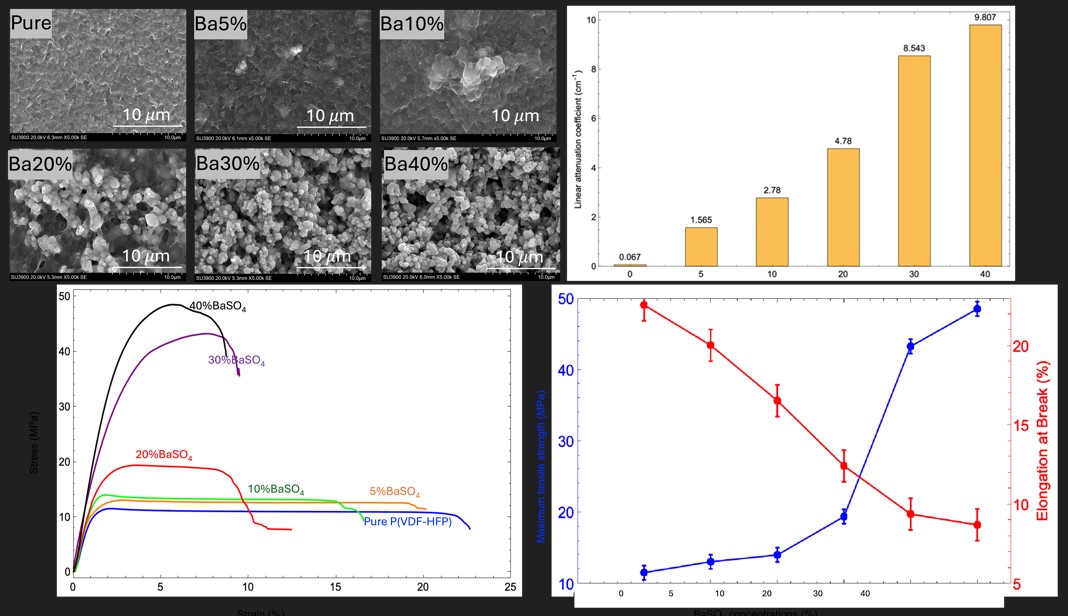
Figure 2 illustrates SEM micrographs showing the surface morphology of both pure P(VDF-HFP) and BaSO4/P(VDF-HFP) composite films. The micrograph of pure P(VDF-HFP) reveals a relatively smooth and uniform surface, with no visible filler particles or significant texture at the micro-scale. This smooth and dense morphology can be attributed to the solution casting method used during film fabrication, where the evaporation of DMF solvent leaves behind a uniform polymer layer [27]. As BaSO4 was incorporated into the P(VDF-HFP) polymer matrix, the surface morphology becomes progressively rougher and more granular. At lower concentrations (5% BaSO4 to 10%BaSO4), the gradual addition of BaSO4 introduces small, scattered clusters of particles. The BaSO4 particles are generally more evenly dispersed and embedded within the polymer, allowing the matrix to maintain a relatively smooth surface, though some aggregation begins to appear at 10%BaSO4. This initial level of aggregation does not significantly disrupt the overall smoothness of the polymer. For 20%BaSO4 and higher, the surface becomes increasingly granular, with more pronounced clustering of BaSO4 particles. At higher concentrations (30%BaSO4 to 40%BaSO4), the particles are densely packed, resulting in rougher surfaces with agglomerated particles. The polymer matrix struggles to encapsulate and uniformly disperse the high volume of BaSO4, causing the particles to cluster together due to attractive forces, such as van der Waals interactions [14]. These aggregated clusters cannot be fully integrated into the polymer chains, leading to a granular and rough morphology. The agglomerates act as micro-sized protrusions, giving the surface its rough, grainy appearance.
AFM analysis
AFM analysis was conducted to further support the surface morphology observations. The AFM images reveal that the BaSO4/P(VDF-HFP) composite films exhibit significant surface roughness on a large scale. Figure 3 presents the AFM images of pure P(VDF-HFP) and BaSO4/P(VDF-HFP) composite films, with the corresponding surface roughness values (Rq) listed in Table 1. The AFM image of pure P(VDF-HFP) shows a relatively smooth surface with minimal height variation. The surface roughness is quite low (Rq: 66.57 nm), which is consistent with the SEM micrographs and the previously reported morphology of pure P(VDF-HFP) films. This smoothness suggests that in the absence of BaSO4 fillers, the polymer chains form a dense and uniform film during the solution casting process. It also indicates that the polymer matrix can crystallize uniformly when there is no filler particles present to disrupt the structure. For BaSO4/P(VDF-HFP) composites, surface roughness increases with BaSO4 content. The results clearly show a progressive increase in surface roughness as the BaSO4 content rises from 5% to 40%. This is due to the growing number of BaSO4 particles in the polymer matrix, which raises the likelihood of particle aggregation, leading to an uneven topography.
At lower concentrations of BaSO4, such as 5% to 10%, the AFM images reveal a slight increase in surface roughness compared to the pure polymer. Small bumps or granular formations appear, indicating the presence of dispersed BaSO4 particles. However, the surface remains relatively smooth, suggesting that the BaSO4 particles are well-dispersed and have not significantly disrupted the uniformity of the polymer matrix. At 20%BaSO4, the Rq value (180.66 nm) increases significantly compared to the 10% BaSO4/P(VDF-HFP) sample, indicating that larger clusters have formed, introducing greater height variations and resulting in a rougher surface, as also shown in the SEM results. At higher concentrations (30% to 40%), surface roughness is dominated by BaSO4 particle agglomerates, as the polymer matrix can no longer maintain an even dispersion of the filler. This results in a highly irregular and rough surface texture. The polymer’s capacity to encapsulate and uniformly disperse BaSO4 is exceeded at these concentrations, leading to large-scale agglomeration and the formation of surface protrusions. This change in morphology can affect the microstructural and mechanical properties of the composite films, which is important to consider for x-ray absorption applications.
3.2. Hydrophobicity
For X-ray shielding materials, surface wettability can impact performance, particularly in environments where moisture exposure is a concern (e.g., medical devices, protective clothing). Hydrophobic surfaces may resist moisture better, protecting the integrity of the shielding material over time. The water contact angle (WCA) measures how water interacts with a surface. A WCA below 90° indicates hydrophilicity, between 90° and 150° indicates hydrophobicity, and above 150° indicates superhydrophobicity, reflecting how well a surface repels or attracts water [28]. WCA is inversely related to surface energy (SE), which influences a material’s behavior, including adhesion, durability, and interaction with other materials [29].
Figure 4 presents the WCA and SE results for pure P(VDF-HFP) and BaSO4/P(VDF-HFP) composite films. For pure P(VDF-HFP), the WCA is 93.28° ± 1.42°, and the SE is 24.18 ± 0.39 mJ/m2, indicating a slightly hydrophobic surface. The relatively high SE contributes to moderate wettability. This confirms the hydrophobic nature of P(VDF-HFP), attributed to the presence of CF₃ and fluorine atoms, which make fluorine-containing polymers naturally hydrophobic. Additionally, SEM in the previous section showed a smooth polymer surface with minimal roughness, as expected in a uniform polymer matrix. AFM would likely confirm minimal nanoscale roughness, consistent with the relatively low WCA. As BaSO4 was incorporated (from 5% to 20%), the WCA gradually increased from 94.26° ± 1.42° to 99.27° ± 1.37°, suggesting a slow shift toward greater hydrophobicity. Simultaneously, the SE decreased steadily from 23.36 ± 0.44 to 21.48 ± 0.19 mJ/m2, implying that BaSO4 nanoparticles were lowering the surface tension. SEM revealed dispersed BaSO4 particles within the polymer matrix, likely contributing to slight surface roughness. AFM also indicated a subtle increase in nanoscale roughness, correlating with the gradual rise in WCA. For the composite with the highest BaSO4 content (40%), the WCA reached 120.85° ± 0.47° and SE dropped to 12.25 ± 0.06 mJ/m2, indicating significant hydrophobicity approaching superhydrophobic behavior. Although BaSO4 itself is hydrophilic [30], which would normally decrease WCA and increase SE, the observed behavior can be attributed to substantial aggregation of BaSO4 particles, as revealed by SEM and AFM images. These particles likely formed rough micro- and nanoscale structures, increasing surface roughness, which ultimately enhanced WCA and reduced SE. This outcome can be explained by Young's and Wenzel's equations, both of which relate surface roughness to wettability [31]. Young’s equation describes the equilibrium contact angle (θ) for a liquid droplet on an ideal, smooth surface, taking into account interfacial free energies (surface tensions) between solid, liquid, and gas phases. However, this model assumes a homogeneous (for pure P(VDF-HFP)), flat surface, making it less applicable to rough surfaces like those observed in BaSO4/P(VDF-HFP) composites. The Wenzel equation, an extension of Young's model, considers surface roughness by introducing a roughness factor, which amplifies the natural wetting behavior of a surface. According to the Wenzel model, for hydrophobic surfaces, increased roughness leads to higher contact angles as the liquid follows the contours of the rough surface [32]. Thus, the high BaSO4 content created an uneven, rough surface that amplified the material’s hydrophobicity, as predicted by the Wenzel model, even though BaSO4 itself is hydrophilic. This suggests that surface morphology, rather than the inherent chemical nature of BaSO4, dominated the wettability properties of the composite, driving it towards greater hydrophobicity.
3.3. Crystal Structure
FTIR analysis
FTIR spectroscopy is a crucial tool for analyzing the crystal structure of polymers, enabling optimization of mechanical and thermal properties in X-ray shielding materials. By assessing phase composition and crystallinity, FTIR analysis supports the development of durable, efficient materials that meet strict safety and performance standards for radiation protection. Figure 5 shows FTIR peaks associated with the crystal structures of both P(VDF-HFP) and BaSO4 in the composites. For pure P(VDF-HFP), vibrational peaks are observed at 431 cm−2, 481 cm−2, and 531 cm−2 (CF2 bending); 614 cm−2 (CF2 bending and skeletal bending); 762 cm−2 (in-plane bending/rocking); 834 cm−2 (CH2 rocking and CF2 asymmetric stretching); 873 cm−2 (CH2 rocking); 975 cm−2 (CH out-of-plane deformation); 1,069 cm−2 (CF₃ out-of-plane deformation); 1,171 cm−2 (CF2 antisymmetric stretching); 1,231 cm−2 (CF out-of-plane deformation); and 1,402 cm−2 (CH2 scissoring). These peaks confirm the semi-crystalline nature of P(VDF-HFP), with both amorphous and crystalline regions contributing to its structure [33,34,35]. In BaSO4 powder, the sulfate group exhibits four fundamental vibrational modes: one nondegenerate mode (ν₁), one doubly degenerate mode (ν2), and two triply degenerate modes (ν₃ and ν4). The FTIR spectrum of BaSO4 typically displays several significant bands, with intense bands associated with asymmetric stretching and bending (ν₃ and ν4) and weaker bands representing symmetric stretching and bending (ν₁ and ν2). In the FTIR spectrum, bands in the range of 1,078–1,193 cm−2 and a shoulder at 983 cm−2 correspond to the symmetric stretching of the SO42− group. The SO42− group’s stretching vibration is identified at 1,543 cm−2 (ν₃), while peaks at 610 and 639 cm−2 correspond to out-of-plane bending. The absorption peak at 3,401 cm−2, attributed to the H2O stretching vibrations at vacant Ba sites, is evident in pure BaSO4 but absent in the composites, suggesting the elimination of hydration-related vibrations during composite formation. Peaks around 2,064 cm−2 represent overtones and combinations of stretching and bending of sulfur-oxygen bonds powder [36,37].
In the FTIR spectra of BaSO4/P(VDF-HFP) composites with varying BaSO4 concentrations, characteristic peaks from both the P(VDF-HFP) polymer matrix and BaSO4 are retained, indicating successful integration of BaSO4 within the polymer structure. Key BaSO4 peaks, including SO42− bending vibrations at 610 and 639 cm−2 and symmetric stretching vibrations within the range of 1078–1193 cm−2, remain stable across all concentrations, underscoring the sulfate group’s structural integrity in the composite. As BaSO4 concentration increases, these specific peaks grow in intensity, reflecting the increasing BaSO4 content. Meanwhile, prominent P(VDF-HFP) polymer peaks, such as CF2 bending at 531 and 614 cm−2, CF2 in-plane bending or rocking at 762 cm−2, and CH2 rocking at 834 cm−2, are preserved across all composite spectra. This suggests that BaSO4 does not significantly disrupt the semi-crystalline structure of P(VDF-HFP), allowing the composite to retain the structural and mechanical properties of the polymer [21]. Additionally, peak shifts observed in the FTIR spectra of the composites may indicate changes in the chemical environment around specific functional groups due to interactions between BaSO4 and the P(VDF-HFP) matrix. When BaSO4 is incorporated into P(VDF-HFP), the particles interact with the polymer chains, potentially altering local electronic distributions and bond strengths within the polymer. Such interactions could cause slight shifts in the CF2 bending or CH2 rocking peaks of P(VDF-HFP), reflecting molecular-level interactions that affect the polymer’s structural arrangement. These shifts provide valuable insight into the compatibility and interaction between the phases within the composite material [20]. The absence of the 3401 cm−2 peak, associated with H2O stretching vibrations in pure BaSO4 powder, suggests that hydration in BaSO4 is eliminated during composite formation, likely due to thermal processing or polymer-particle interactions that reduce the presence of free water molecules. This elimination of water content is advantageous as it enhances the composite's durability and thermal stability, making it suitable for applications in environments where moisture could affect material performance. Overall, the composite retains the structural integrity of P(VDF-HFP) alongside the stability of BaSO4, highlighting its balanced integration, mechanical flexibility, and suitability for radiation-shielding applications.
XRD analysis
The crystalline data for pure P(VDF-HFP) and BaSO4/P(VDF-HFP) composites provides key insights into how BaSO4 loading affects the overall crystalline structure of the composite material. Figure 6 shows the XRD intensity data at various 2θ angles for pure P(VDF-HFP), BaSO4 powder and BaSO4-P(VDF-HFP) composite films with different BaSO4 concentrations. Table 1 lists the crystallinity levels evaluated from XRD results using Equation 2. The P(VDF-HFP) consists of alternating segments of vinylidene fluoride (VDF) and hexafluoropropylene (HFP), with a structural formula represented as: [—CH2—CF2—]ₙ — [—CF2—CF(CF₃)—]ₘ, where n and m represent the relative ratios of the VDF and HFP units in the copolymer. The P(VDF-HFP) structure combines crystalline (from VDF) and amorphous (from HFP) regions, resulting in a semi-crystalline copolymer that can exhibit multiple crystalline phases, primarily the α, β, and γ phases, depending on the polymer chain configuration [38]. The XRD pattern of the pure P(VDF-HFP) film reveals broad, low-intensity peaks, indicating its semi-crystalline structure. These peaks, associated with the crystalline α-phase of P(VDF-HFP), are relatively weak and broad, reflecting a moderate crystallinity level of 60.1% [39]. This semi-crystalline structure combines rigid crystalline domains and flexible amorphous regions. However, with light elements like carbon, hydrogen, and fluorine, pure P(VDF-HFP) has limited X-ray shielding capability due to its low atomic number composition. To investigate the BaSO4 powder, its XRD pattern reveals sharp, high-intensity peaks at specific 2θ values, reflecting its crystalline structure. According to Sifontes's research [36], BaSO4 powder typically exhibits prominent, sharp peaks in its diffraction pattern, characteristic of a crystalline material. This pattern confirms BaSO4's orthorhombic crystal structure and highlights its high degree of crystallinity. The recorded XRD data display peaks at defined 2θ positions, particularly between 10° and 80°, which correspond to characteristic diffraction angles of BaSO4. The peak intensity underscores the material's purity and structural integrity, distinguishing it from less crystalline or amorphous forms. Additionally, the sharpness and intensity of these peaks indicate the stable sulfate ion arrangement within the lattice, which is essential for applications requiring materials with high thermal stability and low reactivity.
The XRD patterns of BaSO4/P(VDF-HFP) composites reveal that increasing BaSO4 content impacts the crystalline structure of the P(VDF-HFP) matrix. When BaSO4 is incorporated, changes in both XRD patterns and crystallinity demonstrate its influence on the composite's structure. Additional peaks, characteristic of BaSO4’s crystalline structure, appear in the XRD patterns, confirming its successful integration and dispersion within the matrix. The broad α-phase peak of P(VDF-HFP) around 2θ ≈ 20° is absent, indicating that slight changes in intensity suggest structural modifications within the polymer matrix. As BaSO4 concentration rises, the XRD peaks sharpen and intensify, particularly around BaSO4’s characteristic angles, indicating an increase in overall crystallinity. This effect is likely due to BaSO4 acting as a nucleating agent, promoting the formation of crystalline domains within the P(VDF-HFP) matrix. This nucleation enhances polymer chain organization, leading to a more crystalline structure. The resulting higher crystallinity improves material rigidity and stability, which is beneficial for applications requiring a more robust composite material. Furthermore, the inclusion of BaSO4, with its high atomic number, increases the composite’s density and enhances its potential for X-ray attenuation. Higher BaSO4 concentrations improve X-ray shielding, as evidenced by the persistent BaSO4 peaks in the XRD patterns, which confirm its presence and distribution within the matrix. The broadening and reduction of P(VDF-HFP) peaks contribute to effective X-ray absorption and shielding. This increased density and enhanced X-ray shielding make these composites suitable for lightweight radiation protection applications.
3.4. Mechanical Property
The mechanical properties of polymer composite films are crucial for practical applications, especially in contexts such as X-ray shielding, where both strength and flexibility are required. Adding BaSO4 filler to P(VDF-HFP) significantly affects these properties. To achieve optimal mechanical performance, it’s essential to control BaSO4 content and ensure uniform particle dispersion within the composite. Standard tensile tests reveal the impact of BaSO4 on tensile properties, including Young’s modulus, tensile strength, and elongation at break, as illustrated in Figure 7. For pure P(VDF-HFP), the stress-strain curve displays an initial elastic phase, followed by a peak stress that transitions into plastic deformation, with minimal resistance to further stretching. This behavior is characteristic of a ductile polymer. The tensile properties show a Young’s modulus of 6.3 MPa, indicating low stiffness and high flexibility. With a tensile strength of 11.59 MPa and an elongation at break of 22.61%, pure P(VDF-HFP) is highly flexible but lacks mechanical strength due to the absence of reinforcement. Upon introducing BaSO4, the stress-strain curves of the 5%-20% BaSO4/P(VDF-HFP) composites retain a similar shape to that of pure P(VDF-HFP), but with enhanced stiffness and tensile strength. As BaSO4 content increases, both Young’s modulus and tensile strength rise, reaching 12.6 MPa and 19.45 MPa, respectively, at 20% BaSO4. The elongation at break, however, decreases from 20.07% to 12.45%, indicating reduced ductility as the material stiffens. These composites offer a balance of strength and flexibility, suitable for X-ray shielding applications where moderate stiffness and improved tensile strength are advantageous. For the 30%-40% BaSO4/P(VDF-HFP) composites, the stress-strain curves indicate substantial increases in tensile strength and rigidity, showcasing the strong reinforcing effect of BaSO4. With Young’s modulus values of 13.4 MPa and 16.4 MPa, and tensile strengths reaching 43.33 MPa and 48.59 MPa for 30% and 40% BaSO4, respectively, these composites demonstrate high load-bearing capacity. However, the elongation at break falls to 9.41% and 8.73%, signifying increased brittleness and decreased flexibility. These properties make the 30%-40% BaSO4 composites ideal for applications requiring robust strength and rigidity, although they may be less suitable for environments needing flexibility. As noted by Xiaolei Chen et al. [18] in studies on BaSO4-filled polymer composites, the Young’s modulus and tensile strength of these materials increase progressively, while elongation decreases with higher BaSO4 content. When inorganic particles are embedded within a polymer matrix, stress can transfer from the matrix to the particles, thereby enhancing yield stress. The tensile yield stress in particle-filled polymers is primarily influenced by filler content and interfacial interactions, which depend on factors such as interfacial adhesion, particle size, and dispersion within the matrix. BaSO4 nanoparticles, in particular, benefit from their hydrophobic surface, which allows better dispersion in the polymer matrix. Consequently, as BaSO4 content rises, the interface transfers more stress from the matrix to the inorganic particles, leading to increased tensile yield stress in the composite.
3.5. Thermal Stability
The thermal stability evaluation of P(VDF-HFP) and BaSO4/P(VDF-HFP) composites for X-ray shielding involves key measurements such as decomposition temperature, weight loss rate, and residual mass. A high decomposition temperature and low weight loss rate indicate heat resistance, while stable residual mass reflects BaSO4's durability as a filler. This analysis helps optimize BaSO4 content to enhance thermal resilience and ensure reliable shielding performance. The thermogravimetric (TGA) and derivative thermogravimetric (DTG) curves for pure P(VDF-HFP) and BaSO4/P(VDF-HFP) composite films, shown in Figure 8 and summarized in Table 2, reveal differences in decomposition temperature, weight loss rate, and residual mass between the pure polymer and the composites. From Figure 8(a), pure P(VDF-HFP) begins significant weight loss around 469°C, indicating relatively low thermal stability. In contrast, BaSO4/P(VDF-HFP) composites display higher decomposition temperatures, starting at approximately 479°C for the 5% BaSO4 composite and reaching around 498°C for the 40% BaSO4 composite. Additionally, the weight loss rate decreases with increased BaSO4 content, indicating slower degradation in composites. For instance, the 30% BaSO4 composite shows a more gradual weight loss, demonstrating BaSO4’s heat-resistant effect, which stabilizes the polymer matrix and delays degradation. Furthermore, the residual mass after thermal decomposition is minimal for pure P(VDF-HFP), as it fully degrades, leaving little residue. However, BaSO4/P(VDF-HFP) composites retain progressively higher residual mass with increased BaSO4 content, with the 40% BaSO4 composite showing the highest residual mass. This residual mass is mainly due to the thermally stable BaSO4, which remains intact and provides a durable framework even after the polymer matrix has decomposed, enhancing the composite’s suitability for high-temperature applications.
In Figure 8(b), the DTG data provide additional information on the thermal decomposition behavior of pure P(VDF-HFP) and BaSO4/P(VDF-HFP) composites, specifically peak decomposition temperature, maximum weight loss rate, and final residual mass for each sample. The peak decomposition temperature, where each sample experiences the maximum weight loss rate, is around 499°C for pure P(VDF-HFP), indicating lower thermal stability compared to the composites. With increasing BaSO4 content, the peak decomposition temperature gradually rises, reaching 504°C for the 5% BaSO4 composite, 507°C for the 20% BaSO4 composite, and 513°C for the 30% BaSO4 composite. This increase in peak temperature with higher BaSO4 content suggests that BaSO4 stabilizes the polymer matrix, delaying thermal decomposition. The maximum weight loss rate, representing the steepest part of the decomposition curve, is highest for pure P(VDF-HFP) at 27.55 %/min, suggesting rapid degradation under thermal stress. As BaSO4 content increases, the weight loss rate decreases significantly, with the 5% BaSO4 composite at 21.36 %/min and the 40% BaSO4 composite at 7.73 %/min, indicating improved resistance to sudden thermal breakdown. The residual mass represents the material left after heating to the final temperature, indicating the stability of the inorganic BaSO4 component. Pure P(VDF-HFP) has negligible residual mass, as it fully degrades without leaving a stable residue. In contrast, BaSO4/P(VDF-HFP) composites retain progressively higher residual mass with increased BaSO4 content. For instance, the 5% BaSO4 composite has a small residual mass, while the 30% BaSO4 composite retains a much larger amount. This residual mass is primarily composed of thermally stable BaSO4, which does not decompose and remains intact, providing structural stability even after polymer degradation. These results indicate that increasing BaSO4 content in P(VDF-HFP) composites raises the peak decomposition temperature, reduces the maximum weight loss rate, and increases the final residual mass, demonstrating BaSO4's role in enhancing the thermal stability and durability of the composite for high-temperature X-ray shielding applications requiring prolonged structural integrity.
3.5. Absorption Performance
The linear attenuation coefficient is a parameter that is used to describe the fraction of attenuated incident X-ray photons per unit thickness of material. In general, the primary interactions within shielding materials encompass the photoelectric effect, Compton scattering, and pair production [40]. However, pair production occurs when the energy of the incident photon exceeds 1.022 MeV. Since this study employed low-energy photons (60 – 100 keV), the potential interaction mechanisms should be photoelectric and Compton scattering. In the case of the photoelectric effect, all the energy of the incident photon is absorbed by an atomic electron, unlike Compton scattering, where the energy transferred to an electron depends on the scattering angle [41]. The linear attenuation coefficient (μ), a crucial factor in evaluating the absorption capacity of shielding materials, is influenced by several key variables. These include the atomic number of the shielding material, its physical density, and the energy of the incident X-ray photon. BaSO4 is comprised of a high atomic number of Ba. So, the increase in BaSO4 concentration was found to increase the linear attenuation coefficient, as shown in Figure 9. Each value of μ was calculated from the slope of the linear equation and the thickness of the shielding (. The results show that the increase of BaSO4 causes a linear attenuation coefficient increase. This phenomenon occurs because there is an increased likelihood that X-ray photons will collide with electrons within the atoms of barium, which has a high atomic number, resulting in the photoelectric effect.
Considering the radiation absorption capability of this shielding material, it was found that as the linear attenuation coefficient increases, the radiation passing through the shielding decreases, as shown in Table 3. The percentage attenuation was calculated as the percentage of the difference between the intensity of the incident and the transmittance of X-ray photons. The results show that the performance of X-ray shielding material made of BaSO4/P(VDF-HFP) depends on the concentration of BaSO4 in the shielding sheet. Furthermore, the increase in shielding thickness can increase the efficiency of X-ray protection as well. The results found from this research show that the highest percentage of attenuation from X-ray source of 60 keV and 0.1 mA was 71/52 % at the BaSO4 concentration of 40% and the thickness of 0.12 cm.
The investigation of the percentage attenuation of the shielding at different concentrations of BaSO4 in BaSO4/P(VDF-HFP) shielding sheets by the high energy of X-ray photons is shown in Table 4. These results show that the ability of the shielding decreases while radiating with high energy. It is possible that the main part of X-ray photons was scattered and moved through the shielding. This phenomenon leads to a decrease in the linear attenuation coefficients of the shielding.
4. Conclusions
Polymer composite shielding offers an alternative approach to mitigate the risk of X-ray radiation exposure. In this method, a selected polymer acts as a matrix, while a high atomic number particle serves as a filler to absorb the X-ray photons. BaSO4/P(VDF-HFP) composite shielding sheets were produced with varying concentrations of BaSO4. Post irradiation with X-ray photons, these sheets were analyzed to assess their photon absorption capacity, surface characteristics, and bulk properties. The results indicated that increasing the concentration of BaSO4 in BaSO4/P(VDF-HFP) enhances the linear attenuation coefficient of the shielding sheet when exposed to 60 keV and 0.1 mA X-ray generator. However, the linear attenuation coefficient decreases with higher X-ray generator power at 80 and 100 keV. Additionally, higher BaSO4 concentrations led to rougher and more hydrophobic surfaces on the shielding sheets. Furthermore, tensile and thermal tests showed improvement in the shielding sheet’s properties.
Institutional Review Board Statement
Not applicable
Data Availability Statement
The processed data required to reproduce these findings is available from the authors upon reasonable request.
Acknowledgments
The authors would like to express their gratitude to the Thailand Institute of Nuclear Technology (Public Organization), Thailand, for providing financial support and equipment necessary for this project. The authors also extend their thanks to the Faculty of Science and Technology, Nakhon Si Thammarat Rajabhat University, and the Division of Physical Science (Physics), Faculty of Science, Prince of Songkla University, for their instrumental support.
Conflicts of Interest
The authors have declared that no competing interests exist.
References
- Alghamdi et al., "Radiation Risk Awareness Among Health Care Professionals: An Online Survey," Journal of Radiology Nursing, vol. 39, pp. 132-138, 06/01 2020. [CrossRef]
- D. Miller and D. Schauer, "The ALARA principle in medical imaging," AAPM Newsletter, vol. 40, pp. 38-40, 01/01 2015.
- S. M. J. Mortazavi et al., "Lead-free, multilayered, and nanosized radiation shields in medical applications, industrial, and space research," 2024, pp. 305-322.
- K. Singh, R. K. Singh, B. Sharma, and A. K. Tyagi, "Characterization and biocompatibility studies of lead free X-ray shielding polymer composite for healthcare application," Radiation Physics and Chemistry (1993), pp. 9-15, 2017, doi: DOI:101016/jradphyschem201704016.
- L. Yu, P. L. Yap, A. Santos, D. Tran, and D. Losic, "Lightweight polyester fabric with elastomeric bismuth titanate composite for high-performing lead-free X-ray shielding," Radiation Physics and Chemistry, vol. 205, p. 110726, 12/01 2022. [CrossRef]
- S. Palanisami et al., "Lead-free X-Ray shielding aprons using Zn-doped SnO2 epoxy nanocomposite: A promising alternative to traditional heavy and lead-based materials," Optical Materials, vol. 145, p. 114496, 2023/11/01/ 2023. [CrossRef]
- S. Jayakumar, T. Saravanan, and J. Philip, "A review on polymer nanocomposites as lead-free materials for diagnostic X-ray shielding: Recent advances, challenges and future perspectives," Hybrid Advances, vol. 4, p. 100100, 2023/12/01/ 2023. [CrossRef]
- H. Alsaab and S. Zeghib, "Analysis of X-ray and gamma ray shielding performance of prepared polymer micro-composites," Journal of Radiation Research and Applied Sciences, vol. 16, no. 4, p. 100708, 2023/12/01/ 2023. [CrossRef]
- S.-C. Kim, "Construction of a Medical Radiation-Shielding Environment by Analyzing the Weaving Characteristics and Shielding Performance of Shielding Fibers Using X-ray-Impermeable Materials," Applied Sciences, vol. 11, no. 4. [CrossRef]
- J. Wang et al., "Preparation of eGaIn NDs/TPU Composites for X-ray Radiation Shielding Based on Electrostatic Spinning Technology," Materials, vol. 17, no. 2. [CrossRef]
- Bawazeer et al., "Evaluation of X-ray radiation shielding performance of Bi2O3 and BaTiO3 embedded in PVP and PEG polymer nanocomposite," Radiation Effects and Defects in Solids, 07/22 2024. [CrossRef]
- V. More, Z. Alsayed, M. S. Badawi, A. A. Thabet, and P. P. Pawar, "Polymeric composite materials for radiation shielding: a review," Environmental Chemistry Letters, vol. 19, no. 3, pp. 2057-2090, 2021/06/01 2021. [CrossRef]
- Yao, X. Li, K. G. Neoh, Z. Shi, and E. T. Kang, "Antibacterial activities of surface modified electrospun poly(vinylidene fluoride-co-hexafluoropropylene) (PVDF-HFP) fibrous membranes," Applied Surface Science, vol. 255, no. 6, pp. 3854-3858, 2009/01/01/ 2009. [CrossRef]
- Kazemi and M. R. Yaftian, "PVDF-HFP-based polymer inclusion membrane functionalized with D2EHPA for the selective extraction of bismuth(III) from sulfate media," Scientific Reports, vol. 14, no. 1, p. 11622, 2024/05/21 2024. [CrossRef]
- L. Shi, R. Wang, Y. Cao, D. Liang, and J.-H. Tay, "Effect of additives on the fabrication of poly(vinylidene fluoride- co-hexafluropropylene) (PVDF-HFP) asymmetric microporous hollow fiber membranes," Journal of Membrane Science - J MEMBRANE SCI, vol. 315, pp. 195-204, 05/01 2008. [CrossRef]
- M. J. Toh, P. C. Oh, and M. I. S. Mohd Shaufi, "Preparation of Highly Hydrophobic PVDF-HFP Membrane with Anti-Wettability Characteristic," IOP Conference Series: Materials Science and Engineering, vol. 778, no. 1, p. 012176, 2020/04/01 2020. [CrossRef]
- M. S. Gharissah et al., "Composites cement/BaSO4/Fe3O4/CuO for improving X-ray absorption characteristics and structural properties," Scientific Reports, vol. 12, no. 1, p. 19169, 2022/11/10 2022. [CrossRef]
- X. Chen, L. Wang, J. Shi, H. Shi, and Y. Liu, "Effect of Barium Sulfate Nanoparticles on Mechanical Properties and Crystallization Behaviour of HDPE," Polymers and Polymer Composites, vol. 18, no. 3, pp. 145-152, 2010/03/01 2010. [CrossRef]
- H. A. Maghrabi, A. Vijayan, F. Mohaddes, P. Deb, and L. Wang, "Evaluation of X-ray radiation shielding performance of barium sulphate-coated fabrics," Fibers and Polymers, vol. 17, no. 12, pp. 2047-2054, 2016/12/01 2016. [CrossRef]
- H. Agarwal, S. Yadav, and G. Jaiswar, "Effect of nanoclay and barium sulfate nanoparticles on the thermal and morphological properties of polyvinylidene fluoride nanocomposites," Journal of Thermal Analysis and Calorimetry, vol. 129, no. 3, pp. 1471-1479, 2017/09/01 2017. [CrossRef]
- L. A. Silva, A. M. S. Batista, T. Serodre, A. T. B. Neto, C. A. Furtado, and L. O. Faria, "Enhancement of X-ray Shielding Properties of PVDF/BaSO4 Nanocomposites Filled with Graphene Oxide," MRS Advances, vol. 4, no. 3, pp. 169-175, 2019/01/01 2019. [CrossRef]
- S. Banerjee, "Simple derivation of Young, Wenzel and Cassie-Baxter equations and its interpretations," 09/11 2008. [CrossRef]
- D. K. Owens and R. C. Wendt, "Estimation of the surface free energy of polymers," Journal of Applied Polymer Science, vol. 13, no. 8, pp. 1741-1747, 1969/08/01 1969. [CrossRef]
- J. Yuennan, N. Tohluebaji, C. Putson, N. Muensit, and P. Channuie, "Enhanced electroactive β-phase and dielectric properties in P(VDF-HFP) composite flexible films through doping with three calcium chloride salts: CaCl, CaCl·2HO, and CaCl·6HO," Polymers for Advanced Technologies, vol. 35, no. 6, p. e6437, 2024. [CrossRef]
- K. Selvakumar and R. Manimuthu, "Investigation on meta-polybenzimidazole blend with sulfonated PVdF-HFP proton conducting polymer electrolytes for HT-PEM fuel cell application," Journal of Materials Science: Materials in Electronics, vol. 29, 09/01 2018. [CrossRef]
- H. Li and S. Lim, "Boosting Performance of Self-Polarized Fully Printed Piezoelectric Nanogenerators via Modulated Strength of Hydrogen Bonding Interactions," (in eng), Nanomaterials (Basel), vol. 11, no. 8, Jul 25 2021. [CrossRef]
- T. Mälzer, L. Mathies, T. Band, R. Gorgas, and H. S. Leipner, "Influence of Different Solvents and High-Electric-Field Cycling on Morphology and Ferroelectric Behavior of Poly(Vinylidene Fluoride-Hexafluoropropylene) Films," Materials, vol. 14, no. 14. [CrossRef]
- Y. Guo and H. Zhao, "Femtosecond laser processed superhydrophobic surface," Journal of Manufacturing Processes, vol. 109, pp. 250-287, 2024/01/17/ 2024. [CrossRef]
- P. S. Souza, A. J. Santos, M. A. P. Cotrim, A. M. Abrão, and M. A. Câmara, "Analysis of the surface energy interactions in the tribological behavior of ALCrN and TIAlN coatings," Tribology International, vol. 146, p. 106206, 2020/06/01/ 2020. [CrossRef]
- H. Bala et al., "In situ preparation and surface modification of barium sulfate nanoparticles," Colloids and Surfaces A: Physicochemical and Engineering Aspects, vol. 274, no. 1, pp. 71-76, 2006/02/15/ 2006. [CrossRef]
- Wang, Y. Zhang, L. Shi, J. Li, and Z. Guo, "Advances in the theory of superhydrophobic surfaces," Journal of Materials Chemistry, 10.1039/C2JM32780E vol. 22, no. 38, pp. 20112-20127, 2012. [CrossRef]
- Li et al., "A Review on Superhydrophobic Surface with Anti-Icing Properties in Overhead Transmission Lines," Coatings, vol. 13, p. 301, 01/28 2023. [CrossRef]
- Y. Bormashenko, R. Pogreb, O. Stanevsky, and E. Bormashenko, "Vibrational spectrum of PVDF and its interpretation," Polymer Testing - POLYM TEST, vol. 23, pp. 791-796, 10/01 2004. [CrossRef]
- Ramesh, "One-step fabrication of biomimetic PVDF-BaTiO3 nanofibrous composite using DoE," Materials Research Express, vol. 5, p. 085308, 07/05 2018. [CrossRef]
- Siva, T. Shakthi, and J. Hemalatha, "Synthesis and ferroelectric investigations of poly(vinylidene fluoride- co -hexafluoropropylene)-Mg(NO 3 ) 2 films: ARTICLE," Journal of Applied Polymer Science, vol. 133, 06/01 2016. [CrossRef]
- Á. B. Sifontes et al., "Obtaining Highly Crystalline Barium Sulphate Nanoparticles via Chemical Precipitation and Quenching in Absence of Polymer Stabilizers," Journal of Nanomaterials, vol. 2015, no. 1, p. 510376, 2015/01/01 2015. [CrossRef]
- L. Staicu, T. Bajda, L. Drewniak, and L. Charlet, "Power Generation: Feedstock for High-Value Sulfate Minerals," Minerals, vol. 10, p. 188, 02/19 2020. [CrossRef]
- D. Li and M. Liao, "Study on the dehydrofluorination of vinylidene fluoride (VDF) and hexafluoropropylene (HFP) copolymer," Polymer Degradation and Stability, vol. 152, pp. 116-125, 2018/06/01/ 2018. [CrossRef]
- Mohammed, S. Salman, and F. M.Noori, "Preparation and Characterizations of Poly (vinylidene fluoride)(PVDF)/Ba0. 6Sr0. 4TiO3 (BST) Nanocomposites," International Journal of Applied Engineering Research, vol. 13, pp. 5008-5013, 01/01 2018.
- Q. Chang, S. Guo, and X. Zhang, "Radiation shielding polymer composites: Ray-interaction mechanism, structural design, manufacture and biomedical applications," Materials & Design, vol. 233, p. 112253, 08/01 2023. [CrossRef]
- Baeyens et al., "Basic Concepts of Radiation Biology," in Radiobiology Textbook, S. Baatout Ed. Cham: Springer International Publishing, 2023, pp. 25-81.
Figure 1.
Schematic illustration of (a) the film preparation process and (b) the X-ray absorption experimental setup for BaSO4/P(VDF-HFP) composites.
Figure 1.
Schematic illustration of (a) the film preparation process and (b) the X-ray absorption experimental setup for BaSO4/P(VDF-HFP) composites.
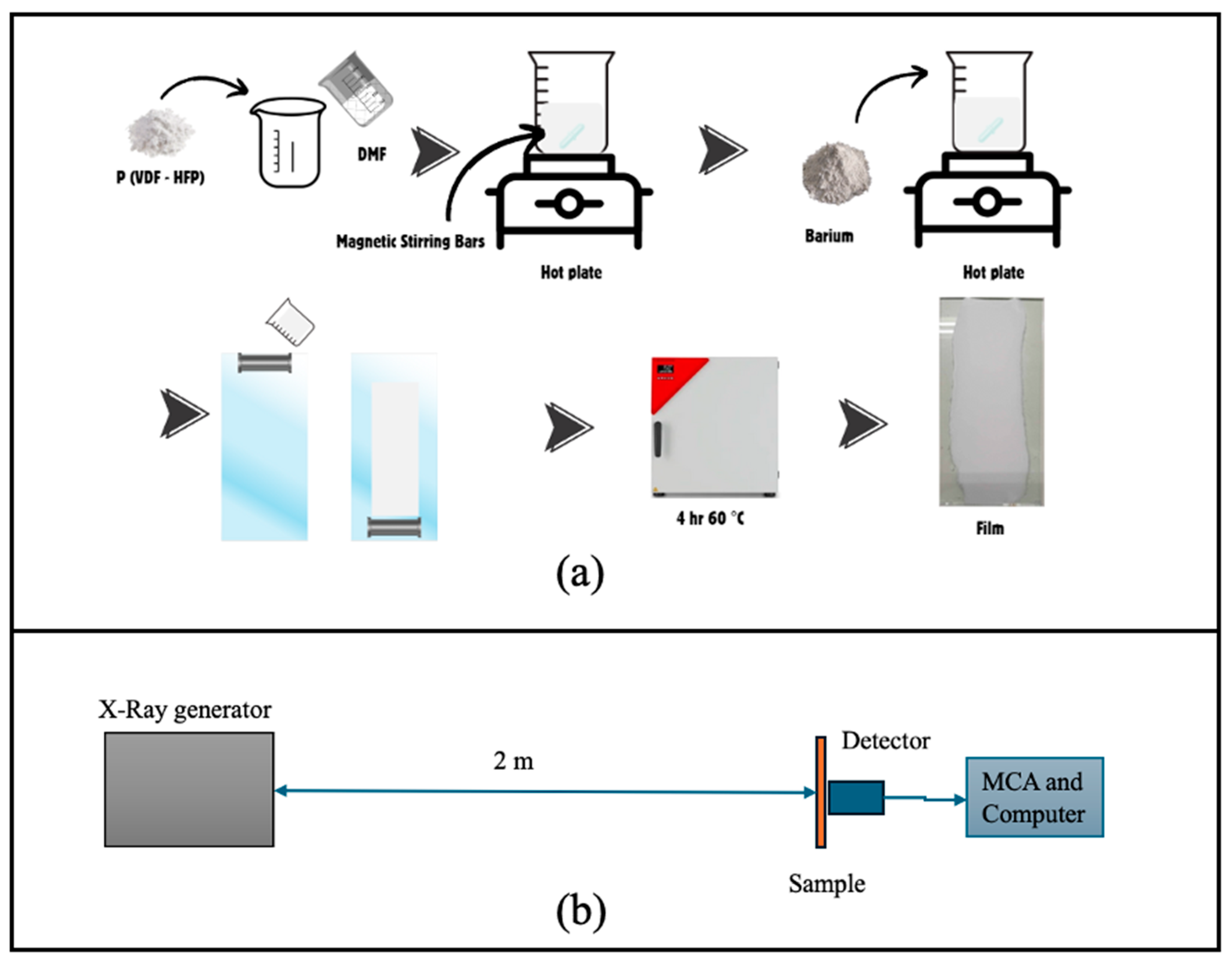
Figure 2.
SEM micrographs of pure P(VDF-HFP) and BaSO4/P(VDF-HFP) composite films.
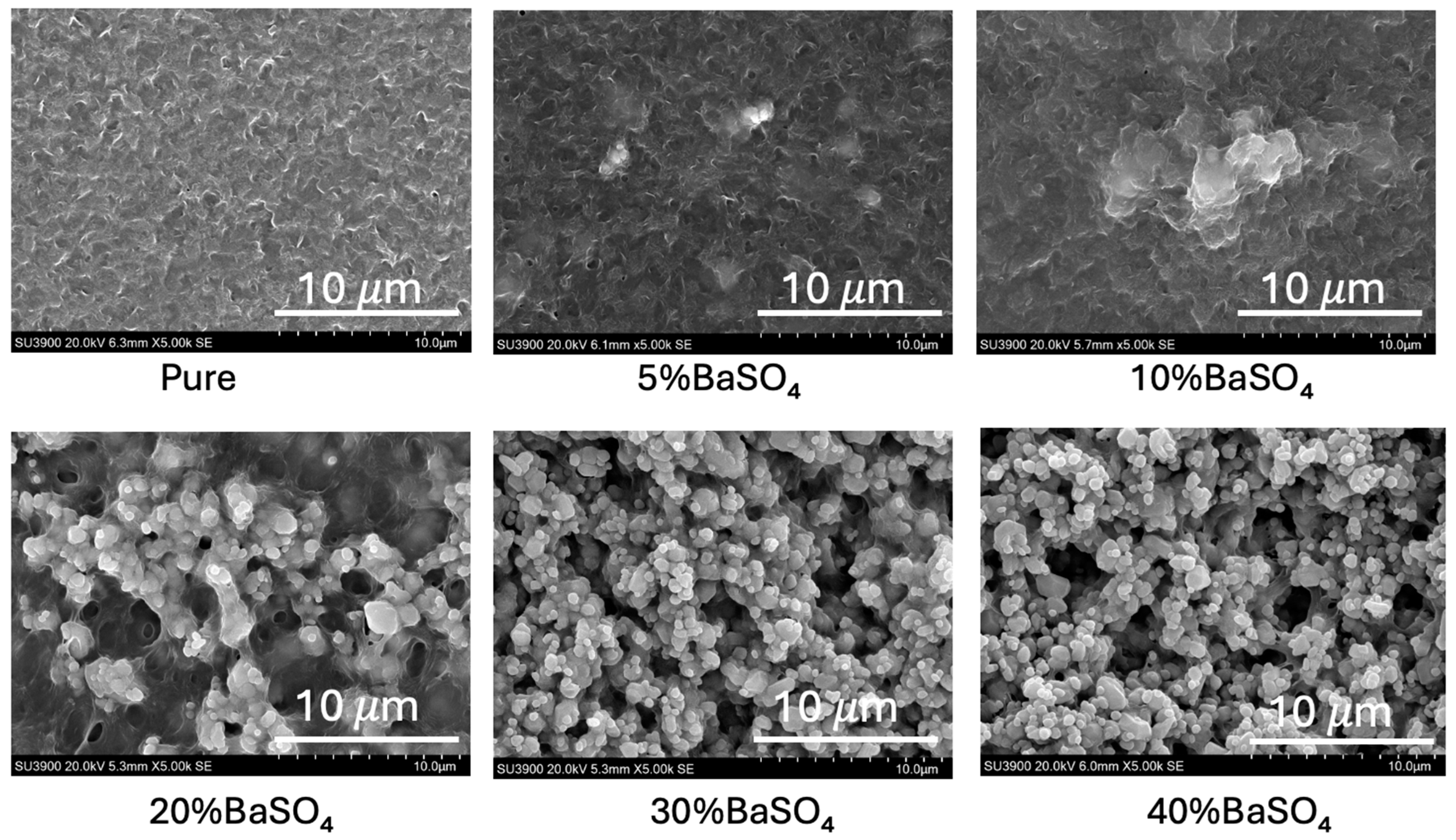
Figure 3.
AFM images of pure P(VDF-HFP) and BaSO4/P(VDF-HFP) composite films.
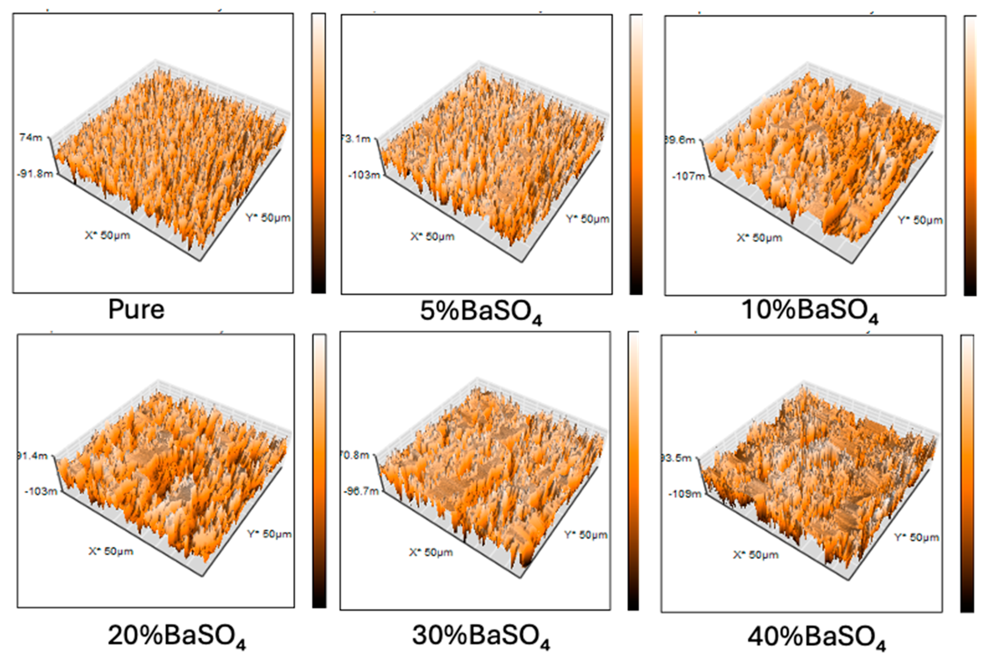
Figure 4.
Water contact angle (WCA) and surface energy (SE) of pure P(VDF-HFP) and BaSO4/P(VDF-HFP) composite films.
Figure 4.
Water contact angle (WCA) and surface energy (SE) of pure P(VDF-HFP) and BaSO4/P(VDF-HFP) composite films.
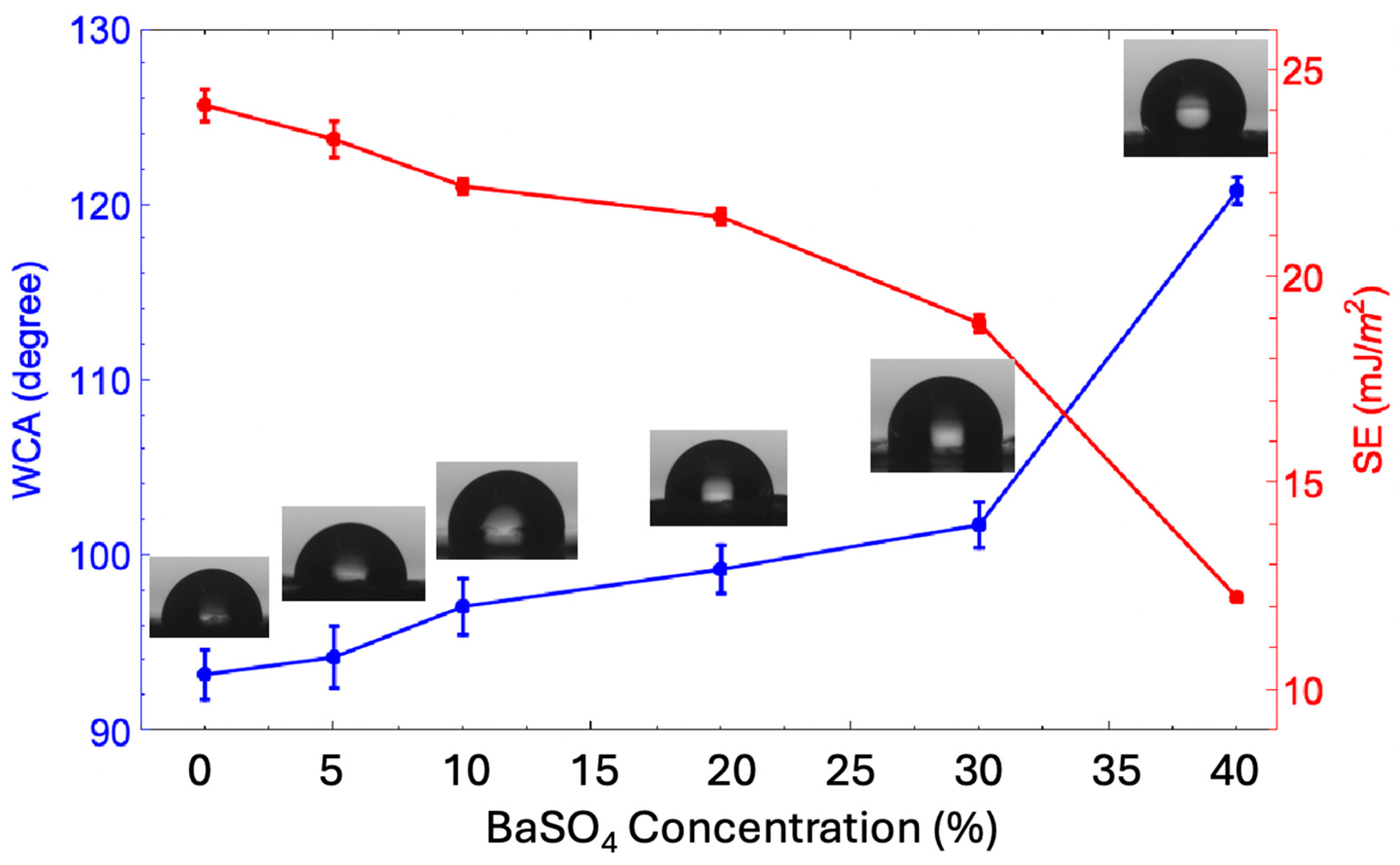
Figure 5.
FTIR patterns of pure P(VDF-HFP) and BaSO4P(VDF-HFP) composite films.
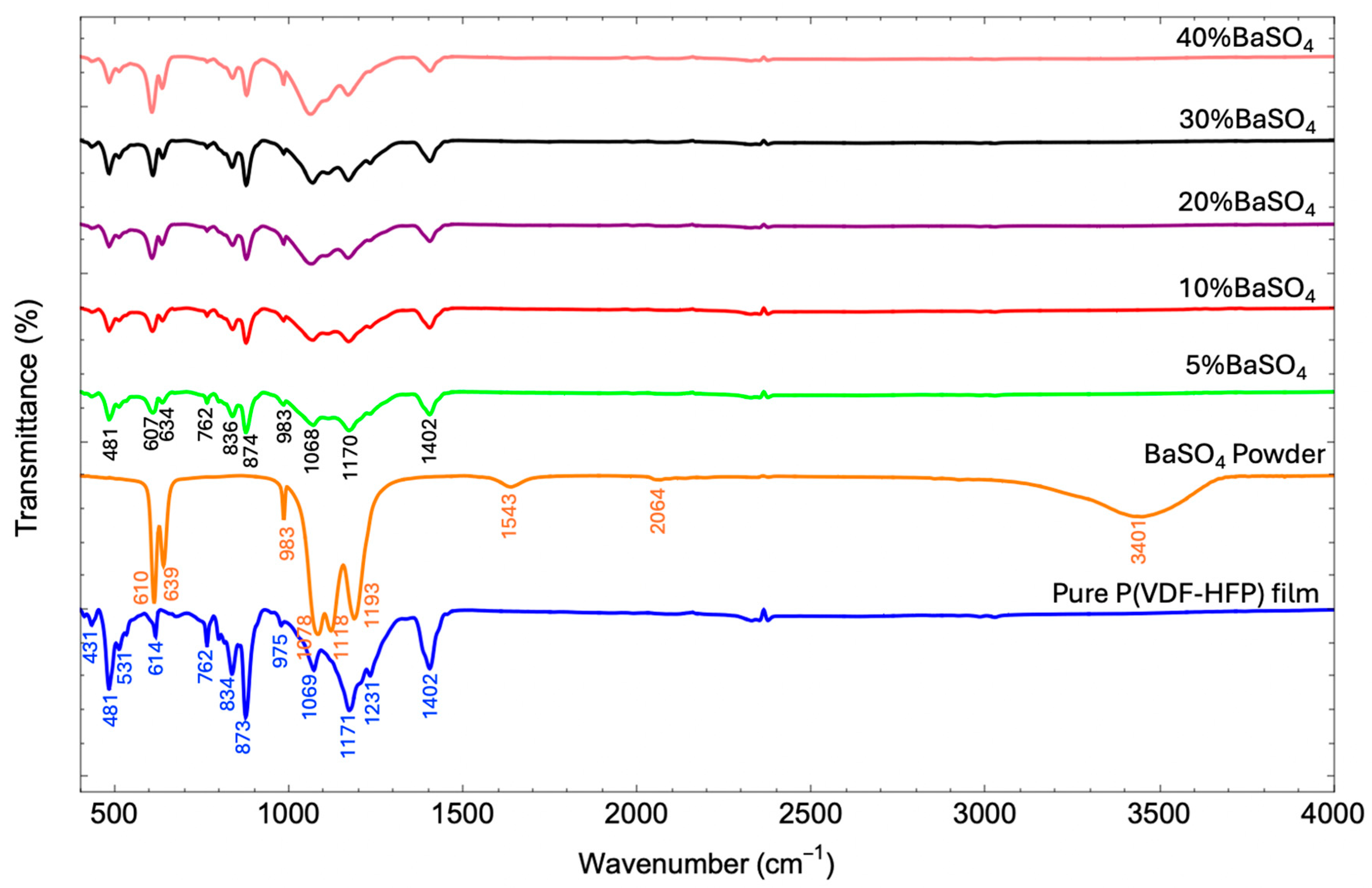
Figure 6.
XRD patterns of pure P(VDF-HFP) and BaSO4P(VDF-HFP) composite films.
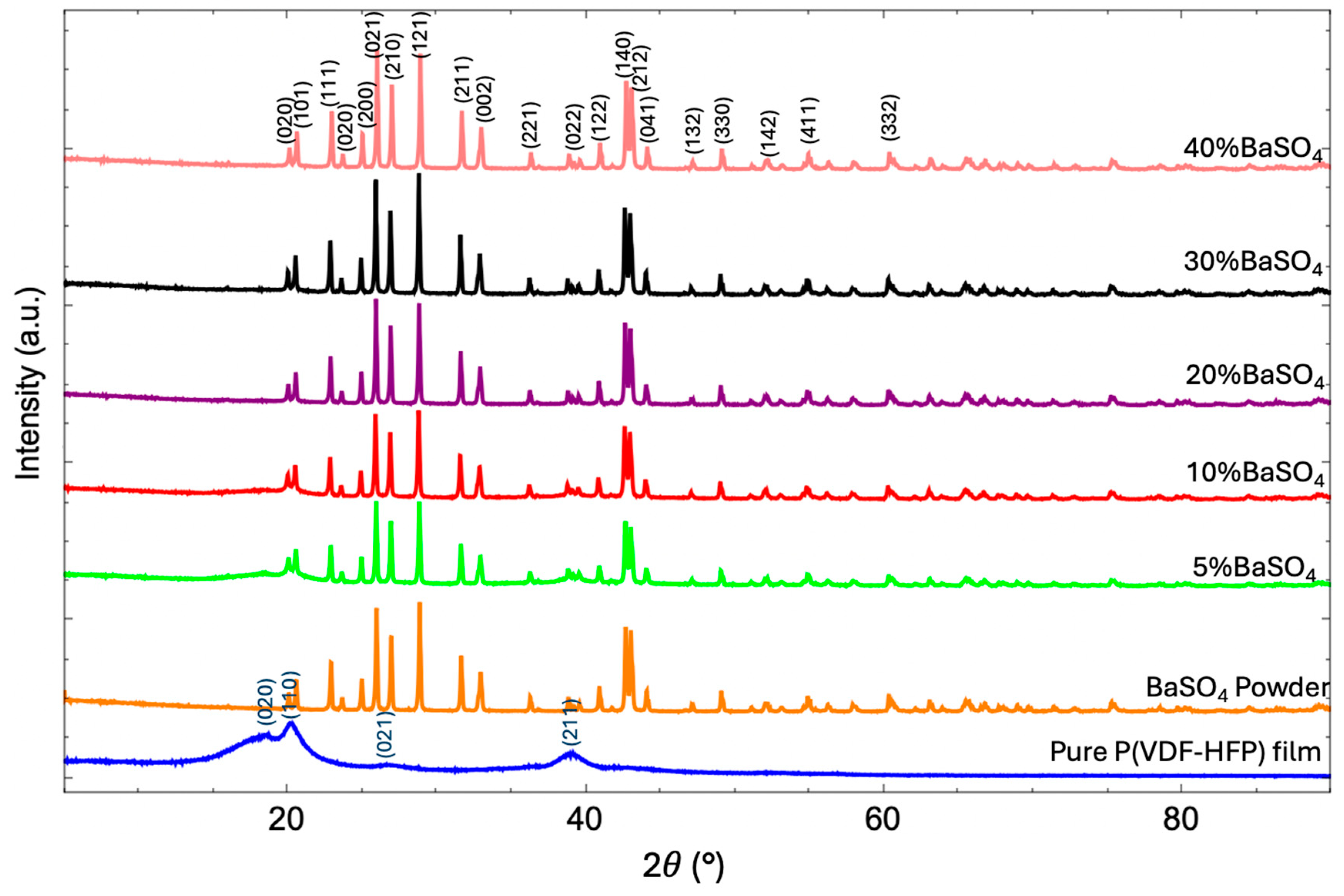
Figure 7.
(a) Stress-strain curves and Young's modulus (inserted table) and (b) Values of tensile properties (Tensile strength and elongation at break) of pure P(VDF-HFP) and BaSO4/P(VDF-HFP) composite films.
Figure 7.
(a) Stress-strain curves and Young's modulus (inserted table) and (b) Values of tensile properties (Tensile strength and elongation at break) of pure P(VDF-HFP) and BaSO4/P(VDF-HFP) composite films.
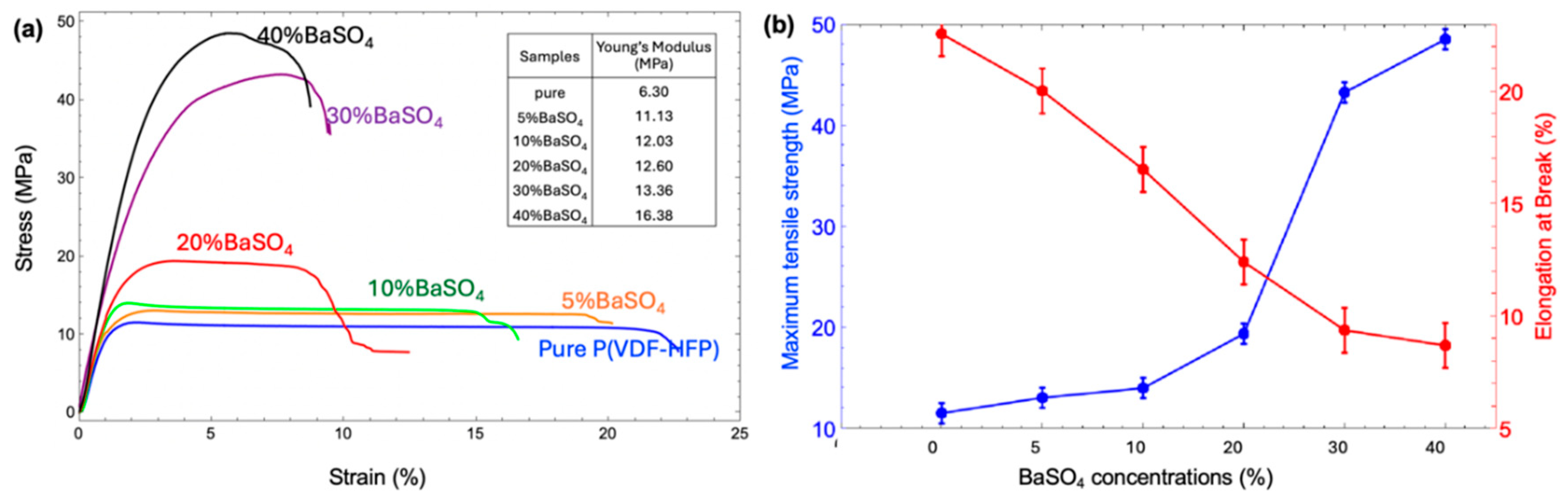
Figure 8.
(a) TGA and (b) DTG thermograms of pure P(VDF-HFP) and BaSO4/P(VDF-HFP) composite films.
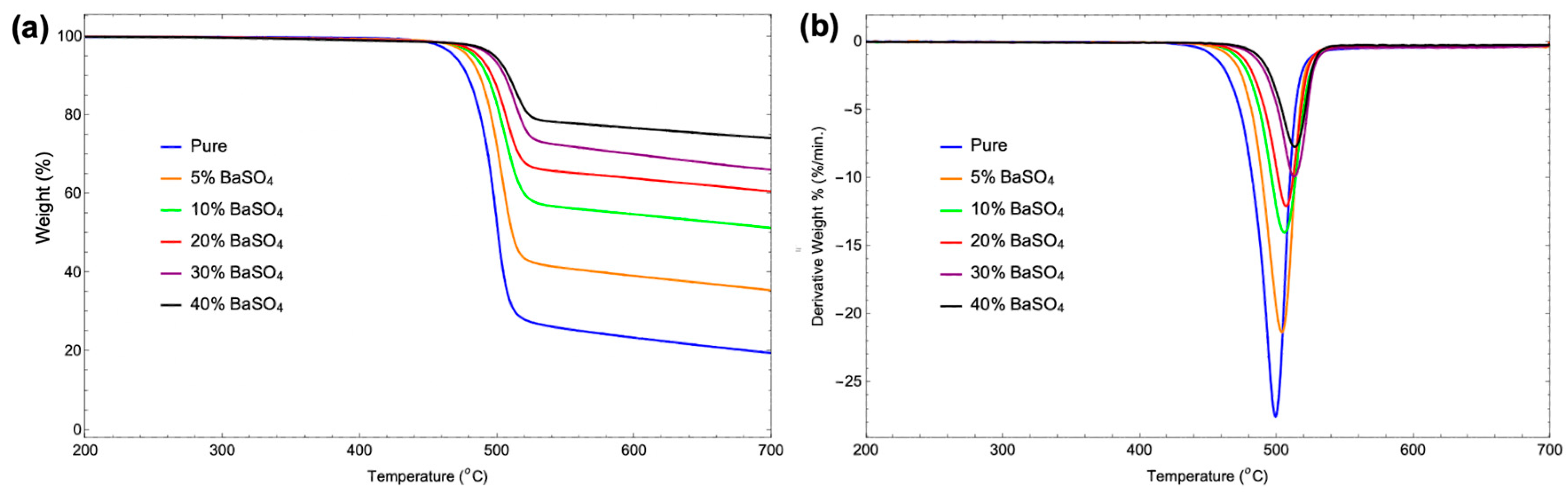
Figure 9.
Relation between BaSO4 concentration in PVDF-HFP composite film and linear attenuation coefficient (μ).
Figure 9.
Relation between BaSO4 concentration in PVDF-HFP composite film and linear attenuation coefficient (μ).
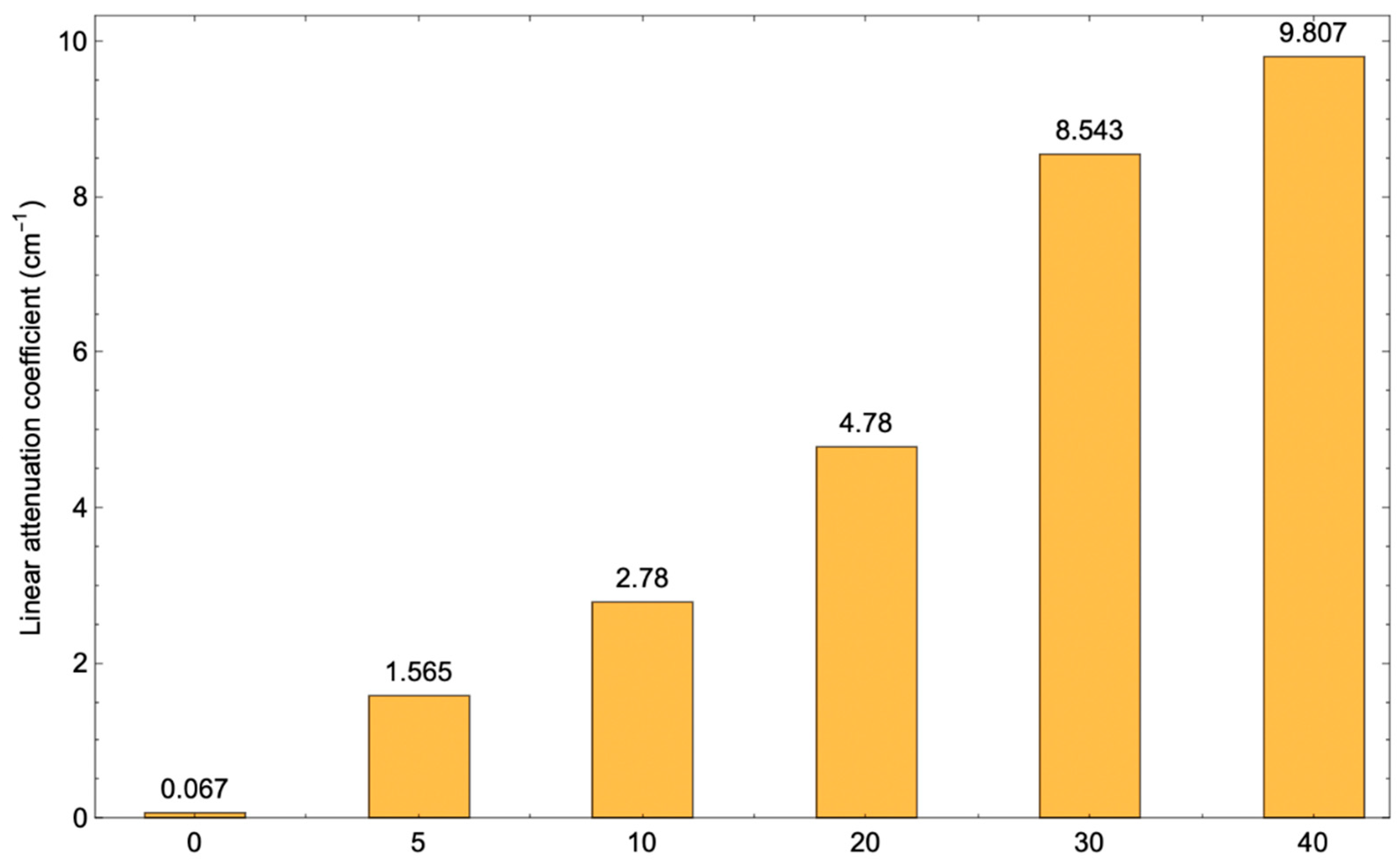

Table 1.
Porosity, root mean square roughness, crystallinity values of pure P(VDF-HFP) and BaSO4/P(VDF-HFP) composite films.
Table 1.
Porosity, root mean square roughness, crystallinity values of pure P(VDF-HFP) and BaSO4/P(VDF-HFP) composite films.
| Sample | Porosity (%) |
Root means square roughness (Rq) (nm) |
Crystallinity (Xc) (%) |
|---|---|---|---|
| Pure | 0.30 | 66.57 | 60.10 |
| 5% BaSO4 | 1.60 | 138.93 | 71.24 |
| 10% BaSO4 | 4.67 | 139.96 | 73.81 |
| 20% BaSO4 | 15.05 | 180.66 | 79.00 |
| 30% BaSO4 | 20.40 | 237.45 | 80.44 |
| 40% BaSO4 | 25.21 | 590.32 | 83.53 |
Table 2.
Decomposition temperature, weight loss rate, and residual mass evaluated from TGA and DTG thermograms of pure P(VDF-HFP) and BaSO4/P(VDF-HFP) composite films.
Table 2.
Decomposition temperature, weight loss rate, and residual mass evaluated from TGA and DTG thermograms of pure P(VDF-HFP) and BaSO4/P(VDF-HFP) composite films.
| Sample | TGA Analysis Results | DTG Analysis Results | ||||
|---|---|---|---|---|---|---|
| Decomposition Temperature (°C) | Weight Loss Rate (%/°C) |
Residual Mass (%) |
Peak Decomposition Temperature (°C) | Maximum Weight Loss Rate (%/min) |
Residual Mass (%) |
|
| pure | 468.87 | 0.55 | 19.58 | 499.09 | 27.55 | -0.06 |
| 5% BaSO4 | 478.78 | 0.46 | 35.50 | 503.72 | 21.36 | -0.07 |
| 10% BaSO4 | 483.38 | 0.34 | 51.34 | 505.7 | 14.03 | -0.28 |
| 20% BaSO4 | 486.69 | 0.27 | 60.60 | 506.86 | 12.10 | -0.16 |
| 30% BaSO4 | 495.29 | 0.24 | 66.15 | 512.65 | 9.93 | -0.19 |
| 40% BaSO4 | 498.26 | 0.18 | 74.11 | 513.15 | 7.73 | -0.23 |
Table 3.
Attenuation of X-ray photons due to the component of BaSO4 in BaSO4/P(VDF-HFP) composites.
| Thickness (cm) | Attenuation (%) | ||||
|---|---|---|---|---|---|
| 5%BaSO4 | 10%BaSO4 | 20%BaSO4 | 30%BaSO4 | 40%BaSO4 | |
| 0.02 | 1.48 | 4.95 | 10.99 | 19.48 | 22.71 |
| 0.04 | 4.92 | 8.96 | 19.05 | 36.65 | 39.81 |
| 0.06 | 7.65 | 14.65 | 26.11 | 47.27 | 49.77 |
| 0.08 | 10.47 | 19.26 | 31.68 | 54.54 | 58.49 |
| 0.1 | 13.87 | 24.02 | 39.32 | 61.39 | 65.64 |
| 0.12 | 15.50 | 27.41 | 44.95 | 66.25 | 71.52 |
Table 4.
Attenuation of X-ray photons due to the component of BaSO4 in BaSO4/P(VDF-HFP) composites at different X-ray energy photon.
Table 4.
Attenuation of X-ray photons due to the component of BaSO4 in BaSO4/P(VDF-HFP) composites at different X-ray energy photon.
| X-ray energy (keV) | Attenuation (%) of the 0.2 mm thickness | ||||
|---|---|---|---|---|---|
| 5%BaSO4 | 10%BaSO4 | 20%BaSO4 | 30%BaSO4 | 40%BaSO4 | |
| 60 | 1.48 | 4.95 | 10.99 | 19.48 | 22.71 |
| 80 | 2.28 | 2.12 | 7.33 | 10.96 | 13.95 |
| 100 | 0.04 | 1.20 | 3.74 | 6.29 | 7.89 |
Disclaimer/Publisher’s Note: The statements, opinions and data contained in all publications are solely those of the individual author(s) and contributor(s) and not of MDPI and/or the editor(s). MDPI and/or the editor(s) disclaim responsibility for any injury to people or property resulting from any ideas, methods, instructions or products referred to in the content. |
© 2025 by the authors. Licensee MDPI, Basel, Switzerland. This article is an open access article distributed under the terms and conditions of the Creative Commons Attribution (CC BY) license (http://creativecommons.org/licenses/by/4.0/).
Copyright: This open access article is published under a Creative Commons CC BY 4.0 license, which permit the free download, distribution, and reuse, provided that the author and preprint are cited in any reuse.


