Preprint
Review

This version is not peer-reviewed.

Unlocking the Power Within: Exploring the Role of Microbiota in Human Health

Submitted:

07 February 2025

Posted:

07 February 2025

You are already at the latest version

Abstract

The human microbiota, consisting of trillions of microorganisms in various organ systems, plays a pivotal role in maintaining health and preventing disease. Recent research underscores the signif-icance of gut microbiome in processes such as metabolism, immune regulation, and neuroprotection. Methods: This review explores the multi-system impact of microbiota, emphasizing its roles in human health and its involvement in conditions affecting the digestive, immune, integumentary, respiratory, urinary, reproductive, and nervous systems. We synthesized findings from recent studies, including observational research, clinical trials, and meta-analyses, focusing on key areas such as the gut-brain axis, short-chain fatty acid metabolism, immune modulation, and microbial diversity across organ systems. Results: Our findings reveal that microbiota composition influences systemic health by enhancing gut barrier function, regulating immune homeostasis, and synthe-sizing essential nutrients. Dysbiosis is implicated in metabolic disorders, autoimmune diseases, or neurological conditions, including Alzheimer's and Parkinson's diseases. Conclusions: Microbiota is a cornerstone of human health, with essential roles in maintaining physiological balance and mit-igating disease risk. Therapeutic interventions such as probiotics, prebiotics, and fecal microbiota transplantation show promise for restoring microbial equilibrium and treating various conditions. However, further research is needed to elucidate specific mechanisms and optimize microbio-ta-based therapies.

Keywords: 
;  ;  ;  ;  ;  

1. Introduction

The total genetic material contained within the microbiota, which includes bacteria, viruses, and fungi residing within and on the body, is referred to as the microbiome [1]. The human microbiota refers to the trillions of microorganisms, including commensal, symbiotic and pathogenic microbes that live in the gastrointestinal tract, skin, respiratory tract, and other parts of the body [2].
Several factors can positively influence the composition of the microbiota. For instance, the mode of delivery, whether vaginal or via cesarean section, significantly influences the initial composition and development of the newborn's microbiota. During vaginal delivery, infants encounter maternal bacteria, which alters their gastrointestinal (GI) microbiota and stimulates immune system development [3]. Moreover, due to the unsterile properties of breast milk, breastfeeding plays another notable role, having bacteria that influence the GI tract of the neonate, affecting growth and development while also providing immunoglobulin A antibodies that protect against a range of pathogens, significantly contributing to infant health [3]. Additionally, the genetic configuration of the carrier influences the composition of digestive microbiota. In monozygotic twins, the intestinal flora is more like that in dizygotic twins; similarly, relatives tend to have more similar microbial populations than unrelated individuals [3]. Furthermore, microbial communities have been shown to influence both bacterial and viral infections. For instance, the GI microbiota plays a pivotal role in eradicating hepatitis B virus infection [3]. Lastly, intestinal flora is affected by specific diets. Low-fat and high-fibre diets are thought to be beneficial due to their role in regulating the gut microbiome. Fermented dairy products, such as yoghurt, are favorable to health due to their ability to modify microbial communities in the gut. Lactic acid bacteria, delivered to the GI tract via yoghurt consumption, improve the intestinal ecosystem by enhancing tight junctions in the GI epithelium and limiting harmful enzyme production [3].
Recent research highlights the significance of microbiota across various aspects of human health. The proper functioning of the host organism depends on the microbiota, which is involved in processes such as metabolism, immune responses, digestion, intestinal permeability, bone growth, and development, highlighting its importance in maintaining health and preventing diseases [4]. For instance, it is well-established that the respiratory tract acts as a protective barrier against potential respiratory pathogens. In addition to its protective role, the respiratory microbiota is believed to contribute to homeostasis and immune maintenance of the respiratory pathway [5]. The GI microflora also regulates bone metabolism, although the specific mechanisms remain unclear [6]. Furthermore, short-chained fatty acids (SCFAs) generate the fermentation of non-digestible carbohydrates by the gut microbiota, influencing immunomodulation, GI barrier function, and glucose homeostasis [7]. Additionally, evidence reveals the involvement of gut microbiota in xenobiotic and drug metabolism [8]. The antimicrobial protection of the small intestine is attributed to a pattern recognition receptor - microbe-associated molecular patterns (PRP-MAMP) complex that activates some signaling pathways to promote mucosal barrier function and antimicrobial protein (AMP) production [8].
This review aims to provide a comprehensive understanding of the role of microbiota in human health. Specifically, it investigates how different microbial communities across multiple organ systems contribute to health and disease prevention, focusing on their involvement in metabolic, immune, and protective functions. By exploring these interactions, this review highlights the importance of microbiome in maintaining overall well-being and its potential implications for diagnostic and therapeutic interventions.

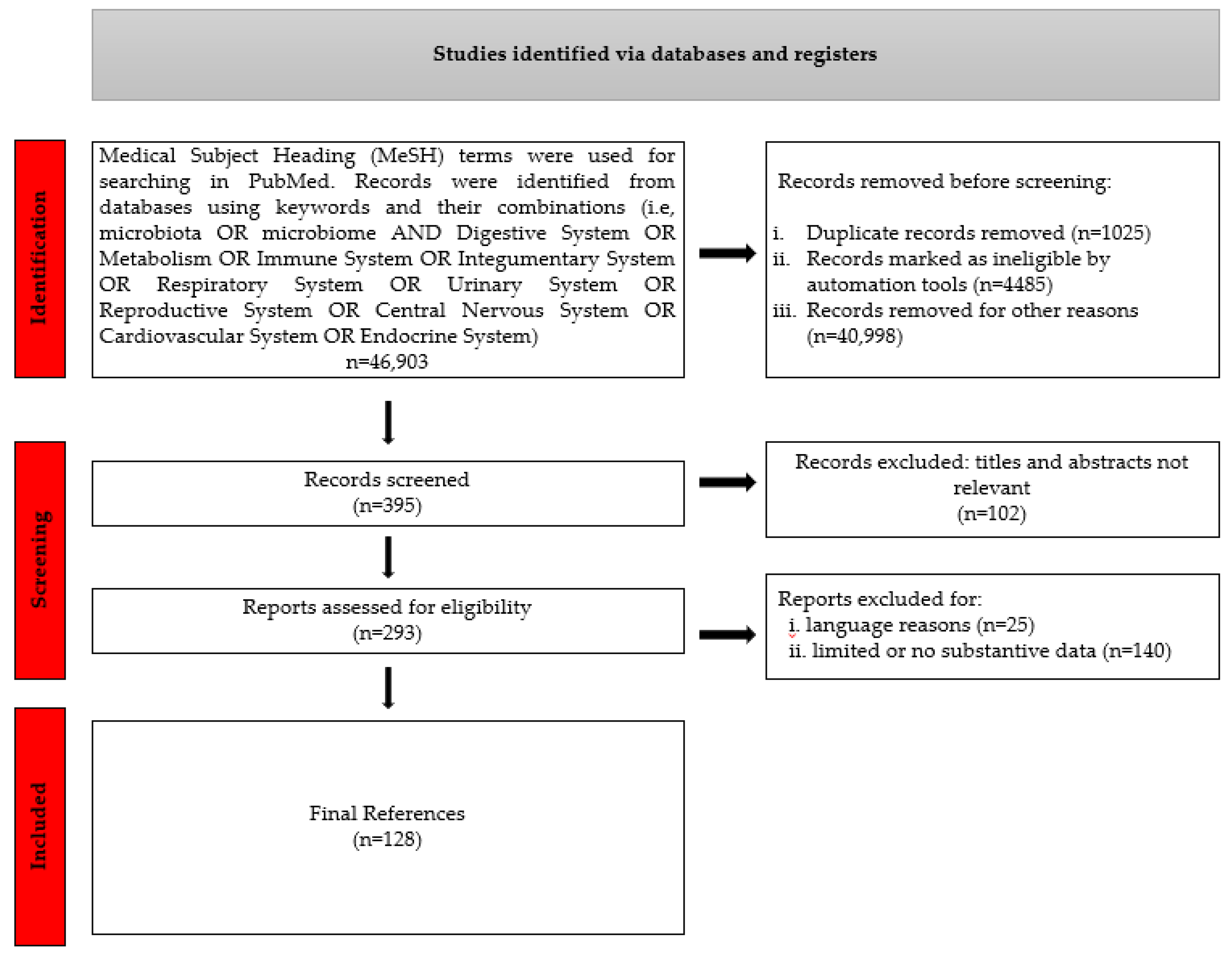
2. Materials and Methods

References were found in relevant databases such as the National Center for Biotechnology Information (PubMed), the Centers for Disease Control and Prevention, the Scientific Electronic Library Online, Scopus, Web of Science, and the Cochrane Database. For this review, utilizing searches within the publication interval of 1 January 2014-31 December 2024, along with linking the term "Microbiota" or" Microbiome" with one of the following: "digestive system or metabolism or immune system or integumentary system or respiratory system or urinary system or reproductive systems or central nervous system or cardiovascular system or endocrine system".
Peer-reviewed articles, reviews, clinical trials, observational studies, or meta-analyses, written in English, Italian or Romanian, focused on microbiota's impact on health and disease in humans were included. Records identified as duplicates, marked as ineligible by automation tools, deemed irrelevant based on titles and abstracts, not published in English, Italian, or Romanian, lacking substantive data to support conclusions, without full-text availability, or with a context outside human microbiota research were excluded, Figure 1.

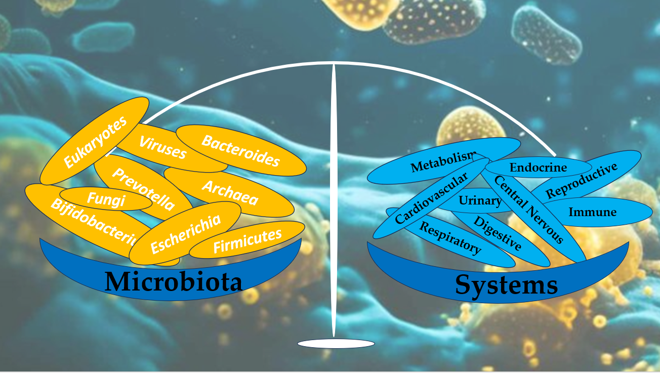
3. The Multisystem Impact of Microbiota.

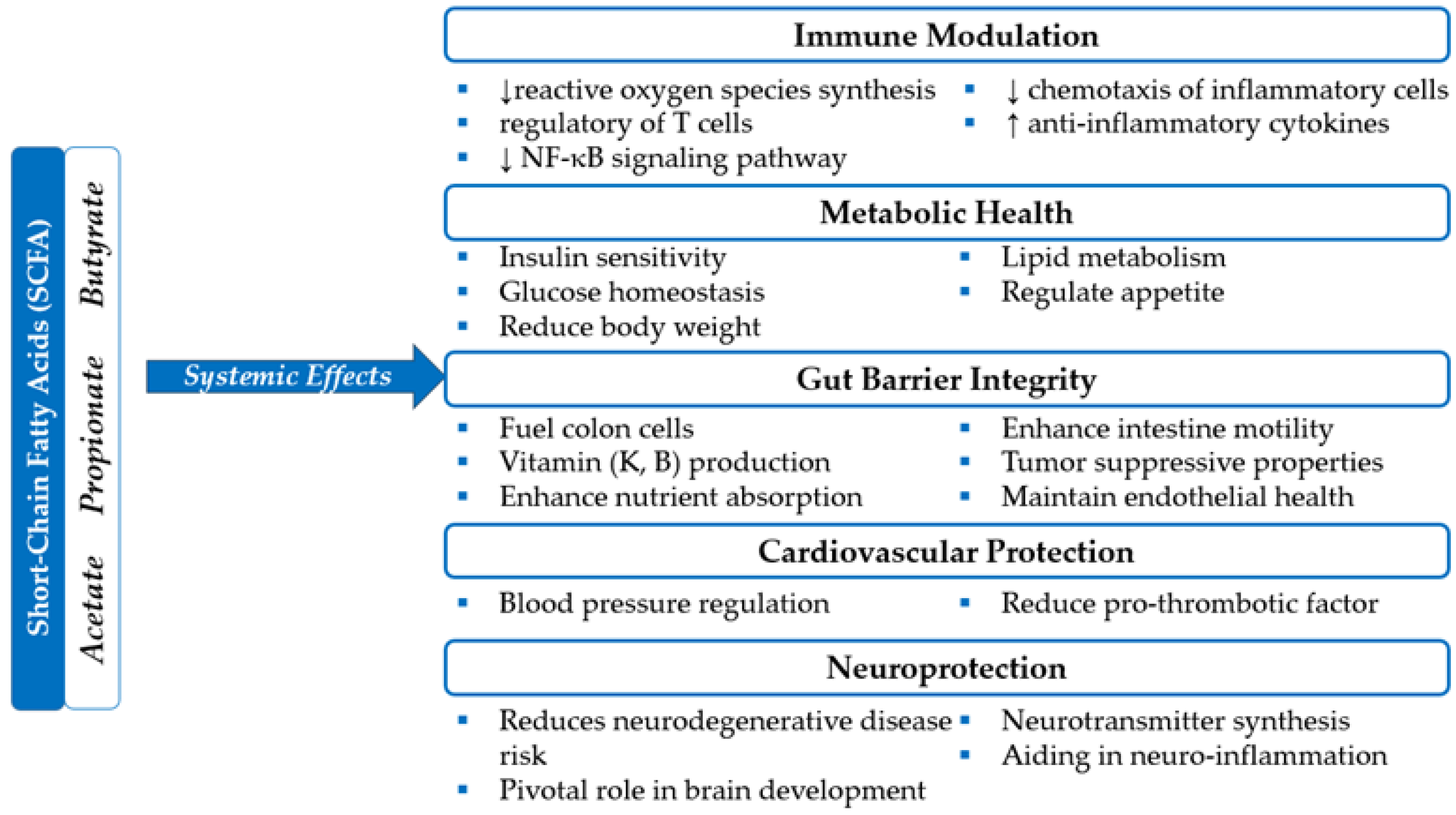
3.1. Digestive System And Metabolism

The human digestive system harbors a vast and complex community of microorganisms known as microbiota. These microbes are essential for overall health, aiding digestion, supporting immune function, and maintaining metabolic balance. Predominantly composed of bacteria, these microbial communities support digestive health by breaking down indigestible food and synthesizing essential vitamins and SCFAs. Complex polysaccharides are broken down into absorbable substrates by these microbes, which coexist in the digestive system without causing clinical ailments. The gut flora also plays a crucial role in regulating the immune system by protecting against pathogens and modulating immune responses [9]. Many modern studies highlight the role of gut microbes in behavioural modulation through the gut-brain axis. Maintaining microbial equilibrium is important to prevent inflammation caused by increased intestinal lining permeability, which can trigger heightened immune reactions.
Fermentation of complex polysaccharides, such as dietary fibres, which human enzymes cannot digest, is one of the primary functions of gut microbes [10]. SCFAs such as butyric, propionic, and acetic acids are produced during this process in the large intestine. These SCFAs are essential for energy production, serving as the primary energy source for colonocytes, regulating intestinal motility, protecting the integrity of the intestinal mucosal layer, improving nutrient absorption, regulating glucose and lipid metabolism, and modulating systemic inflammatory responses, Figure 2 [10,11,12]. Butyrate, produced by bacteria such as Faecalibacterium prausnitzii, Eubacterium rectale, and Roseburia spp., has been shown to have anti-inflammatory and antitumor properties, contributing to the maintenance of intestinal mucosal health, and reducing the risk of conditions such as leaky gut syndrome [12,13]. Additionally, studies indicate that butyrate stimulates the production of mucin and antimicrobial peptides, which protect the intestinal mucosa from infectious agents [12]. The degradation of glycoproteins in the intestinal mucus, carried out by bacteria such as Akkermansia muciniphila, Bacteroides fragilis, and Ruminococcus gnavus, provides essential nutrients for the gut microbiota and contributes to maintaining the integrity of the intestinal barrier [14,15,16].
The gut microbiota is involved in protein digestion processes (dietary, host-derived, or produced by microorganisms). Species include Clostridium spp., Bacteroides spp., Proteus spp., and Peptostreptococcus spp. break down undigested proteins into secondary metabolites, such as amino acids and small peptides, which are further transformed, through fermentation and deamination processes, into end products like SCFAs, ammonia, biogenic amines, phenols, and indoles [17]. These metabolites can either be eliminated by the host or used in specific biochemical reactions to synthesise other compounds.
Furthermore, a high-protein diet, as well as the type of protein ingested, leads to variations in the concentrations of different bacterial families and major phyla (Actinobacteria, Bacteroidetes, Firmicutes, Proteobacteria, Tenericutes, Spirochaetes), which are responsible for initiating and sustaining local or systemic pathological processes, such as inflammatory bowel diseases, colonic cancer, and metabolic disorders (e.g., type 2 diabetes) [17].
SCFA - Short-Chain Fatty Acids; NF-κB - Nuclear Factor kappa-light-chain-enhancer of activated B cells.
The microbiota plays an important role in the metabolism of xenobiotics. These processes may include direct metabolism into various inactive products (as in the case of digoxin in some individuals colonized with Eggerthella lenta) or, conversely, their activation through conversion into prodrugs (e.g., humimycin antibiotics synthesized by Rhodococcus spp. or the transformation of soy-derived isoflavones into equol, an estrogen-like substance). Additionally, the microbiota can contribute to synthesising toxic metabolites, especially following their re-entry into the intestine through the enterohepatic circulation of compounds previously detoxified by the host. Species such as Lactobacillus spp. and Bifidobacterium spp. can bind aflatoxins and heavy metals like mercury, cadmium, and lead, ensuring their elimination, while Akkermansia muciniphila, by supporting the mucus layer, can limit the absorption of certain xenobiotics, acting as a physical barrier [18].
Gut microbiota is essential for the synthesis of B-complex vitamins such as thiamine (B1), riboflavin (B2), niacin (B3), pantothenic acid (B5), pyridoxine (B6), biotin (B7), folate (B9), and cobalamin (B12), and K (K2) which collectively support critical metabolic processes including carbohydrate and fat metabolism, DNA synthesis, neurotransmitter production, red blood cell formation, blood clotting, bone health, and maintaining vascular integrity. Vitamin synthesis varies by age. In infants, species such as Escherichia coli, Bacteroides spp., Veillonella parvula, Prevotella copri, Phocaeicola dorei, Bifidobacterium breve, Bifidobacterium bifidum, and Faecalibacterium prausnitzii are involved in the production of B-complex vitamins, while Escherichia coli, Bacteroides spp., Veillonella parvula, and Phocaeicola dorei contribute to the synthesis of vitamin K [19]. In adults, the synthesis of B-complex vitamins is supported by Bacteroides spp., Eubacterium rectale, Phocaeicola dorei, Bifidobacterium adolescentis, Faecalibacterium prausnitzii, Ruminococcus gnavus, and Prevotella copri, whereas vitamin K production involves Bacteroides spp., Eubacterium rectale, and Phocaeicola dorei [19]. In elderly individuals, species such as Escherichia coli, Bacteroides spp., Klebsiella pneumoniae, Prevotella copri, Phocaeicola dorei, Ruminococcus gnavus, and Alistipes putredinis contribute to B-complex vitamin synthesis, while Escherichia coli, Bacteroides spp., Klebsiella pneumoniae, and Phocaeicola dorei are responsible for vitamin K production [19]. These microbial dynamics highlight the age-dependent role of the gut microbiota in supporting metabolic and physiological functions. Any disruption in this balance can negatively impact metabolic health, as the symbiosis between the gut microbiome and humans is vital for optimal digestive health [20]. This also underscores the significance of a healthy and diverse microbiota for the overall well-being of a human being.
The gut microbiome's impact extends beyond the digestive system, profoundly affecting the human body's metabolic regulation. Metabolism involves catabolism and anabolism, ensuring the production of necessary energy and constructing complex structures essential for the body. The gut microbiota plays a significant role in regulating and supporting these processes.
The gut microbiota plays a key role in lipid metabolism through various mechanisms. Processes such as deconjugation and dihydroxylation, carried out by species of human microbiota, transform primary bile acids (BA) into secondary BA, which are essential for emulsifying, breaking down, and absorbing lipids [21,22]. Cholesterol metabolism is regulated through the interaction between the gut microbiota and BA [23]. Bacteria such as Eubacterium coprostanoligenes, Bifidobacterium spp., and Lactobacillus spp. convert cholesterol into coprostanol, a less absorbable compound excreted in feces [22]. Trimethylamine N-oxide (TMAO) disrupts cholesterol and lipoprotein metabolism, contributing to vascular inflammation, plaque formation, and an increased risk of cardiovascular disease and diabetes [24,25,26,27,28]. TMAO is produced in the liver from trimethylamine (TMA), which is generated in the gut by bacterial species such as Firmicutes spp., Proteobacteria spp., Clostridium spp., Desulfovibrio spp., and Anaerococcus spp. through dietary choline and carnitine metabolism, primarily sourced from high-fat foods like red meat and eggs [28]. Bacteroides spp. and Bifidobacterium spp. metabolize dietary sphingolipids and simple sphingoid bases from the diet, improving intestinal barrier function, reducing exposure to bacterial toxins, and supporting immunity, with a potential role in regulating metabolic health and neurodevelopment [22]. Additionally, bacteria such as Bacteroides spp. and Lactobacillus spp. modulate the production of eicosanoids (prostaglandins and leukotrienes) by metabolizing essential fatty acids (omega-3 and omega-6), influencing inflammatory processes and overall metabolic balance [22].
The gut-brain axis, a bidirectional communication route between the digestive and central nervous systems, highlights the microbiota's role in regulating appetite, satiety, and energy production. Gut hormones, such as glucagon-like peptide-1 (GLP-1) and PYY, are released in response to microbial metabolites, including SCFAs. These hormones are critical in maintaining appetite, glucose balance and insulin sensitivity [24].
Studies have shown that metabolic disorders such as heart disease, hypertension, obesity, insulin resistance, gastrointestinal disorders, and type 2 diabetes, as well as an increased risk of cancers, are associated with dysbiosis [25,26]. Obese individuals show a specific microbial signature, characterized by reduced microbial diversity and an elevated Firmicutes-to-Bacteroidetes ratio, associated with a higher capability to harvest energy from the diet [27]. A 2022 study by Zhou et al. stated a decline in gut microbial diversity and deterioration of butyrate-producing bacteria in individuals with type 2 diabetes [25].
The gut microbiome can be balanced in several ways, with diet being the most influential factor. People who consume fibre-rich diets, specifically those based on plants, have higher concentrations of Bifidobacteria and Lactobacillus species [29]. On the contrary, diets high in fats and sugars are associated with a lower variety of microbial communities and increased concentrations of pro-inflammatory bacteria, leading to metabolic dysfunction [30].
Prebiotics and probiotics have emerged as promising advancements towards improving digestive health. Prebiotics are non-digestible food compounds such as inulin, which nourish beneficial microbes and foster their growth, and probiotics are live microorganisms found in fermented foods and supplements that help replenish the microbial balance in the gut [31,32]. The introduction of both has shown a positive effect on overall human well-being by potentially reducing metabolic diseases by decreasing inflammation, enhancing gut barrier functions, and modulating glucose uptake.
In addition to dietary interventions, fecal microbiota transplantation (FMT) has proven to be an effective method for normalizing the gut microbiota, as demonstrated in treating recurrent Clostridioides difficile infection [33].

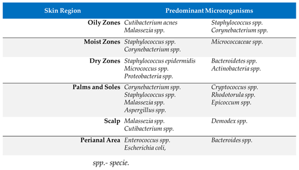
3.2. Integumentary System

Skin, a multidimensional organ, is vital in preserving physical harmony by defending against external threats, regulating fluid balance, controlling temperature, sensing stimuli, and combating pathogens. Humans represent a minority compared to many microbes inhabiting our skin. The microbial communities on the skin are remarkably diverse and complex, Table 1 [34,35]. The skin interaction begins with vaginal birth, while c-section offers a more sterile environment [36]. The relationship between the human skin and its colonizing microorganisms differs, encompassing mutualistic, communal, saprophytic, and parasitic interactions. The skin microbiome varies significantly across different body regions and changes with genome, gender, age, and environmental factors such as living conditions, antibiotic use, sun exposure, and diet.
Table 1. Skin Microbiota Diversity.
Preprints 148601 i001
The stratum corneum, consisting of corneocytes surrounded by lipid layers, creates a resilient physical barrier that prevents water loss and protects against external threats [37,38]. Lower temperature, fatty acid production and sweat gland secretion of lactic acid maintain acidity, supporting beneficial bacteria while inhibiting pathogenic strains like Staphylococcus aureus [39,40]. Luckily, most skin areas are dry, which is generally an unfavorable environment for bacterial growth. The continuous shedding of epithelial cells in the stratum corneum renews the skin surface and removes adhering microorganisms [41]. The skin's defence also includes a chemical barrier of antimicrobial peptides, proteases, cytokines, and chemokines, which can directly inhibit microbial growth or activate immune responses [42]. Age significantly affects the diversity and composition of skin microbiota. Studies suggest that adults have higher microbial diversity than adolescents and the elderly. Skin moisture retention and sebum production are influenced not only by age but also by gender. Lipophilic bacteria like Propionibacterium increase in abundance during adolescence, peaking in the third decade of life, which correlates with sebum levels [43].
Physiological variances in hormone metabolism, perspiration rate, and skin surface pH between males and females contribute to gender variations in skin environments [44]. While previous research highlighted significant alpha diversity differences between genders at the palm only, current studies identify these distinctions across all body sites, particularly the glabella. The specific reasons for gender variations in the glabella remain theoretical, but differences in facial cosmetic usage within the genders could potentially play a role.
Urban and rural environments differ significantly, as do the lifestyles of their inhabitants. For instance, rural adults and the elderly are more likely to engage in agricultural work, while urbanites typically have indoor occupations. These lifestyle disparities can affect skin conditions and the microbial communities present there. Indoor microbiomes predominantly originate from humans, whereas outdoor microbial sources include soil, water, animals, atmosphere, and plant-associated microbes, which can influence the composition of an outdoor worker's skin microbiome [45,46].
The primary commensal microbes on the skin are Staphylococcus epidermidis (S. epidermidis), Cutibacterium acnes (C. acnes), formerly Propionibacterium acne, and Corynebacterium. Unfortunately, our knowledge about other bacterial species on the skin is limited due to their low quantity and typically harmless nature [47]. The successful colonization of human skin by S. epidermidis is due to its commensal lifestyle, which prioritizes traits favoring persistence without harming the host. With low cytotoxicity and the ability to evade host defences, S. epidermidis minimizes immune responses, resembling a mutual non-aggression pact. Certain characteristics suggest a symbiotic relationship, aiding the defense against transient pathogens. However, in compromised host immunity scenarios such as injury or surgery, S. epidermidis may exhibit opportunistic pathogenic behavior [48]. A reduced presence of commensal strains is often linked with an increased risk of pathogenic colonization [49]. Studies show that coagulase-negative S. epidermidis produces its AMPs that protect against colonization by the pathogenic Staphylococcus aureus [50]. C. acnes exhibits both non-pathogenic and pathogenic characteristics. It shares some properties with the ‘Generally Recognized as Safe’ (GRAS) status microbiome [51]. The relationship between the host and C. acnes consists of both mutualistic and parasitic elements, depending on the host’s tolerance, genetics, hormones, age and the anatomical site of infection [49]. The sebaceous glands of the scalp, face, chest, and back produce large amounts of sebum, where the lipophilic anaerobe C. acnes predominates [52].
Coryneform spp. bacteria include several species, with Corynebacterium being the most common. Corynebacterium is a gram-positive pleomorphic rod divided into diphtheria and non-diphtheria species (diphtheroids). They are harmless bacteria that inhabit moist areas such as armpits and groins [53]. However, some species can cause opportunistic infections. Corynebacterium jeikeium, a diphtheroid, part of the normal flora, has become a significant source of hospital-acquired infections, particularly in immunocompromised or skin-barrier defect patients. Upon breaching the skin barrier, it may lead to severe conditions, including sepsis and endocarditis [54].
The fungal variety within the skin's microbiota is limited, with the most predominant species being Malassezia spp. Studies estimate that Malassezia spp. account for 60-90% of the total fungal population on human skin [55]. Due to their inability to synthesize lipids, they are primarily found in sebaceous skin areas such as the scalp, face, back, and chest. Other fungal residents of the skin, like Candida albicans and dermatophytes, are only distantly related to Malassezia. Despite over a century of known associations with various diseases, the role of Malassezia in health and disease remains debated. Culture-based assessments indicate that Candida spp. infrequently inhabit human skin but may cause clinical infections, particularly in individuals with compromised immune systems, diabetes, or following antibiotic therapy. Another additional fungal genus reported to be present in the skin microbiota is Debaryomyces hansenii. [56].
Viruses inhabit healthy skin and are often considered pathogenic agents. This was evidenced by the asymptomatic presence of β and γ-human papillomaviruses and viruses from the Polyomavirus genus [49,57]. Merkel cell polyomavirus (MCPyV), originally isolated from an aggressive neuroendocrine skin tumor known as Merkel cell carcinoma (MCC), was discovered on the skin surface of most healthy individuals, as well as on normal or pathological skin in patients with benign or non-MCC malignant skin conditions [57]. Viruses can positively impact homeostasis by regulating microbial populations and controlling overgrowth [58].
Skin is the body's largest organ, extending from head to toe. The roles of the cutaneous microbiota are multiple. The commensal microorganisms at this level form a natural protective barrier against skin colonisation by pathogenic microorganisms, competing for space and nutrients and synthesizing antibacterial molecules (natural antibiotics, free fatty acids, AMPc) that inhibit pathogens [35]. The microbiota stimulates the immune system by maintaining the local pro- and anti-inflammatory balance and activating local immune cells. These microorganisms regulate the skin's pH, maintain the integrity of the stratum corneum, control local inflammatory processes, and preserve the skin's homeostasis [35]. Some microorganisms can stimulate cellular regeneration processes, promoting the repair of skin lesions. A balanced cutaneous microbiota reduces the risk of skin conditions such as acne, atopic dermatitis, psoriasis, chronic wounds, and bacterial or fungal local infections [35]. Lastly, the microbiota helps the skin adapt to various environmental conditions, such as temperature or humidity variations and exposure to chemical agents.
The skin covering the hands is exposed the most to microbes. Proper hygiene is crucial for preventing many microbial-based diseases. Good hand hygiene measures have been intensified since COVID-19. The skin also needs protection from excessive sunlight, avoiding exposure to the strongest sun’s rays and using sun protection products as often as possible. It has been shown that ultraviolet radiation can influence both the phylogenetic and genotypic composition, as well as the activity of the microbial communities [60]. Restoring and maintaining a healthy skin microbiome is vital for overall skin health and the effective management of skin diseases. The normal skin microbiota evolves along with the human host. Understanding the mutualistic roles of these organisms could enhance our knowledge of the delicate balance of our skin microbiota and could be utilized to develop therapeutic strategies to treat skin diseases or to fight more harmful skin-associated microbes, such as Methicillin-resistant Staphylococcus aureus (MRSA) [61].

3.3 Respiratory System

The healthy lung was traditionally considered a sterile organ, particularly the distant alveoli [62]. We now know that it supports a rich and varied microbiota. A large part of these microbial communities, which are made up of bacteria, viruses, and fungi, is responsible for maintaining normal physiological activities of the respiratory system [63]. Previously, numerous studies have been done on the impact of gut microbiota on human health and disease; however, in recent years, the impact of microbiome on lung health and disease has been increasingly discussed [64,65]. Only recently have the functional mechanisms and consequences of the microbiome on lung health been explained. Respiratory tract microbiota has multiple roles in maintaining respiratory health, the most important being to serve as a gatekeeper to prevent respiratory infections from colonizing the system [66,67,68].
The lungs are continually exposed to germs suspended in the air and upper airways [69]. Since over 80% of the bacteria that inhabit the human body are undetectable by conventional culture-based methods, it is challenging to demonstrate the presence of the microbiome in the lungs. However, recent advances in sequencing technologies like amplicon have increased our comprehension of the lung microbiome [62,70,71].
A wide variety of microorganisms colonize the lung's mucosal tissue. The lungs and airways are colonized by microorganisms from the oral cavity and environment [72]. The most common class of microorganisms in the lungs of healthy people are Prevotella, Streptococcus, Veillonella, Neisseria, Haemophilus, and Fusobacterium [73,74,75]. This diverse microbiota forms a complex and heterogeneous ecosystem that supports several key physiological processes [5,62,76]. Most flora is aerobic or facultatively anaerobic, except for Clostridium, Veillonella, and Porphyromonas, specialized anaerobes [63].
The lung microbiota plays essential roles in maintaining immune homeostasis, modulating inflammatory responses, and protecting against respiratory pathogens. The host's immune system is modulated predominantly by the lung bacteria [77]. The lining of the lungs is covered in a thin layer of mucus with bacteriostatic properties, which offers microorganisms a low-nutrient habitat. The lung microenvironment is characterized by high immunological "tolerance," which is primarily maintained by subpopulations of alveolar macrophages (AMs) and dendritic cells (DCs) [66]. These cells demonstrate their immunoregulatory capabilities by triggering the production of regulatory T cells (Treg) and releasing prostaglandin E2 (PGE2), tumour growth factor-beta (TGF-β), and interleukin-10 (IL-10). There is growing evidence that the lung microbiota plays a crucial role in fostering lung immunological tolerance by influencing resident immune cells [76,77]. These microorganisms help the lung react to dangerous infections correctly, preventing excess inflammatory reactions to benign antigens [68,72,78]. Microbial compounds and immune cell pattern recognition receptors (PRRs) interact to accomplish this immunological control [66]. These interactions prevent excessive immune responses and regulate inflammatory pathways. Additionally, immunoglobulin A (IgA), which covers mucosal surfaces and maintains immunological homeostasis by neutralizing pathogens before they can infiltrate deeper into lung tissues, is partly produced by microbiota [68,79].
The lung microbiota supports the respiratory system's physical defences, such as the mucosal barrier and mucociliary clearance. Inhaled particles and microorganisms are trapped by a mucus layer that protects the epithelial cells lining the airways. The synthesis and composition of this mucus are influenced by microbial communities, which maximize its ability to capture dangerous particles while permitting effective gas exchange. Furthermore, the microbiota facilitates the movement of cilia, which are tiny hair-like projections on the surface of airway epithelial cells that aid in removing mucus and debris from the lungs [63,67,68,80]. The host's mucosal barriers and microbial communities must balance to avoid infections and preserve lung health. Because the microbiota impacts mucociliary function, pathogens are effectively eliminated, lowering the risk of respiratory infections and enhancing the respiratory system's overall resistance [67,68].
Despite the structural differences between the gut and lungs, possible anatomic communications and intricate networks involving their respective bacteria have supported the possibility of a gut-lung axis. According to the gut-lung axis theory, changes in the gut microbiome's components brought on by illness, food, or medications are associated with altered immune responses and airway homeostasis [64,77,81,82,83]. Even though our understanding of the gut-lung axis is still developing, new research suggests lung disorders may be treated by modifying the gut microbiota [77]. The gut microbiota produces microbial metabolites, like SCFAs, which can enter the circulation or lymphatic system and reach the lungs, influencing immunological responses and supporting lung homeostasis [84]. Numerous studies show that the gut and lung microbiota are important for maintaining pulmonary health, and as such, they could be targets for nutrition-based preventive strategies against lung disease in older populations. This is because communication along the gut-lung axis becomes more pronounced with age or in elderly patients [82].

3.4 Urinary and Reproductive Systems

The urinary microbiota is the microbial community in the urinary system within the bladder or in bladder-obtained urine [85,86]. Previously, urine was said to be sterile, but bacterial communities have been discovered in the bladder, which has a potential role in urinary health [85,87,88]. Recent studies documented a wide spectrum of microbiota associated with healthy, culture-negative urine [85,89]. Many of these urinary microbial communities are like those seen in the human vagina, colon, and skin [90].
The five predominant bacteria in the urinary system of healthy males were Streptococcus, Lactobacillus, Prevotella, Corynebacterium, and Pseudomonas, and these were also seen in the urethral swab, although at varying degrees [89,90,91]. The microbiota at the penile sulcus is predominantly Pseudomonadaceae spp., which is rare in urine, whereas Sneathia spp. and Lactobacillus spp., which are common in urine, are rare in the sulcus of uncircumcised men [90]. It was observed that some of the penile microbiota, such as Pseudomonadaceae and Oxalobactericeae, were the same irrespective of circumcision status, but before circumcision, the putative anaerobic bacterial microbiota belonging to the families of Clostridiales and Prevotellaceae were more prevalent [91].
The five most predominant urinary microbiota in healthy females were Lactobacillus, Corynebacterium, Gardnerella, Prevotella, and Bacillus [87]. For the female reproductive system, many of the same urinary microbiota were cultured from the vaginal specimen, such as the Lactobacillus, Gardnerella, Bifidobacterium, Enterococcus, Actinomyces, Prevotella, and Atopobium [87]. This shows that both urinary and vaginal tracts may have a common urogenital microbial community. Although another study reported that the urethra and bladder may have distinct microbial communities from the vagina [91]. A healthy vaginal microbial community, however, consists of Lactobacillus, and it seems to play a key role in preventing several urogenital diseases by lowering the environmental pH through lactic acid production [87,89,90,91,92,93,94]. It was also observed that most microbial communities are commonly shared by men and women, except Lactobacillus crispatus, found only in healthy women [92]. In summary, the most common microbiota detected in males' urinary and reproductive systems was Streptococcus, while in females, it was Lactobacillus [87,88,89,90,92,93,94].
There are several ways in which these microbial communities impact the urinary and reproductive systems. The abundance of vaginal lactobacilli prevents urinary tract infections (UTI) as it has been shown that depletion of Lactobacillus in the microbiota is associated with UTI risk, and replacement with Lactobacillus crispatus intravaginal suppository probiotics reduced recurrent UTI in premenopausal women [94,95].
The female microbial community protects against bacterial vaginosis, which has been shown to result from replacing the microbiota rich in Lactobacillus crispatus with anaerobic species; thus, the urinary microbiota in healthy females differ significantly from those with bacterial vaginosis [92,96].
The urinary microbiota plays a role in preventing urge urinary incontinence (UUI) in females [89]. Both Lactobacillus spp. were cultured in the females with and without UUI. In contrast, Lactobacillus gasseri was prevalent in the UUI cohort, Lactobacillus crispatus was the prevalent species in the non-UUI females [89]. Also, Gardnerella was more frequently cultured in the urine specimen of the females with UUI than in the non-UUI cohort [89].
The urinary microbiota also reduces the risk of UTI in those with neurogenic bladder who showed a decrease in Lactobacillus and an increase in Enterobacteriales, indicating the possibility that this dysbiosis likely predisposed them to UTI [94].
The vaginal microbiome promotes healthy pregnancy outcomes, while Escherichia coli causes adverse events in pregnancy, including preterm birth [96,97]. Secretions from the female genital tract inhibit Escherichia coli. In health, the microbiota rich in Lactobacillus crispatus is associated with healthy pregnancy outcomes as it promotes Escherichia coli inhibitory activity of vaginal secretions, preventing its colonization and subsequently reducing the risk of preterm birth [96]. The Lactobacillus microbiota has also promoted fertility as its abundance in the female urogenital tract decreased in patients with infertility [98].
The microbial community in the urinary tract is important in preventing cellular rejection, as Lactobacillus in females and Streptococcus in males were reduced in those with interstitial fibrosis and tubular atrophy 6-8 months after renal transplantation. In contrast, lactobacillus normalized in females and streptococcus increased in males, with excellent transplant function [89].
The decrease in the putative anaerobic microbial communities (Clostridiales and Prevotellaceae) was more prevalent post-circumcision. It may be a contributory factor in the protection against Human Immunodeficiency virus (HIV) and other sexually transmitted diseases [91].

3.5 Central Nervous System

The gastrointestinal microbiome has emerged as a promising avenue for managing and improving neurological well-being. These microorganisms have been shown to influence the effects of neurological disorders through the gut-brain axis. This axis enables bi-directional communication between the central nervous system and gastrointestinal microflora through neural, endocrine, immune, and metabolic circuits, Figure 3. These interactions influence neuroprotection, blood-brain barrier permeability, cognitive function, motor skills, and overall brain health. Dysbiosis has been linked to various mental and neurological disorders. The recovery and symptom management of diseases like Alzheimer's Disease (AD), Parkinson's Disease (PD), autism spectrum disorders (ASD), and multiple sclerosis (MS) can be influenced by these microbial factions.
The vagus nerve represents the primary communication pathway between the gut microbiota and the brain. Its intestinal receptors are indirectly stimulated by chemical substances produced by the microbiota, such as SCFAs, neurotransmitters, intestinal hormones [cholecystokinin, peptide YY(YY)], and mechanical stimuli (changes in pressure and intestinal wall distension) [99,100]. Many of these signals are mediated by enteroendocrine cells, which release signalling molecules that activate the afferent fibres of the vagus nerve. Microbial metabolites, such as SCFAs, are vital for optimal CNS functioning. SCFAs, such as acetate and butyrate, are pivotal in brain development, neurotransmitter synthesis, and neuroinflammation [99]. Gastrointestinal microbiota directly produces or regulates the production of neurotransmitters such as serotonin, dopamine, and gamma-aminobutyric acid (GABA). The afferent component of the vagus nerve transmits the information received from the intestine to the nucleus of the solitary tract in the brainstem, where it is processed and relayed to brain regions involved in emotion regulation (amygdala, prefrontal cortex) and autonomic functions (hypothalamus). The hypothalamic-pituitary-adrenal (HPA) axis is modulated by signals transmitted from the gut through the vagus nerve, reducing stress and systemic inflammation. Studies have shown that probiotic strains such as Lactobacillus and Bifidobacterium elevate serotonin levels, aiding in the reduction of depressive and anxiety symptoms associated with AD and PD [99,100]. The production of dopamine, which is crucial for motor regulation, is influenced by the gut microbes. Following probiotic treatment, an increase in dopamine signalling has been observed in PD patients [101].
The enteric nervous system (ENS), the "second brain," consists of a complex autonomous neuronal network that plays a vital role in regulating gastrointestinal functions. It maintains a close bidirectional communication with the central nervous system through the gut-brain axis. This communication involves the vagus nerve and neurotransmitters (serotonin, dopamine, acetylcholine), influencing intestinal activity, emotion regulation, behaviour, and autonomic responses. Moreover, the ENS plays a fundamental role in coordinating immune responses in the gut by interacting with immune cells, contributing to immunological tolerance and protection against pathogens. ENS dysfunctions can lead to the development of gastrointestinal and neuropsychiatric disorders.
HPA - hypothalamic-pituitary-adrenal; gamma-aminobutyric acid - GABA
Neurodegenerative diseases are aggravated by systemic inflammation, often caused by dysbiosis. Treatment with prebiotics and probiotics helps reduce inflammatory cytokine levels by restoring gastrointestinal microbial equilibrium. This mechanism has shown potential in conditions like MS, where lowering inflammation leads to a slowdown in disease progression [102]. The bacterial community of the stomach produces butyrate, which helps reduce inflammation by modulating immunity and supporting regulatory T cells [103]. In PD, FMT has demonstrated promise in reducing inflammatory markers [104]. In MS, inflammation markers declined in patients given SCFAs through prebiotics or supplemented with dietary fibre [101,105].
In AD, the gut microbial community can reduce amyloid-beta deposition by regulating systemic inflammation and oxidative stress. Studies have shown that prebiotic supplementation (which promotes the growth of Bifidobacteria) in individuals with AD is linked to enhanced memory and cognitive scores [105]. Dietary polyphenols broken down by gut bacteria can amplify neuroplasticity and cognitive resilience [106].
Gut dysbiosis is often evident in patients with PD. Supplementing them with Lactobacillus plantarum containing prebiotics has been linked to improved motor function. This effect is achieved by reducing alpha-synuclein buildup and boosting gut motility [101,107].

3.6 Cardiovascular System

Microbial metabolites affect the cardiovascular system, impacting vessels, vascular tone, lipids, and blood pressure. SCFAs produced from microbial fermentation of dietary fibres in the colon are one of the main interactions between the microbiota and the cardiovascular system [108,109]. SCFAs, absorbed into the bloodstream, induce vasodilatory effects and contribute to regulating blood pressure. They interact with specific receptors on vascular smooth muscle cells, such as free fatty acid receptor (FFAR) 2 and FFAR3 [109,110]. Activation of these receptors promotes blood vessel dilation, thereby improving blood flow. Additionally, butyrate and acetate can stimulate the production of nitric oxide (NO), an essential mediator of vasodilation [111]. NO regulates vascular tone by activating soluble guanylate cyclase, increasing cyclic guanosine monophosphate levels, and activating protein kinase G, which leads to the relaxation of smooth muscle cells. NO also plays a significant role in endothelial protection and preventing thrombosis by inhibiting platelet aggregation and reducing the risk of atherosclerosis [112]. Moreover, SCFAs might modify the renin-angiotensin system, the key regulator of blood pressure. SCFAs can also reduce systemic inflammation, contributing to protection against cardiovascular diseases such as hypertension and atherosclerosis [109].
A second important microbial metabolite is TMA, which gut bacteria make from dietary choline, phosphatidylcholine, and L-carnitine. The production of TMA and its conversion to TMAO in the liver is associated with cardiovascular disease, but the production of TMAO is also regulated during normal, physiological states of lipid metabolism. The modulated cholesterol and lipid absorption in the intestine by microbiota supports healthy blood lipid levels (essential to maintaining vascular integrity and function) [113,114].
Additionally, the gut-brain axis, the linkage of the gut microbiota with central nervous system signalling, also has implications for cardiovascular regulation. For instance, the vagus nerve may convey some effects of microbial metabolites on heart rate and autonomic control of the cardiovascular system. This neural pathway hints that microbiota can affect the cardiovascular response indirectly through the modulation of heart rate variability as a measure of autonomic balance and cardiovascular fitness [115].
Recent research indicates that gut microbiota can influence salt sensitivity, a crucial factor in blood pressure regulation. The effect is mediated by gut microbial metabolites such as SCFAs and arachidonic acid [116].

3.7. Endocrine Function

The microbiota also heavily impacts the endocrine system, which governs hormone production and regulation. Several of these hormones, involved in metabolic and stress responses, are synthesized or regulated by gut bacteria. SCFAs synthesized by gut bacteria activate FFAR2 and 3 receptors on intestinal L cells, stimulating the secretion of glucagon-like peptide-1 (GLP-1 and PYY), which play a crucial role in glucose metabolism and appetite regulation [117].
The synthesis of ghrelin, a peptide secreted by X/A-like neuroendocrine cells in the gastric mucosa, can be indirectly stimulated by the presence of intestinal bacteria such as Bifidobacterium, Lactobacillus or Akkermansia [118,119]. Ghrelin subsequently stimulates appetite and promotes the release of growth hormone. Akkermansia modulates the secretion of other key gut hormones, such as leptin, GLP-1, PYY, and cholecystokinin. It also increases satiety and improves metabolism [119,120].
In addition to that, the gut microbiota also interacts with the HPA axis, which comprises the core region for the body's response to stress. The cortisol release that successively characterizes the HPA axis represents the primary regulator of the adaptive response to stress and of metabolism. Some gut bacteria, such as Lactobacillus, Bifidobacterium and Akkermansia muciniphila, can activate this axis, affecting the release of the corticotropin-releasing hormone from the hypothalamus [121,122,123].
We have found that microbial communities play an essential role in the metabolism of iodine, an essential component in thyroid hormone synthesis. Small intestine gut bacteria contribute to modulating iodine absorption to maintain normal thyroid gland functioning by producing thyroxine and triiodothyronine, which are responsible for regulating metabolism. Directly supporting endocrine balance and metabolic regulation, the efficiency with which microbes contribute to nutrient absorption, including iodine, indirectly [124].
The gut microbiota is known to influence both metabolic and steroid hormones, particularly oestrogens and androgens. A key mechanism involves the production of enzymes, such as β-glucuronidase, by specific bacterial phyla, including Bacteroidetes, Firmicutes, Verrucomicrobia, and Proteobacteria [125]. These enzymes facilitate the deconjugation of oestrogens in the gut, enabling their reabsorption into the bloodstream via enterohepatic recycling. This process is crucial in maintaining balanced circulating oestrogen levels, essential for reproductive health, bone density, and cardiovascular function [126].
Bile acids’ potential to modulate the gut microbiome is essential for regulating the cardiovascular and endocrine systems. Bile acids derived from the liver are primary, while those altered by the bacteria found in the intestine are secondary forms. These secondary bile acids serve as ligands binding to the FXR and TGR5, which modulate glucose and lipid metabolism and energy expenditure. The right regulation of these pathways from the microbiota benefits the metabolic health and cardiovascular system. [118]

3.8. Immune System

The human microbiota interacts with the immune system from the first moments of life, forming a bidirectional axis essential for maintaining a balance between the host and it. The immune system influences the composition of the microbiota, while the microbiota shapes the functions of innate and adaptive immunity.
Immediately after birth, the newborn begins to be colonized by a microbial flora that varies depending on environmental factors, nutrition, and the mode of delivery (vaginal or cesarean section). The microbiota tends to mature within the first three years of life, reaching a stable composition like an adult's. During this "window of opportunity," the dynamic relationship between the developing microbiota and the immune system plays a critical role in immune development.
At birth, newborns lack a fully developed immune system, making them vulnerable to microbial agents. The microbiota contributes to the maturation of T lymphocytes [Treg, T helper (Th1, Th2, Th17)] and B lymphocytes, the synthesis of IgA (which plays a role in local defense) and serum IgG, as well as the inhibition of IgE production (involved in allergic reactions) [127,128]. Additionally, the microbiota regulates the development and activity of antigen-presenting cells (APCs), neutrophils, natural killer (NK) cells, and mast cells.
In germ-free animals, the absence of microbiota results in structural lymphoid deficiencies and immunosuppression [127]. Immune homeostasis depends on the balance between pro-inflammatory responses (Th1, Th17) and anti-inflammatory responses (Treg, IgA). A diverse and balanced microbiota maintains this balance, producing metabolites such as SCFAs, essential for Treg regulation and the control of systemic inflammation.
The microbiota also contributes to the integrity of the epithelial barrier by stimulating mucus production, antimicrobial peptides (AMPs produced by Paneth cells), and strengthening intercellular junctions. Dysbiosis compromises this barrier, leading to bacterial translocation, activation of inflammatory responses, and an increased risk of systemic inflammation.
Commensal microorganisms significantly induce local immune tolerance, reducing the risk of excessive inflammatory reactions. Polysaccharides synthesized by species such as Bacteroides fragilis modulate Th1/Th2 responses, promoting immune balance [127,128].
Dysbiosis caused by unbalanced diets, stress, or antibiotic treatments is associated with chronic inflammation and an increased risk of autoimmune diseases [128].
The human microbiota is pivotal in maintaining overall health and influences multiple physiological systems. Table 2 summarises the effects of the human microbiota on various body systems.

3.9. Microbiota - A New Frontier in Diagnosis and Treatment

The individual composition of the microbiota presents opportunities for a personalized approach to various pathologies, offering diagnostic, treatment, and prevention solutions tailored to each patient. Genomic sequencing and metabolomic analysis allow for the detailed characterization of the individual microbiome and the identification of specific microbial biomarkers that guide clinicians toward the early detection of predisposition to or presence of pathologies such as metabolic syndrome, autoimmune diseases, or neurodegenerative conditions, Table 3.
In clinical practice, the personalization of microbiota-based therapeutic strategies has proven effective in multiple fields. Targeted probiotics can stimulate beneficial bacterial species such as Lactobacillus, Bifidobacterium, and Akkermansia, reducing inflammation and improving epithelial barrier function. Prebiotics, substances that promote the growth of beneficial bacteria, are already being used to support metabolic and immune health. Fecal microbiota transplantation is an efficient treatment method, demonstrating success rates of over 90% in recurrent Clostridioides difficile infections, more than 60% in inflammatory bowel diseases, and less than 60% in extraintestinal conditions [129].
Moreover, the microbiota plays a key role in the body's response to various therapies by metabolizing medications, influencing their bioavailability and efficacy. Personalized drug therapy, tailored to the individual microbiota profile of each patient, can prevent adverse reactions and increase therapeutic response rates.
Adapting diet based on microbiota analysis can reduce the risk of developing and complicating certain metabolic diseases, such as obesity or diabetes mellitus.
Identifying the unique characteristics of the microbiota paves the way for precise, personalized therapies, to optimized health and an improved quality of life.

4. Conclusions

The microbiota represents a unique ecosystem essential for maintaining human health and is involved in multiple physiological functions, from metabolism to regulating immune responses and neuroprotection. It serves as the individual fingerprint of each human organism, enabling the development of personalized therapies, a promising solution for treating debilitating acute and chronic conditions such as Clostridioides difficile infection, autoimmune diseases, neurodegenerative disorders, and metabolic syndromes. In-depth studies are required to understand the mechanisms through which the microbiota interacts with different bodily systems.
Understanding the role of the microbiota in regulating interactions between various organs and systems could open new perspectives in disease prevention through nutritional and pharmaceutical interventions. An integrated approach to microbiota research could significantly contribute to revolutionizing personalized medicine and improving patients' quality of life.

Author Contributions

All authors have read and agreed to the published version of the manuscript.

Funding

The APC was funded by the University of Oradea, Romania.

Institutional Review Board Statement

Not applicable.

Informed Consent Statement

Not applicable.

Data Availability Statement

Not applicable.

Acknowledgements

All authors thank the University of Oradea for providing the logistic facilities they used.

Conflicts of Interest

The authors declare no conflicts of interest.

References

  1. Shanahan, F.; Ghosh, T.S.; O'Toole, P.W. The Healthy Microbiome—What Is the Definition of a Healthy Gut Microbiome? Gastroenterology 2021, 160, 483–494. [CrossRef]
  2. El-Sayed, A.; Aleya, L.; Kamel, M. Microbiota's Role in Health and Diseases. Environ. Sci. Pollut. Res. Int. 2021, 28, 36967–36983. [CrossRef]
  3. Wen, L.; Duffy, A. Factors Influencing the Gut Microbiota, Inflammation, and Type 2 Diabetes. J. Nutr. 2017, 147, 1468S–1475S. [CrossRef]
  4. Gomaa, E.Z. Human Gut Microbiota/Microbiome in Health and Diseases: A Review. Antonie Van Leeuwenhoek 2020, 113, 2019–2040. [CrossRef]
  5. Man, W.H.; de Steenhuijsen Piters, W.A.; Bogaert, D. The Microbiota of the Respiratory Tract: Gatekeeper to Respiratory Health. Nat. Rev. Microbiol. 2017, 15, 259–270. [CrossRef]
  6. Lu, L.; Chen, X.; Liu, Y.; Yu, X. Gut Microbiota and Bone Metabolism. FASEB J. 2021, 35, e21740. [CrossRef]
  7. Portincasa, P.; Bonfrate, L.; Vacca, M.; De Angelis, M.; Farella, I.; Lanza, E.; Khalil, M.; Wang, D.Q.; Sperandio, M.; Di Ciaula, A. Gut Microbiota and Short Chain Fatty Acids: Implications in Glucose Homeostasis. Int. J. Mol. Sci. 2022, 23, 1105. [CrossRef]
  8. Jandhyala, S.M.; Talukdar, R.; Subramanyam, C.; Vuyyuru, H.; Sasikala, M.; Nageshwar Reddy, D. Role of the Normal Gut Microbiota. World J. Gastroenterol. 2015, 21, 8787–8803. [CrossRef]
  9. James, K.R.; Gomes, T.; Elmentaite, R.; Kumar, N.; Gulliver, E.L.; King, H.W.; Stares, M.D.; Bareham, B.R.; Ferdinand, J.R.; Petrova, V.N.; et al. Distinct Microbial and Immune Niches of the Human Colon. Nat. Immunol. 2020, 21, 343–353. [CrossRef]
  10. Koh, A.; De Vadder, F.; Kovatcheva-Datchary, P.; Bäckhed, F. From Dietary Fiber to Host Physiology: Short-Chain Fatty Acids as Key Bacterial Metabolites. Cell 2016, 165, 1332–1345. [CrossRef]
  11. Fusco, W.; Lorenzo, M.B.; Cintoni, M.; Porcari, S.; Rinninella, E.; Kaitsas, F.; Lener, E.; Mele, M.C.; Gasbarrini, A.; Collado, M.C.; et al. Short-Chain Fatty-Acid-Producing Bacteria: Key Components of the Human Gut Microbiota. Nutrients 2023, 15. [CrossRef]
  12. Hodgkinson, K.; El Abbar, F.; Dobranowski, P.; Manoogian, J.; Butcher, J.; Figeys, D.; Mack, D.; Stintzi, A. Butyrate’s role in human health and the current progress towards its clinical application to treat gastrointestinal disease. Clinical Nutrition 2023, 42, 61-75. [CrossRef]
  13. Louis, P.; Hold, G.L.; Flint, H.J. The Gut Microbiota, Bacterial Metabolites, and Colorectal Cancer. Nat. Rev. Microbiol. 2014, 12, 661–672. [CrossRef]
  14. Patrick, S. A tale of two habitats: Bacteroides fragilis, a lethal pathogen and resident in the human gastrointestinal microbiome. Microbiology 2022, 168. [CrossRef]
  15. Coletto, E.; Latousakis, D.; Pontifex, M.G.; Crost, E.H.; Vaux, L.; Perez Santamarina, E.; Goldson, A.; Brion, A.; Hajihosseini, M.K.; Vauzour, D.; et al. The role of the mucin-glycan foraging Ruminococcus gnavus in the communication between the gut and the brain. Gut Microbes 2022, 14, 2073784. [CrossRef]
  16. Collins, S.L.; Patterson, A.D. The Gut Microbiome: An Orchestrator of Xenobiotic Metabolism. Acta Pharm. Sin. B 2020, 10, 19–32. [CrossRef]
  17. Bartlett, A.; Kleiner, M. Dietary protein and the intestinal microbiota: An understudied relationship. iScience 2022, 25, 105313. [CrossRef]
  18. Tarracchini, C.; Lugli Gabriele, A.; Mancabelli, L.; van Sinderen, D.; Turroni, F.; Ventura, M.; Milani, C. Exploring the vitamin biosynthesis landscape of the human gut microbiota. mSystems 2024, 9, e00929-00924. [CrossRef]
  19. Ríos-Covián, D.; Ruas-Madiedo, P.; Margolles, A.; Gueimonde, M.; de Los Reyes-Gavilán, C.G.; Salazar, N. Intestinal Short-Chain Fatty Acids and Their Link with Diet and Human Health. Front. Microbiol. 2016, 7, 185. [CrossRef]
  20. Ridlon, J.M.; Kang, D.J.; Hylemon, P.B. Bile salt biotransformations by human intestinal bacteria. J. Lipid Res. 2016, 47, 241–259. [CrossRef]
  21. Morales-Sánchez, A.; Fuentes-Pananá, E.M. Human Viruses and Cancer. Viruses 2014, 6, 4048–4049.
  22. Brown, E.M.; Clardy, J.; Xavier, R.J. Gut microbiome lipid metabolism and its impact on host physiology. Cell Host & Microbe 2023, 31, 173-186. [CrossRef]
  23. Li, T.; Chiang, J.Y.L. Bile acid signaling in metabolic disease and drug therapy. Pharmacol. Rev. 2014, 66, 948–983. [CrossRef]
  24. Arora, T.; Vanslette, A.M.; Hjorth, S.A.; Bäckhed, F. Microbial regulation of enteroendocrine cells. Med 2021, 2, 553-570. [CrossRef]
  25. Zhou, Z.; Sun, B.; Yu, D.; Zhu, C. Gut Microbiota: An Important Player in Type 2 Diabetes Mellitus. Front Cell Infect Microbiol 2022, 12, 834485. [CrossRef]
  26. DeGruttola, A.K.; Low, D.; Mizoguchi, A.; Mizoguchi, E. Current Understanding of Dysbiosis in Disease in Human and Animal Models. Inflamm Bowel Dis 2016, 22, 1137-1150. [CrossRef]
  27. Zhang, K.; Zhang, Q.; Qiu, H.; Ma, Y.; Hou, N.; Zhang, J.; Kan, C.; Han, F.; Sun, X.; Shi, J. The complex link between the gut microbiome and obesity-associated metabolic disorders: Mechanisms and therapeutic opportunities. Heliyon 2024, 10, e37609. [CrossRef]
  28. Ebrahimzadeh Leylabadlo, H.; Sanaie, S.; Sadeghpour Heravi, F.; Ahmadian, Z.; Ghotaslou, R. From role of gut microbiota to microbial-based therapies in type 2-diabetes. Infection, Genetics and Evolution 2020, 81, 104268. [CrossRef]
  29. Sonnenburg, J.L.; Bäckhed, F. Diet–microbiota interactions as moderators of human metabolism. Nature 2016, 535, 56–64. [CrossRef]
  30. Scheithauer, T.P.M.; Rampanelli, E.; Nieuwdorp, M.; Vallance, B.A.; Verchere, C.B.; van Raalte, D.H.; Herrema, H. Gut Microbiota as a Trigger for Metabolic Inflammation in Obesity and Type 2 Diabetes. Front Immunol 2020, 11, 571731. [CrossRef]
  31. Gebrayel, P.; Nicco, C.; Al Khodor, S.; Bilinski, J.; Caselli, E.; Comelli, E.M.; Egert, M.; Giaroni, C.; Karpinski, T.M.; Loniewski, I.; et al. Microbiota medicine: towards clinical revolution. J Transl Med 2022, 20, 111. [CrossRef]
  32. Hill, C.; Guarner, F.; Reid, G.; Gibson, G.R.; Merenstein, D.J.; Pot, B.; Calder, P.C. The International Scientific Association for Probiotics and Prebiotics consensus statement on the scope and appropriate use of the term probiotic. Nat. Rev. Gastroenterol. Hepatol. 2014, 11, 506–514. [CrossRef]
  33. Drwiega, E.N.; Nascimento, D.C. A Comparison of Currently Available and Investigational Fecal Microbiota Transplant Products for Recurrent Clostridioides difficile Infection. Antibiotics 2024, 13, 436. [CrossRef]
  34. Carmona-Cruz, S.; Orozco-Covarrubias, L.; Sáez-de-Ocariz, M. The Human Skin Microbiome in Selected Cutaneous Diseases. Front Cell Infect Microbiol 2022, 12, 834135. [CrossRef]
  35. Yang, Y.; Qu, L.; Mijakovic, I.; Wei, Y. Advances in the human skin microbiota and its roles in cutaneous diseases. Microbial Cell Factories 2022, 21, 176. [CrossRef]
  36. Oh, J.; Byrd, A.L.; Park, M.; Kong, H.H.; Segre, J.A. Temporal Stability of the Human Skin Microbiome. Cell 2016, 165, 854–866. [CrossRef]
  37. Sinha, S.; Lin, G.; Ferenczi, K. The skin microbiome and the gut-skin axis. Clin. Dermatol. 2021, 39, 829–839. [CrossRef]
  38. Kengmo Tchoupa, A.; Kretschmer, D.; Schittek, B.; Peschel, A. The epidermal lipid barrier in microbiome-skin interaction. Trends Microbiol 2023, 31, 723-734. [CrossRef]
  39. Lee, H.J.; Kim, M. Skin Barrier Function and the Microbiome. Int J Mol Sci 2022, 23. [CrossRef]
  40. Swaney, M.H.; Nelsen, A.; Sandstrom, S.; Kalan, L.R. Sweat and Sebum Preferences of the Human Skin Microbiota. Microbiol Spectr 2023, 11, e0418022. [CrossRef]
  41. Smythe, P.; Wilkinson, H.N. The Skin Microbiome: Current Landscape and Future Opportunities. Int J Mol Sci 2023, 24. [CrossRef]
  42. Lunjani, N.; Ahearn-Ford, S.; Dube, F.S.; Hlela, C.; O’Mahony, L. Mechanisms of microbe-immune system dialogue within the skin. Genes & Immunity 2021, 22, 276-288. [CrossRef]
  43. Ying, S.; Zeng, D.N.; Chi, L.; Tan, Y.; Galzote, C.; Cardona, C.; Lax, S.; Gilbert, J.; Quan, Z.X. The Influence of Age and Gender on Skin-Associated Microbial Communities in Urban and Rural Human Populations. PLoS One 2015, 10, e0141842. [CrossRef]
  44. Boxberger, M.; Cenizo, V.; Cassir, N.; La Scola, B. Challenges in exploring and manipulating the human skin microbiome. Microbiome 2021, 9, 125. [CrossRef]
  45. Leung, M.H.Y.; Tong, X.; Lee, P.K.H. Indoor Microbiome and Airborne Pathogens. Comprehensive Biotechnology 2019, 31, 96–106. [CrossRef]
  46. Hayleeyesus, S.F.; Manaye, A.M. Microbiological quality of indoor air in university libraries. Asian Pac. J. Trop. Biomed. 2014, 4 Suppl 1, S312–S317.
  47. Flowers, L.; Grice, E.A. The Skin Microbiota: Balancing Risk and Reward. Cell Host Microbe 2020, 28, 190-200. [CrossRef]
  48. Gallo, R.L. S. epidermidis influence on host immunity: more than skin deep. Cell Host Microbe 2015, 17, 143-144. [CrossRef]
  49. Christensen, G.J.; Brüggemann, H. Bacterial skin commensals and their role as host guardians. Benef. Microbes 2014, 5, 201–215. [CrossRef]
  50. Severn, M.M.; Horswill, A.R. Staphylococcus epidermidis and its dual lifestyle in skin health and infection. Nat Rev Microbiol 2023, 21, 97-111. [CrossRef]
  51. Gagliardi, A.; Totino, V.; Cacciotti, F.; Iebba, V.; Neroni, B.; Bonfiglio, G.; Trancassini, M.; Passariello, C.; Pantanella, F.; Schippa, S. Rebuilding the Gut Microbiota Ecosystem. Int J Environ Res Public Health 2018, 15. [CrossRef]
  52. Rozas, M.; Hart de Ruijter, A.; Fabrega, M.J.; Zorgani, A.; Guell, M.; Paetzold, B.; Brillet, F. From Dysbiosis to Healthy Skin: Major Contributions of Cutibacterium acnes to Skin Homeostasis. Microorganisms 2021, 9. [CrossRef]
  53. Chen, Y.E.; Fischbach, M.A.; Belkaid, Y. Skin microbiota-host interactions. Nature 2018, 553, 427-436. [CrossRef]
  54. Casciaro, B.; Loffredo, M.R.; Cappiello, F.; Verrusio, W.; Corleto, V.D.; Mangoni, M.L. Frog Skin-Derived Peptides Against Corynebacterium jeikeium: Correlation between Antibacterial and Cytotoxic Activities. Antibiotics 2020, 9, 448. https://doi.org/10.3390/antibiotics9080448 Grice, E.A.; Dawson, T.L.J. Host-microbe interactions: Malassezia and human skin. Curr. Opin. Microbiol. 2017, 40, 81–87 . [CrossRef]
  55. Kanna, B.V.; Latha, R.; Kavitha, K.; Jeyakumari, D. Phenotypic Characterization of Malassezia spp. Isolated from Healthy Individuals. Res. J. Biotech. 2023, 18, 74–78. [CrossRef]
  56. Schmid, B.; Künstner, A.; Fähnrich, A.; Busch, H.; Glatz, M.; Bosshard, P.P. Longitudinal Characterization of the Fungal Skin Microbiota in Healthy Subjects Over a Period of 1 Year. Journal of Investigative Dermatology 2022, 142, 2766-2772.e2768. [CrossRef]
  57. Silling, S.; Kreuter, A.; Gambichler, T.; Meyer, T.; Stockfleth, E.; Wieland, U. Epidemiology of Merkel Cell Polyomavirus Infection and Merkel Cell Carcinoma. Cancers 2022, 14, 6176. [CrossRef]
  58. Harper, A.; Vijayakumar, V.; Ouwehand, A.C.; ter Haar, J.; Obis, D.; Espadaler, J.; Binda, S.; Desiraju, S.; Day, R. Viral Infections, the Microbiome, and Probiotics. Front. Cell. Infect. Microbiol. 2021, 10, 596166. [CrossRef]
  59. Willmott, T.; Campbell, P.M.; Griffiths, C.E.M.; O'Connor, C.; Bell, M.; Watson, R.E.B.; McBain, A.J.; Langton, A.K. Behaviour and sun exposure in holidaymakers alters skin microbiota composition and diversity. Front. Aging 2023, 4, 1217635. [CrossRef]
  60. Grant, G.J.; Kohli, I.; Mohammad, T.F. A Narrative Review of the Impact of Ultraviolet Radiation and Sunscreen on the Skin Microbiome. Photodermatol. Photoimmunol. Photomed. 2024, 40, e12943. [CrossRef]
  61. Marrella, V.; Nicchiotti, F.; Cassani, B. Microbiota and immunity during respiratory infections: Lung and gut affair. Int. J. Mol. Sci. 2024, 25, 4051. [CrossRef]
  62. Li, R.; Li, J.; Zhou, X. Lung microbiome: New insights into the pathogenesis of respiratory diseases. Signal Transduct. Target. Ther. 2024, 9. [CrossRef]
  63. Fabbrizzi, A.; Amedei, A.; Lavorini, F.; Renda, T.; Fontana, G. The Lung Microbiome: Clinical and Therapeutic Implications. Intern. Emerg. Med. 2019, 14, 1241–1250. [CrossRef]
  64. Natalini, J.G.; Singh, S.; Segal, L.N. The dynamic lung microbiome in health and disease. Nature Reviews Microbiology 2023, 21, 222-235. [CrossRef]
  65. Sommariva, M.; Le Noci, V.; Bianchi, F.; Camelliti, S.; Balsari, A.; Tagliabue, E.; Sfondrini, L. The lung microbiota: Role in maintaining pulmonary immune homeostasis and its implications in cancer development and therapy. Cell. Mol. Life Sci. 2020, 77, 2739–2749. [CrossRef]
  66. Santacroce, L.; Charitos, I.A.; Ballini, A.; Inchingolo, F.; Luperto, P.; De Nitto, E.; Topi, S. The Human Respiratory System and its Microbiome at a Glimpse. Biology 2020, 9, 318. [CrossRef]
  67. Prinzi, A. Normal respiratory microbiota in health and disease. Available online: https://asm.org/Articles/2020/February/Normal-Respiratory-Microbiota-in-Health-and-Diseas (accessed on 24 November 2024).
  68. Wu, B.G.; Segal, L.N. Lung microbiota and its impact on the mucosal immune phenotype. Microbiol. Spectrum 2017, 5. [CrossRef]
  69. Whiteside, S.A.; McGinniss, J.E.; Collman, R.G. The Lung Microbiome: Progress and promise. J. Clin. Investig. 2021, 131. [CrossRef]
  70. Chen, J.; Li, T.; Ye, C.; Zhong, J.; Huang, J.-D.; Ke, Y.; Sun, H. The Lung Microbiome: A new frontier for lung and brain disease. Int. J. Mol. Sci. 2023, 24, 2170. [CrossRef]
  71. Mathieu, E.; Escribano-Vazquez, U.; Descamps, D.; Cherbuy, C.; Langella, P.; Riffault, S.; Remot, A.; Thomas, M. Paradigms of lung microbiota functions in health and disease, particularly in asthma. Front. Physiol. 2018, 9. [CrossRef]
  72. Hérivaux, A.; Willis, J.R.; Mercier, T.; Lagrou, K.; Gonçalves, S.M.; Gonçales, R.A.; Maertens, J.; Carvalho, A.; Gabaldón, T.; Cunha, C. Lung microbiota predict invasive pulmonary aspergillosis and its outcome in immunocompromised patients. Thorax 2021, 77, 283–291. [CrossRef]
  73. Yagi, K.; Huffnagle, G.B.; Lukacs, N.W.; Asai, N. The Lung Microbiome during Health and Disease. Int. J. Mol. Sci. 2021, 22, 10872. [CrossRef]
  74. Huffnagle, G.B.; Dickson, R.P.; Lukacs, N.W. The Respiratory Tract Microbiome and Lung Inflammation: A Two-Way Street. Mucosal Immunol. 2017, 10, 299–306. [CrossRef]
  75. Dickson, R.P.; Erb-Downward, J.R.; Martinez, F.J.; Huffnagle, G.B. The Microbiome and the Respiratory Tract. Annu. Rev. Physiol. 2016, 78, 481–504. [CrossRef]
  76. Budden, K.F.; Gellatly, S.L.; Wood, D.L.; Cooper, M.A.; Morrison, M.; Hugenholtz, P.; Hansbro, P.M. Emerging pathogenic links between microbiota and the gut–lung axis. Nat. Rev. Microbiol. 2016, 15, 55–63. [CrossRef]
  77. Campbell, C.D.; Barnett, C.; Sulaiman, I.A. A clinicians’ review of the respiratory microbiome. Breathe 2022, 18, 210161. [CrossRef]
  78. King, A. Exploring the lung microbiome’s role in disease. Nature 2024. [CrossRef]
  79. Ruane, D.; Chorny, A.; Lee, H.; Faith, J.; Pandey, G.; Shan, M.; Simchoni, N.; Rahman, A.; Garg, A.; Weinstein, E.G.; Oropallo, M.; Gaylord, M.; Ungaro, R.; Cunningham-Rundles, C.; Alexandropoulos, K.; Cerutti, A.; Mehandru, S.; Mucida, D.; Merad, M. Microbiota Regulate the Ability of Lung Dendritic Cells to Induce IgA Class-Switch Recombination and Generate Protective Gastrointestinal Immune Responses. J. Exp. Med. 2016, 213, 53–73. [CrossRef]
  80. Khatiwada, S.; Subedi, A. Lung microbiome and coronavirus disease 2019 (COVID-19): Possible link and implications. Human Microbiome Journal 2020, 17, 100073. [CrossRef]
  81. Mayo Clinic: The Gut-Lung Axis. Available online: https://www.mayoclinic.org/medical-professionals/pulmonary-medicine/news/the-gut-lung-axis-intestinal-microbiota-and-inflammatory-lung-disease/mqc-20483394 (accessed on 24 November 2024).
  82. Enaud, R.; Prevel, R.; Ciarlo, E.; Beaufils, F.; Wieërs, G.; Guery, B.; Delhaes, L. The gut-lung axis in health and respiratory diseases: A place for Inter-organ and inter-kingdom crosstalks. Frontiers in Cellular and Infection Microbiology 2020, 10. [CrossRef]
  83. Dang, A.T.; Marsland, B.J. Microbes, metabolites, and the gut–lung axis. Mucosal Immunol. 2019, 12, 843–850. [CrossRef]
  84. Rastogi, S.; Mohanty, S.; Sharma, S.; Tripathi, P. Possible Role of Gut Microbes and Host’s Immune Response in Gut–Lung Homeostasis. Front. Immunol. 2022, 13, 954339. [CrossRef]
  85. Perez-Carrasco, V.; Soriano-Lerma, A.; Soriano, M.; Gutiérrez-Fernández, J.; Garcia-Salcedo, J.A. Urinary Microbiome: Yin and Yang of the Urinary Tract. Front. Cell. Infect. Microbiol. 2021, 11, 617002. [CrossRef]
  86. Pearce, M.M.; et al. The female urinary microbiome: A comparison of women with and without urgency urinary incontinence. mBio 2014, 5(4), e01283-14. [CrossRef]
  87. Robino, L.; Sauto, R.; Morales, C.; Navarro, N.; González, M.J.; Cruz, E.; Neffa, F.; Zeballos, J.; Scavone, P. Presence of intracellular bacterial communities in uroepithelial cells, a potential reservoir in symptomatic and non-symptomatic people. BMC Infectious Diseases 2024, 24, 590. [CrossRef]
  88. Modena, B.D.; et al. Changes in Urinary Microbiome Populations Correlate in Kidney Transplants With Interstitial Fibrosis and Tubular Atrophy Documented in Early Surveillance Biopsies. Am. J. Transplant. 2017, 17, 712–723. [CrossRef]
  89. Pearce, M.M.; Hilt, E.E.; Rosenfeld, A.B.; Zilliox, M.J.; Thomas-White, K.; Fok, C.; Kliethermes, S.; Schreckenberger, P.C.; Brubaker, L.; Gai, X.; Wolfe, A.J. The Female Urinary Microbiome: A Comparison of Women with and without Urgency Urinary Incontinence. mBio 2014, 5, e01283-14.
  90. Onywera, H.; Williamson, A.-L.; Ponomarenko, J.; Meiring, T.L. The Penile Microbiota in Uncircumcised and Circumcised Men: Relationships With HIV and Human Papillomavirus Infections and Cervicovaginal Microbiota. Front. Med. 2020, 7, 383. [CrossRef]
  91. Gottschick, C.; Deng, Z.L.; Vital, M.; et al. The urinary microbiota of men and women and its changes in women during bacterial vaginosis and antibiotic treatment. Microbiome 2017, 5, 99. [CrossRef]
  92. Wright, M.L.; Fettweis, J.M.; Eaves, L.J.; Silberg, J.L.; Neale, M.C.; Serrano, M.G.; Jimenez, N.R.; Prom-Wormley, E.; Girerd, P.H.; Borzelleca, J.F.; et al. Vaginal microbiome Lactobacillus crispatus is heritable among European American women. Communications Biology 2021, 4, 872. [CrossRef]
  93. Pohl, H.G.; Groah, S.L.; Pérez-Losada, M.; Ljungberg, I.; Sprague, B.M.; Chandal, N.; Caldovic, L.; Hsieh, M. The Urine Microbiome of Healthy Men and Women Differs by Urine Collection Method. Int Neurourol J 2020, 24, 41-51. [CrossRef]
  94. Ng, Q.X.; Peters, C.; Venkatanarayanan, N.; Goh, Y.Y.; Ho, C.Y.X.; Yeo, W.-S. Use of Lactobacillus spp. to prevent recurrent urinary tract infections in females. Medical Hypotheses 2018, 114, 49-54. [CrossRef]
  95. Ghartey, J.P.; et al. Lactobacillus crispatus dominant vaginal microbiome is associated with inhibitory activity of female genital tract secretions against Escherichia coli. PLoS ONE 2014, 9, e96659. [CrossRef]
  96. Liu, X.; Si, S.; Huang, L.; Zhang, M.; Chen, W.; Wang, L.; Yu, Y. Vaginal flora during pregnancy and subsequent risk of preterm birth or prelabor rupture of membranes: a nested case–control study from China. BMC Pregnancy and Childbirth 2023, 23, 244. [CrossRef]
  97. Dong, Y.-H.; et al. Urogenital tract and rectal microbiota composition and its influence on reproductive outcomes in infertile patients. Front. Microbiol. 2023, 14, 1051437. [CrossRef]
  98. Souza, S.V.; Monteiro, P.B.; Moura, G.A.; Santos, N.O.; Fontanezi, C.T.B.; Gomes, I.A.; Teixeira, C.A. Vaginal microbioma and the presence of Lactobacillus spp. as interferences in female fertility: A review system. JBRA Assist Reprod 2023, 27, 496-506. [CrossRef]
  99. Morais, L.H.; Schreiber, H.L.; Mazmanian, S.K. The gut microbiota–brain axis in behaviour and brain disorders. Nat. Rev. Microbiol. 2021, 19, 241–255. [CrossRef]
  100. Scheperjans, F.; et al. Gut microbiota are related to Parkinson's disease and clinical phenotype. Mov. Disord. 2015, 30, 350–358. [CrossRef]
  101. Doroszkiewicz, J.; Groblewska, M.; Mroczko, B. The Role of Gut Microbiota and Gut–Brain Interplay in Selected Diseases of the Central Nervous System. Int. J. Mol. Sci. 2021, 22, 10028. [CrossRef]
  102. Cekanaviciute, E.; et al. Gut bacteria from multiple sclerosis patients modulate human T cells and exacerbate symptoms in mouse models. Proc. Natl. Acad. Sci. USA 2017, 114, 10713–10718. [CrossRef]
  103. Xue, L.J.; et al. Fecal microbiota transplantation therapy for Parkinson's disease: A preliminary study. Medicine 2020, 99, e22035. [CrossRef]
  104. Akbari, E.; Asemi, Z.; Daneshvar Kakhaki, R.; Bahmani, F.; Kouchaki, E.; Tamtaji, O.R.; Taghizadeh, M. Effect of Probiotic Supplementation on Cognitive Function and Metabolic Status in Alzheimer’s Disease. Front. Aging Neurosci. 2016, 8, 256. [CrossRef]
  105. Martín, R.; et al. Gut microbiota modulation: Another way to treat neurological diseases. Biochem. Pharmacol. 2018, 149, 181–197. [CrossRef]
  106. Pellegrini, C.; et al. Gut microbiota in Parkinson's disease: Preclinical and clinical evidence. Transl. Res. 2018, 204, 17–30. [CrossRef]
  107. Clemente-Suárez, V.J.; Redondo-Flórez, L.; Rubio-Zarapuz, A.; Martín-Rodríguez, A.; Tornero-Aguilera, J.F. Microbiota Implications in Endocrine-Related Diseases: From Development to Novel Therapeutic Approaches. Biomedicines 2024, 12, 221. [CrossRef]
  108. Lymperopoulos, A.; Suster, M.S.; Borges, J.I. Short-Chain Fatty Acid Receptors and Cardiovascular Function. Int. J. Mol. Sci. 2022, 23, 3303. [CrossRef]
  109. Yu, F.; Zong, B.; Ji, L.; Sun, P.; Jia, D.; Wang, R. Free Fatty Acids and Free Fatty Acid Receptors: Role in Regulating Arterial Function. Int. J. Mol. Sci. 2024, 25, 7853. [CrossRef]
  110. Robles-Vera, I.; Toral, M.; de la Visitación, N.; Aguilera-Sánchez, N.; Redondo, J.M.; Duarte, J. Protective Effects of Short-Chain Fatty Acids on Endothelial Dysfunction Induced by Angiotensin II. Front Physiol 2020, 11, 277. [CrossRef]
  111. Roy, R.; Wilcox, J.; Webb, A.J.; O’Gallagher, K. Dysfunctional and Dysregulated Nitric Oxide Synthases in Cardiovascular Disease: Mechanisms and Therapeutic Potential. International Journal of Molecular Sciences 2023, 24, 15200.
  112. Shanmugham, M.; Bellanger, S.; Leo, C.H. Gut-Derived Metabolite, Trimethylamine-N-oxide (TMAO) in Cardio-Metabolic Diseases: Detection, Mechanism, and Potential Therapeutics. Pharmaceuticals 2023, 16, 504. [CrossRef]
  113. Nesci, A.; Carnuccio, C.; Ruggieri, V.; D’Alessandro, A.; Di Giorgio, A.; Santoro, L.; Gasbarrini, A.; Santoliquido, A.; Ponziani, F.R. Gut Microbiota and Cardiovascular Disease: Evidence on the Metabolic and Inflammatory Background of a Complex Relationship. Int. J. Mol. Sci. 2023, 24, 9087. [CrossRef]
  114. Canyelles, M.; Borràs, C.; Rotllan, N.; Tondo, M.; Escolà-Gil, J.C.; Blanco-Vaca, F. Gut Microbiota-Derived TMAO: A Causal Factor Promoting Atherosclerotic Cardiovascular Disease? Int. J. Mol. Sci. 2023, 24, 1940. [CrossRef]
  115. Wang, L.; Hu, J. Unraveling the gut microbiota's role in salt-sensitive hypertension: current evidences and future directions. Front Cardiovasc Med 2024, 11, 1410623. [CrossRef]
  116. Christiansen, C.B.; Gabe, M.B.N.; Svendsen, B.; Dragsted, L.O.; Rosenkilde, M.M.; Holst, J.J. The impact of short-chain fatty acids on GLP-1 and PYY secretion from the isolated perfused rat colon. Am J Physiol Gastrointest Liver Physiol 2018, 315, G53-g65. [CrossRef]
  117. Zhang, Y.; Meng, X.; Ma, Z.; Sun, Z.; Wang, Z. Effects of Probiotic Supplementation on Nutrient Intake, Ghrelin, and Adiponectin Concentrations in Diabetic Hemodialysis Patients. Altern Ther Health Med 2023, 29, 36-42.
  118. Hamamah, S.; Hajnal, A.; Covasa, M. Influence of Bariatric Surgery on Gut Microbiota Composition and Its Implication on Brain and Peripheral Targets. Nutrients 2024, 16. [CrossRef]
  119. Canyelles, M.; Borràs, C.; Rotllan, N.; Tondo, M.; Escolà-Gil, J.C.; Blanco-Vaca, F. Gut Microbiota-Derived TMAO: A Causal Factor Promoting Atherosclerotic Cardiovascular Disease? Int. J. Mol. Sci. 2023, 24, 1940. [CrossRef]
  120. Rusch, J.A.; Layden, B.T.; Dugas, L.R. Signalling cognition: The gut microbiota and hypothalamic-pituitary-adrenal axis. Front. Endocrinol. 2023, 14, 1130689. [CrossRef]
  121. Vagnerová, K.; Vodička, M.; Hermanová, P.; Ergang, P.; Šrůtková, D.; Klusoňová, P.; Balounová, K.; Hudcovic, T.; Pácha, J. Interactions between gut microbiota and acute restraint stress in peripheral structures of the hypothalamic–pituitary–adrenal axis and the intestine of male mice. Front. Immunol. 2019, 10, 2655. [CrossRef]
  122. Xu, L.; Chen, D.; Zhao, C.; Jiang, L.; Mao, S.; Song, C.; Gao, F. Decreased abundance of Akkermansia after adrenocorticotropic hormone therapy in patients with West syndrome. BMC Microbiology 2021, 21, 126. [CrossRef]
  123. Shulhai, A.-M.; Rotondo, R.; Petraroli, M.; Patianna, V.; Predieri, B.; Iughetti, L.; Esposito, S.; Street, M.E. The Role of Nutrition on Thyroid Function. Nutrients 2024, 16, 2496. [CrossRef]
  124. Hu, S.; Ding, Q.; Zhang, W.; Kang, M.; Ma, J.; Zhao, L. Gut microbial beta-glucuronidase: a vital regulator in female estrogen metabolism. Gut Microbes 2023, 15(1), 2156157. [CrossRef]
  125. Sui, Y.; Wu, J.; Chen, J. The role of gut microbial β-glucuronidase in estrogen reactivation and breast cancer. Front. Cell Dev. Biol. 2021, 9, 631552. [CrossRef]
  126. Choden, T.; Cohen, N.A. The gut microbiome and the immune system. Exploration of Medicine 2022, 3, 219-233. [CrossRef]
  127. Zheng, D.; Liwinski, T.; Elinav, E. Interaction between microbiota and immunity in health and disease. Cell Research 2020, 30, 492-506. [CrossRef]
  128. Tian, H.; Wang, X.; Fang, Z.; Li, L.; Wu, C.; Bi, D.; Li, N.; Chen, Q.; Qin, H. Fecal microbiota transplantation in clinical practice: Present controversies and future prospects. hLife 2024, 2, 269-283. [CrossRef]
Figure 1. The PRISMA Flowchart.
Preprints 148601 g001
Figure 2. The Role of SCFA in Systemic Health.
Preprints 148601 g002
Figure 3. The Gut-Brain Axis 
Preprints 148601 g003
Table 2. The multisystem impact of microbiota.
System Microbiota-system relationship


1. Digestive System
  • Provides a protective barrier against pathogens.
  • Supports digestion (polysaccharides and proteins).
  • Enhances gut barrier integrity and reduces inflammation.
  • Produces vitamins B-complex and K.
  • Metabolizes dietary fibers
  • Regulates glucose and lipid metabolism.
  • Detoxifies xenobiotics and binds heavy metals.
  • Influences systemic immune responses through modulation of gut-associated lymphoid tissue.


2. Integumentary System
  • Provides a protective barrier against pathogens.
  • Regulates skin pH and supports lipid production.
  • Stimulates skin regeneration and wound healing.
  • Adapts to environmental factors.
  • Modulates immune responses to maintain local homeostasis.


3. Respiratory System
  • Prevents colonization by respiratory pathogens.
  • Modulates immune responses via alveolar macrophages and regulatory T cells.
  • Supports mucosal barrier integrity and facilitates mucociliary clearance.
  • Influences respiratory health through the gut-lung axis, producing SCFAs that modulate lung immunity.
  • Contributes to IgA production, enhancing mucosal defences.


4. Urinary System
  • Maintains urinary tract health by balancing microbial populations and preventing infections.
  • Modulates local pH.
  • Shares microbial communities with the reproductive system, supporting urogenital health.


5. Reproductive System
  • Regulates vaginal pH.
  • Supports healthy pregnancy by modulating local immune responses.
  • Protects against sexually transmitted infections through microbial competition and lactic acid production.
  • Enhances fertility.
  • Interacts with urinary microbiota to maintain overall urogenital health.
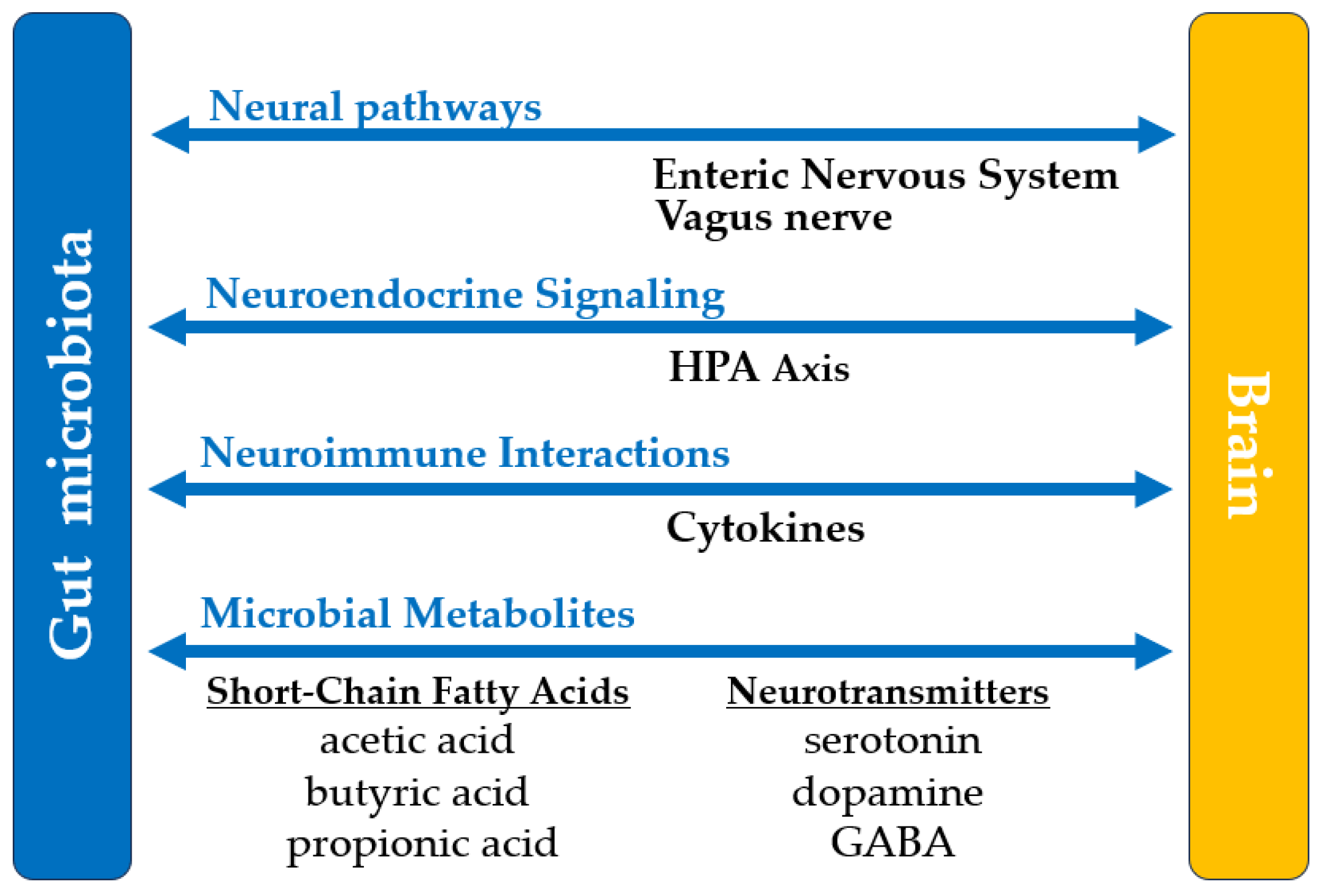
6. Central Nervous System
  • Facilitates gut-brain communication via the vagus nerve.
  • Produces neurotransmitters, influencing mood and cognition.
  • Modulates stress responses.
  • Reduces neuroinflammation.
  • Promotes neuroprotection.
  • Influences the progression and management of neurodegenerative diseases.
7. Cardiovascular System
  • Regulates blood pressure.
  • Modulates lipid metabolism.
  • Reduces systemic inflammation.
  • Protects vascular integrity.
  • Prevents against thrombosis.
  • Produces TMA, linked to cardiovascular health.
  • Supports endothelial function and vascular tone.
8. Endocrine System
  • Regulating appetite and glucose metabolism.
  • Modulates bile acid metabolism, influencing energy expenditure and lipid homeostasis.
  • Affects cortisol production and stress adaptation.
  • Supports thyroid hormone synthesis.
  • Regulates estrogen levels.
9. Immune system
  • Influences the maturation and differentiation of T and B cells.
  • Regulates the production of immunoglobulins.
  • Balancs pro-inflammatory and anti-inflammatory responses.
  • Modulats Treg activity and systemic inflammation.
  • Promoting local immune tolerance.
  • Reinforces the intestinal barrier.
  • Prevents chronic inflammation and autoimmune diseases.
SCFAs - Short-Chain Fatty Acids; IgA - Immunoglobulin A; TMA – Trimethylamine; Treg - Regulatory T cells.
Table 3. Microbial Biomarkers and Relevant Bacteria Across Systems for Diagnostic Applications
System Relevant Microorganisms Products/Biomarkers
1. Digestive System
  • Faecalibacterium prausnitzii
  • Akkermansia muciniphila
  • Roseburia spp.
  • Bacteroides spp.
  • Lactobacillus spp.
  • Eggerthella lenta
  • Firmicutes spp.
  • Firmicutes-to-Bacteroidetes ratio
  • SCFAs
  • mucin
  • lipopolysaccharides
  • GLP-1
  • PYY
2. Integumentary System
  • Cutibacterium acnes
  • Staphylococcus epidermidis
  • Staphylococcus aureus
  • Corynebacterium jeikeium
  • Malassezia spp.
  • Candida spp.
  • Merkel cell polyomavirus
  • free fatty acids
  • AMPs
  • lactic acid
3. Respiratory System
  • Prevotella spp.
  • Streptococcus spp.
  • Veillonella spp.
  • Haemophilus influenzae
  • Moraxella catarrhalis
  • IL-10
  • IL-6
  • IL-8
4. Urinary and Reproductive System
  • Lactobacillus crispatus
  • Lactobacillus gasseri
  • Gardnerella vaginalis
  • Corynebacterium spp.
  • Escherichia coli
5. Central Nervous System
  • Lactobacillus plantarum
  • Bifidobacterium spp.
  • SCFAs
  • PYY
  • serotonin
  • dopamine
  • GABA
6. Cardiovascular System
  • Eubacterium coprostanoligenes
  • Lactobacillus spp.
  • Bifidobacterium spp.
  • Ruminococcus spp.
  • SCFAs
  • TMA
  • arachidonic acid
7. Endocrine System
  • Akkermansia muciniphila
  • Bacteroides spp.
  • Bifidobacterium spp.
  • SCFAs
  • GLP-1
  • PYY
  • β-glucuronidase
  • leptin
  • cholecystokinin
8.Immune System
  • Bacteroides fragilis
  • Lactobacillus spp.
  • Bifidobacterium spp.
  • Faecalibacterium prausnitzii
  • SCFAs
  • IL-10
  • AMPs
SCFAs - Short-Chain Fatty Acids; Ig - Immunoglobulin; TMA – Trimethylamine; GLP-1 – Glucagon-Like Peptide-1; PYY – Peptide YY; AMPs – Antimicrobial Peptides; GABA – Gamma-Aminobutyric Acid
Disclaimer/Publisher’s Note: The statements, opinions and data contained in all publications are solely those of the individual author(s) and contributor(s) and not of MDPI and/or the editor(s). MDPI and/or the editor(s) disclaim responsibility for any injury to people or property resulting from any ideas, methods, instructions or products referred to in the content.
Copyright: This open access article is published under a Creative Commons CC BY 4.0 license, which permit the free download, distribution, and reuse, provided that the author and preprint are cited in any reuse.
Prerpints.org logo

Preprints.org is a free preprint server supported by MDPI in Basel, Switzerland.

Subscribe

Disclaimer

Terms of Use

Privacy Policy

Privacy Settings

© 2025 MDPI (Basel, Switzerland) unless otherwise stated