Preprint
Article

This version is not peer-reviewed.

First Detection of Bluetongue Virus Type 3 in Poland, 2024—A Case Study in European Bison (Bison bonasus)

A peer-reviewed article of this preprint also exists.

Submitted:

06 March 2025

Posted:

06 March 2025

You are already at the latest version

Abstract

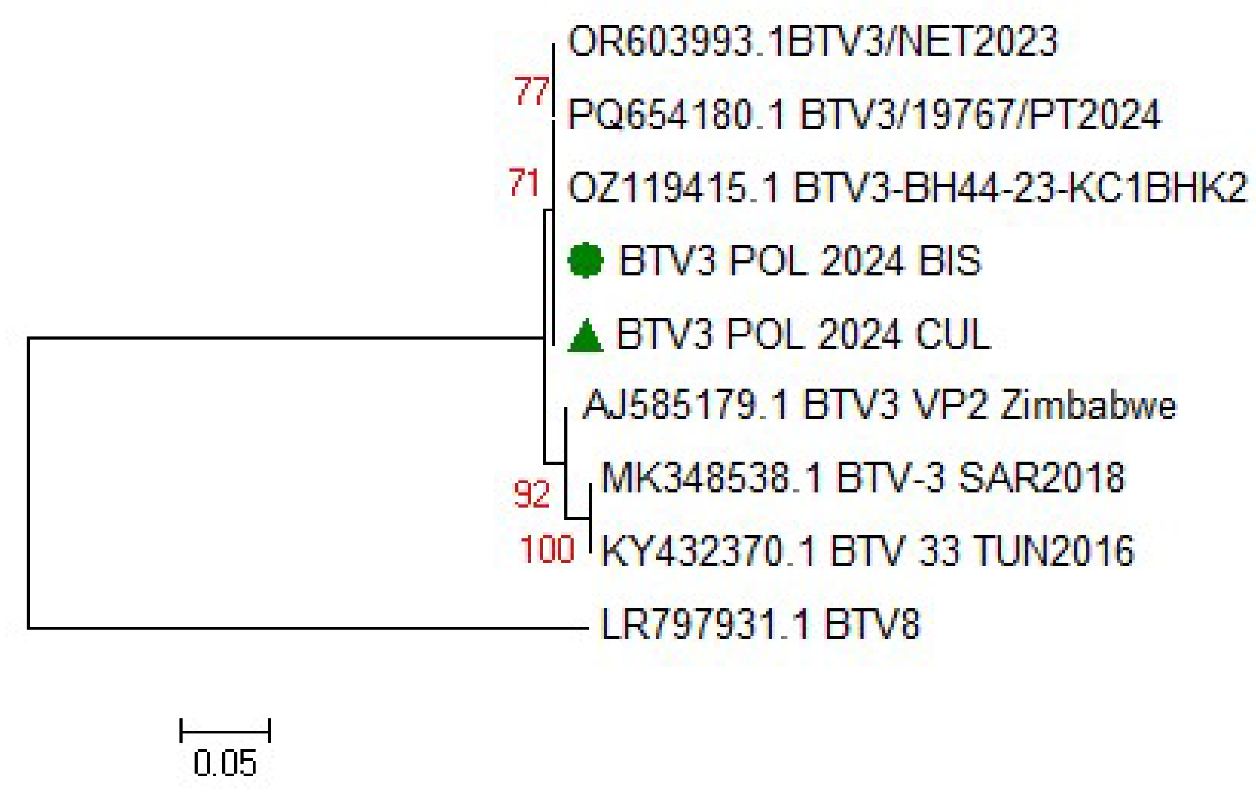
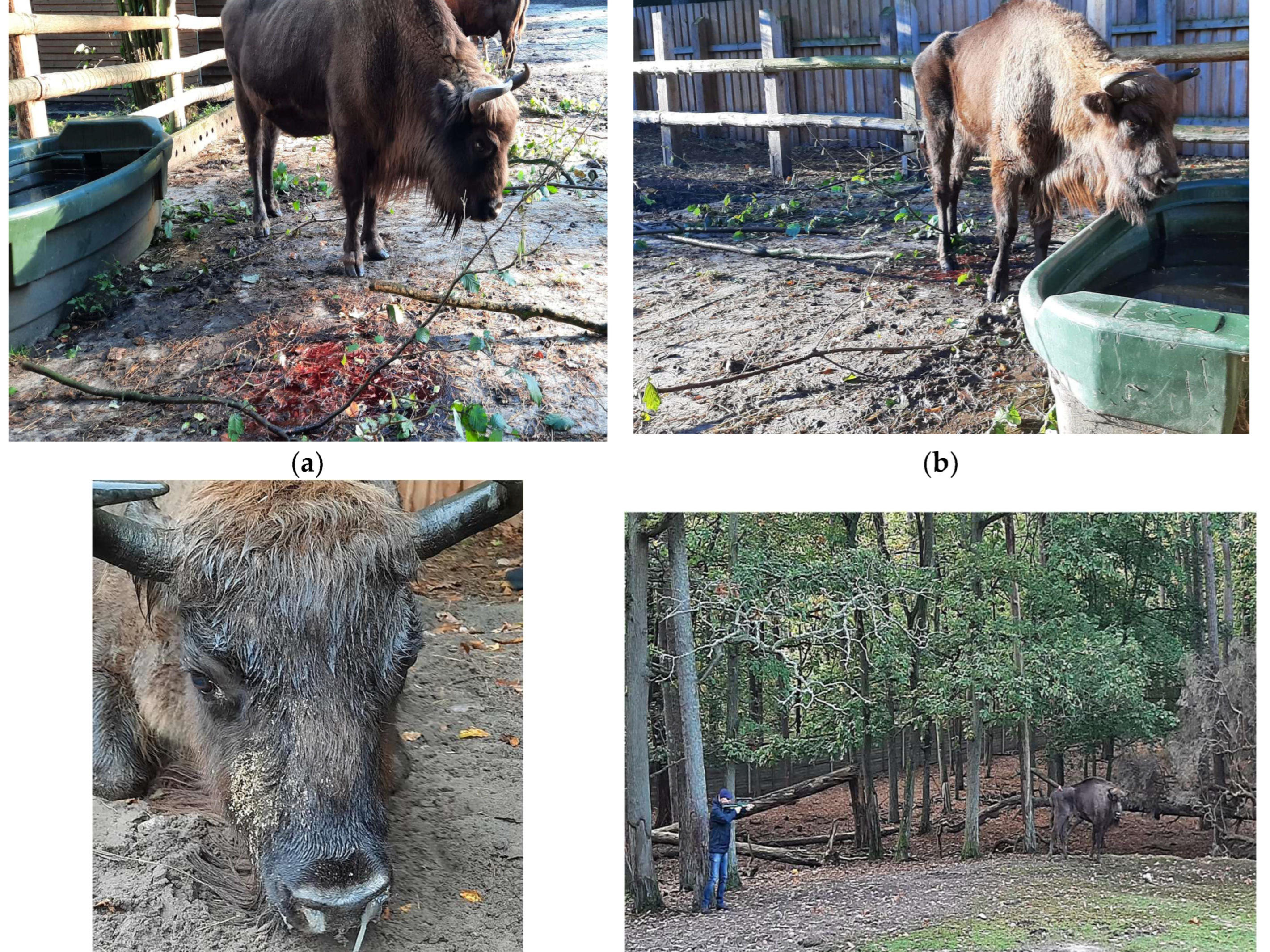
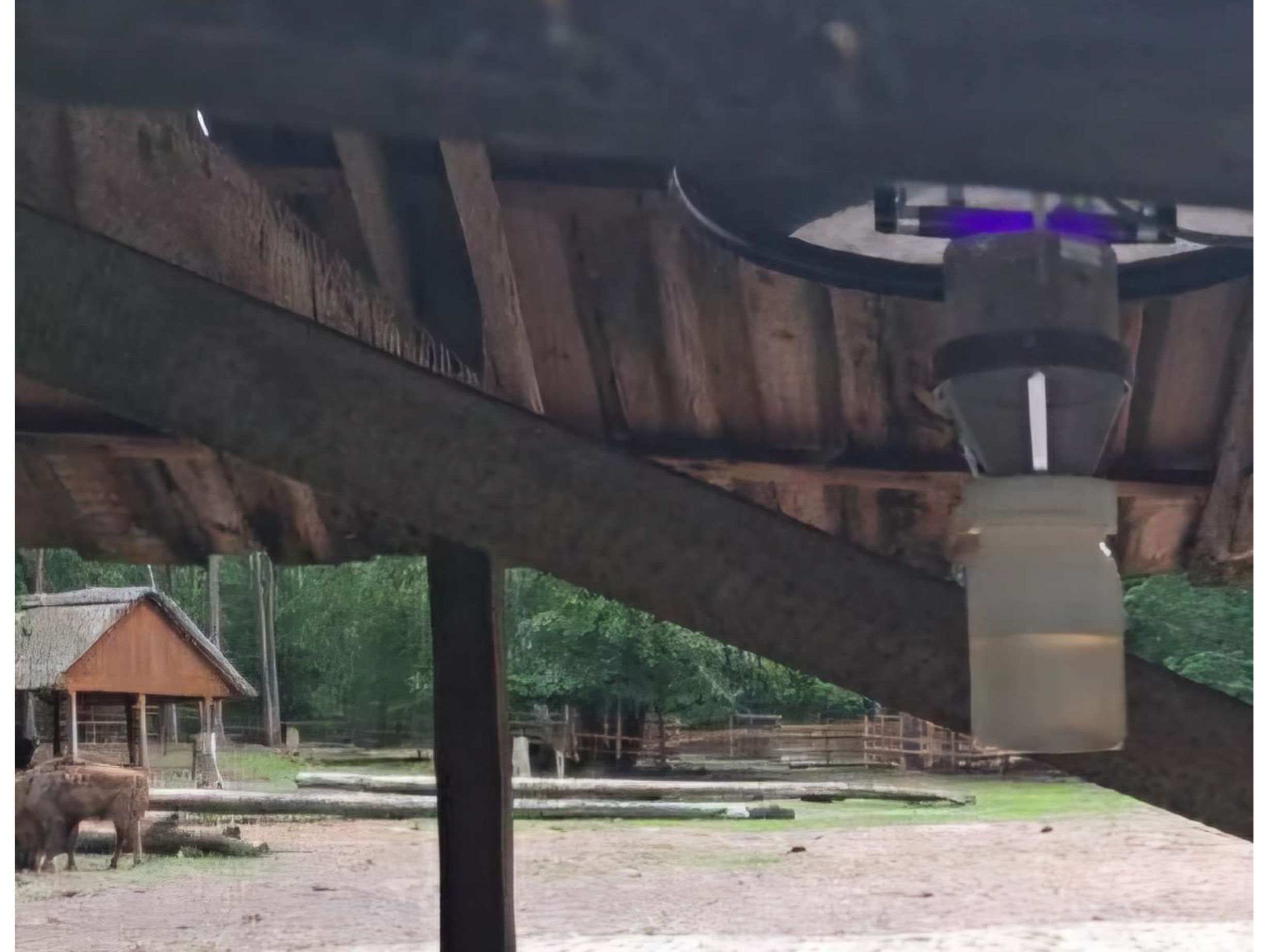
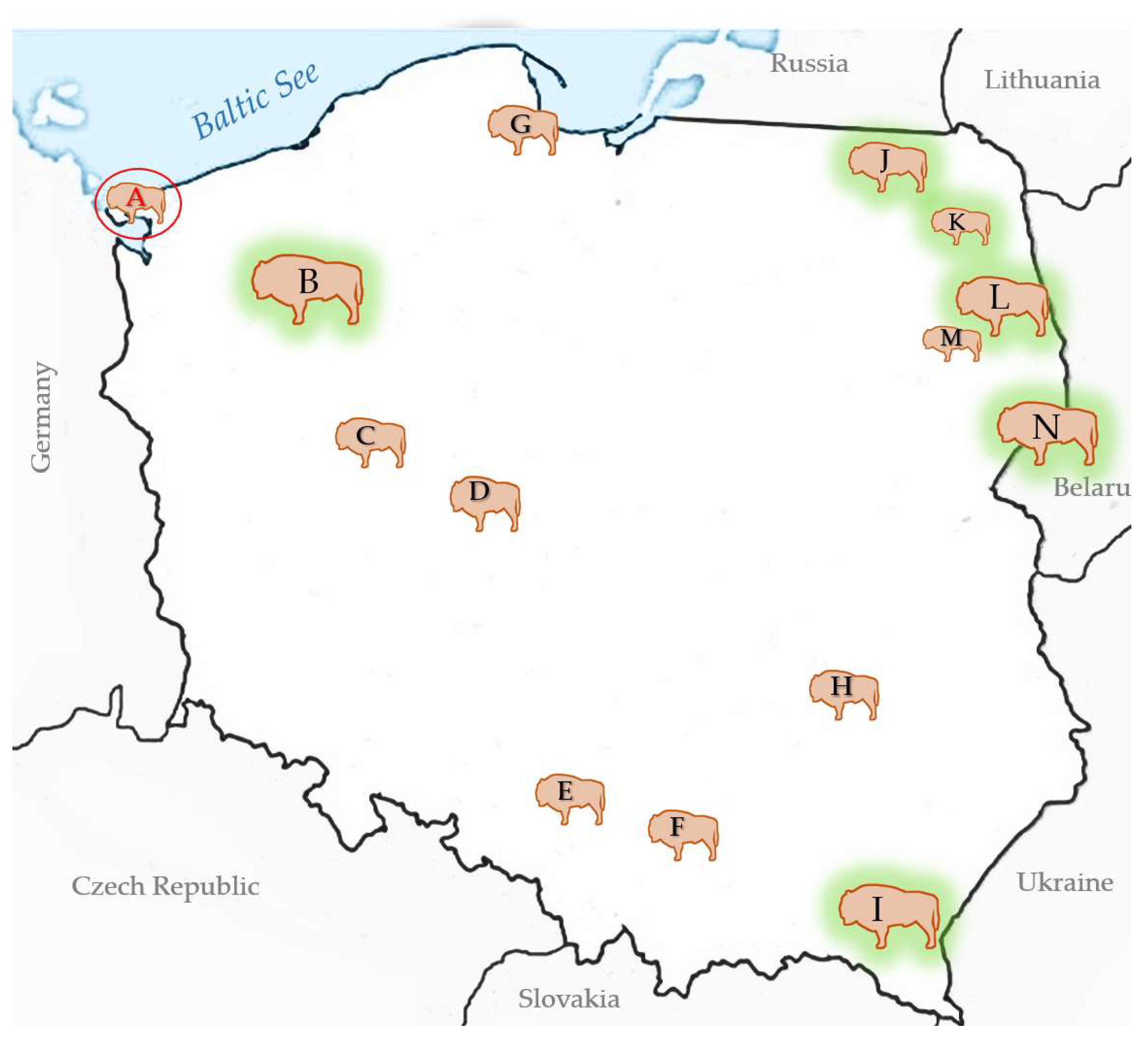
The emergence of an another bluetongue virus serotype BTV-3 has been causing great losses in animal farming in Europe since fall 2023. The virus spreads faster than the epidemic BTV-8, which appeared on the continent nine years earlier. The study describes first case of BTV-3 in Poland detected in European bison (Bison bonasus), just approx. 15 km from the German-Polish border. The animal suffered from severe and fatal hemorrhagic disease. The symptoms included respiratory problems, bloody diarrhea and rapidly progressive cachexia. In addition to confirmation of BTV-3 infection in the blood and spleen of the animal, the virus was also detected in one of pool of blood-fed Culicoides punctatus caught near the enclosure two weeks after the death of European bison. This is the first evidence of BTV-3 detection in C. punctatus what suggests vector competency for this serotype. Phylogenetic analysis based on the segment 2 of the virus revealed homology of Polish isolate to the BTV-3 strains circulating in the Netherlands, Germany and Portugal, and slightly lower similarity the BTV-3 strains detected in sheep in Sardinia (Italy) in 2018 and in Tunisia in November 2016. Retrospective serosurvey of the exposure to BTV in thirteen other European bison populations widespread in the country indicated that the observed case at the Wolin National Park was the first BTV-3 to be detected in Poland.

Keywords: 
;  ;  ;  ;  ;  

1. Introduction

The role of wildlife in the transmission and maintenance of many pathogens in the environment remains highly understudied. The emerging climate-sensitive infections (CSIs) like bluetongue (BT) caused by a virus of the same name are spread transboundary and transcontinentally via vectors whose range and activity are changing with global warming. Bluetongue virus (BTV) is an Orbivirus, which belongs to Reoviridae family and known to affect domestic, wild ruminants and camelids. It is transmitted by nocturnal blood-suckling midges of the genus Culicoides. Historically, BTV circulation was limited to tropical and subtropical climates of Africa and since the mid-20th century had occurred also in the Mediterranean Basin and Americas which was believed to be related to the presence of competent vectors C. imicola and C. sonorensis, respectively. The virus is considered predominantly pathogenic to sheep, but as it occurs in 24 different “classical” serotypes and several novel serotypes referred to as atypical, the viruses may differ in their tropism and virulence to the vertebrae hosts. Interestingly, BTV infections are considered subclinical in the tropical zones, while their emergence in temperate zones or introduction of susceptible animals into BTV endemic areas results in clinical disease [1]. The first major intrusion of BTV serotype 8 into central Europe over latitude 50°N in 2006 created havoc in the animal production sector and has forced the need to reassess the epidemic risks, particularly in terms of climate change [2]. Now, almost two decades later, more and more BTV serotypes are being reported in Europe, and the latest to spread from the center of the continent at a record rate is BTV-3 [3,4,5,6,7,8]. The rapid expansion of these often tropical or neglected diseases in Europe is also linked to the wide range of reservoir species the agent infects or is able to adapt to with the possibility of overwintering while remaining endemic in the area. Vertical transmission of BTV in Culicoides spp. has not been demonstrated to be responsible for interseasonal virus transmission [9,10], as it was suggested for culicoid-borne Schmallenberg virus (SBV)[11]. Therefore, it is very likely that wildlife play an important role in the maintenance of BTV in the environment [12,13]. However, since they are omitted from the official BTV monitoring or eradication programs, it leads to an underestimation of the prevalence of these CSIs.
The European bison or wisent (Bison bonasus) is the largest wild ruminant of Europe, which became extinct in the wild in north-eastern Poland (the lowland E. bison) and the Caucasus in Russia (the Caucasian E. bison) after the First World War. Thanks to the efforts of International Society for the Protection of European Bison supported by the Polish government, the species has been restored from extinction; however, it remains protected and categorized by the International Union for Conservation of Nature (IUCN) at the Red List of Threatened Species as a near threatened species [14]. Nearly a century has gone by since the start of the restitution breeding of Bison bonasus in the Białowieża Forest (now UNESCO World Heritage site), and the European population of the species in 2023 exceeded 11,000 individuals, almost 80% of which remain free-ranging [15]. Most European bison live in Poland, Russia and Belarus (Figure S1). Quite numerous and still thriving populations also exist in Germany, Romania, Ukraine and Lithuania. European bison have even been introduced into the Iberian peninsula, despite discussions that the species is alien to these areas [16]. The evidence of the presence of these emblematic large herbivores in northern Spain and southern France are prehistoric remains and paintings such as those found in the Altamira cave, Cantabria, Spain of the E. bison larger analogue, the steppe bison (Bison priscus). Climate change forced such megafauna to migrate to the northern hemisphere, where they became extinct thousands of years ago. The observations presented here shed some light on the exposure risk in both scenarios: when progressive environmental changes threaten native fauna and when the animals are exposed to threats via human activity, globalization and relocation of animals to drastically different environments. It is interesting to note that this is the first case of BTV-3 infection in Poland, which was detected a few weeks before any other cases found by national active monitoring in subclinically infected cattle. In addition to the first description of the clinical picture and pathological changes of BTV-3 infection in the E. bison, we present the details of entomological monitoring in its sylvatic habitat, identification of the virus in Culicoides vector, and the analysis of the BTV epizootic situation in fourteen E. bison populations between 2023 and 2024.

2. Materials and Methods

2.1. Case Study and Clinical Diagnosis

Fatal case of 9 year-old female European bison was reported on 15 October 2024 in the European bison game reserve at Wolin National Park (WNP; N 53° 56' 2.107'' E 14° 28' 34.402''), which is located at the island at the Baltic See in the immediate vicinity of Germany's eastern border. Prior to its death, the animal suffered from gradual deterioration of health, lack of appetite, separating itself from the herd, which was first observed around ten days earlier. In the last 2-3 days, the animal developed bloody diarrhea, serous and then purulent nasal discharge, debilitation, apathy and impaired mobility (Figure 1a–c). Treatment using Draxxin, Baytril and Naxel injected intramuscularly by a dart gun (Figure 1d) and intravenous infusions of Ringer's lactate solution, 40% Glucose, Biotyl, Duphalyte and Meloven, while already unable to stand up. The postmortem examination was performed onsite not more than few hours after it’s death and samples of parenchymal organs, intestines, EDTA and clotted blood were send chilled at 4°C to the laboratory for virological and bacteriological testing. Fragments of lungs, liver, spleen, kidneys, small and large intestines were fixed in 10% formalin, pH = 7.2 for histopathological examination. Then, the tissues were transferred to alcohol solutions, acetone and xylene to paraffin blocks in a tissue processor (Leica TP-1020). For the analysis, four μm thick tissue sections were stained with hematoxylinand eosin (HE) and observed under microscope.
Some transient nasal discharge and moderate depression was seen in other two European bison in the herd, however, the symptoms quickly resolved without the need for veterinary intervention. Sampling was waived due to the cost and risk associated with the necessary chemical immobilization of the animals.

2.2. Culicoides Monitoring

Due to the increasing threat of BTV and EHDV transmission from the west of Europe, culicoides monitoring also included the WPN area in 2024. The entomological studies were conducted as described previously [17] on the basis of consent with WNP No 42.20.1.2024. The insects were collected using ultraviolet light trap (CDC 1212, John W. Hock Company, Gainesville, Florida, USA) located in the immediate proximity of the European bison enclosure (Figure 2), which was set up every one or two weeks overnight from 16 June until the end of annual activity of these nocturnal insects (2 December). The midges, which were attracted by UV light, sucked in by a fan and finally were trapped in the container with sterile water and a drop of detergent. Then, the insects were sieved and placed in 70% ethanol until being tested under SDF PLAPO 1XPF objective in the Olympus SZX16 microscope. Entomological examination included species identification based on methodology described by Mathieu et al. [18], determination of sex and female gonotrophic cycle stage (nulliparous—virgin, unpigmented abdomen; parous—pigmented, blood fed and reproduced at least once; gravid—abdomen filled with egg batches; and freshly blood fed) and preparation of pools of up to 20 blood fed individuals for molecular testing.

2.3. Surveillance Sampling

The surveillance was carried out as part of a conservation strategy for the species as described previously [19,20,21]. For the monitoring purposes, 160 serum samples European bison collected between 2023 and 2024, retrospectively to the case detected at WNP were tested for the presence of BTV specific antibodies. In the case of a seropositive animal, a virological examination of the full blood sample was performed subsequently. The animals originated from seven free-ranging populations including three largest ones in Europe: Białowieża Forest (N), Bieszczady mountains (I) and Zachodniopomorskie herds (B); and seven captive herds spread across the country (Figure 3).
The samples originated both from female (n=71) and male (n=89) European bison in the age between 2 days and 23 years. European bison were sampled solely on the occasion of other procedures and were not immobilized or euthanised for the tests described hereby. Samples were taken from clinically healthy, which were pharmacologically immobilized for placing collars with telemetric transmitters or official testing of translocated individuals (n=84); fallen meaning found dead (n=25); euthanized (n=40) due to poor health condition in accordance with the corresponding decisions of the Minister of the Environment and the General Director for Environmental Protection; or dead in traffic (car, train) accident (n=7). From the immobilized or recently eliminated animals, the blood was collected through the puncture of the external jugular vein (vena jugularis externa), less often from the tail vein (vena caudalis mediana). Blood from dead, necropsied animals was collected in the form of a clot from the heart or from body cavities. Blood was collected into the sterile 9 ml EDTA-tubes and serum clot activator tubes, which were centrifuged within 24 hours. The full-blood and serum samples were stored at -70°C in the biobank of the Department of Virology and Viral Animal Diseases, NVRI until analysis.

2.4. Virological Testing

Virus isolation. Bluetongue virus was isolated in a baby hamster kidney cell line clone 21 (BHK-21). BHK-21 cell monolayers were overlaid with blood cells extracted from EDTA-treated whole blood after removal of virus-neutralizing antibodies and 20% homogenate (w/v) of spleen as prepared in Eagle’s Minimum Essential Medium (EMEM) and incubated at 37°C in 5% CO2 with humidity. Removal of virus-neutralizing antibodies was performed according to WOAH protocol [22]. The cell monolayers were monitored microscopically for the appearance of cytopathic effects (CPE) up to 5-7 days. If no CPE was observed, subsequent passage in BHK-21 cell culture was performed. Positive CPE or negative cell culture results for BTV isolation were confirmed after each cell culture passage by real-time RT-PCR testing.

2.5. Molecular Testing

Nucleic acid extraction. Nucleic acids were extracted from whole blood and 10% homogenate of spleen tissue collected intravitally or post mortem from European bison, or pools of blood fed Culicoides. Tissue and insect samples were grinded in phosphate-buffered saline (PBS) by mechanical homogenization with 1.4 mm ceramic (zirconium-silicate) beads (Lysing Matrix D, MP Biomedicals) using TissueLyser LT (Qiagen) and two cycles of 45 s at 6500 rpm with an interval of sample cooling on ice as described previously [17]. Homogenates were clarified at 2500 rpm for 5 min at 4° C. Total nucleic acid extraction was performed from 200 ml of EDTA blood sample, spleen homogenate or insect suspension supernatant using IndiMag Pathogen Kit (Indical, Leipzig, Germany) in an IndiMag 48s machine for automated nucleic acid extraction, following manufacturer’s protocol. Nucleic acids were used immediately for RT-PCR or were preserved at -20 ° C until use.
RT-PCR. BTV RNA was detected by pan real-rime RT-PCR described by Hofmann et al. [23] targeting the NS3 segment fragment according to the European Union Reference Laboratory (EURL – BT) standard operating protocol and WOAH recommendation [22]. Briefly, 2 µl of RNA and 10 µM oligonucleotides were denaturized at 95°C for 5 minutes and immediately cooled on ice for 3 minutes. Next, mixture of 0,2 uM BTV probe, 12,5 µl of 2 x buffer RT-PCR, 8 µM oligonucleotides for ß-actin detection of mammals or 18 S rRNA of Culicoides as Internal Control (house keeping gene) and enzyme mix were added. rtRT-PCR was performed at QuantStudio 6 instrument (Applied Biosystems, ThermoFisher Scientific). Samples were classified as positive if the cycle threshold (Ct) value was lower than 40. Samples with Ct higher or equal to 38 were considered as doubtful and retested. For BTV-3 typing commercially available Adiavet BTV Type 3 kit was applied. Typing was carried out according to manufacturer instruction and QuantStudio 6 instruments. To perform phylogenetic studies gel-based RT-PCR targeting the detection of 1000 bp fragment of segment 2 of BTV-3 was designed. The primers sequence were as follows: BTV_3_AO_F: 5’-AATYACCTATTYAATACCGC-3’ and BTV_3_AO_R: 5’-TCATCTCACGATATCTATC-3’. The PCR products were visualized in 2 % horizontal electrophoresis agarose gel and after purification were subjected to Sanger sequencing in two directions on the automated sequencer ABI PRISM 310 Genetic Analyzer (Applied Biosystem) using a BigDye Sequencing Kit (Applied Biosystem) with GeneScan Analysis Software.
Phylogenetic studies. Nucleotide sequences of 1000 bp fragment of segment 2 of BTV were aligned using Clustal W Multiple alignment and a phylogenetic Neighbour Joining tree was generated using an appropriate evolutionary model bootstrapped on the set of 1000 replicates with the Mega 5 software [24]. The similarity matrix was done using BLOSUM62 in BioEdit software v. 7.0.5.3.

2.6. Serological Testing

INgezim BTV DR 12.BTV.K0 (INGENASA, Spain) targeting VP7 protein specific antibodies of 24 BTV serotypes was used. The kit is intended for testing bovine, sheep or goat serum samples. The test was performed in accordance with the instructions provided by the manufacturer. Shortly, 50 µl serum was diluted with 50 µl diluent and mixed. The plate was sealed and incubated for an hour at 37°C and washed 6 times (300 µl/well) using previously diluted in deionised water 1:24 Washing solution. 100 µl Peroxidase conjugate (ready to use) was added to each well, plate was sealed and incubated for 1 hour at 37°C. After rinsing the plate with Washing solution, 100 µl Substrate (TMB) was added to each well, sealed and incubated for 15 min at room temperature. After than added 100 µl Stop solution to each well. The optical density (O.D.) was read at 450 nm. Samples were considered positive if O.D. was higher than the cut-off (15% of positive control) and negative if the OD value was equal or lower than the positive cut-off. The specificity and sensitivity of this ELISA test were 100% and 99,8%, respectively according to the manufacturer.

2.7. Additional Testing

During the investigation into the cause of death, a number of other tests were performed including serological assays for alphaherpesvirus (bovine herpesvirus type 1 (BoHV-1), pestivirus (bovine viral diarrhoea virus – BVDV), Schmallenberg virus (SBV) and bovine coronavirus (BCoV) specific antibodies according to the methods previously described [19,25]. We have used nestedPCR described by VanDevanter et al. [26] for the detection of gammaherpesvirus including malignant catarrhal fever virus (MCFV).
Moreover, culture tests were carried out for aerobic and anaerobic bacterial infections in internal organ tissue and small intestine.

3. Results

3.1. Case Study

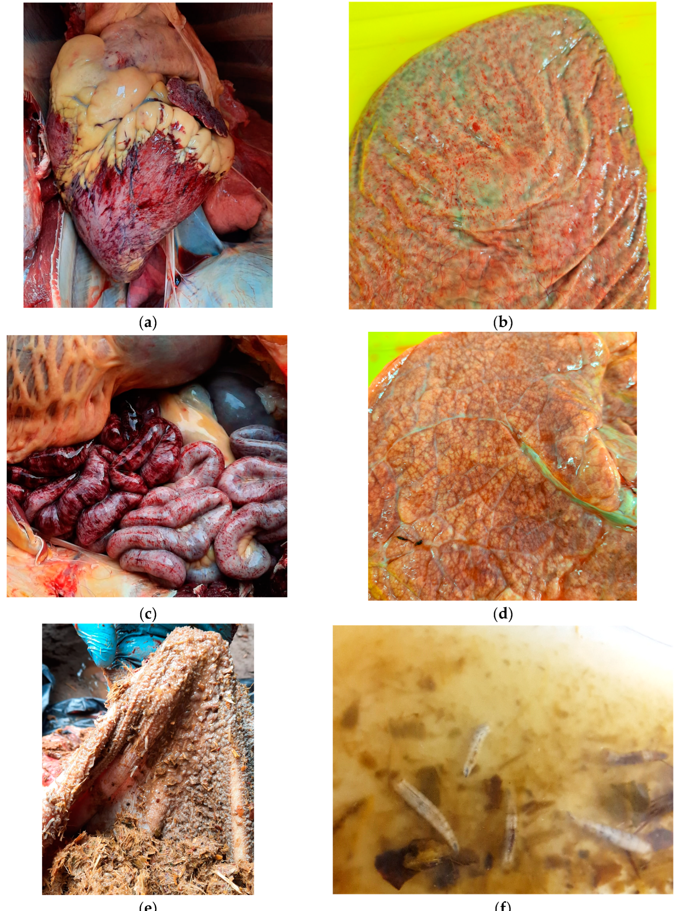
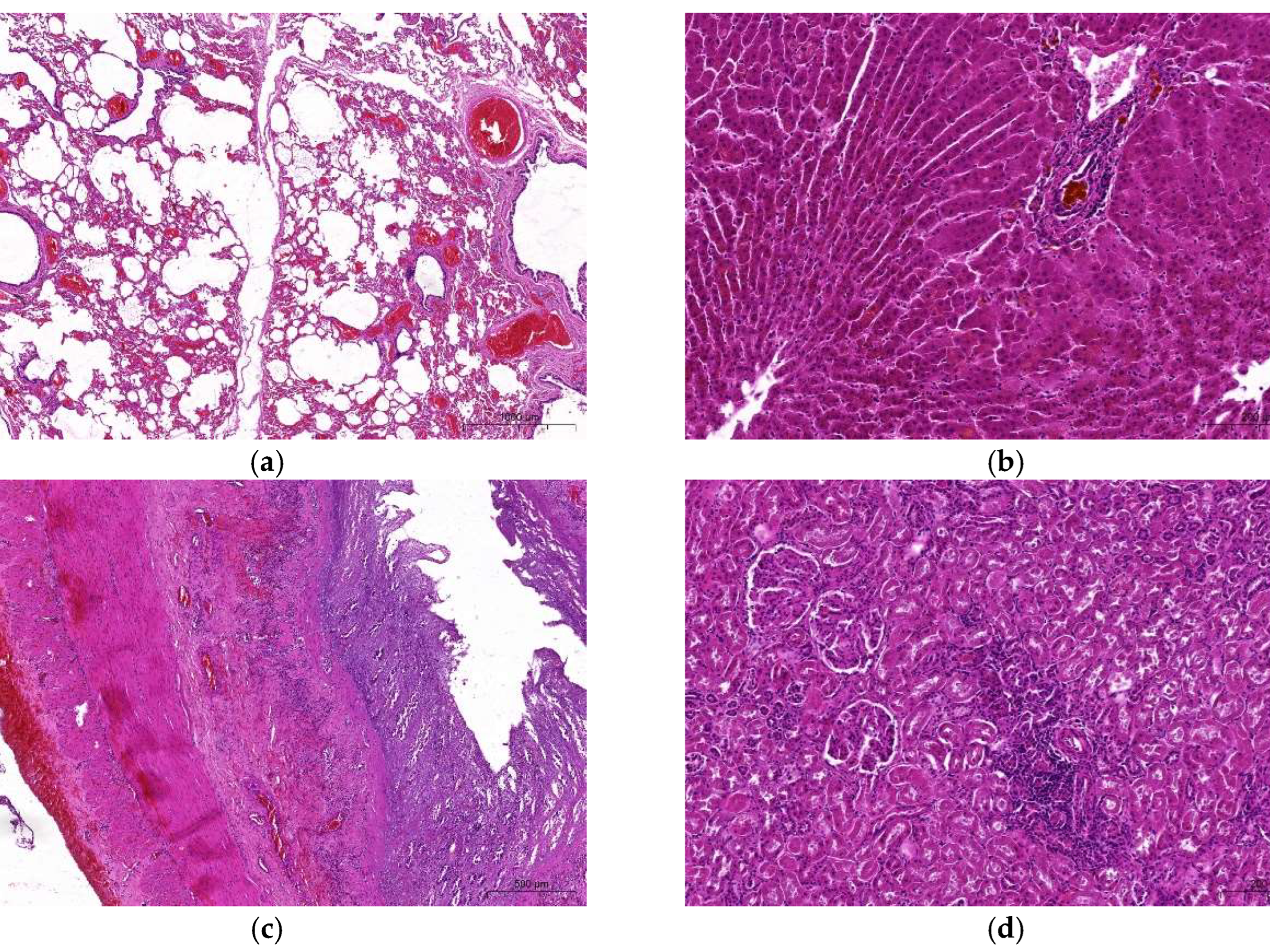
3.1.1. Macro- and Microscopic Findings

External examination of the carcass revealed emaciation (carcass weight 300-320 kg) and generalized dehydration. No edema in the head, nor any changes in the nostrils, oral mucosa, limbs, hooves or udder, which are quite characteristic in BTV infections were observed. However, at the necropsy, some characteristic lesions such as petechiae under the epicardium (Figure 4a), splenic capsule (Figure 4b) and severe hemorrhagic abomasitis and enteritis filled with bloody ingesta (Figure 4c) and multifocal pulmonary emphysema (Figure 4d) were detected, suggesting some hemorrhagic disease. Other lesions involved fatty liver, which could have been related to the animal’s age, and the presence of fly larvae in the contents of rumen and jejunum (Figure 4e), which were later genetically identified as maggots of a blow fly (Lucilia caesar, Linnaeus, 1758) based on the COI gene [27]. Microscopic examination revealed multifocal alveolar lung emphysema characterized by the presence of irregularly distended alveoli and large sac-like air spaces resulting from rupture of their walls with spike-like stumps protruding into their lumen. In the vicinity of the emphysema foci there were areas of atelectasis with collapsed alveoli and severely engorged blood vessels (Figure 5a). There was moderate disruption of hepatic cord architecture with dissociation of hepatocytes and disorganization of sinusoids affecting mostly the midzonal and periportal regions of hepatic lobules. Occasionally, bile canaliculi and ducts contain luminal, brown to bright yellow, globular material (bile) (Figure 5b). Despite marked autolysis of the intestinal mucosa, partial to full thickness mucosal necrosis with hemorrhage, fibrin, eosinophilic cellular and karyorrhectic debris, was observed throughout the entire length of the small intestine and colon. Occasionally, remaining small intestinal crypts were ectatic, lined by attenuated epithelium, and contain sloughed epithelial cells admixed with moderate numbers of macrophages, lymphocytes, plasma cells, and fewer intact and necrotic neutrophils. The inflammatory infiltrate composed mainly of lymphocytes and plasma cells extends through the muscularis mucosa into the submucosa. The submucosa, and serosa were markedly expanded by abundant hemorrhage (Figure 5c). In the kidneys, adjacent to the renal tubules were mild to moderate multifocal aggregates of lymphocytes and plasma cells (Figure 5d).

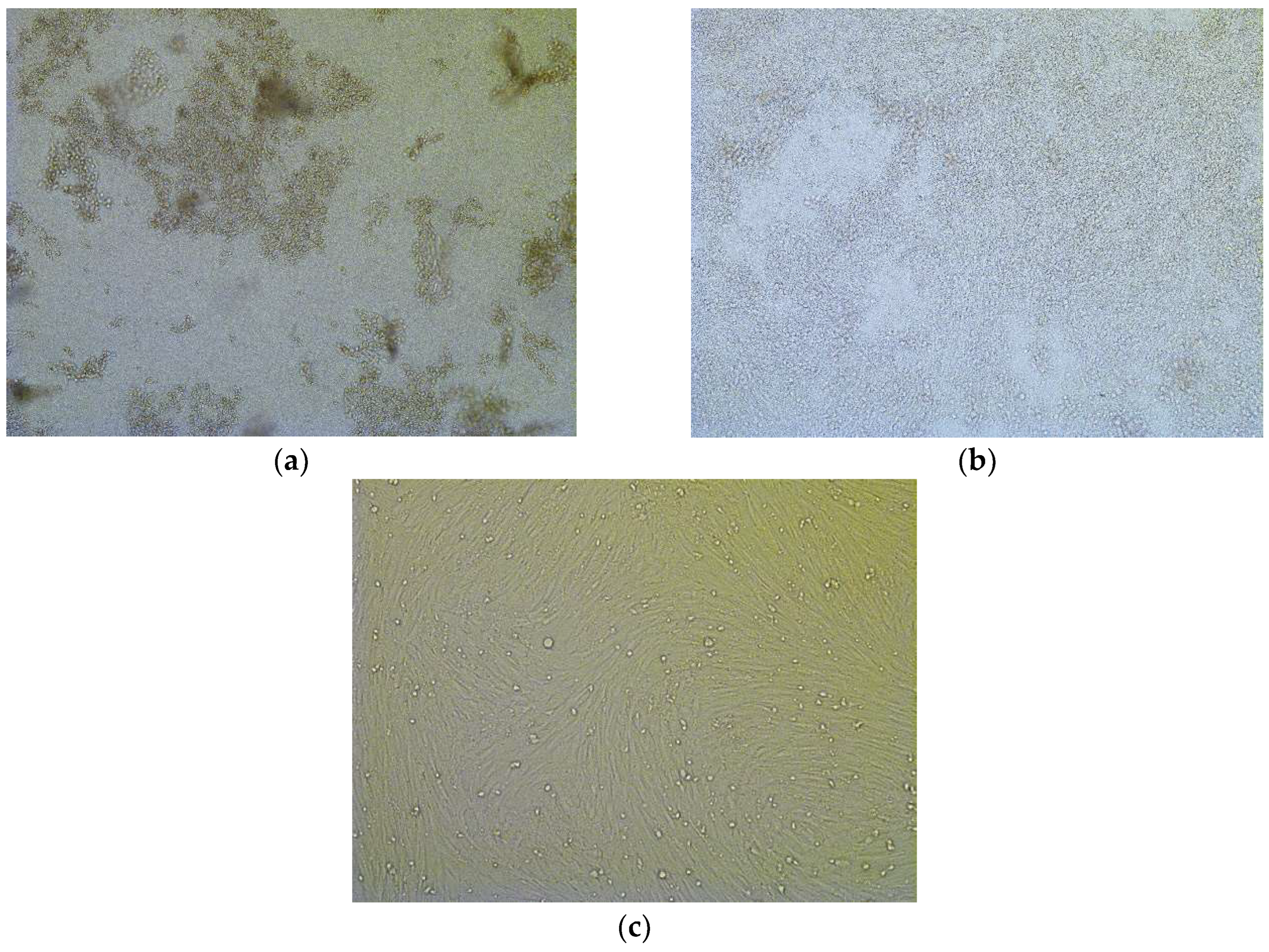
3.1.2. BTV Detection

Both samples (blood and spleen) of suspected European bison tested positive for BTV using pan rtRT-PCR with Ct (threshold cycle) values: 23.8 and 24.2, respectively. Typing of BTV with using commercial kit revealed the presence of BTV type 3 (Ct = 22.1 and 23.5 in blood sample and spleen homogenate, respectively).
Furthermore, BTV was isolated in BHK-21 cells monolayers from suspected European bison blood cells and spleen homogenate. Three subsequent passages were performed. CPE was observed at second and third passage (Figure 6), however, the effect of the virus was much more pronounced in case of full blood sample (Figure 8a). BTV replication in BHK-21 cells was confirmed by rt RT-PCR for blood cells (Ct = 29.3 and 13.4 at passage 2 and 3) and spleen (Ct = 36.3 and 27.6 at passage 2 and 3).
Involvement of other pathogens was excluded as most tests were negative, except for the presence of antibodies against SBV.

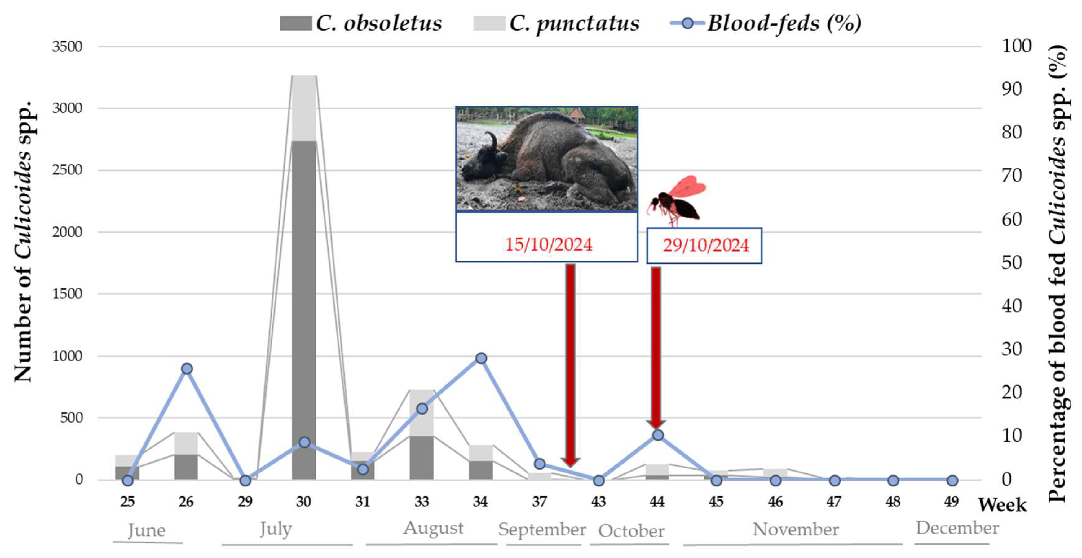
3.1.3. Culicoides spp. Activity and BTV Detection

A total of 5,553 Culicoides individuals was caught during 15 night catches in the European bison enclosure at WNP between June and December 2024. The most frequent were C. obsoletus (n=3833) and C. puntatus (n=1636). Other species included C. achrayi (n=5); C. circumscriptus (n=2); C. fascipennis (n=3); C. festivipennis (n=1); C. furcillatus (n=1); C. grisescens (n=2); C. newsteady (n=27); C. pallidicornis (n=2); and C. pulicaris (n=39). Most of them were females of different gonotrohic forms (nulliparous, n=2809; parous, n=2021; blood-feds, n= 614; gravid, n=85), with small proportion of males (n=33). The midges were most active in June and August (Figure 8), however, based on the presence of blood-fed females, the insects were actively feeding until the end of October. Thirty three pools of blood-fed females were tested for BTV genetic material. Only one pool (3%) of 13 C. puntatus individuals caught two weeks after the dead of the European bison cow (Figure 1) was found BTV positive with Ct value 33.58 (Figure 7). Typing of BTV detected in Culicoides revealed the presence of BTV-3 (Ct = 32.8).

3.1.4. BTV Strain Characterization

Phylogenetic analysis performed for 870 bp fragment of segment 2 BTV-3 revealed high nucleotide sequence similarity (99.7 %) of the BTV-3 isolate detected in European bison and pool of Culicoides to BTV-3 strains detected in the Netherlands (the first BTV-3 isolate detected in Western Europe in 2023), Portugal (a dog on September 2024) and Germany (GenBank, accession nos: OR603992; PQ654180; OZ119415) (Figure 8). The nucleotide sequences of the Polish European bison and culicoid BTV-3 were 96.8 % identical to the BTV-3 SAR2018 strain isolated from a sheep in Sardinia (Italy) in 2018 (GenBank, accession no: MK348538) and in Barbarine ewe in Tunisia in November 2016 (GenBank accession no: KY432370).
Figure 8. Phylogenetic relationship of BTV-3 isolates detected in European bison () and pool of Culicoides () at Wolin National park with reference nt sequences available in GenBank database based on 870 bp fragment of Segment 2 BTV-3. Phylogenetic tree was generated using the Neighbour joining method (Kimura2 parameter) as implemented in Mega5 software [24]. Bootstrap value (1000 replicates) over 70% indicating significant support for the tree topology are shown next to the branches.
Preprints 151479 g008

3.2. BTV Survey of European Bison in Poland

BTV antibodies were detected in 37 (23.1%) out of 160 European bison sampled in 2023 and 2024. Most seropositive animals were found in the north-eastern free-rangin populations of Białowieża and Knyszyńska Forests, with only one single reactor in the enclosure in Pszczyna (6-year old healthy bull born at the origin), on the south of the country (Table 1). The seroprevalence increased with age . Most of seropositive animals were over 9 years of age. No BTV was detected in the blood of any of the seropositive European bison, except for the index case at WNP (3.1.).

4. Discussion

The appearance of BTV-3 infections on Polish territory was not surprising, as the disease had been reported in western Germany since the fall of 2023, but continuous monitoring in the country did not confirm cases of infection until the fall of 2024. The BTV-3 case found as the first case in a European bison was about 20 km away from the German outbreak registered in October 2024 [28], what indicates the virus migrated most likely through Culicoides. Those midges are able to fly several kilometers over a few days by themselves, while a heavy wind can make them travel hundreds of kilometers. The suspicion was reported to the NVRI, which has been involved in the monitoring of viral infections in the E. bison population as part of a scientific collaboration with free-ranging populations and captive herd managers since 2012 [20,21], while also being the National Reference Laboratory (NRL) for BTV. Shortly thereafter, further outbreaks in domestic cattle (7 in 2024 on the western side of the country and 3 in 2025) were identified in the national monitoring supervised by the state veterinary service (Figure S2). In early 2025, BTV unexpectedly appeared in the northeastern part of the country, likely related to entry of BTV-3 infected animals. The virus is expected to spread further across the country in 2025. So far, however, in majority of BT outbreaks, no clinical cases of BT have been identified in domestic ruminants. Despite the fact that E. bison are related to domestic cattle, in which symptoms of BTV infections are rather less severe than in sheep, they appear to be more susceptible to infection with this virus. A relatively high mortality rate (30%) of BTV-8 infections was described in E. bison in Germany in 2007 [20,21]. For BTV-3, clinical signs and mortality have been observed in E. bison in the Netherlands, Denmark and Germany in the last two years [29]. In addition to the relatively severe course of BT in E. bison, it should be noted that based on seroprevalence data, arbovirus exposure rate in the species is higher than in other wild or farmed ruminants. This applies to culicoides-borne BTV and SBV, as well as to tick-borne encephalitis virus (TBEV). Infections with BTV and SBV have emerged in European bison in Poland simultaneously in 2012 [30,31]. While SBV has spread all over the country, BTV infections were limited almost exclusively to the north-eastern populations [19]. The seroprevelence of SBV and BTV in European bison at the beginning of epizootic reached 75 and 25 %, respectively and were significantly higher as compared to the cervids sampled in the same locations [31]. In this study, the presence of antibodies was also observed in northeastern E. bison populations (Białowieża and Knyszyńska forests) in 2023-2024. Since no virus has been detected in any of the seropositive bison, these infections are most probably associated with BTV-14, which was identified as circulating locally in cattle [32]. Its unexpected appearance in the east of the country was with a transmission from the east [32,33,34], while BTV-8, which emerged in Western Europe has never reached Poland. The circulating BTV-14 was characterized by a very low pathogenicity, and the contamination of some illegally used vaccine as a source of infection was suspected as the virus was probably attenuated and closely related to the reference BTV -14 strain [32,33]. The presence of antibodies in calves and older than 9 years old E. bison suggests either maternal immunity or persistence of antibodies as no virus was detected. Still some limited circulation of BTV-14 in E. bison population could not be excluded, despite no evidence of it in the official monitoring in domestic ruminants. High tropism of arthropods to the species was also revealed in TBEV serosurvey, where seroprevalence reached as high as 63%, which made the E. bison more sensitive indicatory species than deer considered to be sentinels of this zoonotic virus in the endemic foci [35]. Two factors should be considered relevant: firstly, due to their size and secretion of large amounts of CO2 and other gases and odors, E. bison are more attractive for insects, and secondly, E. bison naturally inhabit humid forests, which provide an excellent habitat for arthropod vectors to thrive. With the global warming, insect activity season can be significantly extended, as observed in this study, where Culicoides spp. were feeding on hosts until late October and operating until November, and perhaps even longer given the effectiveness of the trap used. Previously, we have observed that the number of Culicoides biting midges caught in the E. bison reserve at the Białowieża Forest outnumbered, by several dozen times, the numbers of those insects collected in the neighboring cattle farms [36]. Similarly, studies of ticks residing on the skin of E. bison in recent years have shown a change in tick activity from seasonal to year-round [37]. With prolonged arthropod activity, the risk of exposure to arthropod-born pathogens also increases because prevention of ectoparasite invasion is limited and highly inefficient in the wild animals. Our study is the first one to provide evidence of the vector competence to BTV-3 of one of the more abundant species of C. puntatus in Europe, the second most numerous caught at the location of BTV-3 outbreak in E. bison. In the recent study, Voight et al. [5] have detected BTV-3 RNA in a pool of midges of mixed species of C. obsoletus, C.scoticus and C. chiopterus and undetermined parity status. However, since 1,603 pools were tested in total, monitoring for the presence of the virus in the vector has a limited predictive value for BTV-3 circulation, as concluded by the researchers. The effectiveness of such detection may increase if only parous and/or blood-fed midge pools are tested, as in our case. However, here again, BTV-3 could not be detected earlier, but only after a case occurred in the E. bison. Nevertheless, the virus was observed to circulate even two weeks later, which suggests further transmission that will be verified by continuation of entomological monitoring in 2025. Parous C. puntatus have been demonstrated to contain BTV-1 RNA in Italy, in Sardinia in 2013 [38], and Abruzzo and Apulia in 2014 [39]. The species has been shown to be possibly involved in BTV transmission in Turkey in 2007 [40] and previous BTV-8 epidemic in Germany [41]. In the latter study, extant Culicoides activity was also observed in the winter of 2007/2008, which , explaining the possibility of overwintering BTV in the insect vector.
To conclude, it is difficult to predict for the time being the impact of BTV-3 on the protection of this iconic species, one of the major symbols of the rewildling initiatives and biodiversity conservation in Europe. It is important that we begin to look at climate risks comprehensively, also taking into account the health of the environment as a part of ecosystem well-being and us, together constituting OneHealth.

Supplementary Materials

The following supporting information can be downloaded at: www.mdpi.com/xxx/s1, Figure S1: Numbers by country and distribution of European bison (Bison bonasus) populations worldwide registered by the European bison Pedigree Book 2023 [Raczyński, 2024]. Additionally, six European bison are reared in two locations in Indonesia. Figure S2: The distribution of BTV-3 cases in cattle in Poland recorded until March 1, 2025 by the Central Veterinary Office (CVO) at the map available at: https://bip.wetgiw.gov.pl/bt/mapa/. The black and red cow figures represent the locations of the outbreaks in 2024 and 2025, respectively, while the case of the infected European bison is marked with a green asterisk. The blue areas indicate the restriction zones.

Author Contributions

Conceptualization, M.L. and M.K.K.; methodology, M.L., A.O., W.Ł., Ł.S., A.N. and P. T.; software, M.L., A.O. and W.Ł.; investigation, M.L. and J.R.; resources, M.L. and M.K.K.; data curation, M.L. and Ł.S.; writing—original draft preparation, M.L., A.O., M.S. and W.Ł.; writing—review and editing, M.L. and A.O.; visualization, M.L. and W.Ł.; supervision, M.L.; project administration, M.L.; funding acquisition, M.S. and J.R. All authors have read and agreed to the published version of the manuscript.” Please turn to the CRediT taxonomy for the term explanation. Authorship must be limited to those who have contributed substantially to the work reported.

Funding

The research was carried out within the framework of the implementation of the Ministry of Agriculture and Rural Development's special purpose subsidy entitled ‘Wild animals as reservoirs of infectious and invasive pathogens dangerous to human and livestock health’ in accordance with contract No. DŻW.zlf.070.6.2024.

Institutional Review Board Statement

The study was conducted in accordance with the permits issued by the Minister of Climate and Environment (DOP—1.61.21.2021.ZK) and the General Director of Environmental Protection (DZP—WG.6401.9.2021.EB).

Data Availability Statement

Data are available from Magdalena Larska (NVRI, Puławy, POLAND) on request.

Acknowledgments

We would like to acknowledge the involvement of all veterinarians supervising European bison populations, especially Dr. Jan Mruk supervising who provided necropsy protocol and sampling. We would also like to acknowledge the invaluable assistance of conservation officers Marek Dylawerski and Konrad Wrzecionkowski from Wolin National Park who prepared a chronological case file including photographic documentation. We would like to thank Sven Reiche (FLI, Riems, Germany) for providing reference virus strains as positive controls for RT-PCR and PCR.

Conflicts of Interest

The authors declare no conflicts of interest.

References

  1. Walton, T. E. The history of bluetongue and a current global overview. Vet. Ital. 2004, 40, 31–38. [Google Scholar] [PubMed]
  2. Purse, B. V.; Brown, H. E.; Harrup, L.; Mertens, P. P.; Rogers, D. J. Invasion of bluetongue and other orbivirus infections into Europe: the role of biological and climatic processes. Rev. Sci. Tech. 2008, 27, 427–442. [Google Scholar] [PubMed]
  3. Boender, G. J.; Hagenaars, T. J.; Holwerda, M.; Spierenburg, M. A. H.; van Rijn, P. A.; van der Spek, A. N.; Elbers, A. R. W. Spatial Transmission Characteristics of the Bluetongue Virus Serotype 3 Epidemic in The Netherlands, 2023. Viruses 2024, 16, 625. [Google Scholar] [CrossRef] [PubMed]
  4. van den Brink, K. M. J. A., Santman-Berends, I. M. G. A., Harkema, L., Scherpenzeel, C. G. M., Dijkstra, E., Bisschop, P. I. H., Peterson, K., van de Burgwal, N. S., Waldeck, H. W. F., Dijkstra, T., Holwerda, M., Spierenburg, M. A. H., van den Brom, R. Bluetongue virus serotype 3 in ruminants in the Netherlands: Clinical signs, seroprevalence and pathological findings. Vet. Rec. 2024, 195, e4533.
  5. Voigt, A., Kampen, H., Heuser, E., Zeiske, S., Hoffmann, B., Höper, D., Holsteg, M., Sick, F., Ziegler, S., Wernike, K., Beer, M., Werner, D. Bluetongue Virus Serotype 3 and Schmallenberg Virus in Culicoides Biting Midges, Western Germany, 2023. Emerg. Infect. Dis. 2024, 30, 1438–1441.
  6. Newbrook, K., Obishakin, E., Jones, L. A., Waters, R., Ashby, M., Batten, C., Sanders, C. Clinical disease in British sheep infected with an emerging strain of bluetongue virus serotype 3. Vet. Rec. 2025, 196, e4910. [CrossRef]
  7. Barros, S. C., Henriques, A. M., Ramos, F., Luís, T., Fagulha, T., Magalhães, A., Caetano, I., Abade Dos Santos, F., Correia, F. O., Santana, C. C., Duarte, A., Villalba, R., Duarte, M. D. Emergence of Bluetongue Virus Serotype 3 in Portugal. Viruses 2024, 16, 1845.
  8. Barua, S. , Rana, E. A., Prodhan, M. A., Akter, S. H., Gogoi-Tiwari, J., Sarker, S., Annandale, H., Eagles, D., Abraham, S., Uddin, J. M. The Global Burden of Emerging and Re-Emerging Orbiviruses in Livestock: An Emphasis on Bluetongue Virus and Epizootic Hemorrhagic Disease Virus. Viruses 2024, 17, 20. [Google Scholar]
  9. Mayo, C. , Mullens, B., Gibbs, E. P., MacLachlan, N. J. Overwintering of Bluetongue virus in temperate zones. Vet. Ital. 2016, 52, 243–246. [Google Scholar]
  10. Wilson, A. , Darpel, K., Mellor, P. S. Where does bluetongue virus sleep in the winter? PLoS Biol. 2008, 6, e210. [Google Scholar] [CrossRef]
  11. Larska, M. , Lechowski, L., Grochowska, M., Żmudziński, J. F. Detection of the Schmallenberg virus in nulliparous Culicoides obsoletus/scoticus complex and C. punctatus - the possibility of transovarial virus transmission in the midge population and of a new vector. Vet. Microbiol. 2013, 166, 467–473. [Google Scholar] [PubMed]
  12. Falconi, C. , López-Olvera, J. R., Gortázar, C. BTV infection in wild ruminants, with emphasis on red deer: a review. Vet. Microbiol. 2011, 151, 209–219. [Google Scholar] [PubMed]
  13. Talavera, S. , Muñoz-Muñoz, F., Verdún, M., Pujol, N., Pagès, N. Revealing potential bridge vectors for BTV and SBV: a study on Culicoides blood feeding preferences in natural ecosystems in Spain. Med. Vet. Entomol. 2018, 32, 35–40. [Google Scholar] [PubMed]
  14. Plumb, G., Kowalczyk, R. Hernandez-Blanco, J.A. Bison bonasus. The IUCN Red List of Threatened Species 2020: e.T2814A45156279. (accessed on 27 February 2025).
  15. Nores, C. , Álvarez-Laó D., Navarro A., Pérez-Barbería F. J., Castaños P. M., Castaños de la Fuente J., Morales Muñiz A., Azorit C., Muñoz-Cobo J., Fernández Delgado C., Granado Lorencio C., Palmqvist P., Soriguer R., Delibes M., Vilà M., Simón M., Cabezudo B., Galán C., García-Berthou E., Almodóvar A., Elvira B., Brufao Curiel P., Casinos A., Herrero J., Blanco J. C., García-González R., Nogués-Bravo D., Margalida A., Fisher B., Arlettaz R., Gordon I. J., Ludwig A., Lovari S., Cook B. D., Carranza J., Csányi S., Apollonio M., Kowalczyk R., Demarais S., López-Bao J. V. Rewilding through inappropriate species introduction: The case of European bison in Spain. Conserv. Sci. Pract. 2024, 6, e13221. [Google Scholar]
  16. Raczyński, J. European Bison Pedigree Book 2023; Białowieża National Park: Białowieża, Poland, 2024; p. 9. Available online: https://bpn.gov.pl/pliki-do-pobrania/pobierz/7cc95262-0985-45df-9b39-493b293e87da.pdf (accessed on 27 February 2025).
  17. Mathieu, B. , Cêtre-Sossah, C., Garros, C., Chavernac, D., Balenghien, T., Carpenter, S., Setier-Rio, M. L., Vignes-Lebbe, R., Ung, V., Candolfi, E., Delécolle, J. C. (2012). Development and validation of IIKC: an interactive identification key for Culicoides (Diptera: Ceratopogonidae) females from the Western Palaearctic region. Parasites vectors 2012, 5, 137. [Google Scholar]
  18. Kęsik-Maliszewska, J. , Larska, M., Collins, Á. B., Rola, J. Post-Epidemic Distribution of Schmallenberg Virus in Culicoides Arbovirus Vectors in Poland. Viruses 2019, 11, 447. [Google Scholar] [CrossRef]
  19. Larska, M. , Tomana, J., Socha, W., Rola, J., Kubiś, P., Olech, W., Krzysiak, M. K. Learn the Past and Present to Teach the Future—Role of Active Surveillance of Exposure to Endemic and Emerging Viruses in the Approach of European Bison Health Protection. Diversity 2023, 15, 535. [Google Scholar]
  20. Larska, M.; Krzysiak, M. K. Infectious diseases monitoring as an element of Bison bonasus species protection. In Compendium of the European Bison (Bison bonasus) Health Protection, National Veterinary Research Institute: Puławy, Poland, 2022; pp. 71–96.
  21. Larska, M. , Krzysiak M.K. Infectious Disease Monitoring of European Bison (Bison Bonasus). In Wildlife Population Monitoring, Ferretti M. Ed.; IntechOpen Limited, London, UK, 2019; pp. 1-21.
  22. WOAH Terrestrial Manual 2021, Bluetongue (Infection with Bluetongue Virus), chapter 3.3.3.
  23. Hofmann, M. , Griot, C., Chaignat, V. P., Erler, L., Thür, B. Bluetongue disease reaches Switzerland. Schweiz. Arch. Tierheilkd. 2008, 150, 49–56. [Google Scholar]
  24. Tamura, K., Dudley, J., Nei, M., Kumar, S.: MEGA4: Molecular evolutionary genetics analysis (MEGA) software version 4.0. Mol Biol Evol 2007, 24, 1596–1599.
  25. Larska, M. , Tomana, J., Krzysiak, M. K., Pomorska-Mól, M., Socha, W. Prevalence of coronaviruses in European bison (Bison bonasus) in Poland. Sci. Rep. 2024, 14. [Google Scholar] [CrossRef]
  26. VanDevanter, D. R. , Warrener, P., Bennett, L., Schultz, E. R., Coulter, S., Garber, R. L., Rose, T. M. Detection and analysis of diverse herpesviral species by consensus primer PCR. J. Clin. Microbiol. 1996, 34, 1666–1671. [Google Scholar] [PubMed]
  27. Videvall, E. , Bensch, S., Ander, M., Chirico, J., Sigvald, R., Ignell, R. Molecular identification of bloodmeals and species composition in Culicoides biting midges. Med. Vet. Entomol. 2013, 27, 104–112. [Google Scholar] [PubMed]
  28. Friedrich-Loeffler-Institut (FLI) webpage on Bluetongue situation. Available online: https://www.fli.de/de/aktuelles/tierseuchengeschehen/blauzungenkrankheit/ (accessed on 1 March 2025).
  29. Rodrigues, E. (PWN Waterleidingbedrijf Noord-Holland, Velserbroek, the Netherlands); Rasmussen, T. B. (Statens Serum Institut, Copenhagen, Denmark). Personal communication, 2024.
  30. Larska, M. , Krzysiak, M. K., Smreczak, M., Polak, M. P., Żmudziński, J. F. First detection of Schmallenberg virus in elk (Alces alces) indicating infection of wildlife in Białowieża National Park in Poland. Vet. J. 2013, 198, 279–281. [Google Scholar] [PubMed]
  31. Krzysiak, M. K. , Iwaniak, W., Kęsik-Maliszewska, J., Olech, W., Larska, M. Serological study of exposure to selected arthropod-borne pathogens in European bison (Bison bonasus) in Poland. Transbound. Emerg. Dis. 2017, 64, 1411–1423. [Google Scholar]
  32. Orłowska, A. , Trębas, P., Smreczak, M., Marzec, A., Żmudziński, J. F. First detection of bluetongue virus sero-type 14 in Poland. Arch. Virol. 2016, 161, 1969–1972. [Google Scholar]
  33. Koltsov, A. , Tsybanov, S., Gogin, A., Kolbasov, D., Koltsova, G. Identification and characterization of bluetongue virus serotype 14 in Russia. Front. Vet. Sci. 2020, 7, 26. [Google Scholar]
  34. Flannery, J. , Frost, L., Fay, P., Hicks, H., Henstock, M., Smreczak, M., Orłowska, A., Rajko-Nenow, P., Darpel, K., Batten, C. BTV-14 Infection in Sheep Elicits Viraemia with Mild Clinical Symptoms. Microorganisms 2020, 8, 892. [Google Scholar]
  35. Krzysiak, M.K. , Anusz, K., Konieczny, A., Rola, J., Salat, J., Strakova, P., Olech, W., Larska, M. The European bison (Bison bonasus) as an indicatory species for the circulation of tick-borne encephalitis virus (TBEV) in natural foci in Poland. Ticks Tick Borne Dis, 1799. [Google Scholar]
  36. Kęsik-Maliszewska, J., Krzysiak, M. K., Grochowska, M., Lechowski, L., Chase, C., Larska, M. EPIDEMIOLOGY OF SCHMALLENBERG VIRUS IN EUROPEAN BISON ( BISON BONASUS) IN POLAND. J. Wildl. Dis. 2018; 54, 272–282.
  37. Juszczyk, A., Larska, M., Krzysiak, M.K. Frequency of tick infestation in European bison species in the context of changing environmental conditions as an indicator of potential risk of pathogen exposure. In Proceedings of the Conference 100 years of European bison restitution, Niepołomice, Poland, 6-8 March 2023; pp. 37–38.
  38. Foxi, C., Delrio, G., Falchi, G., Marche, M. G., Satta, G., & Ruiu, L. Role of different Culicoides vectors (Diptera: Ceratopogonidae) in bluetongue virus transmission and overwintering in Sardinia (Italy). Parasit. Vectors 2016, 9, 440.
  39. Goffredo, M. , Catalani, M., Federici, V., Portanti, O., Marini, V., Mancini, G., Quaglia, M., Santilli, A., Teodori, L., Savini, G. Vector species of Culicoides midges implicated in the 2012–2014 Bluetongue epidemics in Italy. Vet. Ital. 2015, 51, 131–138. [Google Scholar]
  40. Yavru, S., Dik, B., Bulut, O., Uslu, U., Yapici, O., Kale, M., Avci, O. New Culicoides vector species for BTV transmission in Central and Central West of Anatolia. Annu. Res. Rev. Biol. 2018, 27, 1–9.
  41. Hoffmann, B. , Bauer, B., Bauer, C., Bätza, H. J., Beer, M., Clausen, P. H., Geier, M., Gethmann, J. M., Kiel, E., Liebisch, G., Liebisch, A., Mehlhorn, H., Schaub, G. A., Werner, D., Conraths, F. J. Monitoring of putative vectors of bluetongue virus serotype 8, Germany. Emerg. Infect. Dis. 2009, 15, 1481–1484. [Google Scholar]
Figure 1. The case of 9-year-old European bison cow in the enclosure of Wolin National Park: (a) bloody diarrhea, reduced appetite; lethargy; (b) bloody diarrhea, loss of appetite, impaired water intake, gradual emaciation; (c) nasal discharge, stupor; (d) drug administration by dart pneumatic riffle.
Preprints 151479 g001
Figure 2. UV-light trap CDC 1212 (John W. Hock Company, Gainesville, Florida, USA) for Culicoides spp. collection installed at the European bison enclosure of Wolin National Park (WNP, Poland in 2024.
Preprints 151479 g002
Figure 3. Distribution of the European bison populations in Poland. The free-living population are marked in green halo. The location of the index case at Wolin National Park (WNP) - A is indicated with a red oval.
Preprints 151479 g003
Figure 4. Necropsy findings of 9-year-old European bison cow found dead in the enclosure of Wolin National Park: (a) hemorrhages in the epicardium; (b) hemorrhages under the splenic capsule; (c) hemorrhagic enteritidis; (d) multifocal pulmonary emphysema; (e, f).
Preprints 151479 g004
Figure 5. Microscopic examination: (a) lung; alveolar dilation and fragmentation of alveolar walls adjacent to the areas of collapsed alveoli, significant dilatation and congestion of the pulmonary blood vessels. HE, bar = 1000 µm; (b) liver; dissociation of hepatic cords with loss of hepatic cord architecture, bile ducts expanded by variably-sized accumulations of brown bile pigment. HE, bar = 200 µm; (c) small intestine; extensive necrosis of the intestinal mucosa with submucosal and subserosal hemorrhage HE, bar = 500 µm; (d) kidney; mild lymphoplasmacytic interstitial nephritis. HE, bar = 200 µm.
Preprints 151479 g005
Figure 6. Cytopathic effect of BTV-3 isolated in BHK-21 cells from blood cells (a) and spleen (b) of suspected European bison (passage 3) compared to non-infected cells (c).
Preprints 151479 g006
Figure 7. Timeline of Culicoides activity and BTV-3 case confirmation in European bison and in Culicoides punctatus blood-fed pool, which were caught two weeks later at Wolin Nationa Park. The grey collumns represet abudnance of the two most common midge species: C. obsoletus and C. punctatus. The blue dots connected wityh a line represent the persentage of blood fed insects (secondary axis on ritght) meaning the proportion of all females having blood meal in their abdomen to all the females caught for each time point.
Preprints 151479 g007
Table 1. Descriptive statistics of European bison exposure to BTV based on the presence of specific antibodies followed by RT-PCR for detectable RNAemia of seropositive animals in relation to their origin, population type, age, gender and sanitary status.
Variable BTV seropositive Pan-BTV RT-PCR
n/N1 % (CI2) n/N1 strain
Origin (total population size3)
A - Wolin National Park (9) 1/3 33.3 (0.8-90.6) 13/1 BTV-3
B – Zachodniopomorskie herds (349) 0/4 0 (0-60.2) 0/0
I – Bieszczady (750) 0/10 0 (0-30.8) 0/0
C - ZOO Poznań (9) 0/1 0 (0-97.5) 0/0
D – Gołuchów (7) 0/2 0 (0-84.1) 0/0
E – Pszczyna (53) 1/51 2 (0.05-10.5) 0/1
F – Niepołomice (16) 0/1 0 (0-97.5) 0/0
G - ZOO Gdańsk (8) 0/2 0 (0- 0/0
H – Bałtów (9) 0/4 0 (0-60.2) 0/0
J - Borecka Forest (127) 0/3 0 (0-70.8) 0/0
K - Augustowska Forest (23) 0/1 0 (0-97.5) 0/0
L - Knyszyńska Forest (298) 3/16 18.8 (4.0-45.6) 0/3
M – Kopna Góra 0/1 0 (0-97.5) 0/0
N - Białowieża Forest (829) 33/62 53.2 (40.1-66.0) 0/33
Population type
free-living 35/82 42.7 (31.8-54.1)
captive 3/79 3.8 (0.7-10.7)
Age group
≤ 1 year old 2/26 7.7 (1.0-25.1) 0/1
2-3 years old 1/38 2.6 (0.056-13.8) 0/1
≥ 4 years old 34/97 35.0 (25.9-45.8) 14/34 BTV-3
Gender
female 22/72 30.6 (20.5-43.0) 14/22 BTV-3
male 15/89 16.8 (9.7-26.3) 0/15
Sanitary status
immobilized (healthy) 7/84 8.3 (3.4-16.4) 0/7
eliminated 17/40 42.5 (27.0-59.1) 0/17
fallen 11/26 42.3 (23.4-63.1) 14/11 BTV-3
dead in traffic accident 2/7 28.6 (3.7-71.0) 0/2
missing data 1/4 25.0 (0.6-80.6) 0/1
1number of seropositive European bison/all tested (missing data were excluded); 2binomial exact 95% or one-sided 97.5% confidence interval; 3according to the most recent data from 2022 [15]; 3this is the BTV-3 case discribed in this case report. Significant differences marked in bold (P<0.05).
Disclaimer/Publisher’s Note: The statements, opinions and data contained in all publications are solely those of the individual author(s) and contributor(s) and not of MDPI and/or the editor(s). MDPI and/or the editor(s) disclaim responsibility for any injury to people or property resulting from any ideas, methods, instructions or products referred to in the content.
Copyright: This open access article is published under a Creative Commons CC BY 4.0 license, which permit the free download, distribution, and reuse, provided that the author and preprint are cited in any reuse.
Prerpints.org logo

Preprints.org is a free preprint server supported by MDPI in Basel, Switzerland.

Subscribe

Disclaimer

Terms of Use

Privacy Policy

Privacy Settings

© 2025 MDPI (Basel, Switzerland) unless otherwise stated