Preprint
Review

This version is not peer-reviewed.

Tick-Borne Colpodella Species Infections: Time for a New Integrated Approach to Understand Transmission and Pathogenicity

A peer-reviewed article of this preprint also exists.

Submitted:

06 March 2025

Posted:

07 March 2025

You are already at the latest version

Abstract

Colpodella species are free-living predatory protists that prey on algae, ciliates and bodonids using myzocytosis. Colpodella species have been reported in human and animal infections. Polymerase chain reaction (PCR) using primers targeting 18S rRNA genes of Cryptosporidium and piroplasms has identified Colpodella species in arthropods, host blood, and feces demonstrating the phylogenetic closeness of Colpodella species to the apicomplexa. However, in both human and animal infections, life cycle stages of Colpodella present in arthropods and infected hosts are unknown. In this review we provide an overview of widespread occurrence of Colpodella species in ticks, and pathogenicity in humans and animals. We discuss methods for culture and microscopy that can aid diagnosis. Phylogenetic tree analysis of Colpodella species identified using 18S rRNA demonstrates that the Colpodella species identified in different geographic regions represent different species and strains that may impact virulence and zoonotic transmission. There is a pressing need to culture Colpodella species, and to stain cells for morphological identification. This will aid molecular investigations aimed at identifying molecular markers of Colpodella spp. facilitating transmission, survival and pathogenesis in hosts, and determining which species and strains to prioritize for the risk of zoonotic infections to humans and for infections in animals.

Keywords: 
;  ;  ;  ;  ;  ;  ;  ;  

1. Introduction

Colpodella species are free-living protist predators that are found in a variety of environmental locations. Colpodella spp. are phylogenetically related to the pathogenic apicomplexa like Plasmodium spp. and Babesia spp. [1]. Several Colpodella species are recognized based on ultrastructure and the presence of an apical complex, such as Colpodella angusta, C. edax, C. pseudoedax, C. pugnax, C. vorax, C. unguis, C. tetrahymenae, C. gonderi and C. perforans [2,3,4,5,6]. Colpodella turpis has been identified using differential interference contrast microscopy (DIC) but has not been characterized by ultrastructure [2]. Colpodella pontica is now known as Voromonas pontica due to phylogenetic and morphological characterizations that led to a change in the genus name [6]. Additional efforts aimed at redefining taxonomic nomenclature and positions have been initiated for some of the other Colpodella species [6]. However, additional life cycle characteristics, including morphological, molecular, cell biology, genetic and biochemical data will be needed to correctly assign the new nomenclature to Colpodella spp. The life cycle of Colpodella species consists of trophozoite and cyst stages [2,3,7,8]. Colpodella trophozoites are fusiform (“banana shaped”) in shape with a curved rostrum at the anterior end and have a length of between 5 µm-22 µm depending on the species [2]. For example, Colpodella gonderi is approximately 5 µm, C. vorax is 8-12 µm, C. pugnax is 7-15 µm, C. turpis is 14-22 µm and C. edax is 10-18 µm [2,4]. The width of the trophozoites varies in size from 2 µm-10 µm. Colpodella sp. ATCC 50594 maintained in Hay medium culture containing its prey Parabodo caudatus, was shown to have juvenile trophozoites that emerge from cysts, and feed by myzocytosis on P. caudatus [7,8]. The trophozites are 7-9 µm long and the posterior food vacuole formed after feeding is 2.5 µm to 6.4 µm in diameter. The round to oval cyst stages of Colpodella spp. are 5-6.7 µm in diameter [4]. After the trophozoites differentiate into pre-cysts still containing large posterior food vacuoles, the cyst divides to release juvenile trophozoites that repeat the cycle [7,8]. Cell division results in the production of two or more trophozoites. In Colpodella sp. ATCC 50594 asymmetric cell division takes place and asynchronous development of the trophozoites occurs within the cyst [7]. Some Colpodella species such as C. pseudoedax do not encyst and divide by longitudnal fission. Similarly, oblique-transversal cell fission was identified in C. unguis which also possesses trichocysts [4,5]. Two types of cysts are formed, transient and resting cysts. The former excysts during the most active stage of the culture resulting in the release of juvenile trophozoites, while the latter can persist in culture for up to fourteen days and then they are used for subculture to seed new cultures [7,8].

1.1. Geographic Distribution of Colpodella Species

Once thought to be rare in the environment, Colpodella species have been detected globally in arthropods [9,10,11,12,13,14,15], soil, grass, among dead leaves, cattle manure, fecal samples, on damp wood chips and surface beach sand [16,17,18,19], in dairy cattle manure [20], thrombolitic microbialites [21], wastewater, freshwater, ponds and marine environments [16] and in blood, cerebrospinal fluid (CSF), urine and skin [22,23,24,25,26,27] (Table 1). The global range of Colpodella species is cosmopolitan and includes countries in North, South, East and West Africa, North, Central and South America, Asia, Europe, Greenland in the Arctic [28], Antarctica [29] and in Australia [30], and New Zealand (Table 1). Colpodella species have been identified in six genera of ticks and in a biting fly found on several types of animals including horses, cattle, goats, sheep, dogs, cats, pangolins, fox, duck, Eurasian Coot, in cultures of mosquito larvae and in association with sea bass [19,31] (Table 1). Colpodella species have been reported in four human infections consisting of two cases of relapsing fever, a tick-borne infection and a case of urinary tract infection [22, 23, 26, NCBI accession number MF594625]. Colpodella spp. infections may be zoonotic since the same Colpodella species and strains have been identified in both human, animals and ticks [11,12,14,19,22,23]. Roughly 75 % of human infectious and parasitic diseases are zoonotic [32] ( https://www.who.int/news-room/fact-sheets/detail/zoonoses accessed 1/11/25). In order to develop preventive measures and appropriate treatments, the status of Colpodella spp. as opportunistic or zoonotic parasites will need to be established. Molecular markers including virulence factors of Colpodella species are unknown, host specificity is unknown and the significance of co-infections with bacteria, piroplasms and Cryptosporidium spp. in hosts are also unknown. We recently identified Kelch 13 and coronin genes in Colpodella sp. ATCC 50594 as markers for endocytosis [33]. However, additional markers will need to be identified to aid characterization of stages involved in transmitting infection and contributing to pathogenesis. Colpodella spp. prey on bodonids, ciliates and algae as free-living protists, using the process of myzocytosis to aspirate nutrients from their prey. Cytoplasmic contents of the prey or whole prey can be aspirated into a posterior food vacuole of the predator [2,3,4,5,6,7]. The ectoparasitic Colpodella, consisting of C. tetrahymenae and C. gonderi prey on ciliates and attach for periods of prolonged feeding on their prey [6,34]. Trophozoites of the model Colpodella sp. ATCC 50594 carry out endocytosis, in addition to myzocytosis demonstrating that nutrient uptake can occur through predation and endocytosis [35]. The mode of Copldella spp. survival in arthropod and vertebrate hosts is unknown. During myzocytosis, Colpodella spp. can aspirate cytoplasmic contents along with organelles or whole prey into a posterior food vacuole suggesting that attachment to host cells may occur and that uptake of whole cells or destruction and aspiration of host cell contents may occur. Colpodella spp. can also feed intermittently on different prey by moving from one prey to another aspirating cytoplasmic contents [2,3,35]. The identification of protists serving as prey for Colpodella species has not been described in the arthropod and vertebrate hosts reported. Whether Colpodella spp. feeds by myzocytosis, trogocytosis or phagotrophy within hosts is unknown. Colpodella spp. have been identified in ticks co-infected with the piroplasms Babesia spp. and Theileria spp. and detected in fecal samples from diarrheic small ruminants, ostrich and calves that were also infected with Cryptosporidium spp. (Table 1). Colpodella spp. were also detected in the feces of a goat, fox, duck and Eurasian Coot [18].

1.2. Colpodella Species Infections in Human and Animal Hosts

A blood infection was reported in a 57 year old woman from Yunnan Province in China with relapsing illness and an immunodeficiency of natural killer cells [22]. Giemsa staining of blood cells demonstrated intracellular infection in erythrocytes [22]. In a second case of relapsing fever reported in a male patient, Colpodella DNA sequences (NCBI accession number MF594625) were also identified. However no further details were provided regarding pathogenesis or treatment. A 55 year old female patient from Heilongjiang Province of Northeast China developed neurological symptoms following a tick-bite [23]. Polymerase chain reaction of DNA in blood and cerebrospinal fluid (CSF) using primers targeting Babesia spp. identified Colpodella spp. in CSF but not in the blood sample [23]. It is unclear if the tickborne infection described for the patient was zoonotic [23]. Colpodella gonderii along with its prey Colpoda steinii was identified in the urine of a 70 year old female patient with a history of chronic diseases, in Cluj-Napoca, Romania by wet-mount microscopy and Giemsa staining [26]. The patient was admitted for respiratory symptoms including breathing difficulties, dyspnea with orthopnea but no urinary symptoms. Giemsa staining identified Colpodella gonderi and Colpoda steinii trophozoites. After treatment with ceftriaxone and metronidazole, both protists were no longer detected in the urine [26]. In each of the human cases, no other parasites or pathogens were detected. In animal infections, pathology was described in a South China Tiger (Panthera tigris amoyensis Hilzheimer), that developed symptoms following a tick bite [12] and in pangolins heavily infested with ticks [11]. In both animal cases multiple organ failure was described upon anatomical examination and histological evaluation of damaged animal tissues [11,12]. The ticks infesting the pangolins also contained bacterial pathogens. However, contributions to the pathology by Colpodella spp. is in question since the characteristics of Colpodella spp. pathogenesis among animal hosts has not been described or defined. What remains unclear is whether the increased reports of Colpodella spp. detection in ticks and animals reflect increased incidence of infection or whether the recent increased reports are due to the result of more epidemiological screenings being carried out to evaluate piroplasm and Cryptosporidium infections in domestic animals, live stock and wild life. It is possible that Colpodella spp. infections have always been occurring but have been misdiagnosed in humans and animals, maybe the hosts have been asymptomatic, and no clinical presentations manifested so there was no need to identify an infecting pathogen. Asymptomatic hosts can serve as reservoirs of infection and can pose a problem with potential infections in the elderly, susceptible hosts, immunocompromised individuals and hosts with varying immunodeficiencies. Multiple species and strains of Colpodella have been identified in arthropod and vertebrate hosts. Phylogenetic analysis shows a clustering of species and strains in groups related to the species and strains identified in human and animal infections showing symptoms and pathogenesis, as well as in clusters associated with specific animals and ticks [12,18,19,22,23]. The presence of the same species and strains of Colpodella spp. identified in ticks and animals and also in humans is suggestive of zoonosis, but this has to be investigated and confirmed. Microscopy demonstrated intracellular infection within erythrocytes for the human infection from Yunnan Province, China. In infected animals where blood was examined, microscopy was not performed at the time of sample collection, so that it is unclear if Colpodella spp. infection was intracellular or extracellular in the animal blood examined [10,12,24,25,36,37,38]. In a cat infection where Colpodella spp. was the only agent identified, Colpodella trophozoites or cysts were not detected in damaged tissues processed for histology and staining by hematoxylin and eosin (H&E) staining and by Giemsa stain [25]. Were life cycle stages present in the tissues and they were not recognized due to a lack of knowledge regarding the morphology of Colpodella trophozoite and cyst stages? It is also unclear if Colpodella spp. was responsible for the gastrointestinal disturbances leading to diarrhea in small ruminants, an ostrich and calves with symptoms of diarrhea [18] (Table 1). What life cycle stages of Colpodella spp. were present in the fecal samples? Knowledge of whether cyst or trophozoite stages are present in samples collected from arthropod and vertebrate hosts will be essential for understanding the dynamics and epidemiology of Colpodella spp. transmission and spread within individual hosts and among hosts, in sites of infection and for the mode of pathogenesis.

2. Is Colpodella Species an Opportunistic or Zoonotic Parasite?

It is unknown if all Colpodella species are parasitic and equally pathogenic. Therefore, arthropod competency to function as biological vectors, and the putative lack of competency in Colpodella spp. to enhance their parasitism needs to be investigated. Furthermore, the mode of survival of Colpodella spp. in the arthropod and vertebrate hosts and their source of nutrients also needs to be investigated. Due to the type of infections reported for Colpodella species in human and animal hosts, Colpodella spp. may behave like the free living amoeba that can cause highly pathogenic infections “occassionally” and have been referred to as opportunistic, accidental or amphizoic [39]. It is also unclear if Colpodella spp. are zoonotic. Due to the presence of Colpodella spp. in arthropods and the potential to infect human hosts through tick bites, Colpodella spp. infections pose a public health problem. Many emerging diseases currently described in humans are zoonotic [32]. Zoonotic pathogens are represented in all groups of pathogens, including viruses, bacteria, fungi, parasites and prions [32]. Mathison and Sapp [40] include Colpodella spp. among the eukaryotic parasites of humans. Whether Colpodella species are true pathogens is a serious matter as ticks carry and spread many pathogens and are capable of transmitting pathogens to humans and animals. Most significantly, and of most importance to public health is that ticks are capable of transmitting zoonotic pathogens from animals to humans [41]. From the reported cases, it appears, Colpodella spp. have been transmitted through ticks and as direct transmissions (Table 2). Colpodella species have been detected in six tick genera, a biting fly and in host tissues and body fluids (Table 2). No transmission stages for Colpodella species have been described in any of the reports published so far and the morphological identity of the life cycle stages found in arthropod, human and animal hosts is unknown. If we are to understand and properly manage “emerging” colpodellosis, then an integrated diagnostic protocol that includes staining and microscopy to identify the morphology of life cycle stages must be employed along with molecular diagnostic protocols, genotyping and immunoassays to identify stage specific antigens and markers for Colpodella species. Taber’s medical dictionary [https://www.tabers.com/tabersonline/view/Tabers-Dictionary/736397/all/_osis (Accessed 1/29/25)] defines the suffix “-osis” as a condition, status or process, whether normal or diseased, or sometimes an increase. We are confronted with such a situation with the recent increased reporting of Colpodella spp. in animal infections, in humans and in arthropods (Table 3). This increased detection merits assigning to Colpodella spp. the designation of an infection, namely, “colpodellosis”. A level of protocol standardization will have to be developed to ensure accurate detection and description of Colpodella species and strains, with a recognition of the morphology of the life cycle stages identified in the host. Such protocols are used for human parasitic infections, including infections caused by apicomplexans like
babesiosis and cryptosporidiosis, and include cell culture, staining and microscopy, nucleic acid amplification & hybridization employing polymerase chain reaction (PCR) and clustered regularly interspaced short palindromic repeats (CRISPR)-Cas (CRISPR associated protein) protocols, immunoassays and informed treatment stategies [42]. Co-infections of Babesia spp. and Colpodella spp. have been reported, thereby necessitating the need to apply similar diagnostic protocols used for babesiosis which include microscopic evaluation of thick and thin blood smears as microscopy is the gold standard for laboratory diagnosis in cases of human infection. Polymerase chain reaction, in vitro culture and xenodiagnosis in lab animal models are also used to detect different Babesia spp. [42]. In investigations resulting in the identification of a new species of Babesia known as Babesia galileei, Romanowsky staining was performed along with PCR using primers targeting piroplasmid 18S rRNA, cytochrome B and Heatshock protein 70 genes for amplification of DNA extracted from cat blood and salivary glands of ticks [43].

2.1. Colpodellosis in the Making

What type of association exists between Colpodella spp. and its arthropod, human and animal hosts? Does the association meet the definition of a parasite where Colpodella spp. and its hosts are in a one-sided symbiotic relationship resulting in Colpodella spp. benefiting at the expense of its vertebrate hosts [44,45,46]? What virulence factors are present in Colpodella species? Olano et al. [44] describe factors involved in the evolution of virulence in pathogens and these factors include natural selection, direct selection, coincidental evolution, short-sighted within-host selection, pathogenic manipulations of host immunity, and source-sink model. It is not a premature exercise to speculate and to pose theoretical questions aimed at determining the parasitic nature of Colpodella spp. If the cases of single infections by Colpodella spp. caused the symptoms and pathogenesis described in human and animal hosts and the pathology described in animal infections, then we must investigate further to know what life cycle stages are present in the arthropods and the blood, CSF and fecal samples of the hosts reported. Futhermore, we must identify stages contributing to pathogenesis. What factors are contributing to the increased detection of Colpodella spp. in ticks, humans and animals? Is it too early to make categorizations about the virulence and pathogenicity of Colpodella species? Are we beginning to see dependence of Colpodella spp. on arthropod and vertebrate hosts, away from dependence on protist prey in the environment, or a decrease in the abundance of protist prey in the environment, due to the increased temperatures occuring as a result of climate changes across the globe? In the pathology described for the South China tiger bitten by a tick, only Colpodella spp. was identified in the ticks [12]. If we consider the development of virulence in this case, has transmission become “enhanced” and are the ticks and biting flies true biological vectors for Colpodella spp. and not mere mechanical vectors or accidental hosts? If so, what life cycle stages develop in the ticks and biting flies, and are prey organisms for Colpodella spp. also present in the arthropods? How is Colpodella spp. transported from the site of entry into the host to tissue sites within the host i.e., movement of Colpodella spp. from point A to point B as it were? So far there is no evidence for direct selection where the virulence of Colpodella spp. is directly related to its transmission rate [44]. Colpodella spp. have been reported in arthropods, humans, animals and birds (Table 1). It is unclear if we are dealing with host specificity among the ticks and vertebrate hosts carrying Colpodella spp. A vexing problem that is preventing progress in determining whether Colpodella spp. are true pathogens is that we do not know what life cycle stages are entering the host, establishing infection, mediating spread within the host, enhancing parasitism and pathogenesis and allowing for exit or egress of Colpodella spp. from the host back into the enviroinment. We need to identify the life cycle stages mediating transmission and determine if infection is intracellular or extracellar in host blood and tissues. Are Colpodella spp. in arthropods and vertebrate hosts accidental with no benefit for Colpodella spp. but detrimental to the host in the “right” circumstance of host susceptibility due to immunosupression or immunodeficiency? Again, we currently have no answers, but we have the opportunity to find out as Colpodella spp. continue to be detected in different vertebrate hosts and in different tick species, increasing the potential for zoonotic infections.

2.2. From Free-Living Predators to Opportunistic Parasites

Opportunistic human infections by free-living protists are not without precedent. The thermotolerant Naegleria fowleri is a free-living bi-flagellate amoeba consisting of a cyst, amoeboid and flagellated trophozoite stages found in the soil and in ponds, lakes, and other aquatic environments, including some tap water [47,48,49,50]. When water contaminated with N. fowleri is forced into the nasal cavities through activities like diving or splashing of water in lakes or contaminated pools, N. fowleri can penetrate the cribriform plate and enter the brain where it causes primary meningococcal encephalitis (PAM) [39,47,51,52]. Understanding differential gene expression in life cycle stages of parasites is critical to identifying genes essential for transmission, invasion, virulence, pathogenesis and for metabolic adaptations in the environment, in vectors and in host tissue. These types of investigations are ongoing to understand infections caused by N. fowleri and other opportunistic free-living parasites [51]. Such an approach will be necessary to understand parasitism and pathogenesis by Colpodella spp. Acanthamoeba spp. are also free-living protists, and part of a group of free-living amoeba (FLA) along with Naegleria fowleri, Balamuthia mandrillaris, Sappinia spp. and Vermamoeba vermiformis that have trophozoite and cyst stages in their life cycles [39,53,54,55,56,57,58] and cause acute life-threathening infections as opportunistic parasites [39,59]. Among the three most studied FLA, high mortality rates result from infection with B. mandrillaris at 90 %, Acanthamoeba at 98 % and N. fowleri at 99% [39]. Among forty-seven species of Naegleria known, only N. fowleri causes fatal PAM infections [51]. However, there are several strains of N. fowleri resulting in investigations to identify virulent strains. Identification of the thermotolerant Naegleria species such as N. australiensis in water samples represents an indicator for the presence of the more serious N. fowleri in environmental, wastewater and tap water samples [49,55]. Acanthamoeba spp. cause serious diseases like granulomatous inflammation, keratitis, encephalitis and primary meningitis in humans [53,54]. Infections can become severe in immunocompromised individuals where disseminated infections can occur [54]. In the FLA, an integrated approach for diagnosis is used for identification and includes staining, microscopy, cell culture, nucleic acid amplification and immunoassays [52,59,60,61]. Different strains of Acanthamoeba cause different levels of infection severity, pathogenesis and tissue sites infected in the host [58]. Genotyping has aided epidemiological investigations and transmission dynamics of FLA and piroplasms to identify potential hosts at risk for infection, resulting in protocols that provide preventive measures for public safety [50,52]. As more cases of colpodellosis caused by single or coinfections are reported, diagnosis of infection will require an integrated approach to accurately identify Colpodella spp. present in arthropods and host specimens. The application of multiplex real-time PCR (MPL-rPCR) in combination with microscopy and sequencing was instrumental in diagniosing PAM [52], and the use of a combination of molecular techniques such as PCR, phylogenetic analysis, microscopic examination of wet mounts and trichrome staining was useful in identifying Naegleria species in water samples investigated in a Sri Lankan study [62]. Identification of the flagellated trophozoite stage of N. fowleri in CSF by microscopy provided important information regarding the morphological form of the parasite present during infection and aided in treatment [62].

2.3. Pathogenic Protists Are Vectors for Other Pathogens

Acanthamoeba spp. and other amoebas act as reservoirs, vectors and hosts for bacteria, viruses, other protists and fungi [54,63,64,65]. Infection with FLA exposes the human host to other pathogens contained within the amoeba, enhances lateral gene transfer among pathogens and recombination of virulence genes among bacteria and amoeba and impacts the development of antibiotic resistance among bacteria [39,54,63,64]. This is an important consideration for Colpodella species transmitted by ticks to animal and human hosts and in direct non-tick mediated Colpodella spp. infections. Colpodella spp. may harbor pathogens obtained from bacteriovorous prey that enhance infectivity and virulence within hosts during coinfections and single infections. There is no evidence presently to indicate that Colpodella spp. are host to protist, bacterial, viral and fungal pathogens but it is another important factor that must be considered as the pathogenic nature of Colpodella spp. is explored. Screening for the presence of bodonid, ciliate or algal prey in ticks and animals that harbor Colpodella spp. will be important to demonstrate if a coincidental evolution model is at play where virulence factors developed in Colpodella spp. are not required solely for host parasitism but may have other advantages for Colpodella spp. within ecosystems in the environment [44]. In the cases where pathogenesis has been reported, strains of Colpodella species identified in humans have also been identified in ticks and in the animals showing tissue pathology, suggestive of the development or presence of virulent strains of Colpodella spp. and a selective mechanism at work that may enhance zoonosis [44]. However, Colpodella spp. strains identified in human and animal infections have also been described in various geographic environmental locations globally, from different tick species in different environments and in environmental samples such as from soil, dead leaves and aqueous sources (Table 1). This is in contrast to what would be expected in the short-sighted within-host evolution model for virulence proposed by Olano et al. [44]. There are no human deaths reported from colpodellosis and so far there is no evidence of host selection for any of the Colpodella species and strains among the many that have been identified by PCR and DNA sequencing. Death was reported for the South China Tiger and pangolins, although the contributions to pathogenesis by Colpodella spp. in the pangolins is unknown [11]. Similarly, severe tissue damage was reported in a domestic cat with putative Colpodella spp. infection [25]. There is also no evidence of host site selection or site preferences by Colpodella spp. once inside human and animal hosts, so we cannot consider site location changes within the host by Colpodella spp. that will lead to more fulminant and lethal infections within the host. If such a scenario is possible within human and animal hosts we must investigate it, in the likelihood that Colpodella spp. transmitted by arthropods from animal to human hosts, as a zoonosis, may support this model.

2.4. Culturing Colpodella Species for Identification of Virulence Markers

In previous studies we identified exon 7 of the RhopH3 gene, that encodes the RhopH3 rhoptry protein in Colpodella sp. ATCC 50594 using primers targeting Plasmodium falciparum RhopH3 [66]. We recently identified the Kelch 13 and coronin genes in Colpodella sp. ATCC 50594 using oligonucleotide primers targeting the Kelch 13 and coronin genes in P. falciparum [33]. Both genes encode proteins that function in endocytosis and serve as markers for endocytosis in P. falciparum [67,68,69,70,71,72,73,74]. As reports of colpodellosis continues to increase, important antigens recognized by the host immune system would need to be identified. Human cases reporting blood infections, with an intracellular location for Colpodella spp. within erythrocytes, and Colpodella spp. in CSF and urine have been described [22, 23, 26, NCBI accession number MF594625]. Are these the only known infection sites within the body where Colpodella is found? Blood samples collected during screenings of domestic animals, livestock and wildlife may contain antibodies to Colpodella spp. antigens and should be checked using immunoassays. This will mean that the samples containing Colpodella spp. will need to be cultured to obtain pellets for protein extractions. In the cases where the same Colpodella spp. strains are identified in humans, ticks, flies and animals, does this represent an ecological switch of habitats representative of the “source-sink” model of Olano et al. [44] where virulence evolution is taking place as Colpodella species move from the environment, through arthropod hosts, into human and animal hosts and back into the environment in a parsitic life cycle that still needs to be confirmed? The life cycle of Colpodella sp. ATCC 50594 in culture has been described [7,8] and the consistency of the life cycle in culture allows for experimental design and planning, which has led to ultrastructural studies that identified how Colpodella sp. ATCC 50594 aspirates cytoplasmic contents of the prey during myzocytosis [7,75] and revealed previously unknown developmental stages of Colpodella spp. Knowledge of the life cycle in diprotist culture also allowed investigations of nutrient uptake using nanometer beads [35]. The presence of Colpodella spp. in a diprotist culture with Parabodo caudatus poses a challenge to investigations since culture collections for protein and nucleic acid extractions contain macromolecules from both protists. However, P. caudatus can grow in a monoprotist culture in Enterobacter aerogenes bacterized Hay medium, and so extractions of protein and nucleic acids are also performed from the monoprotist culture to provide controls in experiments. Both protists have distinct morphological forms based on the difference in their taxonomic position. Colpodella spp. are related to the Apicomplexans and have many features found in the apicomplexa including an apical complex and conserved genes found in the apicomplexa and P. caudatus is a kinetoplastid with the trophozoites possessing a nucleus and kinetoplast (mitochondrion) that can be distinguished by staining and microscopy [8]. Hay medium is currently used to culture Colpodella sp. ATCC 50594 allowing for large volumes of culture to be obtained for protein, DNA and RNA extraction. A major advance that will benefit future investigations of Colpodella spp. detected and isolated from arthropods, human and animal hosts will be the development of alternate culture systems to culture Colpodella spp. axenically in a monoprotist culture. The current diprotist culture conditions in Hay medium will aid in the identification of additional genes of Colpodella spp. and the verification of gene conservation as we have shown in our studies [33]. Moreover, the function of encoded proteins can be investigated during transmission, establishment of infection, and pathogenesis. Encystation and excystation can be investigated to better understand the life cycle of Colpodella spp. Immune response to Colpodella spp. antigens, and the mechanisms of Colpodella spp. exit from the host can be investigated in culture and in animal models to gain a better understanding of disease processes in colpodellosis.

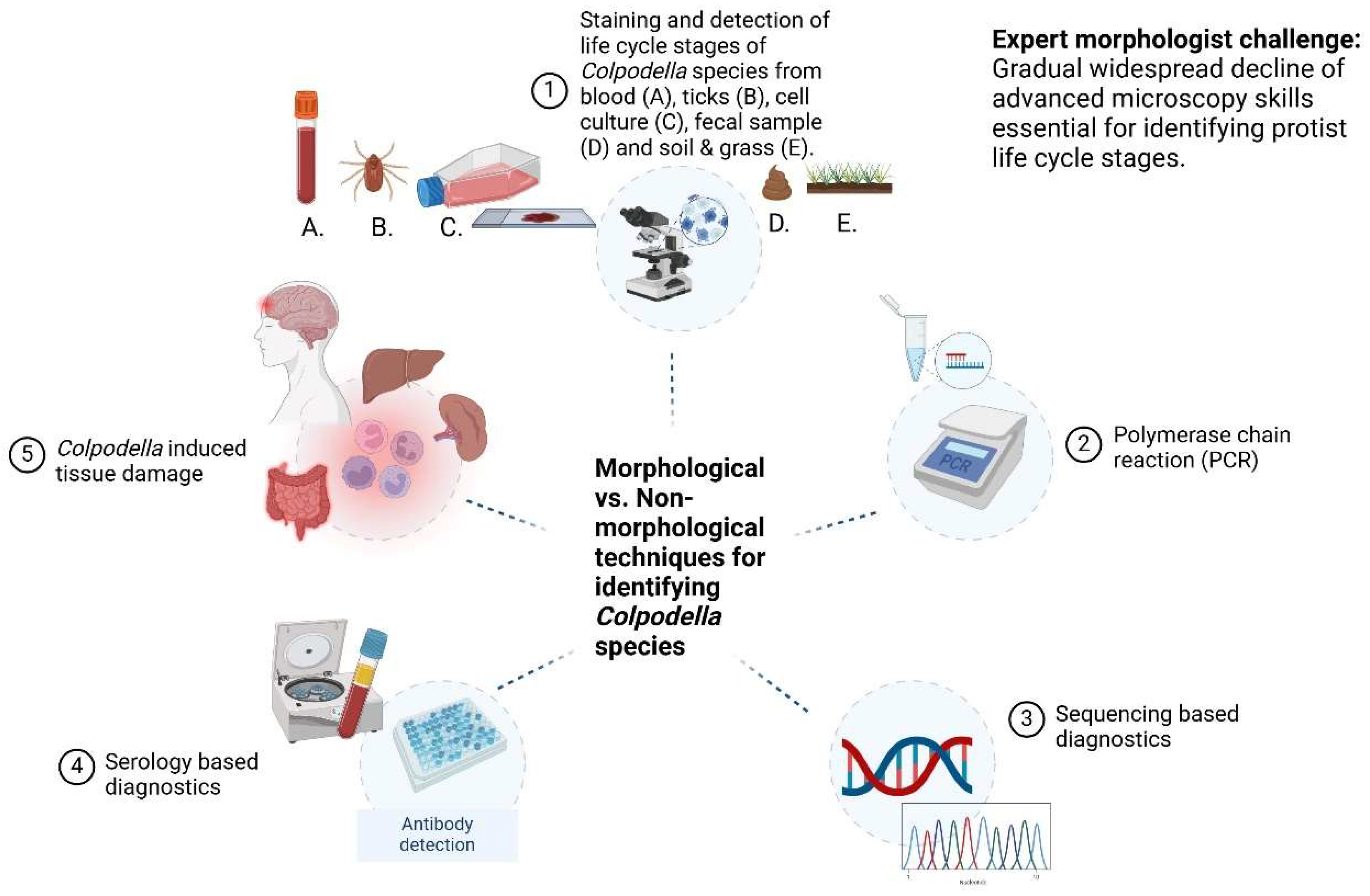
3. Techniques for Detecting Life Cycle Stage Markers of Colpodella spp. in Arthropod and Vertebrate Hosts

Among the apicomplexa, parasites are found in different tissues of the body in both humans and animals. The hemosporidians parasitize intracellular locations, being found within the erythrocyte for Plasmodium spp., Babesia spp., Haemoproteus spp., in lymphocytes for Theileria spp., in macrophages and other nucleated cells such as epithelial cells for Toxoplasma gondii and muscle cells for Sarcocystis spp. Cryptosporidium spp., Cyclospora spp. and Eimeria spp. infect epithelial cells of the gastrointestinal tract [45]. Among the intracellular infections within erythrocytes and lymphocytes, parasites remain within a parasitophorous vacuole (PV) that is formed during host cell invasion or they can exit the PV and develop directly within the cytoplasm. Pathogenic protists including amoeba, flagellates and ciliates range from opportunistic pathogens like Naegleria fowleri and Acanthoameba castellenii [45] to frank zoonotic pathogens like Babesia spp., and pathogens like Plasmodium spp., Entamoeba histolytica, Giardia lamblia, and Trichomonals vaginalis [45]. Sites for pathogenesis range from the brain for N. fowleri, eyes for A. castelleni, and GI tract, skin, brain, liver for E. histolytica. It is not unprecedented for free-living protists to become opportunistic and cause severe acute infections as is the case for the biflagellated N. fowleri, whose flagellated trophozoite stage initiates infection when forced into the nasal cavities and through the cribriform plate to the brain [39,45].

3.1. Mechanisms of Pathogenesis in Colpodellosis. What do We Know?

For Colpodella spp., we need to be able to describe whether trophozoites attach to host cells and express ligands used for adhesion or attachment to receptors on host cells. If cysts are present in the hosts, we need to know if cyst molecules promote inflammatory responses and whether the molecules stimulate innate and adaptive immune responses. If Colpodella spp. trophozoites penetrate host cells, we need to be able to describe specific host cells, receptors expressed on those cells and the participation of apical complex organelle contents from the rhoptries or micronemes acting as ligands for the host cell receptors. Whether endocytosis or phagocytosis is essential for intracellular invasion of host cells would need to be confirmed. Furthermore, Colpodella spp. proteins facilitating establishment of infection and nutrient acquisition would also need to be described. Modes of nutrient uptake such as endocytosis, phagocytosis, myzocytosis and trogocytosis which can be used to obtain nutrients within the host must be described. Pathogenic protists like Entamoeba histolytica and Trichomonas vaginalis feed by trogocytosis which mediates pathogenesis during infection [76,77]. The mechanism of Colpodella spp. multiplication within the host and spread to other tissues, as well as the development of cysts, if this occurs would need to be demonstrated. Presently, none of this is known. Colpodella spp. have been described in blood, CSF, urine and fecal samples and on the skin [22,23,24,25,26,27] (Table 1). Direct damage to host tissue can occur because of physical destruction of the host tissues by parasites physically destroying host cells, inducing inflammation or apoptosis, secretion of proteolytic enzymes, induction of cytokine secretion, or immune mediated damage due to antibodies, cytotoxicity, or cytokines produced in response to the parasitic activities of Colpodella spp. Whether Colpodella spp. are true pathogens remains a critical question that needs to be answered. The mode of nutrient uptake within the host, the spread of life cycle stages within the host and markers of virulence, hold clues for the mechanisms of pathogenesis in the host [78]. In order to fully understand how to prioritize efforts to investigate the biology of Colpodella spp. and identify additional molecular markers that will aid molecular diagnostic methods, identify host sites where Colpodella resides along with the life cycle stages present within the host, sample sources containing Colpodella spp. will need to be cultured, stained for microscopy and the morphology of Colpodella spp. described. Antibodies specific for Colpodella spp. life cycle stages will need to be identified. In animals shown to be positive for Colpodella spp., by PCR, serum samples from the animals can be collected to screen for Colpodella spp. proteins following short-term in vitro culture of samples such as hemolymph from ticks, blood, CSF and fecal samples from hosts as these samples may contain Colpodella spp. along with their prey. It will also be important to detect prey organisms for Colpodella spp. in the examined samples. Parabodo and Bodo spp. were identified with Colpodella spp. in cattle manure [20] and in blood samples of dogs [37] suggesting that prey species for Colpodella spp. may also be present in arthropod, human and animal specimens screened for Colpodella spp. Parabodo caudatus was identified in dog urine [79]. Western blotting can be used to identify proteins reactive with serum antibodies from infected humans and animals. Human antiserum from the female patient with relapsing fever was used in an immunofluorescence assay (IFA) to localize Colpodella spp. proteins within the erythrocyte [22].

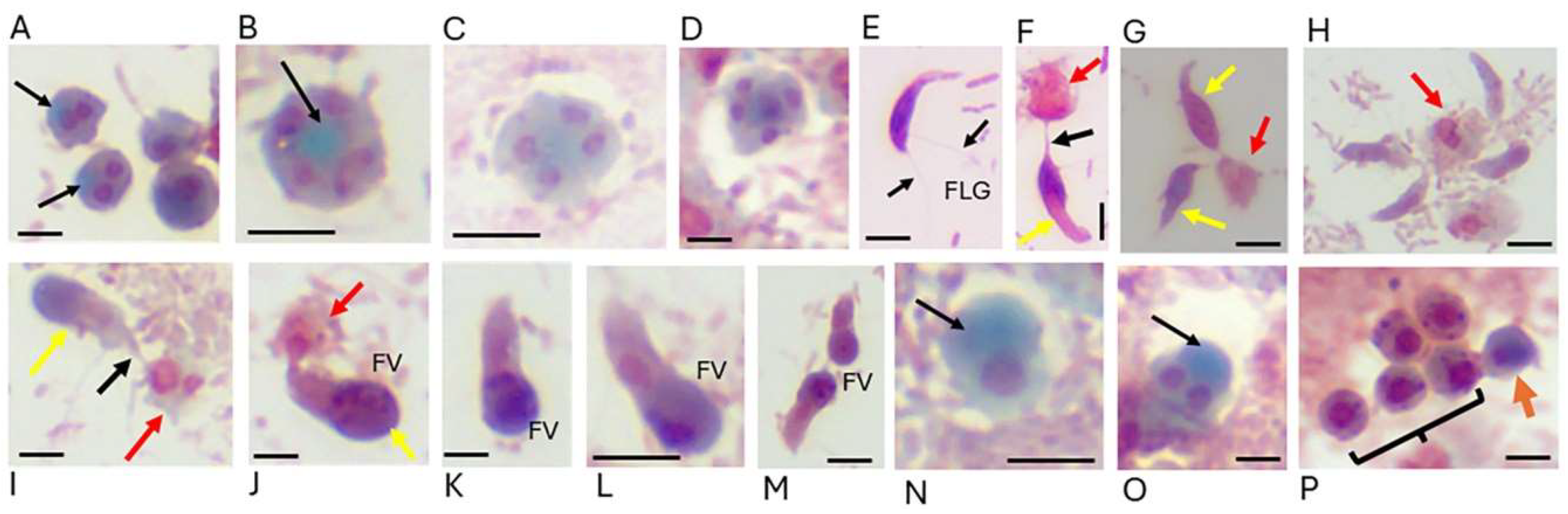
3.2. Microscopy: The Gold Standard for Parasite Identification

Contrary to statements that declare microscopy to be unimportant or unecessary for detecting Colpodella life stages, being able to identify the morphology of infectious disease organisms has been a fundamental feature of infectious disease, parasite investigations and diagnosis [44,80]. At the same time that diagnostic methods are relying more on digitized molecular platforms and less on morphological methods and microscopy, the number of trained microscopists and morphologists in parasitology is declining [80]. In the emergence or re-emergence of infectious disease organisms world-wide, a shortage of trained microscopists is a tremendous set back for public health [80]. Lack of expertise and inadequate morphological identification of parasites in general and emerging parasites like Colpodella spp. in particular can lead to misidentification of parasites. Failure to identify parasites in situations where molecular techniques are inadequate for the sample being tested like in fecal samples may lead to delayed diagnosis. Lack of sufficient genomic, proteomic and transcriptomic data in the databases for organisms like Colpodella spp. and other organisms that are not available in the databases can negatively impact the progress needed to understand the disease causing characteristics of Colpodella spp. [80,81,82]. Additionally, delays in diagnosis and worsening conditions due to delays in appropriate treatment will be detrimental to patient outcomes. For colpodellosis, morphological identification of the protist from collected samples is crucial for identifying life cycle stages initiating transmission and responsible for pathgenesis. In resource poor settings, expensive equipment for molecular testing may not be available, may be expensive and the wait times to receive results from samples submitted to reference labs may result in delays in patient diagnosis. Colpodella spp. have been identified in a variety of global locations and in animals in close contact with humans. PCR may not be available in some settings to make a molecular diagnosis. An integrated diagnostic approach that includes morphological identification of stained pathogens along with serology and molecular identification of DNA is more comprehensive and suitable for reliable identification of pathogens and will provide accurate diagnosis of infections in patients, particularly in identifying life cycle stages within the host [41]. Prevention efforts will be futile if the life cycle stage to target is unknown. The following techniques demonstrate the utility of integrating different diagnostic approaches to identify pathogens (Figure 1).

3.3. Morphology-Based Diagnostic Techniques

Morphology-based techniques utilizing staining of fixed smears prepared from blood, CSF and other body fluids as well as processing of fixed and sectioned tissue for histology needs to be performed to identify life cycle stages of Colpodella spp. Life cycle stages of parasites and other infectious disease organisms can be detected using these approaches. Cell culture of parasites to increase cell density for observations or propagation of the parasites in animal models for morphological evaluation also help confirm the identity of pathogens. This is currently a missing piece in the investigations of Colpodella species. Identifying the morphology of life cycle stages and understanding life cycle stage transitions within the host is essential for determining transmission stage dynamics, the infection's progression and severity, calculating parasite density in the bloodstream and other body fluids, and distinguishing the infection from other diseases with similar symptoms. In the reported cases of Colpodella spp. infections, the specific life cycle stages responsible for transmission and pathogenicity remain unknown.

3.4. Parasite Induced Tissue Damage

Colpodella spp. has been reported to cause inflammation and neurological symptoms in a tickborne human case of Colpodella infection [23]. An immunosupressed female patient with natural killer cell deficiency had intracellular red blood cell infection with Colpodella spp. along with relapsing fever and a male patient also reported to have relapsing fever was shown to have Colpodella spp. infection [22, NCBI accession number MF594625]. In the three cases, only Colpodella species was identified by PCR amplification of the 18S rRNA gene. The role of Colpodella spp. and any secreted/excreted products contributing to symptoms and pathogenesis in the patients is unknown. Colpodella gonderii and its prey Colpoda steinii were detected in the urine of a female patient experiencing chronic diseases along with a urinary tract infection. Treatment resolved the infection leading to the clearance of both protists [26]. However, it is unknown if the protists contributed to the infection. Giemsa staining was used to identify the morphology of predator and prey in the urine sample. It is unclear which type of tissues in the host might be most affected by Colpodella spp. Morphology-based diagnostics are important for assessing parasite-induced tissue damage. In animal infections, a South China tiger developed extensive tissue damage following symptoms that developed after a tick bite containing Colpodella spp. No other pathogen was reported in the tiger’s infection [12]. It is possible that Colpodella spp. by itself can induce immune related pathogenesis or by itself cause tissue damage through the secretion of enzymes and other molecules that promote tissue invasion and destruction. Because life cycle stages have not been identified by microscopy at specific tissue locations, it is unclear how tissues and organs were damaged in the animal host [12] and how Colpodella spp. infection induced the inflammation and neurological symptoms observed in human and animal infections.

3.5. Expert Morphologist Challenge

Increased reliance on non morphology-based detection technologies has led to the gradual, widespread decline of advanced microscopy skills essential for identifying parasite morphology and maintaining morphology expertise in the field. In a period when public health is threathened by emerging and re-emerging diseases, the loss of microscopy expertise is detrimental to accurate parasite and infectious disease agent recognition and identification [80]. Consequently, the objectivity of results remains a limitation due to the need for expert microscopists, parasitologists and pathologists who understand the significance of parasite life cycles in aiding accurate diagnosis. Several studies have evaluated the ultrastructure of different Colpodella spp. [2,3,4,6,7]. Phase contrast microscopy, DIC microscopy and scanning electron microscopy (SEM) have also been applied to examine the morphology of different Colpodella spp. [2,7,8,83]. These types of microscopy require expensive equipment and highly technical expertise along with long times for processing samples, and viewing and capturing images. Light microscopy remains a gold standard for identifying many parasites even in the advent of advanced molecular protocols [84]. This must be applied to Colpodella spp. investigations to aid morphological characterization of Colpodella spp. life cycle stages. We have evaluated traditional staining techniques like Giemsa staining, Wheatley’s trichrome staining and Kinyoun’s staining to identify trophozoites and cysts of Colpodella sp. ATCC 50594 [85]. The traditional staining techniques could allow for identification of trophozoites but were inadequate for distinguishing different developmental stages of Colpodella cysts and differentiating cysts of Colpodella from cysts of the prey, P. caudatus. We developed a new staining protocol, Sam-Yellowe’s trichrome staining that identifies trophozoites, different cyst stages and clearly distinguishes Colpodella sp. ATCC 50594 cysts from P. caudatus cysts [7,85]. The new staining protocols will be useful in identifying Colpodella species present in specimens collected from arthropods, human and animal hosts.

3.6. Polymerase Chain Reaction (PCR) and Sequencing Based Diagnostics

PCR techniques offer high sensitivity, enabling the identification of very small amounts of parasite DNA, which is crucial in scenarios where low levels of parasites in subclinical infections may pose a risk of reactivation or when dealing with cases of asymptomatic infections. Additionally, when the parasite levels are low in clinical samples, isolation of parasites for culture and detection through microscopic examination of smears can be challenging. The availability of different staining protocols, the processing of blood smears as thick or thin smears and the use of different fixation methods has been instrumental in maintaining microscopy as a central technique aiding accurate parasite diagnosis. In order to fully understand the epidemiology and distribution of species and strains of Colpodella spp., samples containing Colpodella spp. must be cultured and stained in addition to extracting DNA for PCR or RNA for qPCR. Among the pathogenic apicomplexa, organisms like Plasmodium have distinct morphological differences among the human, animal and avian species [86,87,88]. Morphological changes yet unknown may occur once Colpodella spp. is present in arthropod, and vertebrate host tissue. If this is the case, we must identify these life cycle stages to aid rational diagnosis and treatment plans. Similarly, distinct morphological differences are seen among different Babesia species [45,89]. Morphological differences also exist among the known Colpodella speices [2,3,4,6,7]. Colpodella species also have different morphological appearances and different sizes. For example DIC images show trophozoites of C. pugnax Cienkowsky, 1865 to be thin, fusiform and 7-15 µm in length with a prominent curved rostrum [2]. Colpodella turpis has a trophozoite that is 14-22 µm in length with a wide body, large hooked rostrum and flagella of equal length [2]. Unfed Colpodella gonderi trophozoites are as small as 5 µm, without a prominent rostrum and two flagella that can be two or more times longer than the body [2]. Trophozoites of Colpodella sp. ATCC 50594 are fusiform shaped, 7-9 µm in length with a slightly hooked rostrum. Morphological differences as well as structural differences observed ultrastructurally contributed to the change in the genus designation of Colpodella pontica to Voromonas pontica [6]. These critical differences underscore the need to culture and stain Colpodella spp. identified in arthropods, animal and human infections. In PCR reactions using primers targeting genes from pathogenic apicomplexans, nested PCR reactions along with DNA sequencing of amplicons have helped in the detection of Colpodella spp. However, PCR tests are not readily available in resource-poor settings where Colpodella spp. might be causing infections along side known apicomplexan, bacterial and viral infections. Sequencing-based diagnostics have an advantage over both morphology-based and non morphology-based methods due to its ability to provide species level identification and geographic strain differentiation. Notwithstanding, the morphology of Colpodella spp. must be identified in specimens collected from arthropod and vertebrate hosts.

3.7. Serology Based Diagnostics

Serology-based tests are useful for rapid detection of antigens and are widely employed to diagnose other Apicomplexan parasites. However, no serology-based testing techniques are available for detecting Colpodella spp. in clinical samples. In previous studies, we showed that antisera containing antibodies specific to P. falciparum rhoptry proteins and Kelch 13 proteins cross-reacted with Colpodella sp. ATCC 50594 antigens in immunofluorescence and western blotting [8,33]. These data along with PCR amplification and sequencing of the genes encoding the proteins demonstrate that rhoptry and Kelch 13 genes are conserved in Colpodella spp. Transcriptomic analysis identified many apicomplexan genes that are found in the chrompodellids which consists of Chromera velia, Vitrella brassicaformis, Voromonas pontica and Colpodella spp. [16]. The use of serological assays to identify host antibodies and parasite antigens is routinely performed as part of integrated approaches to diagnose parasitic infection and identify etiological agents for infection. Immunoassays like enzyme-linked immunosorbent assays (ELISA) and western blotting are used to screen for antibodies reactive with parasite antigens in infections including opportunistic and zoonotic infections. Human and animal host antisera has been used to detect antigens in various body fluids and samples such as in blood, urine, fecal and CSF samples. Immunofluorescence and immunoelectron microsopy allow for the visualization of parasite antigen distribution in parasites. Antibodies against Colpodella spp. antigens will need to be produced and antisera from human and animal hosts will need to be collected and used to screen for the presence of Colpodella antigens, and allow for additional Colpodella genes to be identified to aid phylogenetic analysis in addition to the 18S rRNA gene currently used in investigations. Colpodella spp., pellets can be obtained from Hay medium cultures for use in protein extraction for immunoassays. Evaluation of antibodies to N. fowleri in a Houston-Galvaston Texas (USA) healthy population found 89% seropositivity to N. fowleri [59]. In a study performed in northwestern Mexico, antibodies to the FLA, N. fowleri, Acanthamoeba and Balamuthia were identified in asymptomatic children and adults [90]. Immunoglobulin (Ig) M and IgA antibodies have also been detected in saliva with higher titers obtained in individuals with upper respiratory tract infections [90]. With the ubiquity of Colpodella spp. as indicated by the global distribution that has now emerged, more humans and animals may be exposed to Colpodella spp. than is currently understood. Serological assays as an intergrated component of epidemiological screening and clinical diagnosis cannot be ignored.

3.8. Cost Effectiveness

Diagnostic methods in parasitology consist of microscopy, immunoassays and molecular biology techniques. Each techniques varies in the level of sophistication, equipment cost and the need for highly skilled laboratory personnel. These techniques are used in integrated approaches to identify parasite stages in blood, CSF, urine, sputum, human and animal tissues and fecal samples. Parasites in wet mounts, stained smears and in histological samples can be identified by microscopy which remains the gold standard for parasite detection even though sensitivity may be weaker than that of immunoassays and nucleic acid amplification protocols [84]. Two major advantages of microscopy include the identification of different life cycle stage of parasites following staining and the simultaneous identification of multiple parasite species in one smear [84]. For example, among apicomplexans different species and life cycle stages of Plasmodium spp., Babesia spp., and Theilaria spp. can be identified by microscopy. Blood and sexual stage differences of Plasmodium species are a standard feature of malaria diagnosis [https://www.cdc.gov/dpdx/malaria/index.html accessed 2/10/25]. The detection of Colpodella spp. from arthropod, human and animal hosts will greatly benefit from microscopy since no life cycle stages have been identified in any of the cases of animal and human colpodellosis reported in the literature. Different life cycle stages may be present in arthropod and vertebrate hosts. Such differences are recognized in Plasmodium spp. where different stages such as sporozoites, oocysts and ookinetes are present in the female Anopheles mosquito and trophozoites and gametocytes are present in the vertebrate host. Light and electron microscopy can differentiate these differences [86,91]. In Babesia spp. different life cycle stages are present in the tick and vertebrate host. There is a pressing need to identify the different Colpodella spp. that have been detected in human and animal blood, feces, CSF, ticks and a biting fly. Many of these newly detected Colpodella spp. represent new species and strains of Colpodella for which additional investigations are needed to identify virulent and zoonotic species and strains. Some are species that have been described previously such as C. gonderi, C. tetrahymenae and C. edax [2,3,4,5,6,34]. However, novel species and strains may also be present in the recent reports. Compared to molecular and immunology techniques with higher sensitivity and specificity, the use of light microscopy is less costly but requires highly skilled microscopists to correctly identify parasites [80,92]. High equipment costs, technically trained staff and reliable infrastructure is a requirement for immunoassays and nucleic acid amplification protocols. These protocols require lengthy sample preparation methods and lengthy times to obtain results which can delay diagnosis. Point-of-care testing (POCT) is widely used for infectious disease and parasite identification and many POCT employ antibody detection of antigens from pathogens using lateral flow of reagents to result in antigen-antibody interactions in POCT devices [41,84,93]. The most widely used immunoassays for parasite antigen detection include ELISA, western blotting and immunofluorescent assay (IFA). Assay specificity and sensitivity is high, although problems with cross-reactivities and false negative and false positive results can occur [41,59]. These are mitigated by the use of controls and stringencies in assay development. Molecular techniques using nucleic acid amplification protocols for DNA and RNA, used for parasite detection include PCR, quantitative real-time PCR (qPCR), digital PCR (dPCR), loop-mediated isothermal amplification (LAMP) and CRISPR/Cas12a [84]. These protocols require reliable infrastructure, costly equipment and skilled personnel to perform and interprete assay results. Combined PCR-ELISA protocols are also in use with different mutiplex assays under development for multiple antigen detection assays. 18s rRNA gene sequences, tandem repeat sequences, internal transcribed spacer (ITS) sequences and mitochondrial genes are used in nucleic acid amplification protocols [84]. With no standardized lab tests yet for Colpodella species detection, and with Colpodella spp. detected in fecal samples, blood and CSF, nucleic acid amplification tests will need to be performed from nucleic acids extracted from appropriately stored and handled samples in order to obtain useful data. Contreras-Ferro et al. [41] indicate that qPCR is most useful if performed within 5-10 days of symptom onset. The incubation period for colpodellosis is unknown. For many tickborne pathogens the incubation period and time for development of symptoms is known. Once these characteristics have been determined for Colpodella spp. it will be crucial to determine this for colpodellosis when other tickborne pathogens are identified due to the co-infections that have been reported with bacteria and the piroplasms Babesia spp. and Theileria spp., and Cryptosporidium spp. Amplification of 18S rRNA gene sequences have been used in most of the human and animal cases reporting Colpodella spp. detection. It is critical that additional genes be identified in Colpodella spp. to aid accurate identification of Colpodella species that may be missed when 18S rRNA gene sequences from piroplasms or Cryptosporidium spp. are used for detection. The availability of Hay medium for culturing Colpodella spp. and staining protocols that can be performed in less than 10 minutes will allow for Colpodella spp. life cycle stages to be identified, and pellets for Colpodella protein and nucleic acid extractions to be collected. Additionally, primers targeting the 18S rRNA genes of bodonid, algae and ciliate prey should also be used for amplification to determine if the prey are present in the arthropod, human and animal hosts. Basic light microscopy and morphology-based detection of parasites is rapid, cost effective, and does not require advanced technologies, costly equipment and higly skilled professional staff, which is beneficial in resource poor settings in low and middle income countries (LMICs) [59,94]. Transmission of Colpodella spp. as a zoonotic or opportunistic parasite along with neglected parasites of poverty in endemic areas, poses an additional burden to poor communities. In the absence of costly molecular biology and immunoassay equipment, Colpodella spp. cannot be detected. At minimum, microscopy should be performed on collected samples to aid morphological identification of Colpodella spp.

4. Culture Conditions for Colpodellids and Chromerids

Efforts to cultivate Colpodella spp. along with staining for morphological identification is needed to aid molecular investigations aimed at identifying molecular markers of Colpodella spp. that facilitate transmission, survival and pathogenesis in hosts, and determining which species to prioritize for the risk of zoonotic infections to humans and for infections in animals (domestic and live stock, recreational animals and wildlife) [95]. The chromerids Chromera velia and Vitrella brassicaformis are included in this section because some of the same culture media used for colpodellids has been used for chromerids.

4.1. Culture Media for Cultivating Colpodella Species

Colpodella spp. can be cultured in various types of media to allow for cell biology, molecular biology and microscopy investigations. Detection of Colpodella spp. genes and antigens is highly feasible, as large volumes of culture can be obtained for protein and nucleic acid extraction, and for antibody production. The availability of reliable and consistent culture conditions will aid the idetification of Colpodella spp. antigens that will be crucial for developing diagnostic tests. The availability of reliable culturing techniques allowed us to investigate and describe the life cycle of Colpodella sp. ATCC 50594 in culture [8]. Colpodella pseudoedax and C. unguis have been cultured in Pratt medium and Schmaltz-Pratt medium consisting of 1L H2O, 28.15 g NaCl, 0.67g KCl, 5.51g MgCl2 6H2O, 6.92g MgSO4 7H2O, 1.45g CaCl 2H2O, 0.1g KNO3, 0.01g K2HPO4 3H2O), 20-22% salinity with protist prey [1]. Spumella sp. and Procryptobia sorokini served as prey in the medium. Voromonas pontica (formerly Colpodella pontica) isolated from the coastal waters of the Black Sea has also been cultured in Schmaltz-Pratt medium [2] with Escherichia coli and Bodo sorokini provided in the medium as a nutrient source for the prey. The bacteria serve as food for the protist prey and B. sorokini is the prey for V. pontica. We have cultured V. pontica successfully in concentrated Hay medium (VWR, Inc.) mixed with sea water, bacterized with Enterobacter aerogenes and cultured at 24°C with Percolomonas cosmopolitus as prey [96].
Colpodella gonderi isolated from a grassland site, in shallow brown soil with poor minerals was cultured in soil mixed with rainwater in a petri dish with incubation at 15°C for 4 days [34]. Colpoda steinii, Grossglockneria acuta and Pseudoplatyophora nana served as prey in the culture. Colpodella vorax isolated from pond water that had dead leaves and grass was cultured in pond water contained in a petri dish [3] with Bodo caudatus, Spumella sp., Synura peterseni, Chilomonas paramaecium, Euglena gracilis and Colpoda cucullus as prey for C. vorax.
Colpodella pugnax Cienkowski, 1865 isolated from an artificial hypersaline (26% NaCl) lagoon at Whyalla, South Australia was cultured with the prey species Dunaliella virdis at 28°C under varying light conditions [2]. The type of medium used for cultivation was not specified. Similarly, no culture conditions were specified for C. angusta. However, Procryptobia sorokini, Spumella sp. and Parabodo caudatus served as prey for C. angusta [16].
Colpodella tetrahymenae isolated from the rainforest soil from La Selva, Costa Rica was cultured with soil mixed with natural bacteria with the prey Tetrahymena pyriformis [16]. The medium used for suspending the soil was not specified.
Colpodella sp. ATCC 50594 is cultured in ATCC culture medium 802 (Sonneborn’s Paramecium medium) [https://www.atcc.org/products/50594 accessed 2/10/25] and in Hay medium bacterized with Enterobacter aerogenes and Parabodo caudatus present in the diprotist culture as prey [66]. Hay medium is useful for growing large volumes of diprotist and monoprotist cultures in tissue culture flasks. Cultures in tissue culture flasks are examined under an inverted microscope to monitor cell growth. Bacterized Hay medium cultured overnight at 37°C is used for the subculture of Colpodella sp. cysts the following day. Following Colpodella sp. cyst inoculation of the bacterized medium, cultures are incubated at 24°C for up to 36 h post subculture [8]. Active cultures containing many young active trophozoites feeding by myzocytosis, producing transient cysts which excyst to produce juvenile trophozoites can be found within 20 to 30 h post subculture, with resting cysts beginning to form 30 to 36 h post subculture [8]. Resting cysts can remain in culture up to 14 days and can be used to initiate a new round of cultures. Hay medium can be purchased as a 1x working medium ready to be used or a concentrated stock medium can be purchased and used after diluting it with sterile distilled water to obtain a working medium [8,96]. Centrifugation of the culture allows for the collection of pellets for macromolecular extractions. Protists in the cultures can also be fixed using a final concentration of 5% formalin, centrifuged and pellets washed and resuspended for the preparation of smears and staining for light microscopy, fluorescent staining, immunofluorescence and confocal microscopy [7,75]. In many of the environmental sites where Colpodella spp. have been detected, chromerids such as Chromera velia and Viterella brassicaformis have also been detected [16,97,98]. Chromera velia was first isolated from stony coral Pleslastrea versipora in Australia [97] but it has also been obtained from Boothbay Harbour, ME from the culture collection of marine phytoplankton and cultivated in f/2 medium in sea water with a 12/12 or 8/16 hour light and dark cycle at 26°C [97]. Procryptobia sorokini, Spumella sp. and Parabodo caudatus served as prey. Vitrella brassicaformis was isolated from One Tree Island, the Great Barrier Reef from a stony coral Leptastrea purpurea, with Procryptobia sorokini, Spumella sp. and Parabodo caudatus served as prey [98]. Artificial sea water f/2 medium was used for cultivation of V. brassicaformis at 26°C in flat plastic tubes [98]. Solid agar plates containing 50 mg/ml kanamycin were also used to culture V. brassicaformis at 26°C in a 12/12 hour light/dark regime [98]. Depending on the source of Colpodella spp. from the environment, chromerids may also be present in the sample. In the environment, Colpodella spp. have been isolated from the soil, fresh water, waste water and sea water (Table 1).

4.2. Light, Differential Interference Contrast (DIC) Microscopy and Electron Microscopy (EM)

For Colpodella species except for Colpodella sp. ATCC 50594, light microscopy without staining has been used to examine cells. Phase contrast microscopy, DIC, scanning electron microscopy (SEM) and transmission electron microscopy (TEM) have been used to examine the morphology and ultrastructure of cells [2,3,6,83]. The use of 4’,6-diamidino-2 phenylindole (DAPI) in fluorescent and confocal microscopy has been instrumental in identifying the nuclei and kinetoplasts of P. caudatus and of Colpodella sp. [7,8]. Colpodella sp. ATCC 50594 has been examined with wet mounts, staining and light microscopy, DIC and TEM to examine cell morphology and ultrastructure of Colpodella sp. ATCC 50594 [75]. Different staining techniques that we have used to study Colpodella sp. ATCC 50594 have included the use of Giemsa stain, Wrights-Giemsa stain, H&E, Congo red, calcoflour, Acid-Fast, Wheatley’s trichrome stain and Sam-Yellowe’s trichrome staining series [8,35,75,85] developed to identify and distinguish life cycle stages of Colpodella sp. ATCC 50594 and to distinguish cysts of Colpodella sp. from cysts of its prey P. caudatus. Sam-Yellowe’s trichrome staining protocol was instrumental in aiding the identification of previously unknown developmental stages of Colpodella sp. ATCC 50594 and allowed the description of the life cycle of Colpodella sp. ATCC 50594 in culture as well as the identification of asymmetric division in the cyst stage [8]. The staining protocol is completed in less than 10 minutes. The life cycle stages of Colpodella sp. ATCC 50594 stained with Sam-Yellowe’s trichrome stain are shown in the order of life cycle stage transitions in culture (Figure 2). We propose that this staining protocol will be highly useful in staining formalin-fixed hemolymph samples and other specimens from arthropods, blood, CSF, fecal samples and biopsied specimens for the detection and morphological characterization of Colpodella spp. Four staining protocols from a series that has been used in previous studies are shown in Table 3 (85, 99].
Table 3. Sam-Yellowe’s trichrome staining protocols for Colpodella spp. staining.
Sam-Yellowe’s Trichrome Staining Application of dyes in the order of incubation
Sam-Yellowe’s trichrome A 0.3 % Methylene blue (1 min)
1% Brilliant green (5 min)
1% Neutral Red (1 min)
Distilled water washes were performed in between each dye incubation. After the last wash, smears are air-dried before microscope observation using oil immersion at x1000.
Sam-Yellowe’s trichrome D 1% Crystal violet (30 s)
1% Brilliant green (2 min)
1% Neutral red (1 min)
Distilled water washes were performed in between each dye incubation. After the last wash, smears are air-dried before microscope observation using oil immersion at x1000.
Sam-Yellowe’s trichrome E 1 % Crystal violet (30 s)
1% Brilliant green (2 min)
1% Safranin (1 min)
Distilled water washes were performed in between each dye incubation. After the last wash, smears are air-dried before microscope observation using oil immersion at x1000.
Sam-Yellowe’s trichrome J 0.3 % Methylene blue (1 min)
0.5 % Fast green in alcohol (5 min)
1% Neutral Red (1 min)
Distilled water washes were performed in between each dye incubation. After the last wash, smears are air-dried before microscope observation using oil immersion at x1000.
In our previous studies, dyes from different vendors were evaluated to establish the reproducibility and consistency of the appearance of stained cells. The staining protocol allows for the differentiation of both trophozoites and cysts of the predator and prey, and the identification of Cryptosporidium parvum oocysts and blood stages of P. falciparum [99]. Primary dyes such as methylene blue, crystal violet and Kinyoun’s carbol fucshin have also been used to stain life cycle stages of Colpodella sp. Both dyes stain trophozoite and cyst stages but do not discriminate cyst stages of the predator and prey. Both C. velia and V. brassicaformis have also been studied using light microscopy, SEM and TEM [97,98,100], resulting in the identification of life cycle stages for both chromerids.
Life cycle stages are shown as they occur during the life cycle in culture [Getty et al.]. After preying on P. caudatus, Colpodella sp. ATCC 50594 encysts. The developmental stage of the cyst includes young demilune cysts with a single nucleus. The cyst undergoes cell division to form two or more nuclei (A-D) The black arrows shown in A and B identify the remnants of the food vacuole (FV). Excystation leads to egress of young bi-flagellated trophozoites possessing heterodynamic flagella (FLG) (E). The trophozoites attach to P. caudatus and feed through myzocytosis. One or more Colpodella sp. trophozoites can attach to a single prey during myzocytosis. Four trophozoites are shown feeding on one prey in panel H. The yellow arrows identify Colpodella sp., the red arrow identifies the prey P. caudatus, and the black arrow identifies the tubular tether that forms between predator and prey as shown in F (F-H). The trophozoite aspirates cytoplasmic contents of the prey into a posterior FV that enlarges during feeding (I & J). The black arrow in panel I is the tubular tether formed during myzocytosis. Following feeding, the unattached pre-cyst stage still containing the nucleus is shown (K-M). Flagella and cytoplasmic organelles are degraded and encystation begins (N-P). The arrows in N and O identify the remnants of the food vacuole. In panel P, the single nucleus demilune cyst of Colpodella sp. ATCC 50594 (orange arrow) can be distinguished from the five cysts of P. caudatus shown in the bracket. Scale bars: Panels A, D-K, M, O and P; 10 µm. Panels B, C, L, N; 20 µm. Cells from nutrient uptake cultures in panels A-D, and G-P were stained with Sam-Yellowe’s trichrome A. Cells in panels E and F were stained with Sam-Yellowe’s trichrome E. Images shown were captured at x1000 magnification as described in [7,8]. The images shown are from archived images in the Sam-Yellowe lab. They have not been published previously.

5. Colpodella spp. in Coinfections

Colpodella spp. detected in arthropods and vertebrate hosts have been identified coinfecting hosts with the piroplasms Babesia spp. and Theileria spp. or with Cryptosporidium species. Babesiosis is an emerging human tick borne zoonotic disease in different geographic regions globally [101,102]. Human tickborne infections are considered rare events. However, the reported Colpodella spp. cases indicate that coinfections may be more common than expected [103]. Performing culture and microscopy can be challenging in coinfections, and PCR may not amplify target DNA in some specimens when single PCR is performed. Multiplex PCR techniques and next generation sequencing (NGS) techniques may improve detection of mixed pathogens in coinfections [103]. Colpodella spp. have also been detected coinfecting ticks and animal blood with bacteria such as Anaplasma spp., Rickettsia spp. and Erlichia spp. In coinfected hosts, ascribing the resulting pathology and tissue damage to a specific pathogen is fraught with uncertainty. The number of reports of Colpodella spp. infections in humans and animals has increased in the last few years (Table 4). The increased reports of single infections where only Colpodella spp. was identified and coinfections where multiple pathogens were also detected is discussed below and summarized in Table 4. Six genera of ticks; Ixodes persulcatus, Rhipicephalus (Boophilus) microplus, Rh. bursa, Rh. duttoni, Rh. haemaphysaloides, Haemaphysalis longicornis, H. flava, H. bispinosa, H. hystricis, Hyalomma dromedarii, Dermacentor everestianus, D. nuttalli, D. andersoni, D. atrosignatus, D. taiwanensis and Amblyomma javanense [10,11,12,13,14,22,23,36] (Table 2) have been reported to carry Colpodella spp. in addition to Stomoxys indicus that was reported to carry Colpodella tetrahymenae [15].

5.1. Colpodella spp. Detected in Blood

Xu et al. 2022 [24] examined blood from 400 horses in China that were screened for piroplasms using primers targeting 18S rRNA gene in PCR. DNA sequences in 2 out of 400 horses showed homology to Colpodella spp. Phylogenetic analysis showed both identified species closely related to Colpodella ATCC 50594, a model isolate obtained from brown woodland soil at Gambrill State Park, Frederick, Maryland [https://www.atcc.org/products/50594 accessed 2/10/25], and to Colpodella sp. strains HEP (NCBI accession number GQ411073) and HLJ (NCBI accession number KT364261) isolated from two patients in China and a clade containing the apicomplexans, Babesia spp., Theileria spp., Toxoplasma gondii, Cryptosporidium spp., and Plasmodium spp. An additional 132 horses out of 400 were infected with Theileria equi and 2 out of 400 were infected with Babesia caballi. No symptoms were reported in the horses.

5.2. Colpodella spp. Detected in Blood and Ticks from Cattle and Goats

Free grazing cattle and goats were screened for tickborne parasites. Blood and ticks from 98 cattle and 104 goats in the Puglia region, Southern Italy were screened for tickborne parasites [10]. Genomic DNA extracted from the blood and ticks were used in conventional PCR using primers targeting the 18S rRNA gene for Babesia spp., Theileria spp. and primers targeting the gltA gene for Rickettsia spp. From the cattle blood 36 out of 98 were positive for piroplasms, 7 cattle were positive for Babesia bigemina, 29 cattle were positive for the Theileria sergenti/buffeli/orientalis group, and 2 for Sarcocystis cruzi. From the goat blood, 39 goats out of 104 were positive for Babesia spp. Blood from both cattle and goats was negative for Rickettsia spp. Adult male and female ticks collected from cattle and goats were also examined for tick-borne pathogens. Forty-two adult male and female ticks collected from 33 cattle were evaluated. Thirty-one were identified as positive for Rhipicephalus bursa and eleven were positive for Rh. seucundus. One female Rh. bursa was positive for Colpodella spp. with 100 % sequence identity to Colpodella spp. with NCBI accession number OQ540588.1. In phylogenetic analysis, the identified Colpodella spp. DNA sequence clustered with a Colpodella clade from China and was close to a paraphyletic clade containing Theileria spp., Babesia spp. and Cytauxzoon spp. Among forty-seven adult male and female ticks collected from thirty-six goats, twenty-five were identified as Rh. bursa, and eleven as Rh. secondus. Rickettsia spp. were identified in 1 female Rh. bursa. No symptoms were reported in the cattle and goats [10].

5.3. Colpodella spp. Detected in Blood from Dogs, Cats and Ticks

The presence of piroplasms in dogs and cats in Guiyang, southwestern China was investigated. Colpodella spp. was identified along with Theileria spp. Blood samples were collected from pet cats and dogs attending a veterinary hospital in Guiyang, China [36]. Genomic DNA was extracted from dog and cat blood and used for nested PCR using 18S rDNA primers targeting piroplasm parasites. Sequenced PCR products were BLAST searched and aligned with DNA sequences for P. falciparum and P. berghei, retrieved from NCBI and used for phylogenetic analysis. In pet cats Theileria uilenbergi, T. luwenshuni and Colpodella spp. were detected and in pet dogs Theileria uilenbergi and Colpodella spp. were detected [36]. Colpodella spp. from dogs and cats had 94.99% identity to Colpodella spp. with NCBI Accession OQ540589.1 and 81.71 % identity to each other and 81.77 % and 83 % identity to Colpodella sp. ATCC 50594. Cat Colpodella spp. with NCBI Accession OR226256 clustered in a single clade with Colpodella spp. from human blood with 99.60% identity but distant from gene sequences detected in ticks, horses, cattle, and tigers. Dog Colpodella spp. with NCBI Accession OR226258 clustered into a clade with Colpodella spp. NCBI accession number OQ5405891 detected in H. longicornis from Yiyuan County, Shandong, China with 94.99% identity but distant from Colpodella spp. gene sequences detected in humans, horses, cattle and tigers [36].
In another study, goats and dogs were examined in an epidemiological investigation to identify potential parasites in ticks infesting the animals in Yiyuan County in Central Shandong Province, China [14]. Genomic DNA extracted from ticks of Haemaphysalis longicornis was used in nested PCR amplification using oilgonucleotide primers targeting the 18S rRNA gene of Theileria and Babesia spp. Following amplification, a second set of primers designed to distinguish organisms in co-infections and to identify each species of Theilaria and Colpodella were also used in PCR. Coinfections of Theileria spp. and Colpodella spp. as well as individual infections with Theileria luwenshuni or Colpodella spp. were detected in ticks from goats and dogs. Colpodella spp. identified in goattick 168 had 93.66% identity to Cryptosporidium stuthionis NCBI accession number AJ697751.1 identified from farm ostriches and therefore named Colpodella sp. struthionis [14]. Goattick 161 had 92-98% identity with Colpodella tetrahymenae NCBI accession number MH208619.1. However, on phylogenetic analysis with sequences of Theileria spp., Colpodella spp. and Cryptosporidium spp. retrieved from NCBI, it was distant from other Colpodella tetrahymenae and Colpodella angusta suggestive of strain differences [14]. This species was named Colpodella sp. yiyuansis [14]. Morphological identification of the Colpodella species identified in H. longicornis is essential to determine whether the protist identified by DNA is Cryptosporidium or Colpodella. Cryptosporidium spp. develops oocysts which are identified in fecal samples and Colpodella spp. form cysts. Depending on the staining technique used, sporozoites within the oocyst of Cryptosporidium spp. can be discerned. Colpodella species develop cysts in those species that encyst. They also have trophozoite stages. Pre-cyst stages can also be identified for Colpodella spp. Without morphological identification of life cycle stages, errors in identification will be made leading to incorrect identifications and assigned nomenclature. No symptoms were described for the goats, dogs and cats.
In a Cambodian study, next generation sequencing (NGS) was used in investigations of dog populations where screenings were performed on blood collected from 467 dogs to identify DNA sequences of bacterial and blood borne pathogens [37]. Colpodella spp. along with prey protists of Parabodo and Bodo spp. were identified in one dog. The DNA sequence had 95% identity to Colpodella spp. identified in horse blood, NCBI accession number MW261750.1 [37]. No symptoms were reported in the dogs [37]. Other pathogens detected in the dogs screened, include the bacteria, Mycobacterium spp., Coxiella spp., Neisseria spp. Rhodococcus spp., Treponema spp., Prevotella colorans, Mycoplasma spp., Rickettsia spp. and Wolbachia spp. [37]. Pathogens identified from vectors such as fleas, lice, and ticks infesting the dogs include the apicomplexans, Babesia vogeli and Hepatozoon canis, the kinetoplastids Trypanosoma evansi, Bodo spp., and P. caudatus. The bacterial pathogens, Anaplasma platys, Ehrlichia canis, Mycoplasma haemocanis, Bartonella clarridgeiae, and Candidatus Mycoplasma haematoparvum were also identified [37].

5.4. Colpodella spp. Detected in Camel Ticks, Cattle and Wildlife

In investigations of tick infestation in Egyptian camels in Southern Egypt, within the Luxor and Aswan governorates, 200 camels were investigated [13]. Camels were checked for ticks during regular veterinary examinations. The ticks identified belong to the species Hyalomma dromedarii. DNA extracted from the ticks was used as a template in nested PCR with primers targeting 18S rRNA piroplasmid genes. Thirty out of 297 ticks examined carried Colpodella spp. with DNA sequences homologous to Colpodella spp. identified in H. longicornis with NCBI accession number OQ540590Q, Rh. haemaphysaloides, NCBI accession number MH208621 and humans in China, NCBI accession number GQ411073. Babesia bovis was detected in 16 of the 297 ticks using the spherical body protein-4 gene. No symptoms were reported in the camels. Coinfections have also been identified in blood samples from wild animals (impalas, hartebeests, buffalos, sitatungas, lions, sable antelope and wild dogs) and cattle in the greater Kafue ecosystem in Zambia, using the V4 hypervariable region of the 18S rRNA gene of piroplasms [38]. Coinfections of Theilaria spp., Babesia spp., Hepatozoon spp. and Colpodella spp. were detected [38]. None of the animals had symptoms of infection.

5.5. Colpodella spp. Identified in Fecal Samples from Sheep, Goats, Cattle, Duck and Eurasian Coot

Two clusters of Colpodella spp. were identified in phylogenetic tree analysis following detection of Colpodella spp. in fecal samples collected from goats and sheep in the Federal Capital Territory (FCT) and Jos, Nigeria [18]. The fecal samples were positive for Cryptosporidium spp. oocysts following microscopic examination. DNA was extracted from the fecal samples and used in nested PCR with oligonucleotide primers targeting the full length of Cryptosporidium spp. 18S rRNA gene. DNA from Cryptosporidium spp. and Colpodella spp. was amplified. Colpodella spp. was identified in 2 Red Sokoto goats, and 3 sheep of the Yankassa breed and 1 sheep of the Balami breed. Four sheep and 1 goat had diarrhea, and 1 goat had semi-formed stool [18]. DNA sequences from Colpodella species identified in this study had sequence identity to Colpodella spp. strains identified from Cyprus [19]. Two clusters consisting of Colpodella spp. were closely related to strains identified in a duck and fox in Cyprus, ticks infesting cattle from Mozambique [9], and a second cluster related to Colpodella spp. from a horse in China [18]. Fecal samples were collected from 180 animals that included goats, sheep, foxes, ducks, Eurasian Coots and Eurasian Teals in Cyprus [19]. DNA was extracted from the fecal samples and used for PCR using oligonucleotide primers targeting 18S rRNA genes from Cryptosporidium spp. A 600 bp DNA band identified on agarose gels and sequenced identified 1 of 10 goats, 1 of 29 foxes, 1 of 48 Eurasian Coots and 1 of 7 ducks positive for Colpodella spp. following a BLAST search. The four sequences were similar and had high sequence identity to Colpodella sp. strain HEP with NCBI accession number MH208621 from a human relapsing fever infection [22], strain HLJ with NCBI accession number KT364261 from a tickborne human infection with neurological symptoms [23] and to the Colpodella spp. detected in sheep from Nigeria with NCBI accession number OQ876040.1. In a Turkish study, Colpodella spp. was also isolated from calves with diarrhea (Table 1). The DNA sequence identified with NCBI accession number JN245625 had sequence similarity to Colpodella spp. DNA sequence from strain HEP identified in the human infection from Yunnan Province, China [22].

5.6. Colpodella spp. Detected in Pangolin Ticks

Twenty-one rescued and sick Malayan pangolins infested with ticks were examined for ectoparasites at Guangdong Provincial Wildlife Rescue Center at Guangzhou Zoo and the Guangdong Institute of Applied Biological Resources in China [11]. Seventeen pangolins were found to be tick infested with Amblyomma javanense. Following the death of all the pangolins, DNA was extracted from tissues, blood and collected ticks for PCR. RNA was extracted for RT-PCR. Tissue sections of damaged tissues stained with H&E were also examined. No pathogens were detected in the tissues. DNA was examined for viral pathogens, bacteria and protozoan parasites. DNA sequences obtained from amplified DNA was BLAST searched, and homology obtained for Rickettsia spp, Anaplasma spp. Erlichia spp. Babesia spp., and Colpodella spp. were detected in ticks. Colpodella spp. was not detected in pangolin tissues. Theileria spp. and Hepatozoon spp. were not detected. Also, no viruses were detected. The pangolins showed symptoms of poor vigor, cough, drowsiness, anorexia, and edema of the extremities [11]. Coinfections of Colpodella spp. and Rickettsia spp., Colpodella spp., Rickettsia spp. and Ehrlichia spp. were detected. Bloody stools, hematuria, convulsions and other neurological symptoms were observed in the animals before death [11]. Severe organ damage, congestion and edema of major organs, ascites and inflammation were observed upon autopsy of the pangolins [11]. It is unclear what contributions Colpodella spp. had to the pathogenesis observed in the pangolins and whether A. javanense transmitted Colpodella spp. to the animals. Six out of 33 ticks examined carried Colpodella spp. Two Colpodella spp. had sequence identity to Colpodella spp. NCBI accession number MH012046 from Qinghai and Colpodella spp. NCBI accession number MH208621 from Yunnan. Four sequences had identity to Colpodella spp. NCBI accession number KT600661, the Colpodella species isolated from the human infection with neurological symptoms. In a study to identify the potential for the spread of pathogens by mammalian vectors in Poland, raccoons were investigated. One hundred and seventy ear fragments were collected from roadkill or from live-trapped animals in the “Warta Mouth” National Park [27]. DNA extracted from dried ear fragments was used for PCR using highly conserved primers targeting the V4 hypervariable region of the 18S rRNA gene in a semi-nested PCR. Following DNA sequencing of amplified DNA and BLAST searches, different eukaryotic organisms were identified including chlorophyta, fungi, amoeboids, uncultured colpodellidae and Colpodella spp. [27].

6. Colpodella spp. in Single Infections

6.1. Colpodella spp. in Human Infections

A blood infection was reported in a 57 year old woman from Yunnan Province in China with an immunodeficiency of natural killer cells [22]. Symptoms of productive cough, malaise, hemolytic anemia and relapsing illness was reported [22]. Oligonucleotide primers targeting a conserved fragment of Babesia spp. 18S rRNA was used in PCR and the gene amplified matched with 89% identity to Colpodella tetrahymenae [22]. Following treatment with atovaquone and azithromycin, the Colpodella infection was cleared [22]. Anti-Colpodella antibodies from the patient reacted with Colpodella in erythrocytes by IFA. In a second case of relapsing fever reported in a male patient, Colpodella DNA sequences with NCBI accession number MF594625 were also identified. However no further details were provided for the patient. A 55 year old female patient with neurological symptoms following a tick-bite, from Heilongjiang Province of Northeast China was also reported [23]. Her symptoms included fever, dizziness, gait disturbance, and headache [23]. Polymerase chain reaction of blood and CSF using primers targeting Babesia spp. identified Colpodella spp. in CSF but not in the blood sample [23]. Treatment with doxycycline cleared the infection. In both human cases, Babesia spp., Eperythrozoon spp. [Yuan et al.], Borellia spp., Borellia burgdoferi sensu lato (sl), Babesia spp., tick-borne encephalitis virus (TBEV), Anaplasma spp., Ehrlichia spp. and Rickettsia spp. were negative by PCR [23]. Although an IgG titer of 1:126 in the serum of the patient was obtained for B. burgdoferi leading to suspected Lyme disease, PCR was negative [23]. Two Colpodella spp. were detected in 2 out of 474 adult Ixodes persulcatus from woodlands around the patients’ home [23]. Both DNA sequences with NCBI accession numbers KT600661 and KT60062 shared 93.8% identity but had 88.0-89.0% identity to the tick Colpodella sp. strain HLJ from the patient [Jiang et al.]. Colpodella gonderii along with its prey Colpoda steinii was identified in the urine of a 70 year old female patient with a history of chronic diseases, in Cluj-Napoca, Romania by wet-mount microscopy and Giemsa staining [26]. No other parasites were detected. The patient was admitted for respiratory symptoms including breathing difficulties, dyspnea with orthopnea but no urinary symptoms [26]. Giemsa staining identified Colpodella gonderii and Colpoda steinii trophozoites. After treatment with ceftriaxone and metronidazole, both protists could no longer be detected in the urine [26]. Standard symptoms for colpodellosis have not been described. However, in the relapsing infection [22], Babesia-like symptoms of anemia, elevated reticulocyte and lactate dehydrogenase levels were reported [22].

6.2. Colpodella spp. Detected in Fecal Samples from Large Cats

Fifty-six fresh fecal samples from a Siberian tiger, White tiger, Bengal tiger, African tiger, White lion, Lynx, and Jaguar from Harbin Zoo, China were collected to screen for Cryptosporidium spp. [17]. DNA was extracted from the fecal samples and amplified with PCR using oligonucleotide primers targeting 18S rDNA sequences of Cryptosporidium sp. In 7 out of 56 samples Colpodella spp. DNA was amplified and showed a band of 583 bp on agarose gels. DNA sequence identity of the 18S rRNA gene was 97.26% between Cryptosporidium and Colpodella spp. DNA sequences. No symptoms were reported in the large cats [17].
Twenty-two Colpodella species were detected in ticks (3 genera, 8 species) from the Meihua Mountains, China using nested PCR with universal primers targeting 18S rRNA gene and template DNA extracted from the blood of a South China tiger and DNA extracted from ticks [12]. Four hundred and two ticks were examined of which 22 contained Colpodella spp. Among the ticks, Dermacentor andersoni contained 2 Colpodella sp.; D. atrosignatus contained 1 Colpodella sp.; D. taiwanensis contained 1 Colpodella sp.; Haemaphysalis longicornis contained 3 Colpodella sp., (Colpodella sp. strain HLJ); H. hystricis, contained 3 Colpodella sp., (1 Colpodella sp. strain HLJ); H. bispinosa, contained 1 Colpodella sp., (1 Colpodella sp. strain HLJ); H. flava, contained 2 Colpodella sp., 3 Colpodella sp. strain HEP; and R. duttoni, contained 1 Colpodella spp. [Chiu et al.]. The PCR primers used for nested PCR that amplified Colpodella spp. 18S rRNA gene were designed to target the 18S rRNA gene from piroplasms as described by Matsimbe et al. [9]. The study by Chiu et al. [12] highlights an important factor in the investigations to understand the disease characteristics of Colpodella spp. The aim of the study was not to identify Colpodella species but to identify tickborne pathogens like piroplasms, with the potential for zoonotic transmission of tickborne pathogens, due to the close contact between humans and wild life in the region investigated. The PCR was positive for Colpodella with 22 Colpodella spp. identified. The PCR was negative for Mycoplasma suis and T. gondii. In suspected cases of piroplasm infection, the presence of Colpodella spp. should also be determined as a possible coinfecting species and possibly the only infecting species. Similarly, in suspected cases of cryptosporidiosis, Colpodella infection should also be suspected. DNA sequences from the blood of the tiger and DNA sequences from the ticks had 100% similarity. Both had 90.1 % sequence identity to Colpodella sp. HEP (human erythrocyte parasite) from Colpodella sp. infecting human red blood cells and 90.4 % identity to Colpodella sp. strain HLJ (Heilongjiang) from the human patient with neurological symptoms. Based on DNA analysis, Chiu et al. [12] propose that the tiger became infected through the bite of Haemaphysalis flava identified morphologically and by 16S and ITS2 rRNA sequences. Strains HEP and HLJ have been reported in human infections [22,23]. Colpodella spp. was also detected in water samples from ditches around the tiger enclosure but not in soil samples from the same area [12]. Symptoms in the tiger after tick bite included anorexia, runny nose, drool, and bluish-green stool [12]. The tiger was treated with sulfonamide, cephalosporin, and ampicillin. Pathology following death of the tiger included whole-body severe jaundice, including skin, eye conjunctiva, oral mucosa, trachea and coronary fats around the heart. Hepatomegaly, splenomegaly, hemorrhage in the kidney and mesenteric lymph nodes was also observed [12]. The authors propose that the ticks might have sucked Colpodella sp. along with water from the ditches and transmitted Colpodella to the tiger during the tick bite [12]. Sixteen of the 22 ticks carrying Colpodella spp. were of the genus Haemaphysalis. The species Haemaphysalis flava and H. longicornis were the dominant ticks identified. H. flava also transmits Babesia spp. Colpodella spp. was the only pathogen identified in the ticks. Even among the same Colpodella strains, phylogenetic analysis showed that sequences clustered in separate groups. There is an urgent need to define Colpodella species and strain relationships, morphologically, serologically, molecularly and biochemically to better understand strain distributions globally, transmission patterns and the epidemiology of colpodellosis in the different geographic regions.

7. Conclusions and Recommendations

The detection of Colpodella species in arthropods, humans and animals continues to increase. Infections appear to occur by both direct and tickborne routes, with symptoms and pathogenesis reported through both routes. It is unclear if the direct transmission may also be tickborne and if these infections are opportunistic or zoonotic. The increasing number of reports of Colpodella spp. detection in environments associated with human activity and close to human dwellings poses a public health concern due to the vulnerability and susceptibility of immunocompromised or chronically ill individuals that can become infected by Colpodella spp. It is unclear if the human infections are age or gender biased since there have only been four cases reported, with three women and one man. Due to the frequent reports of Colpodella spp. in ticks biting humans and animals, and known to transmit zoonotic bacterial and protozooan infections, Colpodella spp. is also suspected to be a zoonoses. However evidence is currently lacking to confirm zoonotic infections. However, as investigations continue in this area, transmission patterns will emerge. The most pressing issue facing the understanding of colpodellosis and Colpodella spp. as a pathogen, is that the life cycle stages causing disease transmission and pathogenesis are unknown. The morphology of Colpodella spp. has not been described in any of the tick associated infections, in any of the animals that had Colpodella species in blood samples and in any of the fecal samples examined. Colpodella gonderi was identified in the urine of a chronically ill elderly woman by Giemsa stain. Characteristic identifiable symptoms of colpodellosis in humans and animals that can be distinguished in cases of coinfections with bacteria, piroplasms or Cryptosporidium spp. are unknown. While the use of PCR to amplify the 18S rRNA gene is useful and has demonstrated the close phylogenetic relationship of Colpodella species with the piroplasms and Cryptosporidium spp., the morphology of the different Colpodella species and strains are still unknown and molecules participating in the mechanisms of transmission, nutrient uptake and survivial within the arthropod, human and animal hosts, and mechanisms of pathogenesis are unknown. These must be investigated to understand the biochemistry, cell and molecular biology of Colpodella spp. Kelch 13 and coronin genes were recently detected in Colpodella sp. ATCC 50594 as markers of endocytosis. Additional gene discovery investigations are needed to understand the role of encoded proteins in disease transmission and pathogenesis. When Colpodella spp. are identified in arthropods, and in human and animal specimens, the specimens should be stained for microscopy to identify life cycle stages of Colpodella spp. Staining protocols have been developed that can distinguish Colpodella spp. life cycle stages from those of prey that may be present in specimens. Blood should be collected from humans and animals infected for obtaining antiserum to be used for evaluation of antibodies specific to Colpodella spp. antigens. Culture of specimens containing Colpodella spp. will provide cell pellets that can be extracted for protein, DNA or RNA and for immunological and molecular biology investigations. Primers targeting the 18S rRNA genes of the bodonid, algae and ciliate prey species should also be used for PCR amplification to determine the presence of the prey species in arthropods and host samples. Specimens containing Colpodella spp. should be cultured to propagate the identified species. The prey species for Colpodella spp. may be present in the specimen and can be cultured along with Colpodella spp. Media can be bacterized before use and Bodo spp. and Parabodo spp. can be obtained in monoprotist cultures from the ATCC and added to the culture to maintain growth of Colpodella spp. When epidemiological screenings are performed for tickborne bacteria, piroplasms or for Cryptosporidium, the presence of Colpodella spp. should be suspected and screened. Prolonged symptoms non-responsive to conventional treatments following tick bite or biting flies with unknown etiology in humans or animals should be evaluated for Colpodella spp. Similarly, symptoms of diarrhea in suspected cases of cryptosporidiosis should be evaluated for the presence of Colpodella spp. Proper prevention, management, diagnosis and treatment of colpodellosis will require an integrated approach that includes staining and microscopy, morphological charaterization, nucleic acid amplification and immunoassays.

Acknowledgments

The study was supported by funds from NIH Bridges to Baccalaureate Program, Cuyahoga Community College, Cleveland Ohio. Grant numbers 5T34GM137792-01, 5T34GM137792-05 and Cleveland State University Undergraduate Summer Research Award 2021.

Conflicts of Interest

Declare conflicts of interest or state “The authors declare no conflicts of interest.”.

References

  1. Kuvardina, O.N.; Leander, B.S.; Aleshin, V.V.; Myl'Nikov, A.P.; Keeling, P.J.; Simdyanov, T.G. The Phylogeny of Colpodellids (Alveolata) Using Small Subunit rRNA Gene Sequences Suggests They are the Free-living Sister Group to Apicomplexans. J. Eukaryot. Microbiol. 2002, 49, 498–504. [CrossRef]
  2. Simpson, A.; Patterson, D. Ultrastructure and the identification of the predatory flagellate Colpodella pugnax Cienkowski (Apicomplexa) with a description of Colpodella turpis n. sp. and a review of the genus. Syst, Parasitol., 1996, 33, 187-198.
  3. Brugerolle, G. Colpodella vorax: ultrastructure, predation, life-cycle, mitosis, and phylogenetic relationships. Eur. J. Protistol. 2002, 38, 113–125. [CrossRef]
  4. Mylnikov, A.P. Ultrastructure and phylogeny of colpodellids (Colpodellida, Alveolata). Biol. Bull. 2009, 36, 582–590. [CrossRef]
  5. Mylnikov, A.P.; Mylnikova, Z.M. Feeding spectra and pseudoconoid structure in predatory alveolate flagellates. Inland Water Biol. 2008, 1, 210–216. [CrossRef]
  6. Cavalier-Smith, T.; Chao, E. Protalveolate phylogeny and systematics and the origins of Sporozoa and dinoflagellates (phylum Myzozoa nom. nov.). Eur. J. Protistol. 2004, 40, 185–212. [CrossRef]
  7. Sam-Yellowe, T. Y.; Getty, T. A.; Addepalli, K.; Walsh, A. M.; Williams-Medina, A. R.; Fujioka, H.; Peterson, J. W. Novel life cycle stages of Colpodella sp. (Apicomplexa) identified using Sam-Yellowe's trichrome stains and confocal and electron microscopy. Int Microbiol. 2022, 25, 669-678.
  8. Getty, T.A.; Peterson, J.W.; Fujioka, H.; Walsh, A.M.; Sam-Yellowe, T.Y. Colpodella sp. (ATCC 50594) Life Cycle: Myzocytosis and Possible Links to the Origin of Intracellular Parasitism. Trop. Med. Infect. Dis. 2021, 6, 127. [CrossRef]
  9. Matsimbe, A.M.; Magaia, V.; Sanches, G.S.; Neves, L.; Noormahomed, E.; Antunes, S.; Domingos, A. Molecular detection of pathogens in ticks infesting cattle in Nampula province, Mozambique. Exp. Appl. Acarol. 2017, 73, 91–102. [CrossRef]
  10. Jimale, K.A.; Bezerra-Santos, M.A.; Mendoza-Roldan, J.A.; Latrofa, M.S.; Baneth, G.; Otranto, D. Molecular detection of Colpodella sp. and other tick-borne pathogens in ticks of ruminants, Italy. Acta Trop. 2024, 257, 107306. [CrossRef]
  11. Li, B.; Zhai, J.-Q.; Wu, Y.-J.; Shan, F.; Zou, J.-J.; Hou, F.-H.; Que, T.-C.; Chen, W. Molecular identification of tick-borne Rickettsia, Anaplasma, Ehrlichia, Babesia, and Colpodella in confiscated Malayan pangolins. PLOS Neglected Trop. Dis. 2024, 18, e0012667. [CrossRef]
  12. Chiu, H.-C.; Sun, X.; Bao, Y.; Fu, W.; Lin, K.; Chen, T.; Zheng, C.; Li, S.; Chen, W.; Huang, C. Molecular identification of Colpodella sp. of South China tiger Panthera tigris amoyensis (Hilzheimer) in the Meihua Mountains, Fujian, China. Folia Parasitol. 2022, 69. [CrossRef]
  13. Soliman, A.M.; Mahmoud, H.Y.; Hifumi, T.; Tanaka, T. Discovery of Colpodella spp. in ticks (Hyalomma dromedarii) infesting camels in southern Egypt. Ticks Tick-borne Dis. 2024, 15, 102352. [CrossRef]
  14. 14.
  15. Qi, Y.; Wang, J.; Lu, N.; Qi, X.; Yang, C.; Liu, B.; Lu, Y.; Gu, Y.; Tan, W.; Zhu, C.; et al. Potential novel Colpodella spp. (phylum Apicomplexa) and high prevalence of Colpodella spp. in goat-attached Haemaphysalis longicornis ticks in Shandong province, China. Ticks Tick-borne Dis. 2024, 15, 102328. [CrossRef]
  16. Phetkarl, T.; Fungwithaya, P.; Udompornprasith, S.; Amendt, J.; Sontigun, N. Preliminary study on prevalence of hemoprotozoan parasites harbored by Stomoxys (Diptera: Muscidae) and tabanid flies (Diptera: Tabanidae) in horse farms in Nakhon Si Thammarat province, Southern Thailand. Veter- World 2023, 16, 2128–2134. [CrossRef]
  17. Janouškovec, J.; Tikhonenkov, D.V.; Burki, F.; Howe, A.T.; Kolísko, M.; Mylnikov, A.P.; Keeling, P.J. Factors mediating plastid dependency and the origins of parasitism in apicomplexans and their close relatives. Proc. Natl. Acad. Sci. 2015, 112, 10200–10207. [CrossRef]
  18. Hussain, S.; Li, X.; Bukhari, S.M.; Zhou, M.; Ahmad, S.; Javid, A.; Guan, C.; Hussain, A.; Ali, W.; Khalid, N.; et al. Cross-genera amplification and identification of Colpodella sp. with Cryptosporidium primers in fecal samples of zoo felids from northeast China. Braz. J. Biol. 2023, 83, e247181. [CrossRef]
  19. Elochukwu, C. V.; Nnabuife, H. E.; Nicodemus, M.; Ogo, N. I.;Sylvanus, O. S.; Cornelius, J. O.; Kamani, J.; Maxwell, O. N. Molecular detection of Colpodella sp. using Cryptosporidium primers in faecal samples of small ruminants in FCT and Plateau State , Nigeria. J. Vet. Biomed. Sci. 2024, 6, 112-122.
  20. Hasapis, K.A.; Charalambidou, I.; Phanis, C.O.; Kazamia, S.; Kassinis, N.; Schou, C.; Karanis, P. First Detection and Molecular Characterization of Colpodella in Goats, Foxes, and Birds. Acta Parasitol. 2025, 70, 1–6. [CrossRef]
  21. Neupane, S.; Saski, C.; Nayduch, D. House fly larval grazing alters dairy cattle manure microbial communities. BMC Microbiol. 2021, 21, 1–14. [CrossRef]
  22. Myshrall, K.L.; Mobberley, J.M.; Green, S.J.; Visscher, P.T.; Havemann, S.A.; Reid, R.P.; Foster, J.S. Biogeochemical cycling and microbial diversity in the thrombolitic microbialites of Highborne Cay, Bahamas. Geobiology 2010, 8, 337–354. [CrossRef]
  23. Yuan, C. L.; Keeling, P. J.; Krause, P.J. et al. Colpodella spp.–like Parasite Infection in Woman, China. Emerg Infect Dis. 2012, 18, 125-127.
  24. Jiang, J.-F.; Jiang, R.-R.; Chang, Q.-C.; Zheng, Y.-C.; Jiang, B.-G.; Sun, Y.; Jia, N.; Wei, R.; Liu, H.-B.; Huo, Q.-B.; et al. Potential novel tick-borne Colpodella species parasite infection in patient with neurological symptoms. PLOS Neglected Trop. Dis. 2018, 12, e0006546. [CrossRef]
  25. Xu, M.; Hu, Y.; Qiu, H.; Wang, J.; Jiang, J. Colpodella sp. (Phylum Apicomplexa) Identified in Horses Shed Light on Its Potential Transmission and Zoonotic Pathogenicity. Front. Microbiol. 2022, 13, 857752. [CrossRef]
  26. Wheatley, M.A.; Shamoun, J.; Maggi, R.; Breitschwerdt, E.B.; Sommer, S.L.; Cullen, J.M.; Stowe, D.M. Eosinophilic pericardial effusion and pericarditis in a cat. J. Feline Med. Surg. Open Rep. 2023, 9. [CrossRef]
  27. Neculicioiu, V. S.; Colosi, I. A.; Toc, D. A.; Lesan, A.; Costache, C. When a Ciliate Meets a Flagellate: A Rare Case of Colpoda spp. and Colpodella spp. Isolated from the Urine of a Human Patient. Case Report and Brief Review of Literature. Biology (Basel). 2021, 10, 476.
  28. Solarz, W.; Najberek, K.; Wilk-Woźniak, E.; Biedrzycka, A. Raccoons foster the spread of freshwater and terrestrial microorganisms—Mammals as a source of microbial eDNA. Divers. Distrib. 2020, 26, 453–459. [CrossRef]
  29. Stoeck, T.; Kasper, J.; Bunge, J.; Leslin, C.; Ilyin, V.; Epstein, S. Protistan Diversity in the Arctic: A Case of Paleoclimate Shaping Modern Biodiversity?. PLOS ONE 2007, 2, e728. [CrossRef]
  30. Solon, A.J.; Mastrangelo, C.; Vimercati, L.; Sommers, P.; Darcy, J.L.; Gendron, E.M.S.; Porazinska, D.L.; Schmidt, S.K. Gullies and Moraines Are Islands of Biodiversity in an Arid, Mountain Landscape, Asgard Range, Antarctica. Front. Microbiol. 2021, 12. [CrossRef]
  31. Heidelberg, K.B.; Nelson, W.C.; Holm, J.B.; Eisenkolb, N.; Andrade, K.; Emerson, J.B. Characterization of eukaryotic microbial diversity in hypersaline Lake Tyrrell, Australia. Front. Microbiol. 2013, 4, 115. [CrossRef]
  32. Egizi, A.; Morin, P.J.; Fonseca, D.M. Unraveling microbe-mediated interactions between mosquito larvae in a laboratory microcosm. Aquat. Ecol. 2014, 48, 179–189. [CrossRef]
  33. Rahman, M. T.; Sobur, M. A.; Islam, M. S.; Ievy, S.; Hossain, M. J.; El Zowalaty, M. E.; Rahman, A. T.; Ashour, H. M. Zoonotic Diseases: Etiology, Impact, and Control. Microorganisms. 2020, 8, 1405.
  34. Sam-Yellowe, T.Y.; Roy, A.; Nims, T.; Qaderi, S.; Peterson, J.W. Detection of Kelch13 and Coronin Genes in Colpodella sp. ATCC 50594. Parasitologia 2025, 5, 5.
  35. Olmo, J. L.; Esteban, G. F.; Finlay, B. J. New records of the ectoparasitic flagellate Colpodella gonderi on non-Colpoda ciliates. J Int Microbiol. 2011, 14, 207-211.
  36. Sam-Yellowe, T. Y.; Asraf, M. M.; Peterson, J. W.; Fujioka, H. Fluorescent Nanoparticle Uptake by Myzocytosis and Endocytosis in Colpodella sp. ATCC 50594. Microorganisms 2023, 11, 1945.
  37. Wu, S.; Meng, J.; Yu, F.; Zhou, C.; Yang, B.; Chen, X.; Yang, G.; Sun, Y.; Cao, W.; Jiang, J.; Wu, J.; Zhan, L. Molecular epidemiological investigation of piroplasms carried by pet cats and dogs in an animal hospital in Guiyang, China. Front Microbiol. 2023, 14, 1266583.
  38. Huggins, L. G.; Colella, V.; Koehler, A. V.; Schunack, B.; Traub, R. J. A multipronged next-generation sequencing metabarcoding approach unearths hyperdiverse and abundant dog pathogen communities in Cambodia. Transbound Emerg Dis. 2022, 69, 1933-1950.
  39. Squarre, D.; Nakamura, Y.; Hayashida, K.; Kawai, N.; Chambaro, H.; Namangala, B.; Sugimoto, C.; Yamagishi, J. Investigation of the piroplasm diversity circulating in wildlife and cattle of the greater Kafue ecosystem, Zambia. Parasit Vectors. 2020, 13, 599.
  40. Salazar-Ardiles, C.; Paredes Valencia, K.; Andrade, D. C. Amoebas: the omnipotent organism and silent assassin. Mol Biol Rep. 2025, 52, 160.
  41. Mathison, B. A.; Sapp, S. G. H. An annotated checklist of the eukaryotic parasites of humans, exclusive of fungi and algae. ZooKeys. 2021, 1069, 1–313.
  42. Contreras-Ferro, R.; Trueba, J. M.; Sánchez-Mora, P.; Escudero, R.; Sánchez-Seco, M. P.; Montero, E.; Negredo, A.; González, L. M.; Dashti, A. Llorente, M. T.; Gil-Zamorano, J.; Vázquez, A.; Jado, I.; González-Barrio, D. Why an Integrated Approach to Tick-Borne Pathogens (Bacterial, Viral, and Parasitic) Is Important in the Diagnosis of Clinical Cases. Trop Med Infect Dis. 2024, 9, 272.
  43. Chen, F.; Fu, S.; Jiang, J. F.; Feng, H.; Liu, Z.; Sun, Y.; Li, M. Persistent human babesiosis with low-grade parasitemia, challenges for clinical diagnosis and management. Heliyon. 2024, 10, e39960.
  44. Baneth, G.; Nachum-Biala, Y.; Dvorkin, A.; Arogeti, I.; Amiel, S.; Soueid, Y.; Shwartz, D.; Mumcuoglu, K. Y.; Salant, H. Description of Babesia galileei sp. nov. A Piroplasmid species causing severe disease in domestic cats. Parasit Vectors. 2024, 17, 297.
  45. Olano, J. P.; Weller, P. F.; Guerrant, R. L.; Walker, D. H. Principles of parasitism: host–parasite interactions. Tropical infectious diseases: Principles, pathogens and practice, 2011, p.1-7.
  46. Roberts, L. S.; Janovy, J. Jr.; Nadler, S. Gerald D. Schmidt & Larry S. Roberts Foundation of Parasitology, 9th edition, McGraw Hill Publishers, 2012.
  47. Jones, A. W. Introduction to Parasitology, Addison Wesley, Publishing Company, 1967.
  48. Zeeshan, I.; Ijaz, A.; Maghsi, I. A.; Qasim, M.; Amin, S. B. Navigating Naegleria fowleri: Understanding pathogenesis, causes and preventive measures. Medi Clin Case Rep J. 2023, 1, 166-167.
  49. Cope, J. R.; Ratard, R. C.; Hill, V. R.; Sokol, T.; Causey, J. J.; Yoder, J. S.; Mirani, G.; Mull, B.; Mukerjee, K. A.; Narayanan, J.; Doucet, M.; Qvarnstrom, Y.; Poole, C. N.; Akingbola, O. A.; Ritter, J. M.; Xiong, Z.; da Silva, A. J.; Roellig, D.; Van Dyke, R. B.; Stern, H.; Xiao, L.; Beach, M. J. The first association of a primary amebic meningoencephalitis death with culturable Naegleria fowleri in tap water from a US treated public drinking water system. Clin Infect Dis. 2015, 60, e36-42.
  50. Aurongzeb, M.; Fatima, S. Z.; Hussain, S. I.; Rashid, Y.; Aziz, T.; Alhomrani, M.; Alsanie, W. F.; Alamri, A. S. Detection and identification of Naegleria species along with Naegleria fowleri in the tap water samples. BMC Med Genomics. 2025, 18, 6.
  51. Aykur, M.; Dagci, H. Evaluation of molecular characterization and phylogeny for quantification of Acanthamoeba and Naegleria fowleri in various water sources, Turkey. PLoS One. 2021, 16, e0256659.
  52. Dereeper, A.; Allouch, N.; Guerlais, V.; Garnier, M.; Ma, L.; De Jonckheere, J. F.; Joseph, S. J.; Ali, I. K. M.; Talarmin, A.; Marcelino, I. Naegleria genus pangenome reveals new structural and functional insights into the versatility of these free-living amoebae. Front Microbiol. 2023, 13, 1056418.
  53. Phung, N. T. N.; Pham, H. T.; Tran, T. T.; Dinh, V. H.; Tran, N. M.; Tran, N. A. N.; Ngo, M. Q. N.; Nguyen, H. T. T.; Tran, D. K.; Le, T. K. T.; Quek, C.; Pham, V. H.; Pham, S. T. Naegleria fowleri: Portrait of a Cerebral Killer. Diagnostics (Basel). 2025, 15, 89.
  54. Aykur, M.; Dirim Erdogan, D.; Selvi Gunel, N.; Guler, A.; Biray Avci, C.; Celebisoy, N.; Gunduz, C.; Dagci, H. Genotyping and Molecular Identification of Acanthamoeba Genotype T4 and Naegleria fowleri from Cerebrospinal Fluid Samples of Patients in Turkey: Is it the Pathogens of Unknown Causes of Death? Acta Parasitol. 2022, 67, 1372-1383.
  55. Borecka, A.; Bielawska-DrÓzd, A.; Skotarczak, B.; Adamska, M.; CieŚlik, P.; Antos-Bielska, M.; SkopiŃska-RÓŻewska, E.; Donskow-Łysoniewska, K. Acanthamoeba - pathogen and vector of highly pathogenic bacteria strains to healthy and immunocompromised individuals. Cent Eur J Immunol. 2020, 45, 228-232.
  56. Sousa-Ramos, D.; Reyes-Batlle, M.; Bellini, N. K.; Rodríguez-Expósito, R. L.; Piñero, J. E.; Lorenzo-Morales, J. Naegleria australiensis isolated from a wastewater treatment station in Santiago Island, Cape Verde. J Water Health. 2023, 21, 443-450.
  57. Visvesvara, G. S.; Moura, H.; Schuster, F. L. Pathogenic and opportunistic free-living amoebae: Acanthamoeba spp., Balamuthia mandrillaris, Naegleria fowleri, and Sappinia diploidea. FEMS Immunol Med Microbiol. 2007, 50, 1-26.
  58. Walochnik, J.; Wylezich, C.; Michel, R. The genus Sappinia: history, phylogeny and medical relevance. Exp Parasitol. 2010, 126, 4-13.
  59. Otero-Ruiz, A.; Gonzalez-Zuñiga, L. D.; Rodriguez-Anaya, L. Z.; Lares-Jiménez, L. F.; Gonzalez-Galaviz, J. R.; Lares-Villa, F. Distribution and Current State of Molecular Genetic Characterization in Pathogenic Free-Living Amoebae. Pathogens. 2022, 11, 1199.
  60. Rojo, J. U.; Rajendran, R.; Salazar, J. H. Laboratory Diagnosis of Primary Amoebic Meningoencephalitis. Lab Med. 2023, 54, e124-e132.
  61. Shaukat, A.; Khaliq, N.; Riaz, R.; Munsab, R.; Ashraf, T.; Raufi, N.; Shah, H. Noninvasive diagnostic biomarkers, genomic profiling, and advanced microscopic imaging in the early detection and characterization of Naegleria fowleri infections leading to primary amebic meningoencephalitis (PAM). Ann Med Surg (Lond). 2024, 86, 2032-2048.
  62. El-Sayed, N. M.; Hikal, W. M. Several staining techniques to enhance the visibility of Acanthamoeba cysts. Parasitol Res. 2015, 114, 823-30.
  63. Gunarathna, N.; Amarasinghe, A.; Wijesundara, S.; Iddawela, D.; Wickramasinghe, S. Isolation, molecular characterization and phylogeny of Naegleria species in water bodies of North-Western Province, Sri Lanka. PLoS One. 2021, 16, e0248510.
  64. Philippe, N.; Shukla, A.; Abergel, C.; Bisio, H. Genetic manipulation of giant viruses and their host, Acanthamoeba castellanii. Nat Protoc. 2024, 19, 3-29.
  65. Guimaraes, A. J.; Gomes, K. X.; Cortines, J. R.; Peralta, J. M.; Peralta, R. H. Acanthamoeba spp. as a universal host for pathogenic microorganisms: One bridge from environment to host virulence. Microbiol Res. 2016, 193, 30-38.
  66. Arthofer, P.; Panhölzl, F.; Delafont, V.; Hay, A.; Reipert, S.; Cyran, N.; Wienkoop, S.; Willemsen, A.; Sifaoui, I.; Arberas-Jiménez, I.; Schulz, F.; Lorenzo-Morales, J.; Horn M. A giant virus infecting the amoeboflagellate Naegleria. Nat Commun. 2024, 15, 3307.
  67. Sam-Yellowe, T.Y.; Yadavalli, R.; Fujioka, H.; Peterson, J.W.; Drazba, J.A. RhopH3, rhoptry gene conserved in the free-living alveolate flagellate Colpodella sp. (Apicomplexa). Eur. J. Protistol. 2019, 71, 125637. [CrossRef]
  68. Birnbaum, J.; Scharf, S.; Schmidt, S.; Jonscher, E.; Hoeijmakers, W.A.M.; Flemming, S.; Toenhake, C.G.; Schmitt, M.; Sabitzki, R.; Bergmann, B.; et al. A Kelch13-defined endocytosis pathway mediates artemisinin resistance in malaria parasites. Science 2020, 367, 51–59. [CrossRef]
  69. Behrens, H. M.; Schmidt, S.; Spielmann, T. The newly discovered role of endocytosis in artemisinin resistance. Med Res Rev. 2021, 41, 2998-3022.
  70. Sharma, A. I.; Shin, S. H.; Bopp, S.; Volkman, S. K.; Hartl, D. L.; Wirth, D. F. Genetic background and PfKelch13 affect artemisinin susceptibility of PfCoronin mutants in Plasmodium falciparum. PLoS Genet. 2020, 16, e1009266.
  71. Siddiqui, F. A.; Boonhok, R.; Cabrera, M.; Mbenda, H. G. N.; Wang, M.; Min, H.; Liang, X.; Qin, J.; Zhu, X.; Miao, J.; Cao, Y.; Cui, L. Role of Plasmodium falciparum Kelch 13 Protein Mutations in P. falciparum Populations from Northeastern Myanmar in Mediating Artemisinin Resistance. mBio. 2020, 11, e01134-19.
  72. Gnädig, N. F.; Stokes, B. H.; Edwards, R. L.; Kalantarov, G. F.; Heimsch, K. C.; Kuderjavy, M.; Crane, A.; Lee, M. C. S.; Straimer, J.; Becker, K.; Trakht, I. N.; Odom John, A. R.; Mok, S.; Fidock, D. A. Insights into the intracellular localization, protein associations and artemisinin resistance properties of Plasmodium falciparum K13. PLoS Pathog. 2020, 16, e1008482.
  73. Demas, A. R.; Sharma, A. I.; Wong, W.; Early, A. M.; Redmond, S.; Bopp, S.; Neafsey, D. E.; Volkman, S. K.; Hartl, D. L.; Wirth, D. F. Mutations in Plasmodium falciparum actin-binding protein coronin confer reduced artemisinin susceptibility. Proc Natl Acad Sci U S A. 2018, 115, 12799-12804.
  74. Bane, K. S.; Lepper, S.; Kehrer, J.; Sattler, J. M.; Singer, M.; Reinig, M.; Klug, D.; Heiss, K.; Baum, J.; Mueller, A. K.; Frischknecht, F. The Actin Filament-Binding Protein Coronin Regulates Motility in Plasmodium Sporozoites. PLoS Pathog. 2016, 12:e100571.
  75. Chan, K. T.; Creed, S. J.; Bear, J. E. Unraveling the enigma: progress towards understanding the coronin family of actin regulators. Trends Cell Biol. 2011, 21, 481-8.
  76. Sam-Yellowe, T. Y.; Fujioka, H.; Peterson, J. W. Ultrastructure of Myzocytosis and Cyst Formation, and the Role of Actin in Tubular Tether Formation in Colpodella sp. (ATCC 50594). Pathogens, 2022, 11, 455.
  77. Nakada-Tsukui, K.; Nozaki, T. Trogocytosis in Unicellular Eukaryotes. Cells. 2021, 10, 2975.
  78. Xiang, C.; Li, Y.; Jing, S.; Han, S.; He, H. Trichomonas gallinae Kills Host Cells Using Trogocytosis. Pathogens. 2023, 12, 1008.
  79. Salti, M. I.; Sam-Yellowe, T. Y. Are Colpodella Species Pathogenic? Nutrient Uptake and Approaches to Diagnose Infections. Pathogens. 2024, 13, 600.
  80. Vandersea, M. W.; Birkenheuer, A. J.; Litaker, R. W.; Vaden, S. L.; Renschler, J. S.; Gookin, J. L. Identification of Parabodo caudatus (class Kinetoplastea) in urine voided from a dog with hematuria. J Vet Diagn Invest. 2015, 27, 117-20.
  81. Bradbury, R. S.; Sapp, S. G. H.; Potters, I.; Mathison, B. A.; Frean, J.; Mewara, A.; Sheorey, H.; Tamarozzi, F.; Couturier, M. R.; Chiodini, P.; Pritt, B. Where Have All the Diagnostic Morphological Parasitologists Gone? J Clin Microbiol. 2022, 60, e0098622.
  82. Rosenblatt, J. E. Laboratory diagnosis of infections due to blood and tissue parasites. Clin Infect Dis. 2009, 49, 1103-8.
  83. Ricciardi, A.; Ndao, M. Diagnosis of Parasitic Infections: What’s Going On? Journal of Biomolecular Screening. 2015, 20, 6-21.
  84. Leander, B. S.; Kuvardina, O. N.; Aleshin, V. V.; Mylnikov, A. P.; Keeling, P. J. Molecular phylogeny and surface morphology of Colpodella edax (Alveolata): insights into the phagotrophic ancestry of apicomplexans. J Eukaryot Microbiol. 2003, 50, 334-40.
  85. Li, X.; Dang, Z.; Tang, W.; Zhang, H.; Shao, J.; Jiang, R.; Zhang, X.; Huang, F. Detection of Parasites in the Field: The Ever-Innovating CRISPR/Cas12a. Biosensors (Basel). 2024, 14, 145.
  86. Sam-Yellowe, T. Y.; Addepalli, K.; Yadavalli, R.; Peterson, J. W. New trichrome stains identify cysts of Colpodella sp. (Apicomplexa) and Bodo caudatus. Int Microbiol. 2020, 23, 303-311.
  87. Aikawa, M. Variations in structure and function during the life cycle of malarial parasites. Bull World Health Organ. 1977, 55, 139-56.
  88. Mackenstedt, U.; Brockelman, C. R.; Mehlhorn, H.; Raether, W. Comparative morphology of human and animal malaria parasites. I. Host-parasite interface. Parasitol Res. 1989, 75, 528-35.
  89. Fuehrer, H. P.; Campino, S.; Sutherland, C. J. The primate malaria parasites Plasmodium malariae, Plasmodium brasilianum and Plasmodium ovale spp.: genomic insights into distribution, dispersal and host transitions. Malar J. 2022, 21, 138.
  90. Antunes, S.; Rosa, C.; Couto, J.; Ferrolho, J.; Domingos, A. Deciphering Babesia-Vector Interactions. Front Cell Infect Microbiol. 2017, 7, 429.
  91. Lares-Jiménez, L. F.; Borquez-Román, M. A.; Alfaro-Sifuentes, R.; Meza-Montenegro, M. M.; Casillas-Hernández, R.; Lares-Villa F. Detection of serum antibodies in children and adolescents against Balamuthia mandrillaris, Naegleria fowleri and Acanthamoeba T4. Exp Parasitol. 2018, 189, 28-33.
  92. Orfano, A. S.; Nacif-Pimenta, R.; Duarte, A. P.; Villegas, L. M.; Rodrigues, N. B.; Pinto, L. C.; Campos, K. M.; Pinilla, Y. T.; Chaves, B.; Barbosa Guerra, M. G.; Monteiro, W. M.; Smith, R. C.; Molina-Cruz, A.; Lacerda, M. V.; Secundino, N. F.; Jacobs-Lorena, M.; Barillas-Mury, C.; Pimenta, P. F. Species-specific escape of Plasmodium sporozoites from oocysts of avian, rodent, and human malarial parasites. Malar J. 2016, 15, 394.
  93. de Souza, W. Contribution of microscopy to a better understanding of the anatomy of pathogenic protists. Proc Natl Acad Sci U S A. 2024, 121, e2321515121.
  94. Kuupiel, D.; Bawontuo, V.; Mashamba-Thompson, T. P. Improving the Accessibility and Efficiency of Point-of-Care Diagnostics Services in Low- and Middle-Income Countries: Lean and Agile Supply Chain Management. Diagnostics (Basel). 2017, 7, 58.
  95. von Philipsborn, P.; Steinbeis, F.; Bender, M. E.; Regmi, S.; Tinnemann, P. Poverty-related and neglected diseases - an economic and epidemiological analysis of poverty relatedness and neglect in research and development. Glob Health Action. 2015, 8, 25818.
  96. Getty, T. Life Cycle and Morphological Characterization of Colpodella sp. (ATCC 50594) in Hay Medium. Master's Thesis completed December 2020. Cleveland State University, Cleveland Ohio, USA.
  97. Sam-Yellowe, T. Y.; Yadavalli, R. Voromonas Pontica Identified by Giemsa Staining and AntiRhopH3 Protein Reactivity. J Microbiol Modern Tech. 2019, 4, 103.
  98. Oborník, M.; Modrý, D.; Lukeš, M.; Cernotíková-Stříbrná, E.; Cihlář, J.; Tesařová, M.; Kotabová, E.; Vancová, M.; Prášil, O.; Lukeš, J. Morphology, ultrastructure and life cycle of Vitrella brassicaformis n. sp., n. gen., a novel chromerid from the Great Barrier Reef. Protist. 2012, 163, 306-23.
  99. Oborník, M.; Vancová, M.; Lai, D. H.; Janouškovec, J.; Keeling, P. J.; Lukeš, J. Morphology and ultrastructure of multiple life cycle stages of the photosynthetic relative of apicomplexa, Chromera velia. Protist. 2011,162, 115-30.
  100. Sam-Yellowe, T. Y.; Salti, M. I.; Adeloye, O. E. Sam-Yellowe’s Trichrome Staining Identifies Life Cycle Stages of Free-Living Colpodellids. J Appl Microb Res. 2024, 7, 2 (01-09).
  101. Füssy, Z.; Masařová, P.; Kručinská, J.; Esson, H. J.; Oborník, M. Budding of the Alveolate Alga Vitrella brassicaformis Resembles Sexual and Asexual Processes in Apicomplexan Parasites. Protist. 2017, 168, 80-91.
  102. Zhou, X.; Xia, S.; Huang, J. L.; Tambo, E.; Zhuge, H. X.; Zhou, X. N. Human babesiosis, an emerging tick-borne disease in the People's Republic of China. Parasit Vectors. 2014, 7, 509.
  103. Kumar, A.; O'Bryan, J.; Krause, P. J. The Global Emergence of Human Babesiosis. Pathogens. 2021, 10, 1447.
  104. Sanchez-Vicente, S.; Tokarz, R. Tick-Borne Co-Infections: Challenges in Molecular and Serologic Diagnoses. Pathogens. 2023, 12, 1371.
Figure 1. Techniques for identifying Colpodella species in arthropod, human and animal species and in the environment. A, blood, B, ticks, C, culture, D, fecal samples and E, soil and grass. Colpodella spp. can be formalin fixed, smeared on glass slides for staining and microscopy. The figure was created using BioRender.com (accessed 1/4/ 2025).
Preprints 151540 g001
Figure 2. Sam-Yellowe’s trichrome staining of Colpodella sp. ATCC 50594.
Preprints 151540 g002
Table 1. Global distribution of Colpodella species in arthropods, vertebrates and environmental sources, retrieved from NCBI (https://www.ncbi.nlm.nih.gov/nuccore) and from published studies [16].
Organism Name Source Animal Tick/Biting flies Country
1. Colpodella sp. QYi-2023b Ticks infesting goat and dog Goat Haemaphysalis longicornis China: Yiyuan county Shangdong
2. Colpodella sp. QYi-2023a Ticks infesting goat Goat Haemaphysalis longicornis China: Yi yuan county Shangdong
3. Colpodella sp. isolate 103 Blood Horse N/A China
4. Colpodella sp. isolate 115 Blood Horse N/A China
5. Colpodella sp. RRJ-2016 isolate T18 N/A N/A N/A China
6. Colpodella sp. RRJ-2016 isolate T17 N/A N/A N/A China
7. Colpodella sp. HLJ Woman with neurological symptoms N/A Tick China
8. Colpodella sp. HLJ ticks infesting dog Dog Haemaphysalis longicornis China: Yiyuan County, Shandong
9. Uncultured Colpodella Blood N/A N/A (China)
10. Colpodella sp. Blood Dog N/A China: Guiyang
11. Colpodella sp. Blood Cat N/A China: Guiyang
12. Colpodella sp. N/A N/A N/A China
13. Colpodella sp. N/A Panthera tigris altaica (Amur tiger) N/A China
14. Colpodella sp. N/A Panthera tigris altaica (Amur tiger) N/A China
15. Colpodella sp. N/A Panthera tigris altaica (Amur tiger) N/A China
16. Colpodella sp. N/A Panthera tigris altaica (Amur tiger) N/A China
17.Colpodella tetrahymenae N/A Panthera tigris altaica (Amur tiger) N/A China
18. Colpodella sp. N/A N/A Rhipicephalus microplus China
19. Colpodella sp. N/A N/A Rhipicephalus microplus China
20.Colpodella tetrahymenae Wastewater N/A Dermacentor everestianus China: Qinghai
21. Colpodella sp. N/A N/A Dermacentor nuttalli china: Qinghai
22. Colpodella sp. N/A N/A Dermacentor nuttalli china: Qinghai
23. Colpodella sp. N/A N/A Dermacentor nuttalli China: Qinghai
24.Colpodella tetrahymenae N/A N/A Dermacentor everestianus China: Qinghai
25. Colpodellidae sp. man with relapsing fever Homo sapiens N/A China
26. Colpodellidae sp. woman with relapsing Babesia-like illness Homo sapiens N/A China
27. Colpodella sp. clone pangolin 18_Tick2 Amblyomma javanense Pangolin Tick China: Guangzhou
28. Colpodella sp. clone pangolin 17_Tick1 Amblyomma javanense Pangolin Tick China: Guangzhou
29. Colpodella sp. clone pangolin 16_Tick2 Amblyomma javanense Pangolin Tick China: Guangzhou
30. Colpodella sp. clone pangolin 12_Tick1 Amblyomma javanense Pangolin Tick China: Guangzhou
31. Colpodella sp. clone pangolin 11_Tick2 Amblyomma javanense Pangolin Tick China: Guangzhou
32. Colpodella sp. clone pangolin 4_Tick1 Amblyomma javanense Pangolin Tick China: Guangzhou
33. Colpodella sp. N/A N/A Tick Pakistan
34. Colpodella sp. N/A N/A Tick Pakistan
35. Colpodella sp. N/A N/A Tick Pakistan
36. Colpodella sp. N/A N/A Tick Pakistan
37. Colpodella sp. N/A N/A Tick Pakistan
38. Colpodella sp. N/A N/A Tick Pakistan
39. Colpodella sp. tick P03 collected from cattle Cattle Rhipicephalus (Boophilus) microplus; sex: female Russia
40.Colpodella angusta N/A N/A N/A Russia
41. Colpodella sp. N/A Stomoxys indicus Thailand: Nakhon Si Thammarat
42. Colpodella sp. Blood Dog Cambodia
43. Uncultured Colpodella N/A Bos taurus N/A Japan
44. Colpodella sp. Feces Fox (Vulpes vulpes indutus) N/A Nicosia, Cyprus
45. Colpodella sp. Feces Duck
Anas spp.
N/A Nicosia, Cyprus
46. Colpodella sp. Feces Duck
Anas spp.
N/A Nicosia, Cyprus
47. Colpodella sp. Feces Eurasian Coot (Fulica atra)
48. Colpodella sp. Feces Goat (Capra hircus) N/A Nicosia, Cyprus
49.Colpodella tetrahymenae N/A N/A N/A Portugal
Colpodella angusta Identified with Amyloodinium ocellatum (dinoflagellate ectoparasite) Sea bass Dicentrarchus labrax N/A Portugal
50. Uncultured Colpodella clone PL31 Skin, dried ear fragments, Warta Mouth National Park, western Poland Procyon lotor N/A Poland
51. Uncultured Colpodella clone PL31 Skin, dried ear fragments, Warta Mouth National Park, western Poland Procyon lotor N/A Poland
52. Uncultured Colpodella slow sand filter column for wastewater treatment, Leipzig N/A N/A Germany
53.Colpodella angusta Marine sample N/A Germany
Helgoland
54. Uncultured Colpodella lake water filtered through 3 um from lake Esch sur Sure, depth 0m N/A N/A Luxembourg
55. Colpodella sp. Ticks Cattle and goats Rhipicephalus bursa Italy
56. Colpodella sp. Lake water France
57.Colpodella angusta Feces of calves with diarrhea N/A Turkey: Nevsehir
58.Colpodella gonderi Woman with urinary tract infection (identified with Colpoda steinii) N/A Romania
59. Colpodella sp. TT-2023 whole body of Hyalomma dromedarii Camel Hyalomma dromedarii Egypt:Luxor
60. Colpodella sp. TT-2023 whole body of Hyalomma dromedarii Camel Hyalomma dromedarii Egypt:Aswan
61. Colpodella sp. TT-2023 whole body of Hyalomma dromedarii Camel Hyalomma dromedarii Egypt:Luxor
62. Colpodella sp. TT-2023 whole body of Hyalomma dromedarii Camel Hyalomma dromedarii Egypt:Aswan
63. Uncultured Colpodella Feces Sheep N/A Nigeria
64. Uncultured Colpodella Feces Sheep N/A Nigeria
65. Uncultured Colpodella Feces Sheep N/A Nigeria
66. Uncultured Colpodella Feces Sheep N/A Nigeria
67. Uncultured Colpodella Feces Sheep N/A Nigeria
68. Uncultured Colpodella Feces Sheep N/A Nigeria
69. Colpodella sp. tick P03 collected from cattle Cattle Rhipicephalus (Boophilus) microplus; sex: female Mozambique
70. Colpodella sp. Blood Cattle and wildlife Zambia
71. Colpodella sp. N/A Bovine N/A Brazil
72. Colpodella sp. N/A Bovine N/A Brazil
73. Colpodella sp. N/A Bovine N/A Brazil
74. Colpodella sp. N/A Bovine N/A Brazil
75. Colpodella sp. N/A Bovine N/A Brazil
76. Colpodella sp. N/A Bovine N/A Brazil
77. Uncultured Colpodella tropical floodplain lake N/A N/A Brazil
78.Colpodella tetrahymenae N/A N/A N/A Costa Rica
79.Colpodella angusta laboratory culture N/A N/A Canada: Vancouver
80.Colpodella angusta laboratory culture N/A N/A Canada: Vancouver
81.Clopodella angusta Damp wood chip and surface sand, Locarno beach N/A N/A Canada
Vancouver
82.Colpodella angusta Wood chip on the beach N/A N/A Canada
Boundary Bay
83.Colpodella angusta. Soil from UBC endowment lands N/A N/A Canada
Vancouver
84. Uncultured Colpodella cave and mine N/A N/A New York, USA
85. Colpodella sp. ATCC 50594 Brown woodland soil, Gambrill Park N/A N/A Maryland, USA
86. Colpodella angusta FL1 Fresh water laboratory dishes with mosquito larvae, Rutgers University Mosquito larvae N/A New Jersey, USA
87. Colpodella sp. Cat blood N/A N/A North Carolina, USA
88.Colpodella angusta Mucus from Acropora formosa, Birch Aquarium N/A N/A San Diego California, USA
89.Colpodella angusta Anoxic marine sediment, Bolinas Tidal Flat N/A N/A Bolinas, California, USA
90. Uncultured Colpodella Intertidal thrombolites N/A N/A Florida, USA
91. Uncultured Colpodella Intertidal thrombolites N/A N/A Florida, USA
92. Uncultured Colpodella Intertidal thrombolites N/A N/A Florida, USA
93. Uncultured Colpodella Intertidal thrombolites N/A N/A Florida, USA
94. Colpodella sp. Cattle manure, identified with Parabodo sp. N/A N/A Kansas, USA
95.Colpodella angusta Soil, trembling aspen rhizosphere, elevated CO2 conditions N/A N/A Michigan, USA
96. Colpodella sp. Button and pink thrombolithic mats N/A N/A Bahamas
97. Colpodella sp. Hypersaline Lake Tyrrell N/A N/A Australia
98.Colpodella angusta Wastewater N/A N/A Australia
99.Colpodella angusta Wastewater N/A N/A Australia
100.Colpodella angusta Wastewater N/A N/A Australia
101. Colpodella sp. Wastewater N/A N/A Australia
102.Colpodella angusta Megalapteryx didinus coprolite, sample 01098a, animal feces/manure
Dart River Valley
N/A N/A New Zealand
103. Colpodella sp. Non crust habitat, Asgard Range N/A N/A Antarctica
104.Colpodella angusta Soil in front of the Brazilian Antarctic Station N/A N/A Antarctica
105.Colpodella angusta Oxygen-depleted intertidal marine sediment, upper 2 cm, Greenland N/A N/A Arctic
106.Colpodella angusta Composting diary manure N/A N/A
107.Colpodella angusta Marine N/A N/A
108.Colpodella angusta Animal feces/manure, pig manure storage pit N/A N/A
Table 2. Arthropod hosts, tissue and body fluids containing Colpodella spp. associated with symptoms and pathogenesis.
Source found with Colpodella spp. References
Ticks
Ixodes persulcatus [23]
Rhipicephalus (Boophilus) microplus [9]
Rh. bursa [10]
Rh. duttoni [12]
Rh. haemaphysaloides NCBI accession number MH208621
Haemaphysalis longicornis [12,14]
H. flava [12]
H. bispinosa [12]
H. hystricis [12]
Hyalomma dromedarii [13]
Dermacentor everestianus NCBI accession number MH012047
D. nuttalli NCBI accession number MH012045
D. andersoni [12]
D. atrosignatus [12]
D. taiwanensis [12]
Amblyomma javanense [11]
Biting fly
Stomoxys indicus [15]
Host tissue and body fluids
Skin [27]
Blood [22,24,25,36,37,38]
Cerebrospinal fluid [23]
Urine [26]
Fecal samples [17,18,19], NCBI accession number JN245625
Table 4. Increasing Colpodella spp. infections reported in humans and animals.
Colpodella spp. in humans and animals Year Country Reference
Human relapsing fever, non-tick associated blood infection, single infection, female 2012 China [22]
Human relapsing fever, non-tick associated, single infection, male 2017 China NCBI accession number MF594625
Cattle, tick associated, co-infection 2017 Mozambique [9]
Human tickborne infection, neurological symptoms, single infection, female 2018 China [23]
Raccoon, non-tick associated Colpodella spp. in the skin of the ear, co-infection 2019 Poland [27]
Cattle, non-tick associated blood infection, co-infection 2020 Zambia [38]
Human urinary tract infection associated with Colpodella gonderi and its prey Colpoda steinii, female 2021 Romania [26]
Large zoo felids, Colpodella spp. in fecal samples, co-infection 2021 China [17]
Dog, non-tick associated blood infection, co-infection 2021 Cambodia [37]
Tiger (Panthera tigris amoyensis Hizheimer) in blood and ticks, tickborne Colpodella spp. infection, single infection, multiple organ damage 2022 China [12]
Horse non-tick associated blood infection, co-infection 2022 China [24]
Cat, non-tick associated blood infection, inflammation, tissue damage, single infection 2023 USA [25]
Cats and dogs, non-tick associated blood infection, co-infection 2023 China [36]
Horse, Colpopdella spp. in infesting biting fly (Stomoxys indicus), co-infection 2023 Thailand [15]
Goats and dogs, Colpodella spp. in ticks 2024 China [14]
Camels, Colpodella spp. in infesting ticks 2024 Egypt [13]
Cattle and goats, Colpodella spp. in infesting ticks 2024 Italy [10]
Goats and sheep, non-tick associated infection, Colpodella spp. in diarrhetic fecal samples, co-infection 2024 Nigeria [18]
Pangolins, Colpodella spp. in infesting ticks, co-infection 2024 China [11]
Goats, fox, duck, Eurasian Coot, non-tick associated, Colpodella spp. in fecal samples 2025 Cyprus [19]
Disclaimer/Publisher’s Note: The statements, opinions and data contained in all publications are solely those of the individual author(s) and contributor(s) and not of MDPI and/or the editor(s). MDPI and/or the editor(s) disclaim responsibility for any injury to people or property resulting from any ideas, methods, instructions or products referred to in the content.
Copyright: This open access article is published under a Creative Commons CC BY 4.0 license, which permit the free download, distribution, and reuse, provided that the author and preprint are cited in any reuse.
Prerpints.org logo

Preprints.org is a free preprint server supported by MDPI in Basel, Switzerland.

Subscribe

Disclaimer

Terms of Use

Privacy Policy

Privacy Settings

© 2025 MDPI (Basel, Switzerland) unless otherwise stated