
Submitted:
17 March 2025
Posted:
17 March 2025
You are already at the latest version
Abstract
A study was conducted to examine cattle going through abattoirs in Sudan for evidence of contagious bovine pleuropneumonia (CBPP), an important cattle disease of sub-Saharan Africa. Approximately 0.6% of cattle showed lesions resembling CBPP but the causative pathogen, Mycoplasma mycoides subsp. mycoides, could only be isolated from a small number of these cattle suggesting inhibition of isolation by irresponsible antibiotic usage before slaughter or other pathogens causing CBPP-like lesions. However, the mycoplasmas that were isolated were genetically identical to strains previously isolated from Sudan.
Keywords:
CBPP
; cattle
; mycoplasma
; molecular analysis
Keywords contagious bovine pleuropneumonia; Mycoplasma mycoides subspecies mycoides; pneumonic lungs; cattle; Sudan
1. Introduction
Mycoplasma mycoides subspecies mycoides (Mmm), is a causative agent of contagious bovine pleuropneumonia (CBPP) in cattle [1]. Mmm is a member of the M. mycoides cluster comprising four other closely related mycoplasmas, i.e., M. mycoides subspecies capri, M. capricolum subspecies capricolum, M. capricolum subspecies capripneumoniae, and M. leachii, all causing diseases in ruminants [2]. These mycoplasmas are challenging to differentiate because of their genotypic and phenotypic relatedness [3]. Although CBPP mainly affects cattle, Mmm has been isolated from domestic buffalo in some parts of the world. Recently the disease has continued spreading and causing outbreaks in several African countries [4,5].
CBPP is currently regarded as the most important cattle disease in sub-Saharan Africa since the eradication of rinderpest [6]. CBPP is a notifiable disease of the World Organization for Animal Health (WOAH) and has a significant impact on livestock production and a potential for transboundary spread [7].
The incubation period for naturally infected animals developing CBPP can range from 3 weeks to 6 months [3]. The disease is mainly characterized by pyrexia, anorexia, and respiratory signs such as polypnoea, and cough, and during the acute stage of the disease when the causative agent is transmitted through persistent nasal discharges [3]. However, long-term persistence with less or infrequent CBPP signs occurs during the chronic stage of the disease. The chronic stage is pivotal in disease prevention and control since infected animals with CBPP are more challenging to identify [8]. In such circumstances, lungs will contain typical encapsulated lesions called sequestra and are thought to be responsible for unnoticed infections in a herd [9].
CBPP was first recorded in Sudan between 1875 and 1889, appearing in Darfur, in the west of the country, and later spreading to Khartoum causing massive cattle losses. It re-emerged in 1912 in Kordofan [10]. In 1940, over 100 outbreaks were reported from 6 states affecting over 16,000 infected cattle leading to about 250 deaths [10]. Although several outbreaks occurred between 2007 and 2008, none were reported in the northern region of Sudan, which could be attributed to the limited movement and low stock numbers in this harsh environment [11]. However, it might also be a result of a lack of diagnostic capacity or weak surveillance programme for CBPP [12].
Molecular diagnosis based on polymerase chain reaction (PCR), targeting genes on the Mmm genome, has enhanced sensitivity and reliability of the diagnosis and differentiation of Mycoplasma species compared to previous methodologies of culture and serology [13,14,15]. This study aimed to isolate and identify Mmm from CBPP-affected cattle in the main slaughterhouses in Khartoum and Kordofan states of Sudan, using conventional and molecular methods, to determine its prevalence.
2. Materials and Methods
2.1. Study Areas and Sample Collection
A descriptive cross-sectional study conducted in slaughterhouses from June to October 2020 in Khartoum state (15º47′N 32º43′E), North Kordofan state (14º22′N 29º32′E), and South Kordofan state (11º8′N 29º53′E) (Figure 1). Pneumonic lung samples (n=178) were collected from 32,039 cattle; samples were approximately 5 cm3 of lung tissues and were collected using sterile surgical blades and placed into sterile plastic bags, then transported in 4ºC to the Central Veterinary Research Laboratory (CVRL) in Khartoum state for microbiological testing and molecular identification.
2.2. Microbiological Testing
Pneumonic lung tissue samples were minced in Gourlay’s selective broth medium [31], and the suspension was diluted tenfold to minimize bacterial contamination before incubating at 37ºC aerobically for 3 to 10 days. Dilutions were then suspended on a plate containing semisolid mycoplasma medium and incubated aerobically at 37ºC in a humid chamber for 3 to 15 days. Further incubation for up to 21 days was continued if no visible growth was seen. Tubes showing faint turbidity after day 3 of incubation were examined by Olympus BX51 dark field microscopy (Olympus, Japan) for the presence of filamentous microorganisms or bacterial contamination. A Nikon SMZ18 stereomicroscope (Nikon Corporation, Japan) was also used to show the appearance of the fried-egg type colonies of Mmm. A set of biochemical tests including digitonin [32], arginine [33], and tetrazolium reduction tests [34] were performed.
A single colony from each culture showing the presence of fried egg-type colonies was inoculated into a broth medium and incubated aerobically at 37ºC. On the following 3 to 10 days, when the faint turbidity appeared, the culture was re-inoculated onto solid inhibitors-free media (Sigma Aldrich, USA) to exclude the L-form bacteria [35]. After incubation for 8 days, using a stereomicroscope, fried egg-type colonies were then inoculated in broth media and incubated aerobically at 37ºC. Cultures showing growth were then stored at -20ºC until DNA extraction and molecular identification.
2.3. DNA Extraction
DNA was extracted from the cultured samples using the guanidine chloride extraction method, with minor modifications [36]. DNA pellet was re-suspended in 200 µl of deionized water and kept at -20ºC until tested by PCR.
2.4. PCR for the rpsG and vmm Genes Amplification
PCR was performed using the primers published previously by Dedieu et al. [13] to detect rpsG and vmm genes of Mmm. Amplified PCR products were loaded into 2.5% agarose gel (iNtRON Biotechnology, South Korea) and visualized using UV-trans-illuminator (Major Sciences, Taiwan). The lengths of the amplicons for rpsG and vmm genes were measuring approximately 574 bp and 274 bp, respectively, in comparison with a 100 bp molecular ladder (Solis Biodyne, Estonia).
2.5. Sequence and Bioinformatics Analysis
The vmm gene was sequenced to confirm the biochemical and microbiological results using Sanger dideoxy sequencing method using ABI3500 (Applied Biosystems SeqStudio, 3500 series) provided by Beijing Genomics Institute (BGI, China). Identity of amplified vmm products and percentages of similarity to Mmm sequences available in the NCBI GenBank database (https://www.ncbi.nlm.nih.gov/nuccore) was done using BLAST nucleotide algorithm (https://blast.ncbi.nlm.nih.gov/Blast.cgi). The construction of the phylogenetic tree was based on the maximum likelihood method using Tamura 3-parameter model by using MEGA7 software [37, 38].
3. Results
From the three different slaughterhouses, over 32,000 lungs were screened for CBPP signs. Based on the state of sample collection, 39.1% (12,519/32,039) of the screened lungs were from Khartoum, 32.2% (10,316/32,039) were from North Kordofan, and 28.7% (9,204/32,039) were from South Kordofan. According to the presence of CBPP signs and post-mortem lesions, 178 (0.6%) pneumonic cattle lungs were identified; CBPP signs included 153 (85.9%) oedematous lung parenchyma, 97 (54.5%) marbling appearance, 14 (7.9%) sequestrum, 153 (85.9%) consolidation, 13 (7.3%) adhesion of the pleura (Figure 2A). Of the 178 pneumonic lungs, 28 (15.7%) were collected from Khartoum state, 100 (56.2%) were collected from North Kordofan, and 50 (28.1%) were collected from South Kordofan state (Table 1).
3.1. Microbiological Examinations
Only 4 (2.2%) samples showed the growth of the fried-egg appearance colonies, which is a characteristic feature of Mmm (Figure 2B). The 4 isolates also showed positive results for the biochemical testing for Mmm. Using the digitonin test, an inhibition zone of 10 mm around the digitonin disk was noticed. Glucose fermentation and acid production test were positive by turning the media to red colour. The 4 isolates also hydrolysed arginine and shifted the medium to alkaline and reduced 2, 3, 5 Triphenyl tetrazolium chloride to red formazan.
3.2. Molecular Identification
PCR amplicons from the 4 isolates showed the presence of specific products sizes for the rpsG and vmm genes with product sizes of 574 bp and 275 bp, respectively (Figure 3).
3.3. Sequences Blasting and Phylogenetic Tree
The identity of amplified vmm which is a specific gene for Mmm showed high percentages of similarity to sequences available in the NCBI GenBank database with an identity of 100%. No polymorphic sites were detected in the vmm gene when comparing the Sudanese isolates with the reference isolates. The constructed phylogenetic tree based on the maximum likelihood method Tamura 3-parameter model to describe the nucleotides substitution pattern with the reference isolates showed that all Mmm isolates and the study isolates were clustered in only one group with a bootstrap percentage of 100% (Figure 4).
4. Discussion
The detection and identification of Mmm in cattle clinically suggestive of CBPP is essential since it is difficult to differentiate it from other severe pneumonic infections [16]. The initiation of CBPP outbreaks usually starts with the introduction of infected cattle to previously healthy cattle [17]. Herd movement during the grazing seasons or for trading purposes is also one of the major factors for spreading the infection especially if herd movement is across boundaries of CBPP-free to CBPP-endemic zones. Therefore, controlling livestock movement is crucial in preventing the introduction and spread of these economically and epidemiologically significant diseases.
Diagnosis of CBPP by isolation and identification of the causative agent is challenging and requires well-resourced and well-established laboratory facilities [17,21]. The present study was carried out to isolate and identify Mmm from pneumonic lung tissue samples collected from cattle slaughtered at the abattoirs of Khartoum, South Kordofan, and North Kordofan states. Although 178 CBPP-like lung lesions were investigated, only four isolates (2.2%) were obtained. The present low isolation rate could be due to difficulties encountered before slaughtering and specimen collection such as the administration by cattle owners of antibiotics when respiratory disease is detected which can seriously inhibit the isolation of Mmm [22] Another possibility is that other pathogens including Mycoplasma bovis could have caused the lesions which were suggestive of CBPP. However, these mycoplasmas would likely have been isolated in the culture medium used for Mmm.
Therefore, screening herds using immunological assays such as ELISA would be beneficial for identifying infections like CBPP [23]. However, these methods may be limited in detecting genetic diversity among Mmm, especially when considering the development of in-country vaccines. Vaccines that are based only on locally circulating pathogens might not provide sufficient immunity against newly introduced variants or strains of the pathogen. This limitation underscores the need for molecular tools to monitor genetic variations and guide vaccine formulation to ensure broader and more effective protection against diverse or emerging strains.
Although CBPP outbreaks are prevalent in many African countries, the prevalence is variable from country to country. However, there is also difference in the prevalence within each country’s districts such as variation in prevalence observed in several Ethiopian districts [24,25]. Despite the poor isolation rate, Khartoum state showed the highest point prevalence of Mmm (7.1%), while South and North Kordofan prevalence was 2.0% and 1.0%, respectively. Even though the prevalence is variable among the 3 states, the relatively close similarity between the Kordofan states could be due to a lot of animal movement across and between the two states. The higher prevalence in Khartoum state could be indicative of high animal crossing coming from western Sudan or settling in Khartoum during grazing seasons or for trading purposes, which then increases the chances of close contact between the healthy and infected cattle.
Accordingly, these results urge for greater screening of cattle for CBPP especially during trading-targeted movements to reduce stamping out and other economical related losses [26]. Serological testing such as complement fixation test (CFT) or enzyme linked immunosorbent sssay (ELISA) will provide a more rapid, cheaper and more convenient method to mass screen disease presence and spread among cattle herds than molecular testing [23].
Scientific data about Mmm as a causative agent of CBPP in Sudan is extremely scarce. Very few reports have described the situation of CBPP in Sudan since the late 19th century [10,27]. However, the present study is the first in providing information about the occurrence of CBPP in three different states in Sudan.
In the past, Khartoum state was considered a free-CBPP region [10,27,28]. However, with the increased animal movement across state boundaries for grazing or trading purposes, the spread of CBPP has been further facilitated, making massive outbreaks expected. The present on-going civil war will certainly exacerbate the spread of disease as it has done across many parts of war-torn sub-Saharan Africa,
Recently, an outbreak of CBPP was documented in the western districts of Ethiopia, which shares a border with Eastern Sudan, accompanied by significant cross-border animal movements [24]. Although the Ethiopian outbreak are believed to be due to internal movement of herds within the country’s different endemic-CBPP zones, spillover events of the Mmm are highly likely.
The results of bioinformatics analysis conducted in this study show similar information related to species identification and genetic diversity among isolates. Although the investigated states are quite distant from each other, the identical homology of the sequences highlights the close circulation of Mmm within the states. While this information requires more genetic studies and comprehensive population structure analysis to confirm this observation, the phylogenetic analysis of the vmm gene sequences compared with previously published reference sequences supports the hypothesis of the circulation of certain Mmm populations within the country and within other African countries. Moreover, this can also be underscored by the successful implementation of the CBPP vaccination campaigns in Sudan using a locally produced antigen (CVRL, http://www.cvrlsudan.gov.sd/).
Although, molecular investigations in veterinary disease surveillance requires extensive resources and capacity, they show the usefulness in identifying the emergence of novel pathogens or emerging variants [29]. The useful data obtained by this study highlights extending CBPP surveillance to other states to determine the nature of circulating Mmm. This information will be very useful in vaccine interventions and implicate new subspecies to be used in the local production of vaccines [30].
Despite the study having several limitations including the small sample number of strains obtained, failure to obtain sequence data for most of the samples clinically diagnosed as CBPP, and lack of comprehensive population analysis, this study provides essential baseline evidence on the circulating Mmm strains. Documenting and notifying new Mmm subspecies diversities, particularly at the vmm gene, is crucial as these variations might interfere with future vaccine intervention programs. From a One Health perspective, which emphasizes the interconnectedness of human, animal, and environmental health, such studies are vital in highlighting the importance of comprehensive disease surveillance for both human and animal populations. Understanding the genetic diversity of pathogens like Mmm not only aids in designing more effective animal vaccines but also mitigates the risk of zoonotic spillover that could impact human health. Therefore, ongoing surveillance and molecular studies are necessary to provide actionable insights for integrated disease management strategies under the One Health framework, ensuring that both livestock and public health are safeguarded against emerging threats.
Further spread of CBPP in Sudan is expected since Sudan is bordering CBPP-endemic countries including Ethiopia, Chad, and Central African Republic; current cross-bordering movements of human populations with their domesticated animals in response to climate change or war threatening the country for future upsurge in CBPP cases among ruminants. Therefore, establishment of national vaccination and surveillance programmes, assessing cross-border movements of animals to identify infected ones will help in reaching a nationwide coverage and further control and eradicate CBPP from Sudan.
Author Contributions
EA, NSM, and NMEE contributed to the study conception and design. Material preparation, data collection and analysis were performed by EA, NSM, SIA, AMIO, MOH, AMAS and NMEE. RAJN contributed to the writing review, editing and revision of the manuscript. EA and NSM wrote the first draft of the manuscript and all authors commented on previous versions of the manuscript. All authors read and approved the final manuscript.
Funding
This research received no external funding
Institutional Review Board Statement
The study was approved by the Central Veterinary Research Laboratory research ethics committee, Central Veterinary Research Laboratory, Khartoum, Sudan (CVRL-REC-4011-06).
Informed Consent Statement
Not applicable.
Data Availability Statement
The datasets generated during and/or analysed during the current study are available from the corresponding author on reasonable request.
Acknowledgments
The authors would like to thank the staff at the Central Veterinary Research Laboratory and all those who assisted with the field study and laboratory work.
Conflicts of Interest
The authors have no competing interests to disclose.
References
- Westberg J, Persson A, Holmberg A, Goesmann A, Lundeberg J, Johansson K-E, et al. The genome sequence of Mycoplasma mycoides subsp. mycoides SC type strain PG1T, the causative agent of contagious bovine pleuropneumonia (CBPP). Genome Res. 2004;14:221–7. [CrossRef]
- Costas M, Leach R, Mitchelmore D. Numerical analysis of PAGE protein patterns and the taxonomic relationships within the ‘Mycoplasma mycoides cluster.’ Microbiology. 1987;133:3319–29. [CrossRef]
- Stear, MJ. OIE Manual of Diagnostic Tests and Vaccines for Terrestrial Animals (Mammals, Birds and Bees) 5th Edn. Volumes 1 & 2. World Organization for Animal Health 2004. ISBN 92 9044 622 6.€ 140. Parasitology. 2005;130:727–727. [CrossRef]
- Olorunshola ID, Peters AR, Scacchia M, Nicholas RAJ. Contagious bovine pleuropneumonia - never out of Africa? CABI Rev. 2017;:1–7. [CrossRef]
- Thiaucourt F, Nwankpa N, Amanfu W. Contagious Bovine Pleuropneumonia Veterinary Vaccines: Principles and Applications. 2021. [CrossRef]
- Njeumi F, Taylor W, Diallo A, Miyagishima K, Pastoret P-P, Vallat B, et al. The long journey: a brief review of the eradication of rinderpest. Rev Sci Tech Int Off Epizoot. 2012;31:729–46. [CrossRef]
- Contagious bovine pleuropneumonia - WOAH (formerly OIE). WOAH - World Organisation for Animal Health. https://www.woah.org/en/disease/contagious-bovine-pleuropneumonia/. Accessed 30 Aug 2024.
- Regalla J, Caporale V, Giovannini A, Santini F, Martel JL, Gonçalves AP. Manifestation and epidemiology of contagious bovine pleuropneumonia in Europe. Rev Sci Tech Int Off Epizoot. 1996;15:1309–29. [CrossRef]
- Di Teodoro G, Marruchella G, Di Provvido A, D’Angelo AR, Orsini G, Di Giuseppe P, et al. Contagious bovine pleuropneumonia: a comprehensive overview. Vet Pathol. 2020;57:476–89. [CrossRef]
- Williams, H. Annual Report of the Sudan Veterinary Service, 1939. Annu Rep Sudan Vet Serv 1939. 1940.
- Amira S, Farh S. Contagious Bovine Pleuropneumonia Isolation and Seroprevalence in Khartoum State. Sudan Thesis Univ Khartoum. 2009.
- Thiaucourt F, Nwankpa ND, Amanfu W. Contagious bovine pleuropneumonia. Vet Vaccines Princ Appl. 2021;:317–26. [CrossRef]
- Dedieu L, Mady V, Lefèvre P-C. Development of a selective polymerase chain reaction assay for the detection of Mycoplasma mycoides subsp. mycoides SC (contagious bovine pleuropneumonia agent). Vet Microbiol. 1994;42:327–39. [CrossRef]
- Bashiruddin, JB. PCR and RFLP methods for the specific detection and identification of Mycoplasma mycoides subsp. mycoides SC. Mycoplasma Protoc. 1998;:167–78. [CrossRef]
- Monnerat M-P, Thiaucourt F, Poveda JB, Nicolet J, Frey J. Genetic and serological analysis of lipoprotein LppA in Mycoplasma mycoides subsp. mycoides LC and Mycoplasma mycoides subsp. capri. Clin Diagn Lab Immunol. 1999;6:224–30. [CrossRef]
- Caswell, J. Failure of respiratory defenses in the pathogenesis of bacterial pneumonia of cattle. Vet Pathol. 2014;51:393–409. [CrossRef]
- Lesnoff M, Laval G, Bonnet P, Workalemahu A. A mathematical model of contagious bovine pleuropneumonia (CBPP) within-herd outbreaks for economic evaluation of local control strategies: an illustration from a mixed crop-livestock system in Ethiopian highlands. Anim Res. 2004;53:429–38. [CrossRef]
- EFSA Panel on Animal Health and Welfare (EFSA AHAW Panel), Nielsen SS, Alvarez J, Bicout DJ, Calistri P, Canali E, et al. Assessment of the control measures of the Category A diseases of the Animal Health Law: prohibitions in restricted zones and risk-mitigating treatments for products of animal origin and other materials. EFSA J. 2022;20:e07443. [CrossRef]
- Bölske G, Msami H, Gunnarsson A, Kapaga A, Loomu P. Contagious bovine pleuropneumonia in northern Tanzania, culture confirmation and serological studies. Trop Anim Health Prod. 1995;27:193–201. [CrossRef]
- Hussien M, Abdelhabib E, Hamid A, Musa A, Fadolelgaleel H, Alfaki S, et al. Seroepidemiological survey of contagious bovine pleuropneumonia among cattle in El Jazeera State (Central Sudan). Ir Vet J. 2024;77:9. [CrossRef]
- Molla W, Jemberu WT, Mekonnen SA, Tuli G, Almaw G. Seroprevalence and Risk Factors of Contagious Bovine Pleuropneumonia in Selected Districts of North Gondar Zone, Ethiopia. Front Vet Sci. 2021;8:626253. [CrossRef]
- Mamo Y, Bitew M, Teklemariam T, Soma M, Gebre D, Abera T, et al. Contagious bovine Pleuropneumonia: seroprevalence and risk factors in Gimbo district, southwest Ethiopia. Vet Med Int. 2018;2018. [CrossRef]
- Muuka G, Songolo N, Kabilika S, Hang’ombe BM, Nalubamba KS, Muma JB. Challenges of controlling contagious bovine pleuropneumonia in sub-Saharan Africa. Trop Anim Health Prod. 2012;45:9–15. [CrossRef]
- Wilson, RT. Directors of Veterinary Services in the Anglo-Egyptian Sudan: Claude Percy Fisher (Director 1940-1944), 1918-1944. Director. 1944;1940:1918–44. [CrossRef]
- Zessin K-H, Baumann M, Schwabe CW, Thorburn M. Analyses of baseline surveillance data on contagious bovine pleuropneumonia in the southern Sudan. Prev Vet Med. 1985;3:371–89. [CrossRef]
- Gardner I, Colling A, Caraguel C, Crowther J, Jones G, Firestone S, et al. Introduction-Validation of tests for OIE-listed diseases as fit-for-purpose in a world of evolving diagnostic technologies. Rev Sci Tech Int Off Epizoot. 2021;40:19–28. [CrossRef]
- Edwards, S. OIE standards for vaccines and future trends. Rev Sci Tech Int Off Epizoot. 2007;26:373–8.
- Gourlay, R. Significance of mycoplasma infections in cattle. J Am Vet Med Assoc. 1973;163:905–9. [CrossRef]
- Tully, JG. Tests for digitonin sensitivity and sterol requirement. In: Methods in mycoplasmology. Elsevier; 1983. p. 355–62.
- Ernø H, Stipkovits L. Bovine Mycoplasmas: Cultural and Biochemical Studies: I. Acta Vet Scand. 1973;14:436. [CrossRef]
- Senterfit, LB. Tetrazolium reduction. Methods Mycoplasmol V1 Mycoplasma Charact. 2012;1:377.
- Errington, J. L-form bacteria, cell walls and the origins of life. Open Biol. 2013;3:120143. [CrossRef]
- 36. Ciulla TA, Sklar RM, Hauser SL. A simple method for DNA purification from peripheral blood. Anal Biochem. 1988;174:485–8. [CrossRef]
- Tamura K, Nei M. Estimation of the number of nucleotide substitutions in the control region of mitochondrial DNA in humans and chimpanzees. Mol Biol Evol. 1993;10:512–26. [CrossRef]
- Kumar S, Stecher G, Tamura K. MEGA7: molecular evolutionary genetics analysis version 7.0 for bigger datasets. Mol Biol Evol. 2016;33:1870–4. [CrossRef]
Figure 1.
Map of Sudan. States highlighted in red show the states were samples collection have been conducted.
Figure 1.
Map of Sudan. States highlighted in red show the states were samples collection have been conducted.
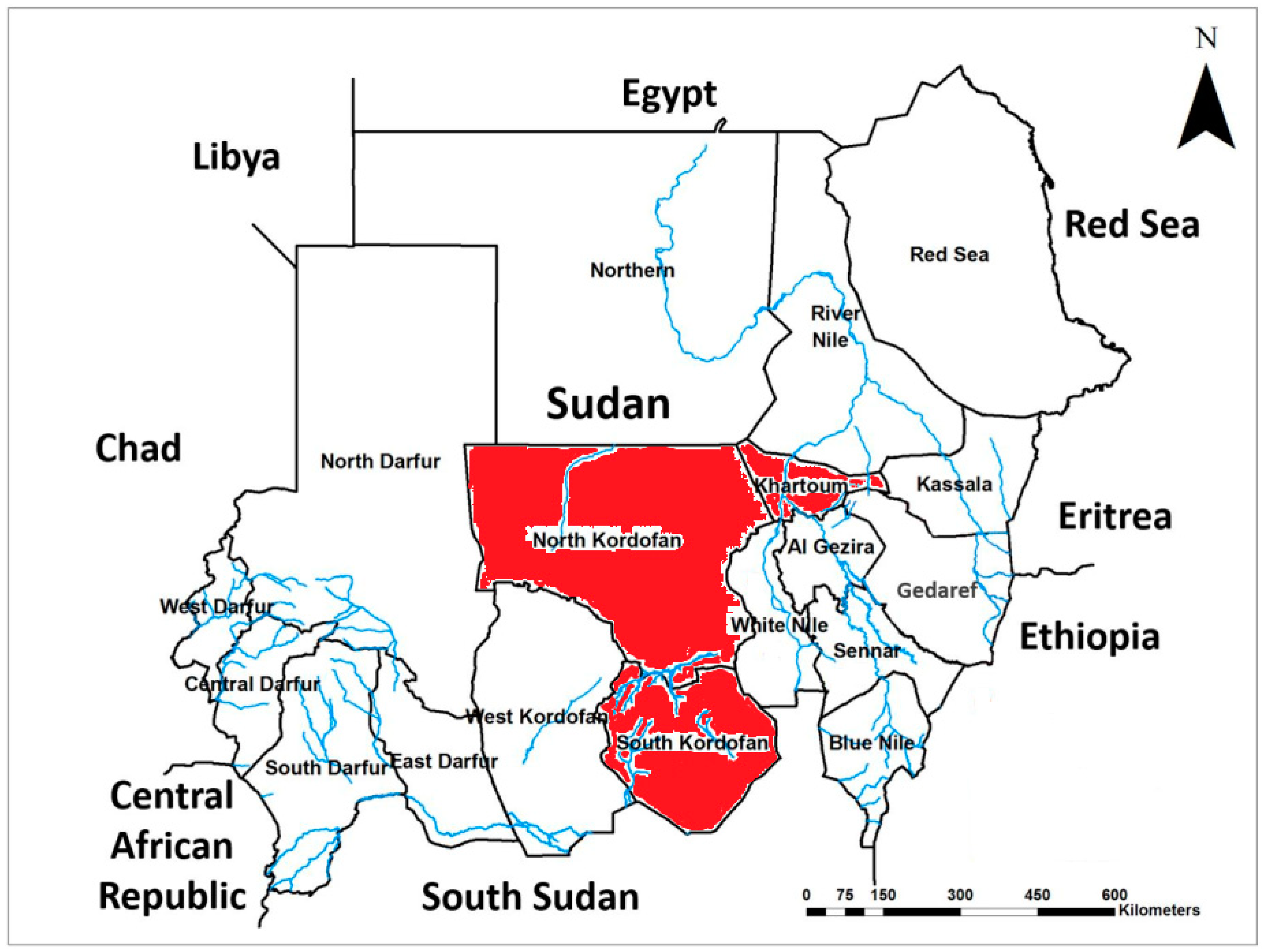
Figure 2.
Post-mortem lesions of CBPP, and microscopic examination of culture. A: The signs of post-mortem lesions of CBPP; including oedematous lung parenchyma, marbling appearance, and consolidation. B: The microscopic examination results for detection of showed fried-egg type appearance colonies of Mycoplasma mycoides subspecies mycoides (Mmm) after incubation for 7 days.
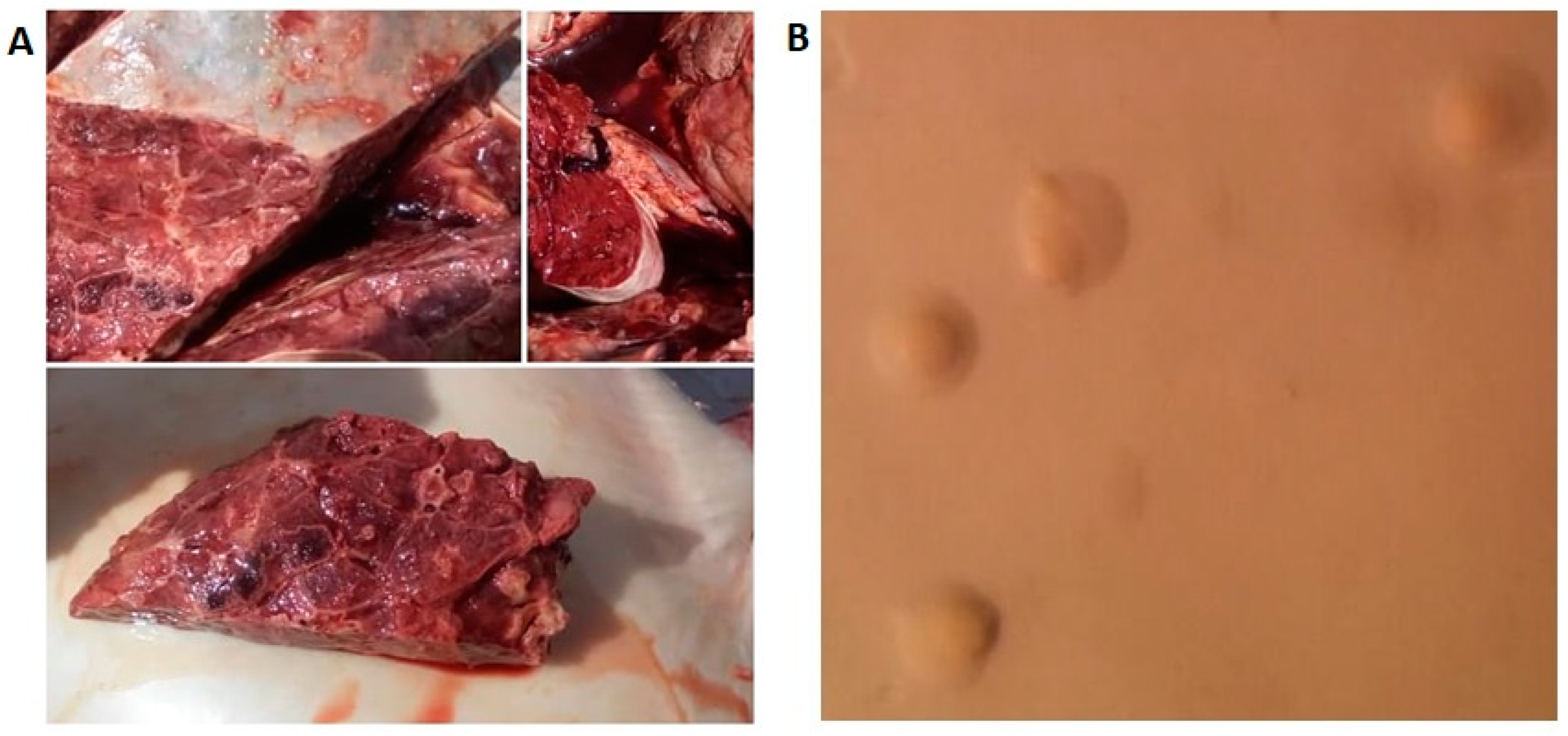
Figure 3.
Molecular detection of rpsG and vmm genes of Mycoplasma mycoides subspecies mycoides using PCR. A: shows the results of rpsG gene amplification, showing a band size of 574 bp. B: shows the results of vmm gene amplification with a band size of 275 bp. Lane MM: Molecular marker (100bp ladder). Lane MNE4 and MNE1: indicate Khartoum state isolates. Lane EK: indicate South Kordofan isolate, and Lane E1B: indicate North Kordofan isolate. Lane M: negative control.
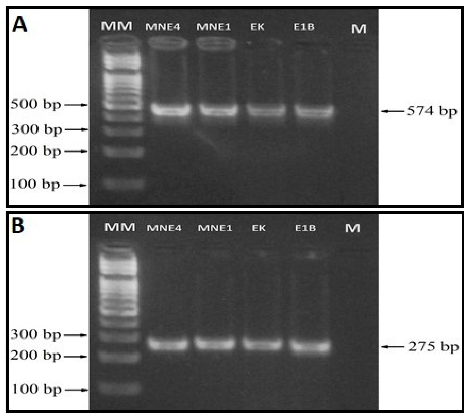
Figure 4.
Phylogenetic tree showing the relation between Mycoplasma mycoides subspecies mycoides (Mmm) reference isolates and the Sudanese isolates. The phylogenetic tree was inferred using the Maximum Likelihood method based on the Tamura 3-parameter model. The bootstrap consensus tree inferred from 1000 replicates. The percentage of tree in which the associated taxa clustered together is shown above the branch. colored boxes indicate the reference sequences of Mmm isolates along with their accession numbers, country of isolation, and year of collection. Red boxes indicate the study isolates with their representative isolation site and year. The black box indicates the reference PG1 isolate of Mmm.

Table 1.
Distribution of pneumonic lungs collected from the different study sites.
| Isolate location | No. lungs screened | Clinically diagnosed as CBPP |
|---|---|---|
| Khartoum | 12519 (39.1%) | 28 (15.7%) |
| North Kordofan | 10316 (32.2%) | 100 (56.2%) |
| South Kordofan | 9204 (28.7%) | 50 (28.1%) |
| Total | 32039 (100%) | 178 (0.6%) |
Disclaimer/Publisher’s Note: The statements, opinions and data contained in all publications are solely those of the individual author(s) and contributor(s) and not of MDPI and/or the editor(s). MDPI and/or the editor(s) disclaim responsibility for any injury to people or property resulting from any ideas, methods, instructions or products referred to in the content. |
© 2025 by the authors. Licensee MDPI, Basel, Switzerland. This article is an open access article distributed under the terms and conditions of the Creative Commons Attribution (CC BY) license (http://creativecommons.org/licenses/by/4.0/).
Copyright: This open access article is published under a Creative Commons CC BY 4.0 license, which permit the free download, distribution, and reuse, provided that the author and preprint are cited in any reuse.


