Submitted:
21 March 2025
Posted:
24 March 2025
You are already at the latest version
Abstract
Leptospirosis is a neglected zoonosis of global significance, associated with flooding and increased precipitation. This study aimed to identify hydro-climatic factors linked to leptospirosis in humans and animals through an integrative literature review. Searches were conducted in August 2024 in the SciELO, Scopus, and PubMed databases using the descriptors “Leptospirosis OR Leptospira” and “flooding” in Portuguese and English. Articles published in the last 10 years (2014-2024), describing the influence of climatic changes, such as heavy rains, floods, and temperature variations, on leptospirosis occurrence, were included. Review articles, conference abstracts, books, and irrelevant studies were excluded. Out of 240 studies, 55 were selected after screening. Findings show urban flooding, driven by high population density and poor drainage infrastructure, facilitates disease spread, while rural flooding increases bacterial dispersion via domestic and livestock animals, inadequate rodent control, and agricultural land contamination. The diversity of serovars highlights the disease's epidemiological complexity, with animals serving as reservoirs and links between environments and humans. Recommendations include strengthening basic sanitation to reduce exposure, rodent population control, implementing early warning systems to monitor climatic conditions, and adopting the One Health approach to integrate human, animal, plant and environmental health for effective leptospirosis control amid climatic changes.
Keywords:
leptospirosis
; Leptospira
; climate changes
; One Health
1. Introduction
Leptospira belongs to the order Spirochaetales, family Leptospiraceae, and genus Leptospira. These are spiral-shaped bacteria resembling corkscrews, distinguished from other spirochetes by the presence of terminal hooks in their structure [1,2]. However, all Leptospira species are morphologically similar, making it challenging to differentiate them based solely on physical characteristics [1].
Despite these morphological similarities, Leptospira species are divided into pathogenic, saprophytic, and intermediate groups. Pathogenic Leptospira have the potential to cause disease in animals and humans, residing in the renal tubules and genital tracts of certain animals (primarily rats) and exhibiting a more complex life cycle. Saprophytic Leptospira are free-living, reproducing in the environment, and are found in surface water, tap water, and moist soil [1]. Additionally, there is a third group, intermediate Leptospira, which is less documented. These species have low pathogenicity and may cause mild clinical signs in mammals [3].
The classification concept evolved after 1989 into two additional schemes: one based on serology, where the serovar is the basic taxonomic unit, and another based on DNA analysis using molecular taxonomy to identify species [4,5]. Serological classification, based on the reactivity of surface antigens, led to the identification of serovars, most of which are pathogenic. These serovars are grouped into serogroups, which may exhibit cross-reactivity [4,5]. Serovars were determined using the Microscopic Agglutination Test (MAT) [5,6].
Alongside this classification, a genotypic classification of the Leptospira genus was established, leading to the description of additional species [7]. Leptospira species (genospecies) are classified based on DNA similarity, and within each species, serology is used to identify surface antigens and classify isolates into serogroups [8]. Currently, over 250 recognized serovars are distributed across 23 serogroups, many of which are associated with clinical diseases [8]. All pathogenic serovars for animals are also pathogenic for humans, with no host-adapted strains identified [7,9].
The primary transmission route of leptospirosis is through contact with water and soil contaminated by the urine of infected animals [10,11,12,13,14]. Infection occurs when the bacteria penetrate tissue barriers, facilitated by wounds, prolonged submersion of intact skin in contaminated water, or mucous membrane exposure [15].
In humans, leptospirosis can present a wide range of symptoms, including fever, headache, chills, muscle or body aches, nausea, vomiting, red eyes, abdominal pain, diarrhea, and skin rashes [16]. The diversity of symptoms often leads to confusion with other diseases, complicating diagnosis. Additionally, some individuals may remain asymptomatic, showing no signs of infection [16]. The incubation period ranges from 2 to 30 days and, in severe cases, the disease can progress to complications such as jaundice, kidney failure, hemorrhage (especially pulmonary), aseptic meningitis, cardiac arrhythmias, pulmonary failure, and hemodynamic collapse [17]. The case fatality rate of leptospirosis ranges from 5% to 15% in severe cases, exceeding 50% in patients with severe pulmonary hemorrhagic syndrome [17].
Animal leptospirosis presents clinical syndromes similar to those observed in humans, with hepatic, renal, and pulmonary involvement as the main manifestations. Common clinical signs include jaundice, hemorrhage, uveitis, uremic syndrome, and reproductive issues such as abortions [18,19,20]. Animals that survive the acute phase of the disease may become carriers, shedding Leptospira in their urine and contaminating the environment [20]. Many Leptospira serovars are associated with maintenance host animals, which acquire the infection easily and excrete the bacteria in urine for prolonged periods [8].
Due to its broad spectrum of clinical manifestations and challenges in early diagnosis, leptospirosis is frequently underreported, despite its significant impact on public health, particularly in tropical and subtropical regions [1]. Globally, the disease accounts for approximately 1,03 million cases and 58900 deaths annually, making leptospirosis one of the leading zoonotic causes of morbidity and mortality [21].
Brazil has the highest number of leptospirosis cases in Latin America, with approximately 4000 cases reported annually [11]. The relationship between flooding and leptospirosis incidence is well-documented [11]. During flood events, contaminated water spreads across urban and rural areas, increasing exposure for humans and animals and creating conditions conducive to bacterial proliferation [11]. Furthermore, the bacterium’s persistence in moist environments and its ability to survive for extended periods in water or flooded soil make floods one of the primary risk factors for leptospirosis transmission [22,23].
The epidemiologic triad of agent, environment, and host is recognized as a key determinant in the occurrence and spread of leptospirosis and other infectious diseases [24]. Consequently, it encompasses all facets of One Health, a collaborative, multisectoral, and transdisciplinary strategy recognizing the inextricable link between human, animal, plant and environmental health [25]. This strategy aims to enhance understanding and strengthen initiatives to confront the challenges in preventing epidemics and epizootics, while also preserving the integrity of ecosystems, benefiting both humans and the biodiversity that supports them [26]. Considering the growing threat of climate change, particularly the increased frequency and intensity of flooding events, which exacerbate leptospirosis, this integrative literature review aims to identify the influence of climate change - focusing on floods, precipitation, and temperature - on the occurrence of leptospirosis in humans and animals. Additionally, it seeks to report the diversity of serovars recorded during flood periods. These findings may contribute to the development of effective prevention and control strategies.
2. Materials and Methods
An integrative literature review was conducted through a systematic search for articles that contribute to answering the research question aimed at identifying the influence of climate change, focusing on flooding, precipitation, and temperature, on the occurrence of leptospirosis in humans and animals.
The search for scientific articles was carried out in the PubMed, SciELO, and Scopus databases, in Portuguese and English, in August 2024, using the Health Sciences Descriptors (DeCS/MeSH) “leptospirosis OR Leptospira” AND “flooding,” identified in the title and/or abstract and/or keywords of the articles, with the same descriptors also being used in Portuguese.
Articles published in Portuguese and English within the last ten years (2014 to 2024), describing the influence of climate change, such as flooding, precipitation, and temperature, on the occurrence of human and animal leptospirosis, were considered. From the articles, the diversity of registered serovars was also identified. Review articles, conference abstracts, and books were excluded, as well as articles that did not meet the inclusion criteria described above. The identified articles were imported into the Rayyan platform, a tool specialized in systematic review screening [27].
After importing the articles, the Rayyan platform was used for detecting and excluding duplicate articles, which were manually reviewed to ensure their removal. Next, two independent reviewers proceeded to read the titles and abstracts of the included articles, classifying them according to the previously established inclusion and exclusion criteria.
In cases of disagreement between the two reviewers, a third researcher was consulted to read the article in question and decide on its inclusion or exclusion. The screening step was conducted blindly on Rayyan to avoid bias among reviewers during the selection process. The information from the articles included in the review was organized into tables highlighting the characteristics of each study.
3. Results
A total of 240 articles were identified in the databases, following the proposed inclusion criteria. After reading the titles and abstracts, 73 duplicate articles and 112 articles that did not meet the inclusion criteria were excluded. Thus, 55 potentially eligible articles were selected and read in full to ensure they met the established criteria. Figure 1 details the process of selecting the studies.
Regarding the type of study, 29 were cross-sectional studies (52.7%), seven ecological studies (12.7%), four epidemiological studies (7.3%), three case studies (5.5%), two case reports (3.6%), two meta-analysis (3.6%), two descriptive studies (3.6%), two experimental studies (3.6%), two mathematical models (3.6%), one cross-over study (1.9%), and one retrospective analytical study (1.9%), as represented in Figure 2.
Table 1 summarizes the databases of the articles with information about the authors, year of publication, country where the study was conducted, the journal in which the article was published, and the main related topic.
The selected studies were conducted primarily on the Asian and American continents (Figure 3).
The articles were published in journals covering various thematic areas, with the most significant representation in Public Health and Epidemiology (27.3%), followed by Agriculture and Veterinary Sciences (20%) and Tropical Medicine and Neglected Diseases (20%) (Figure 4).
4. Discussion
The studies were conducted in 27 countries, highlighting the global public health impact of the disease, with most research conducted in Asia and America. Additionally, the publications are in journals across various thematic areas, demonstrating the interdisciplinary nature of the studies.
The main topics addressed include the relationship between leptospirosis, flooding, and precipitation, and how climatic events contribute to the spread of the disease. The influence of temperature was also discussed, demonstrating how climatic conditions can increase the incidence of leptospirosis. Furthermore, the diversity of serovars found shows the epidemiological complexity of the disease in different regions worldwide. Finally, the studies provide important recommendations for controlling leptospirosis, especially in areas with inadequate sanitation and those vulnerable to flooding.
4.1. Leptospirosis, Flooding, and Precipitation
Floods are common natural events and are the leading cause of death in natural disasters worldwide [44]. With the increasing frequency of extreme weather events, such as floods, as a result of climate change, the risk to health is growing [57,61,64,66].
In the case of leptospirosis, the selected studies report a strong correlation with flood events in urban areas [22,29,34,36,39,40,41,42,46,47,48,50,51,52,53,54,55,56,57,63,64,65,73,77,79,80,81] and rural areas [31,35,37,43,45,59,60,67,68,75,78]. Only one study did not find a relationship between these factors, and its results suggest that, in the eastern region of Poland, water and soil have limited significance in the persistence and spread of Leptospira [28]. However, the authors mention limitations in comparing water reservoirs between areas exposed and not exposed to flooding, as well as a lack of differentiation between Leptospira strains.
The occurrence of leptospirosis in urban areas is directly associated with environmental and structural factors, such as poor sanitation, the accumulation of solid waste in inappropriate locations, and the proliferation of rodents [22,29,33,34,36,39,40,41,42,46,47,48,51,52,53,54,55,56,57,63,64,65,73,77,78,79,80,81,82]. High population density and lack of proper drainage and sewage systems worsen the situation during floods, facilitating the spread of the disease in urban environments [36,41,46]. These combined factors create favorable conditions for increased leptospirosis incidence, especially in vulnerable regions where the impact of floods is more intense and frequent [46].
In rural areas, leptospirosis is related to various environmental and occupational factors. Close contact with domestic and farm animals, which can be hosts for Leptospira, increases the risk of transmission [31,59]. Additionally, agricultural activities such as cattle and pig farming expose rural workers to frequent contact with contaminated soil and water through infected animals’ urine [42,45]. In periods of heavy rain and floods, the inundation of agricultural areas facilitates the spread of the bacteria, contaminating water bodies and soil [67,68,69,75]. The lack of effective rodent control, which also acts as a reservoir, and the absence of adequate sanitation in many rural areas contribute to the persistence and spread of leptospirosis in these environments [29]. These factors, combined with favorable climatic conditions and lack of sanitary infrastructure, intensify the risk of disease outbreaks in rural areas.
There is a significant correlation between high rainfall volumes and leptospirosis outbreaks, particularly evident in areas vulnerable to flooding, where water runoff increases contamination in both urban and rural areas. In India, the peak of leptospirosis cases occurred 17 days after floods [80]. In the Philippines, increased hospital admissions were observed 15 days after increased precipitation, also associated with flooding [47]. Additionally, some studies suggest that leptospirosis incidence may increase one to three months after heavy rainfall [32,65].
Another study conducted in Puerto Rico found the presence of Leptospira in six public networks of surface water supply six months after a hurricane caused extensive flooding [68]. The hurricane, by causing flooding, promoted the spread of waste and urine from infected animals in water bodies, facilitating contamination of water sources, which highlights the relationship between natural disasters, such as hurricanes and subsequent flooding, and the increased risk of leptospirosis contamination in water supply networks [68].
Although floods are a relevant indicator for predicting future leptospirosis cases, floods of lesser severity do not always result in an increase in cases [80]. On the other hand, a meta-analysis on the epidemiology of canine leptospirosis did not find statistically significant evidence of an association between infection occurrence and flooding in the dog’s habitat [39]. In contrast, in horses, the risk of seropositivity triples when animals are exposed to flooding [37], and among humans, exposure to flooding doubles the risk of leptospirosis infection [40,71].
In Brazil, a study found that municipalities that experienced flooding had a leptospirosis incidence rate 77% higher than municipalities that did not report flooding [64]. Studies conducted in various countries link floods to the creation of favorable environmental conditions for the proliferation of the bacteria, particularly through contact with water contaminated by the urine of rodents and other infected animals [31,34,37,48,52,55,58,66,70,77,78].
Living near water bodies is also associated with an increased risk of contracting the disease [36,48]. A study showed that pathogenic Leptospira remain dormant in the soil and can become active for infection under flooding conditions, when soil water content increases [70]. In other words, Leptospira survive at different depths in soils and surface sediments of rivers (depending on water saturation), being transferred to surface water during erosion caused by flooding [22,36]. One study suggests that pathogenic strains of Leptospira are widely distributed in the environment, especially in areas affected by flooding [62].
Precipitation is also a risk factor in the dynamics of leptospirosis and is discussed in several studies that associate heavy rainfall with increased disease spread [32,36,46,47,49,56,63,65,71,72,76]. In the state of Santa Catarina (Brazil), for example, it was reported that each additional day of rain in a given month increases the disease risk by an average of 6% [71]. In northeastern Australia, however, a study suggests that while heavy rainfall likely contributed to increased cases of the disease, the factor was not clearly identified as being associated with a rise in cases in this region [72].
4.2. Influence of Temperature on Leptospirosis Occurrence
Temperature variations also influence leptospirosis cases, particularly in tropical and subtropical climates, with an average 12% increase in risk for every 1°C rise in temperature [71].
An average temperature of 28°C was associated with a higher number of leptospirosis cases in Colombia [79]. In Great Britain, the annual mean temperature was the most significant variable related to the higher incidence of canine leptospirosis, with cases peaking during the autumn season [14]. According to the authors, the likelihood of the disease occurring in dogs was higher at temperatures up to 11°C, although this optimal temperature for canine leptospirosis presence varied depending on the serogroup analyzed.
In Saint Lucia, a Caribbean region, no correlation was found between temperature and disease cases [61]. Meanwhile, in two studies conducted in the state of Santa Catarina, Brazil, leptospirosis showed a seasonal pattern, peaking in summer months, which also coincide with periods of higher rainfall [57].
4.3. Diversity of Serovars Found
The pathogenicity of Leptospira depends on the virulence of the infecting serovar and the susceptibility of the host species, with rodents being the main carriers of Leptospira, followed by wild carnivores. However, all mammals have the potential to be hosts of the bacterium [9]. For instance, L. interrogans is the primary pathogenic species and includes 24 serogroups, such as the Icterohaemorrhagiae serogroup, which encompasses the serovars Icterohaemorrhagiae, Copenhageni, and Lai [82]. In horses, the most recurrent serovar is L. interrogans serovar Pomona, while in pigs, leptospirosis is commonly caused by serovars such as L. interrogans serovars Icterohaemorrhagiae and Copenhageni, as well as L. pomona, L. canicola, L. tarassovi, L. bratislava, and L. muenchen [9]. In cattle, L. borgpetersenii serovar Hardjo is prevalent [9]. Among dogs, the serovars Canicola and Icterohaemorrhagiae were historically the main causative agents of the disease, but vaccination against these serovars has led to the emergence of others, such as Grippotyphosa and Bratislava [8,20].
In the studies included in this review, it was found that in Australia, the most prevalent serovars in human leptospirosis cases were Australis and Zanoni [29,72], as well as Arborea and Hardjo [29]. In Indonesia, the occurrence of serovars such as Bangkinang, Grippotyphosa, Canicola, Robinsoni, Bataviae, and Mini was reported [42]. In the Philippines, the serovars Patoc, Semaranga, Grippotyphosa, Ratnapura, Copenhageni, and Poi were identified [38]. In Colombia, the most frequent serogroups were Australis, Sejroe, Pomona, Bataviae, Pyrogenes, and Grippotyphosa [79]. In the relatively dry region of Sri Lanka, most cases during the 2011 outbreak were caused by L. kirschneri [30].
In dogs, the most frequent serogroups in Great Britain were Australis, Icterohaemorrhagiae, and Sejroe, the latter being absent from commercial vaccines [14]. In Malaysia, Australis, Icterohaemorrhagiae, Canicola, Bataviae, Hardjo, Lai, Grippotyphosa, Ballum, Hardjobovis, and Javanica were identified [58]. In Brazil, serovars such as Icterohaemorrhagiae, Canicola, Pyrogenes, Copenhageni, and Autumnalis were found, with three dogs testing positive for more than one serovar [78]. This study also showed an association between the presence of seropositive dogs and a higher density of human cases. In a study conducted in Bosnia and Herzegovina, the serovars Australis, Bratislava, Sejroe, and Autumnalis were found in dogs, while the highest seroprevalence in foxes was for the serovars Australis, Sejroe, Bratislava, and Bataviae [75].
In sheep, in Uruguay, the serovar isolated in two lamb herds after flooding was Pomona [59]. In cattle in Malaysia, the most frequent serovars were Pomona, Hardjo, and Hebdomadis [67]. In Paraíba State, Brazil, Hardjo was the most prevalent serovar [31,69], followed by Icterohaemorrhagiae and Australis [69]. Therefore, it is necessary to investigate the epidemiological role of domestic animals as reservoirs of leptospirosis for humans [59]. Genotyping studies suggest, for example, that multiple animal reservoirs were implicated in the leptospirosis outbreak in Western Fiji after flooding [51].
In addition to the pathogenic bacteria identified in the studies, intermediate species have also been reported in the environment. The intermediate L. licerasiae was isolated from the environment in Jakarta, Indonesia, and has pathogenic infection potential [52]. In Kelantan, Malaysia, following flooding, L. wolffii, another intermediate Leptospira species, was identified, which could potentially be transmitted to humans and animals [74].
The Leptospira serovars identified decades ago remain the causative agents of leptospirosis in humans [38]. However, the transmission of leptospirosis involves numerous factors, including the impact of climate, whose role is not yet fully understood [32]. The diversity of Leptospira serovars identified in humans and animals highlights the epidemiological complexity of leptospirosis and its multifactorial nature, supporting the interdependence of human, animal, plant and environmental health, a core principle of One Health. The wide serological variability suggests the presence of multiple reservoirs and transmission pathways, emphasizing the need for an integrated approach to epidemiological surveillance, control measures, and risk mitigation. Elucidating this role is essential, as it facilitates epidemic risk analysis and supports preventive efforts, which must involve intersectoral cooperation among public health authorities, promoting improvements in urban infrastructure and intensified surveillance [61,64,65].
4.4. Recommendations for Leptospirosis Control
Integrated human and animal health strategies for controlling leptospirosis in flood-prone areas should include rigorous rodent control measures both before and during the recovery period of natural disasters [51]. Prevention involves the use of personal protective equipment, such as gloves and closed footwear [29], as well as draining flooded areas and implementing sanitary controls before introducing animals [31]. A study in India reported the use of doxycycline as chemoprophylaxis in asymptomatic individuals, at variable doses based on flood exposure [28]. However, this evidence remains inconclusive because the intervention was not implemented as a controlled study, potentially leading to biased results. In contrast, rapid diagnosis, treatment, and community awareness programs are recognized methods of disease control [51].
Nevertheless, early antibiotic treatment is recommended for cases with a high suspicion of leptospirosis, even before laboratory confirmation, as it can reduce the severity and duration of the disease [17]. Therefore, leptospirosis should be included in the differential diagnosis of any patient presenting with acute febrile illness or nonspecific symptoms and a history of potential exposure to floodwaters or rodent-infested areas [83].
Given the high risk of contamination following floods, a permanent recommendation should be to avoid recreational activities in freshwater bodies [35]. Furthermore, implementing environmental interventions targeting transmission sources, such as improved waste disposal management (especially in flooded areas) and efficient basic sanitation, is crucial [41].
Definitive diagnosis of leptospirosis relies on bacterial isolation via culture, DNA detection by polymerase chain reaction (PCR), or demonstration of seroconversion in paired acute and convalescent serum samples [15,84]. The microscopic agglutination test (MAT) remains the gold standard for leptospirosis diagnosis, utilizing a panel of live antigens [1,8]. However, MAT’s limitation lies in its restricted coverage of recognized serovars, potentially missing unknown serovars in specific regions [81]. Real-time PCR has been employed for early Leptospira detection in clinical samples during the acute, leptospiremic phase, which is critical for effective antimicrobial therapy [1]. Despite its utility, a key limitation of PCR-based diagnosis is the current inability to identify the infecting serovar, an important factor for epidemiological and public health purposes [85].
Although the disease’s pathogenesis is well-documented, the cellular and molecular mechanisms remain unclear. Currently, leptospirosis research is entering a “molecular era”, driven by the sequencing of both virulent and saprophytic Leptospira strains conducted by international research groups. This effort aims to identify key bacterial protein virulence factors [2].
Vaccines for animals are licensed worldwide, but their formulations have not significantly changed in recent decades. Vaccine-induced immunity is often restricted to related serovars and is short-lived [18]. In cattle and pigs, pentavalent vaccines typically include L. hardjo, L. pomona, L. canicola, L. icterohaemorrhagiae, and L. grippotyphosa, with some formulations also containing L. bratislava [9]. For dogs, bivalent vaccines (L. icterohaemorrhagiae and L. canicola) or multivalent vaccines including L. pomona and L. grippotyphosa are used. However, most dogs show negative antibody titers against all serovars one-year post-vaccination [86].
In humans, a universal leptospirosis vaccine remains unavailable, despite the testing of several recombinant vaccines that have not yet progressed to clinical trials [82]. Similar to animal bacterin vaccines, these approaches are limited by the lack of an effective cross-protective immune response [87].
5. Conclusions
The reviewed articles emphasize the importance of understanding leptospirosis in the context of climatic events, particularly highlighting the impact of floods on the disease’s dissemination.
Among the main contributions to controlling leptospirosis in flood-prone areas, the review suggests strengthening basic sanitation measures e rodent control to reduce human and animal exposure to contaminated environments. Additionally, implementing early warning systems could enable the monitoring of critical climatic conditions and the anticipation of outbreaks.
Leptospirosis still faces significant underreporting, largely due to insufficient monitoring, with many cases remaining undiagnosed or not reaching healthcare services for mandatory notification. This reveals weaknesses in the surveillance system, which could operate more effectively. Furthermore, it is essential to promote more robust awareness campaigns among the population during flood periods, emphasizing the importance of preventive measures to avoid contamination and minimize the risk of disease spread.
Vaccination of dogs and production animals, particularly in high-risk regions, is also recommended to reduce the role of these animals as Leptospira reservoirs. However, the limited duration and serovar coverage of current animal vaccines, coupled with potential interference with serological testing, restricts their effectiveness in providing broad protection.
Addressing these multifaceted challenges necessitates a One Health approach, fostering more effective strategies for leptospirosis surveillance and control. Ultimately, preventing leptospirosis requires coordinated, interdisciplinary efforts involving physicians, veterinarians, public health specialists, and other stakeholders, particularly in the context of climate change and environmental vulnerabilities.
Author Contributions
Conceptualization, C.C.J., B.F.S. and A.E.S.; methodology, C.C.J., B.F.S. and A.E.S.; software, C.C.J., B.F.S. and A.E.S.; formal analysis, C.C.J. and B.F.S.; writing—original draft preparation, C.C.J., B.F.S. and A.E.S.; writing—review and editing, C.C.J., B.F.S., A.E.S., Á.M., C.P.B. L.A.; funding acquisition, B.F.S. and L.A. This manuscript incorporates content from the dissertation of C.C.J., submitted in partial fulfillment of the requirements for Master’s degree. All authors have read and agreed to the published version of the manuscript.
Funding
This research was funded by Fundação de Amparo à Pesquisa e Inovação do Estado de Santa Catarina (FAPESC) under grant numbers TO2024TR002584 (Universal 21/2024), TO2023TR001418, and TO2023TR001518 (FAPESC nº 15/2023 – Programa de Estruturação Acadêmica para Laboratórios Multiusuários).
Acknowledgments
The authors gratefully acknowledge the research financial support provided by FAPESC. Cintia Cavilha Juppa acknowledges financial support from the Programa Institucional de Bolsas de Pós-Graduação – PIBPG (CNPq Nº 69/2022).
Conflicts of Interest
The authors declare no conflicts of interest.
Abbreviations
The following abbreviations are used in this manuscript:
| DNA | Deoxyribonucleic acid |
| PCR | Polymerase Chain Reaction |
| MAT | Microscopic Agglutination Test |
References
- WHO. World Health Organization. Human leptospirosis: guidance for diagnosis, surveillance and control. https://www.who.int/publications/i/item/human-leptospirosis-guidance-for-diagnosis-surveillance-and-control (accessed on 27 May 2024).
- Duarte, M.I.S.; Duarte Neto, A.N.; Pagliari, C. Doenças infecciosas: visão integrada da patologia, da clínica e dos mecanismos patogênicos; Artmed: Porto Alegre, Brasil, 2024. [Google Scholar]
- Goarant, C. Leptospirosis: risk factors and management challenges in developing countries. Res Rep Trop Med 2016, 7, 49–62. [Google Scholar] [CrossRef] [PubMed]
- Brenner, D.J.; Kaufmann, A.F.; Sulzer, K.R.; Steigerwalt, A.G.; Rogers, F.C.; Weyant, R.S. Further determination of DNA relatedness between serogroups and serovars in the family Leptospiraceae with a proposal for Leptospira alexanderi sp. nov. and four new Leptospira genomospecies. Int J Syst Evol Microbiol 1999, 49, 839–858. [Google Scholar] [CrossRef]
- Marquez, A.; Djelouadji, Z.; Lattard, V.; Kodjo, A. Overview of laboratory methods to diagnose leptospirosis and to identify and to type leptospires. Int Microbiol 2017, 20, 184–193. [Google Scholar] [CrossRef] [PubMed]
- Guernier, V.; Goarant, C.; Benschop, J.; Lau, C.L. A systematic review of human and animal leptospirosis in the Pacific Islands reveals pathogen and reservoir diversity. PLoS Negl Trop Dis 2018, 12, e0006503. [Google Scholar] [CrossRef]
- Mohammed, H.; Nozha, C.; Hakim, K.; Abdelaziz, F. Leptospira: morphology, classification and pathogenesis. J Bacteriol Parasitol 2011, 2. [Google Scholar] [CrossRef]
- Quinn, P.J.; Markey, B.K.; Leonard, F.C. Microbiologia veterinária essencial, 2nd ed; Artmed, 2019.
- Mcvey, S.; Kenneddy, M.; Chengappa, M.M. Microbiologia veterinária, 3rd ed; Editora Guanabara Koogan Ltd.a, 2017.
- Baharom, M.; Ahmad, N.; Hod, R.; Ja’afar, M.H.; Arsad, F.S.; Tangang, F.; Ismail, R.; Mohamed, N.; Mohd Radi, M.F.; Osman, Y. Environmental and occupational factors associated with leptospirosis: a systematic review. Heliyon 2024, 10, e23473. [Google Scholar] [CrossRef]
- Galan, D.I.; Roess, A.A.; Pereira, S.V.C.; Schneider, M.C. Epidemiology of human leptospirosis in urban and rural areas of Brazil, 2000–2015. PLoS ONE 2021, 16, e0247763. [Google Scholar] [CrossRef]
- Philip, N.; Ahmed, K. Leptospirosis in Malaysia: current status, insights, and future prospects. J Physiol Anthropol 2023, 42, 30. [Google Scholar] [CrossRef]
- Rehan, S.T.; Ali, E.; Sheikh, A.; Nashwan, A.J. Urban flooding and risk of leptospirosis; Pakistan on the verge of a new disaster: a call for action. Int J Hyg Environ Health 2023, 248, 114081. [Google Scholar] [CrossRef]
- Taylor, C.; Brodbelt, D.C.; Dobson, B.; Catchpole, B.; O’Neill, D.G.; Stevens, K.B. Spatio-temporal distribution and agroecological factors associated with canine leptospirosis in Great Britain. Prev Vet Med 2021, 193, 105407. [Google Scholar] [CrossRef]
- Brazil. Leptospirose. Saúde de A a Z. https://www.gov.br/saude/pt-br/assuntos/saude-de-a-a-z/l/leptospirose (accessed 03 May 2024).
- CDC. Leptospirosis. https://www.cdc.gov/leptospirosis/infection/index.html (accessed 17 May 2024).
- CDC. Leptospirosis - Fact Sheet for Clinicians. https://www.cdc.gov/leptospirosis/pdf/fs-leptospirosis-clinicians-eng-508.pdf (accessed 04 October 2024).
- Adler, B.; Klaasen, E. Recent advances in canine leptospirosis: focus on vaccine development. Vet Med Res Rep 2015, 245. [Google Scholar] [CrossRef] [PubMed]
- Ellis, W.A. Control of canine leptospirosis in Europe: time for a change? Vet Rec 2010, 167, 602–605. [Google Scholar] [CrossRef]
- Sykes, J.E.; Hartmann, K.; Lunn, K.F.; Moore, G.E.; Stoddard, R.A.; Goldstein, R.E. 2010 ACVIM Small Animal Consensus Statement on Leptospirosis: diagnosis, epidemiology, treatment, and prevention. J Vet Intern Med 2011, 25, 1–13. [Google Scholar] [CrossRef] [PubMed]
- Costa, F.; Hagan, J.E.; Calcagno, J.; Kane, M.; Torgerson, P.; Martinez-Silveira, M.S.; Stein, C.; Abela-Ridder, B.; Ko, A.I. Global morbidity and mortality of leptospirosis: a systematic review. PLoS Negl Trop Dis 2015, 9, e0003898. [Google Scholar] [CrossRef] [PubMed]
- Thibeaux, R.; Genthon, P.; Govan, R.; Selmaoui-Folcher, N.; Tramier, C.; Kainiu, M.; Soupé-Gilbert, M.-E.; Wijesuriya, K.; Goarant, C. Rainfall-driven resuspension of pathogenic Leptospira in a leptospirosis hotspot. Sci Total Environ 2024, 911, 168700. [Google Scholar] [CrossRef]
- Bierque, E.; Thibeaux, R.; Girault, D.; Soupé-Gilbert, M.-E.; Goarant, C. A systematic review of Leptospira in water and soil environments. PLoS One 2020, 15, e0227055. [Google Scholar] [CrossRef]
- Smith, A.M.; Stull, J.W.; Moore, G.E. Potential drivers for the re-emergence of canine leptospirosis in the United States and Canada. Tropical Med 2022, 7, 377. [Google Scholar] [CrossRef]
- CDC. One Health. One Health. https://www.cdc.gov/one-health/about/ (accessed 24 May 2024).
- Artaxo, P. Mudanças Climáticas: caminhos para o Brasil: a construção de uma sociedade minimamente sustentável requer esforços da sociedade com colaboração entre a ciência e os formuladores de políticas públicas. Cienc Cult 2022, 74. [Google Scholar] [CrossRef]
- Ouzzani, M.; Hammady, H.; Fedorowicz, Z.; Elmagarmid, A. Rayyan—a web and mobile app for systematic reviews. Syst Rev 2016, 5, 210. [Google Scholar] [CrossRef]
- Wójcik-Fatla, A.; Zając, V.; Wasiński, B.; Sroka, J.; Cisak, E.; Sawczyn, A.; Dutkiewicz, J. Occurrence of Leptospira DNA in water and soil samples collected in Eastern Poland. Ann Agric Environ Med 2014, 21, 730–732. [Google Scholar] [CrossRef]
- Wynwood, S.J., C., S.B.; Graham, G.C., B., B.R.; Burns, M.A., W., S.L.; Collet, T.A., M., D.B. The emergence of Leptospira borgpetersenii serovar Arborea as the dominant infecting serovar following the summer of natural disasters in Queensland, Australia 2011. Trop Biomed 2014, 31, 281–285. https://www.msptm.org/files/281_-_285_Craig_SB.pdf.
- Agampodi, S.B.; Dahanayaka, N.J.; Bandaranayaka, A.K.; Perera, M.; Priyankara, S.; Weerawansa, P.; Matthias, M.A.; Vinetz, J.M. Regional differences of leptospirosis in Sri Lanka: observations from a flood-associated outbreak in 2011. PLoS Negl Trop Dis 2014, 8, e2626. [Google Scholar] [CrossRef] [PubMed]
- Pimenta, C.L.R.M.; Castro, V.; Clementino, I.J.; Alves, C.J.; Fernandes, L.G.; Brasil, A.W.L.; Santos, C.S.A.B.; Azevedo, S.S. Leptospirose bovina no estado da Paraíba: prevalência e fatores de risco associados à ocorrência de propriedades positivas. Pesq Vet Bras 2014, 34, 332–336. [Google Scholar] [CrossRef]
- Guimarães, R.M.; Cruz, O.G.; Parreira, V.G.; Mazoto, M.L.; Vieira, J.D.; Asmus, C.I.R.F. Análise temporal da relação entre leptospirose e ocorrência de inundações por chuvas no município do Rio de Janeiro, Brasil, 2007-2012. Cien Saude Colet 2014, 19, 3683–3692. [Google Scholar] [CrossRef] [PubMed]
- Gracie, R.; Barcellos, C.; Magalhães, M.; Souza-Santos, R.; Barrocas, P. Geographical scale effects on the analysis of leptospirosis determinants. Int J Environ Res Public Health 2014, 11, 10366–10383. [Google Scholar] [CrossRef]
- Suwanpakdee, S.; Kaewkungwal, J.; White, L.J.; Asensio, N.; Ratanakorn, P.; Singhasivanon, P.; Day, N.P.J.; Pan-Ngum, W. Spatio-temporal patterns of leptospirosis in Thailand: is flooding a risk factor? Epidemiol Infect 2015, 143, 2106–2115. [Google Scholar] [CrossRef]
- Matono, T.; Kutsuna, S.; Koizumi, N.; Fujiya, Y.; Takeshita, N.; Hayakawa, K.; Kanagawa, S.; Kato, Y.; Ohmagari, N. Imported flood-related leptospirosis from Palau: awareness of risk factors leads to early treatment. J Travel Med 2015, 22, 422–424. [Google Scholar] [CrossRef]
- Lau, C.L.; Watson, C.H.; Lowry, J.H.; David, M.C.; Craig, S.B.; Wynwood, S.J.; Kama, M.; Nilles, E.J. Human leptospirosis infection in Fiji: an eco-epidemiological approach to identifying risk factors and environmental drivers for transmission. PLoS Negl Trop Dis 2016, 10, e0004405. [Google Scholar] [CrossRef]
- Sohail, M.L.; Khan, M.S.; Avais, M.; Zahoor, M.Y.; Ijaz, M.; Ullah, A.; Fatima, Z.; Naseer, O.; Khattak, I.; Ali, S. Seroprevalence of Leptospira spp. in horses of distinct climatic regions of Punjab, Pakistan. J Equine Vet Sci 2016, 44, 82–89. [Google Scholar] [CrossRef]
- Gloriani, N.G., V., S.Y.A.M. Post-flooding surveillance of leptospirosis after the onslaught of typhoons Nesat, Nalgae and Washi in the Philippines. Southeast Asian J Trop Med Public Health 2016, 47, 774–786.
- Azócar-Aedo, L.; Monti, G. Meta-analyses of factors associated with leptospirosis in domestic dogs. Zoonoses Public Health 2016, 63, 328–336. [Google Scholar] [CrossRef]
- Ledien, J.; Sorn, S.; Hem, S.; Huy, R.; Buchy, P.; Tarantola, A.; Cappelle, J. Assessing the performance of remotely-sensed flooding indicators and their potential contribution to early warning for leptospirosis in Cambodia. PLoS One 2017, 12, e0181044. [Google Scholar] [CrossRef]
- Hayati, K.S., S.N., S.I.; Salmiah, M.S., E.M.A.; Khin T.D. Hot-spot and cluster analysis on legal and illegal dumping sites as the contributors of leptospirosis in a flood hazard area in Pahang, Malaysia. Asian J Agric Biol 2018, 78–82.
- Syamsuar; Daud, A.; Maria, I.L.; Hatta, Muh.; Widyastuti, D. Identification of serovar leptospirosis in flood-prone areas Wajo district. Indian J Public Health Res Dev 2018, 9, 325. [CrossRef]
- Ijaz, M.; Abbas, S.N.; Farooqi, S.H.; Aqib, A.I.; Anwar, G.A.; Rehman, A.; Ali, M.M.; Mehmood, K.; Khan, A. Sero-epidemiology and hemato-biochemical study of bovine leptospirosis in flood affected zone of Pakistan. Acta Trop 2018, 177, 51–57. [Google Scholar] [CrossRef] [PubMed]
- Supe, A.; Khetarpal, M.; Naik, S.; Keskar, P. Leptospirosis following heavy rains in 2017 in Mumbai: report of large-scale community chemoprophylaxis. Natl Med J India 2018, 31, 19. [Google Scholar] [CrossRef] [PubMed]
- Chadsuthi, S.; Chalvet-Monfray, K.; Wiratsudakul, A.; Suwancharoen, D.; Cappelle, J. A remotely sensed flooding indicator associated with cattle and buffalo leptospirosis cases in Thailand 2011–2013. BMC Infect Dis 2018, 18, 602. [Google Scholar] [CrossRef] [PubMed]
- Vitale, M.; Agnello, S.; Chetta, M.; Amato, B.; Vitale, G.; Bella, C.D.; Vicari, D.; Presti, V.D.M.L. Human leptospirosis cases in Palermo Italy. The role of rodents and climate. J Infect Public Health 2018, 11, 209–214. [Google Scholar] [CrossRef]
- Matsushita, N.; Ng, C.F.S.; Kim, Y.; Suzuki, M.; Saito, N.; Ariyoshi, K.; Salva, E.P.; Dimaano, E.M.; Villarama, J.B.; Go, W.S.; Hashizume, M. The non-linear and lagged short-term relationship between rainfall and leptospirosis and the intermediate role of floods in the Philippines. PLoS Negl Trop Dis 2018, 12, e0006331. [Google Scholar] [CrossRef]
- Radi, M.F.; Hashim, J.H.; Jaafar, M.H.; Hod, R.; Ahmad, N.; Mohammed Nawi, A.; Baloch, G.M.; Ismail, R.; Farakhin Ayub, N.I. Leptospirosis outbreak after the 2014 major flooding event in Kelantan, Malaysia: a spatial-temporal analysis. Am J Trop Med Hyg 2018, 98, 1281–1295. [Google Scholar] [CrossRef]
- Mayfield, H.J.; Lowry, J.H.; Watson, C.H.; Kama, M.; Nilles, E.J.; Lau, C.L. Use of geographically weighted logistic regression to quantify spatial variation in the environmental and sociodemographic drivers of leptospirosis in Fiji: a modelling study. Lancet Planet Health 2018, 2, e223–e232. [Google Scholar] [CrossRef]
- Sohail, M.L.; Khan, M.S.; Ijaz, M.; Naseer, O.; Fatima, Z.; Ahmad, A.S.; Ahmad, W. Seroprevalence and risk factor analysis of human leptospirosis in distinct climatic regions of Pakistan. Acta Trop 2018, 181, 79–83. [Google Scholar] [CrossRef]
- Togami, E.; Kama, M.; Goarant, C.; Craig, S.B.; Lau, C.; Ritter, J.M.; Imrie, A.; Ko, A.I.; Nilles, E.J. A Large leptospirosis outbreak following successive severe floods in Fiji, 2012. Am J Trop Med Hyg 2018, 99, 849–851. [Google Scholar] [CrossRef]
- Widiyanti, D.; Djannatun, T.; Astuti, I.I.P.; Maharsi, E.D. Leptospira detection in flood-prone environment of Jakarta, Indonesia. Zoonoses Public Health 2019, 66, 597–602. [Google Scholar] [CrossRef]
- Ding, G.; Li, X.; Li, X.; Zhang, B.; Jiang, B.; Li, D.; Xing, W.; Liu, Q.; Liu, X.; Hou, H. A time-trend ecological study for identifying flood-sensitive infectious diseases in Guangxi, China from 2005 to 2012. Environ Res 2019, 176, 108577. [Google Scholar] [CrossRef] [PubMed]
- Sumalapao, D.P.; Del Rosario, B.M.; Suñga, L.L.; Walthern, C.; Gloriani, N. Frequency of typhoon occurrence accounts for the poisson distribution of human leptospirosis cases across the different geographic regions in the Philippines. Asian Pac J Trop Med 2019, 12, 38. [Google Scholar] [CrossRef]
- Naing, C.; Reid, S.A.; Aye, S.N.; Htet, N.H.; Ambu, S. Risk factors for human leptospirosis following flooding: a meta-analysis of observational studies. PLoS One 2019, 14, e0217643. [Google Scholar] [CrossRef] [PubMed]
- López, M.S.; Müller, G.V.; Lovino, M.A.; Gómez, A.A.; Sione, W.F.; Aragonés Pomares, L. Spatio-temporal analysis of leptospirosis incidence and its relationship with hydroclimatic indicators in northeastern Argentina. Sci Total Environ 2019, 694, 133651. [Google Scholar] [CrossRef]
- Péres, E.W.; Russo, A.; Nunes, B. The association between hydro-meteorological events and leptospirosis hospitalizations in Santa Catarina, Brazil. Water 2019, 11, 1052. [Google Scholar] [CrossRef]
- Goh, S.H.; Ismail, R.; Lau, S.F.; Megat Abdul Rani, P.A.; Mohd Mohidin, T.B.; Daud, F.; Bahaman, A.R.; Khairani-Bejo, S.; Radzi, R.; Khor, K.H. Risk factors and prediction of leptospiral seropositivity among dogs and dog handlers in Malaysia. Int J Environ Res Public Health 2019, 16, 1499. [Google Scholar] [CrossRef]
- Hamond, C.; Silveira, C.S.; Buroni, F.; Suanes, A.; Nieves, C.; Salaberry, X.; Aráoz, V.; Costa, R.A.; Rivero, R.; Giannitti, F.; Zarantonelli, L. Leptospira interrogans serogroup Pomona serovar Kennewicki infection in two sheep flocks with acute leptospirosis in Uruguay. Transbound Emerg Dis 2019, 66, 1186–1194. [Google Scholar] [CrossRef]
- Blagojević, J.; Šekler, M.; Rajičić, M.; Pejić, B.; Budinski, I.; Jovanović, V.M.; Adnađević, T.; Vidanović, D.; Matović, K.; Vujošević, M. The prevalence of pathogenic forms of Leptospira in natural populations of small wild mammals in Serbia. Acta Vet Hung 2019, 67, 338–346. [Google Scholar] [CrossRef]
- Chery, G.; Francis, L.; Hunte, S.-A.; Leon, P. Epidemiology of human leptospirosis in Saint Lucia, 2010–2017. Rev Panam Salud Publica 2020, 44, 1. [Google Scholar] [CrossRef]
- Baki, N.N.A.; Ali, M.R.M.; Rahman, E.N.S.E.A.; Yusof, N.Y.; Ismail, N.; Chan, Y.Y. Detection and distribution of putative pathogenicity-associated genes among serologically important Leptospira strains and post-flood environmental isolates in Malaysia. Malays J Microbiol 2020, 16, 17–28. [Google Scholar] [CrossRef]
- Silva, A.E.P.; Chiaravalloti Neto, F.; Conceição, G.M.D.S. Leptospirosis and its spatial and temporal relations with natural disasters in six municipalities of Santa Catarina, Brazil, from 2000 to 2016. Geospat Health 2020, 15. [Google Scholar] [CrossRef]
- Gracie, R.; Xavier, D.R.; Medronho, R. Inundações e leptospirose nos municípios brasileiros no período de 2003 a 2013: utilização de técnicas de mineração de dados. Cad Saude Publica 2021, 37, e00100119. [Google Scholar] [CrossRef] [PubMed]
- Syakbanah, N.L.; Fuad, A. Human leptospirosis outbreak: a year after the ‘Cempaka’ tropical cyclone. Jurnal Kesehatan Lingkungan 2021, 13, 211. [Google Scholar] [CrossRef]
- Chadsuthi, S.; Chalvet-Monfray, K.; Wiratsudakul, A.; Modchang, C. The effects of flooding and weather conditions on leptospirosis transmission in Thailand. Sci Rep 2021, 11, 1486. [Google Scholar] [CrossRef]
- Rahman, M.; Khairani Bejo, S.; Zakaria, Z.; Hassan, L.; Azri Roslan, M. Seroprevalence and distribution of leptospiral serovars in livestock (cattle, goats, and sheep) in flood-prone Kelantan, Malaysia. J Vet Res 2020, 65, 53–58. [Google Scholar] [CrossRef]
- Keenum, I.; Medina, M.C.; Garner, E.; Pieper, K.J.; Blair, M.F.; Milligan, E.; Pruden, A.; Ramirez-Toro, G.; Rhoads, W.J. Source-to-tap assessment of microbiological water quality in small rural drinking water systems in Puerto Rico six months after hurricane Maria. Environ Sci Technol 2021, 55, 3775–3785. [Google Scholar] [CrossRef]
- Mgode, G.F.; Mhamphi, G.G.; Massawe, A.W.; Machang’u, R.S. Leptospira seropositivity in humans, livestock and wild animals in a semi-arid area of Tanzania. Pathog 2021, 10, 696. [Google Scholar] [CrossRef]
- Yanagihara, Y.; Villanueva, S.Y.A.M.; Nomura, N.; Ohno, M.; Sekiya, T.; Handabile, C.; Shingai, M.; Higashi, H.; Yoshida, S.; Masuzawa, T.; Gloriani, N.G.; Saito, M.; Kida, H. Leptospira is an environmental bacterium that grows in waterlogged soil. Microbiol Spectr 2022, 10, e02157–21. [Google Scholar] [CrossRef]
- Silva, A.E.P.; Latorre, M.D.R.D.D.O.; Chiaravalloti Neto, F.; Conceição, G.M.D.S. Tendência temporal da leptospirose e sua associação com variáveis climáticas e ambientais em Santa Catarina, Brasil. Cien Saude Colet 2022, 27, 849–860. [Google Scholar] [CrossRef]
- Taunton, C.; Hayek, C.E.; Field, E.; Rubenach, S.; Esmonde, J.; Smith, S.; Preston-Thomas, A. Undetected serovars: leptospirosis cases in the Cairns region during the 2021 wet season. Commun Dis Intell 2022, 46. [Google Scholar] [CrossRef]
- Nardoni Marteli, A.; Guasselli, L.A.; Diament, D.; Wink, G.O.; Vasconcelos, V.V. Spatio-temporal analysis of leptospirosis in Brazil and its relationship with flooding. Geospat Health 2022, 17. [Google Scholar] [CrossRef] [PubMed]
- Rahman, M.S.; Bejo, S.K.; Zakaria, Z.; Hassan, L.; Roslan, M.A. Detection of Leptospira wolffii in water and soil on livestock farms in Kelantan after a massive flood. Sains Malays 2023, 52, 1383–1395. [Google Scholar] [CrossRef]
- Marić, J.S.; Nedić, D.; Vejnović, B.; Velić, L.; Obrenović, S. Seroprevalence of serovars of pathogenic Leptospira in dogs and red foxes (Vulpes vulpes) from Bosnia and Herzegovina. Acta Vet 2023, 73, 389–404. [Google Scholar] [CrossRef]
- Jayaramu, V.; Zulkafli, Z.; De Stercke, S.; Buytaert, W.; Rahmat, F.; Abdul Rahman, R.Z.; Ishak, A.J.; Tahir, W.; Ab Rahman, J.; Mohd Fuzi, N.M.H. Leptospirosis modelling using hydrometeorological indices and random forest machine learning. Int J Biometeorol 2023, 67, 423–437. [Google Scholar] [CrossRef]
- Udayar, S.; Chengalarayappa, N.; Madeshan, A.; Shivanna, M.; Marella, K. Clinico epidemiological study of human leptospirosis in Hilly area of South India - a population-based case control study. Indian J Community Med 2023, 48, 316. [Google Scholar] [CrossRef]
- Sohn-Hausner, N.; Kmetiuk, L.B.; Biondo, A.W. One Health approach to leptospirosis: human–dog seroprevalence associated to socioeconomic and environmental risk factors in Brazil over a 20-year period (2001–2020). Tropical Med 2023, 8, 356. [Google Scholar] [CrossRef]
- Rodríguez-Rodríguez, V.; Castro-Cordero, A.; Calderón-Rangel, A.; Martínez-Ibarra, E.; Yasnot, M.; Agudelo-Flórez, P.; Monroy, F.P. Acute human leptospirosis in a caribbean region of Colombia: from classic to emerging risk factors. Zoonoses Public Health 2024, 71, 107–119. [Google Scholar] [CrossRef]
- Ifejube, O.J.; Kuriakose, S.L.; Anish, T.S.; Van Westen, C.; Blanford, J.I. Analysing the outbreaks of leptospirosis after floods in Kerala, India. Int J Health Geogr 2024, 23, 11. [Google Scholar] [CrossRef]
- Ramos, T.M.V.; Balassiano, I.T.; Silva, T.D.S.M.D.; Nogueira, J.M.D.R. Leptospirose: características da enfermidade em humanos e principais técnicas de diagnóstico laboratorial. RBAC 2022, 53. [Google Scholar] [CrossRef]
- Bharti, A.R.; Nally, J.E.; Ricaldi, J.N.; Matthias, M.A.; Diaz, M.M.; Lovett, M.A.; Levett, P.N.; Gilman, R.H.; Willig, M.R.; Gotuzzo, E.; Vinetz, J.M. Leptospirosis: a zoonotic disease of global importance. Lancet Infect Dis 2003, 3, 757–771. [Google Scholar] [CrossRef]
- Plank, R.; Dean, D. Overview of the epidemiology, microbiology, and pathogenesis of Leptospira spp. in humans. Microbes Infect 2000, 2, 1265–1276. [Google Scholar] [CrossRef] [PubMed]
- Cerqueira, G.M.; Picardeau, M. A century of Leptospira strain typing. Infect Genet Evol 2009, 9, 760–768. [Google Scholar] [CrossRef] [PubMed]
- Haake, D.A.; Levett, P.N. Leptospirosis in humans. In Leptospira and leptospirosis. Current topics in microbiology and immunology; Adler, B., Ed.; Springer Berlin Heidelberg: Berlin, Heidelberg, 2015; Volume 387, pp. 65–97. [Google Scholar] [CrossRef]
- Martin, L.E.R.; Wiggans, K.T.; Jablonski Wennogle, S.A.; Curtis, K.; Chandrashekar, R.; Lappin, M.R. Vaccine-associated Leptospira antibodies in client-owned dogs. J Vet Intern Med 2014, 28, 789–792. [Google Scholar] [CrossRef] [PubMed]
- Felix, C.R.; Siedler, B.S.; Barbosa, L.N.; Timm, G.R.; McFadden, J.; McBride, A.J.A. An overview of human leptospirosis vaccine design and future perspectives. Expert Opin Drug Discov 2020, 15, 179–188. [Google Scholar] [CrossRef]
Figure 1.
Diagram of the studies selected during the integrative review process.
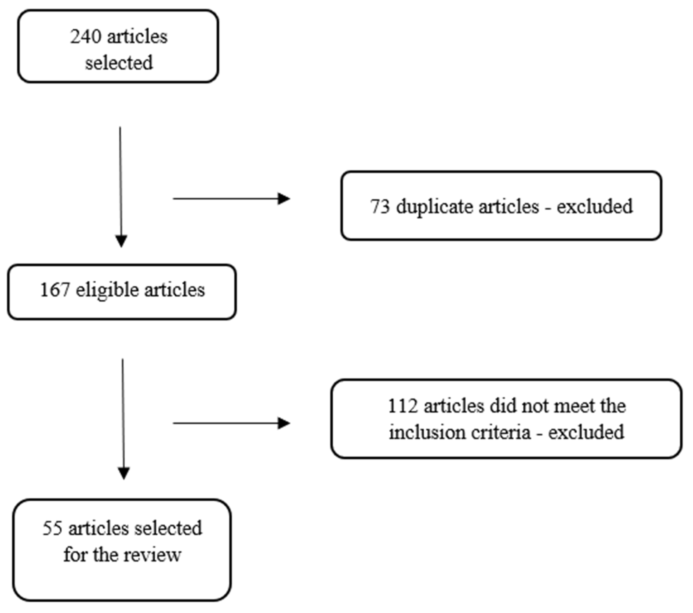
Figure 2.
Illustration representing the types of studies included in the review.
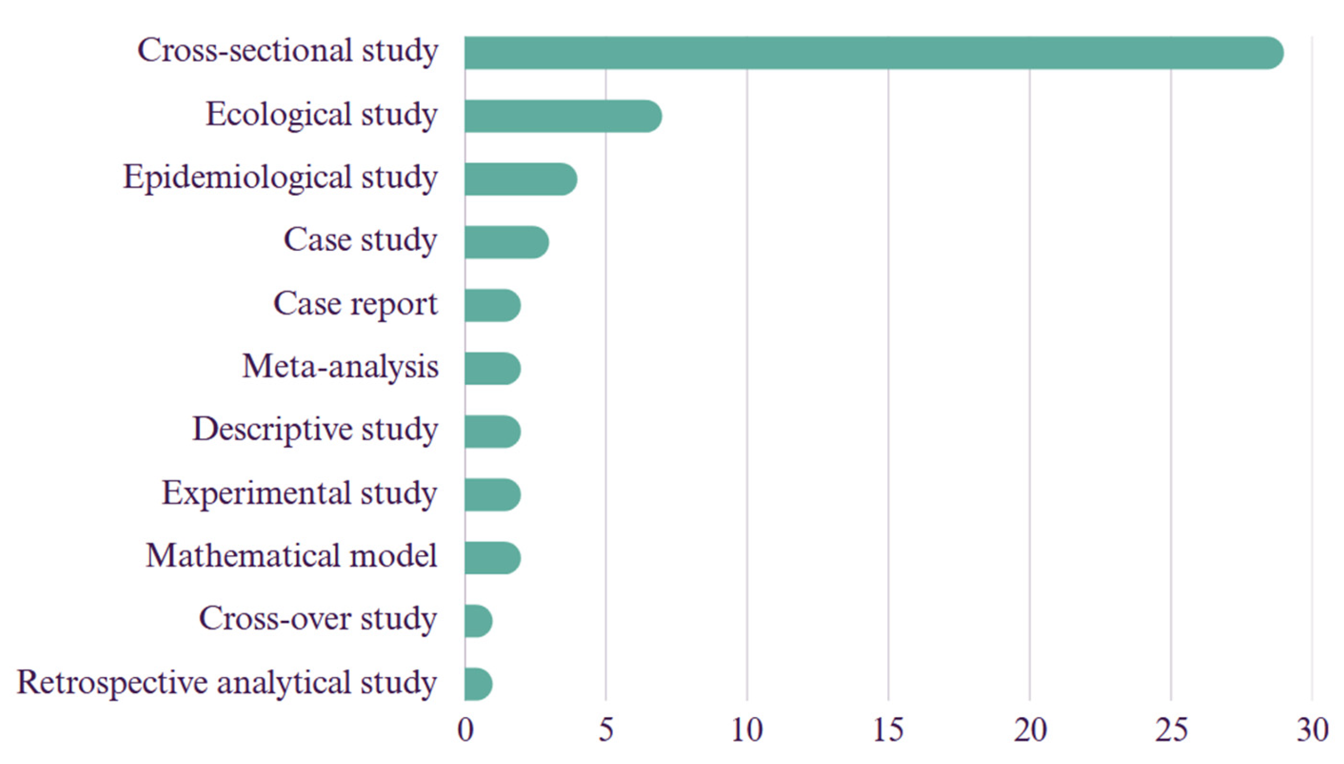
Figure 3.
Illustration representing the number of articles selected for review, classified by continent of publication.
Figure 3.
Illustration representing the number of articles selected for review, classified by continent of publication.
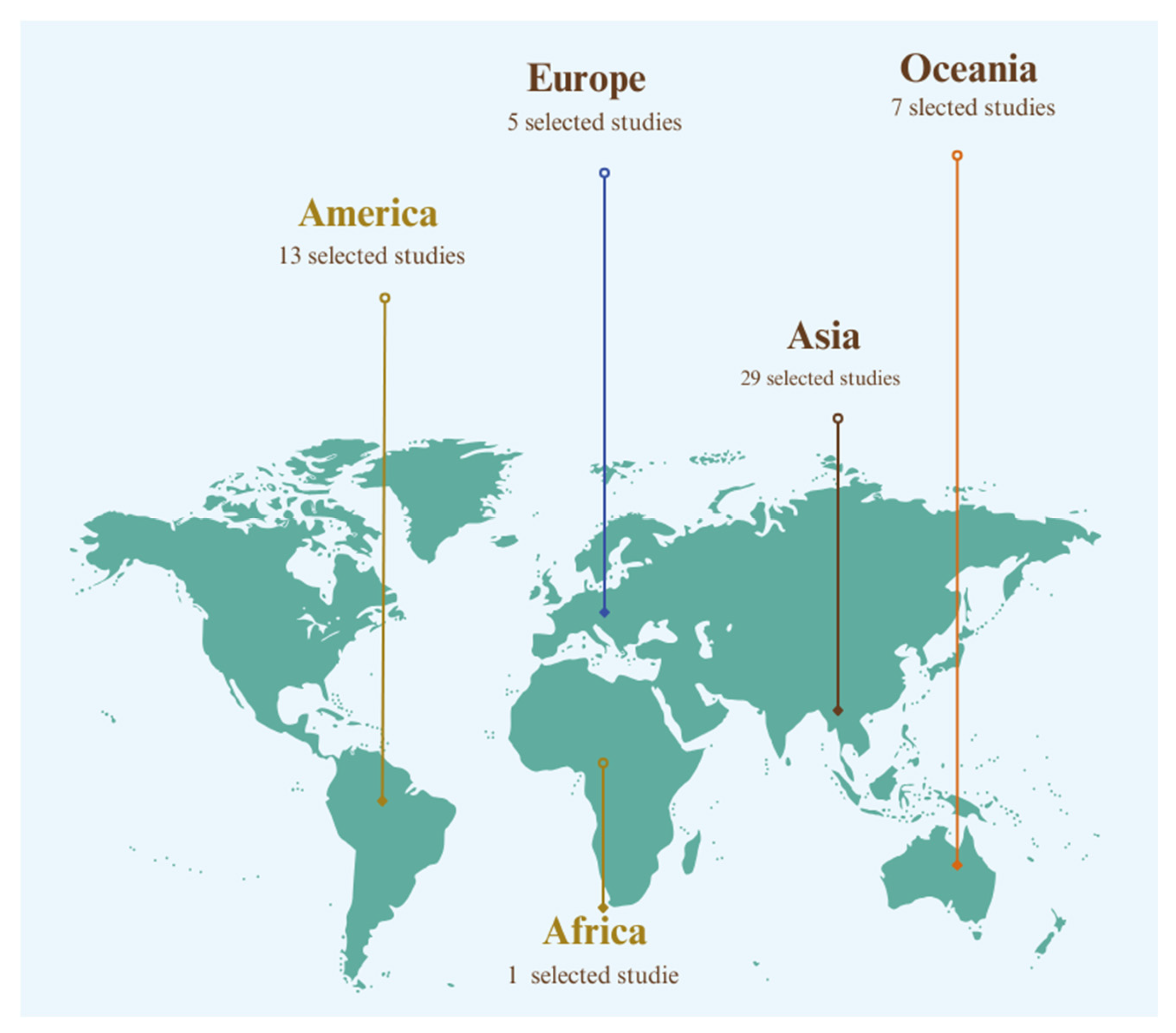
Figure 4.
Illustration representing the journals of publication for the articles selected for review, categorized by related areas.
Figure 4.
Illustration representing the journals of publication for the articles selected for review, categorized by related areas.
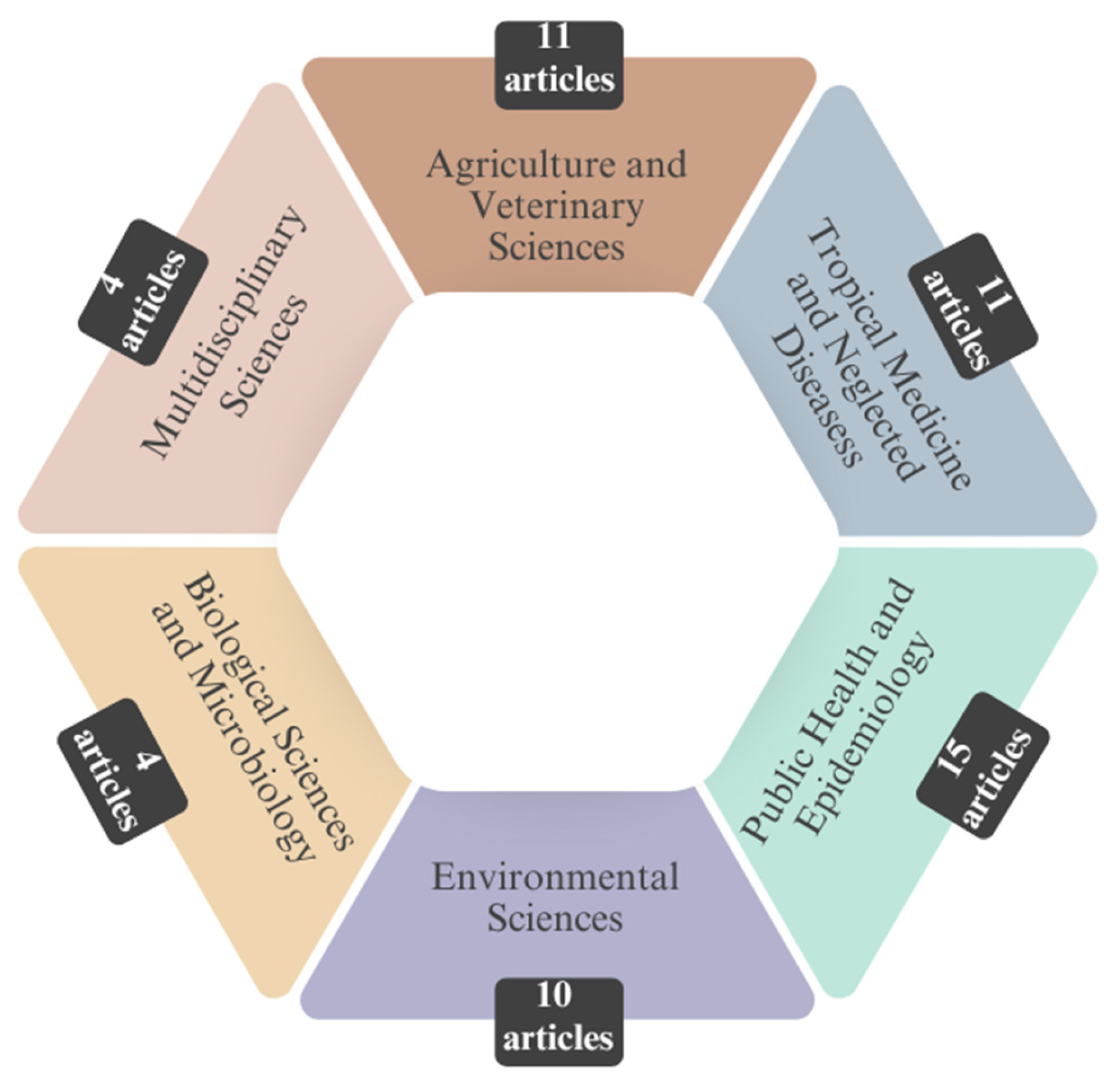

Table 1.
Data on authorship, database affiliation, country, publication journal, and main topic, presented in chronological order.
Table 1.
Data on authorship, database affiliation, country, publication journal, and main topic, presented in chronological order.
| Authors (year) | Database | Country of Study | Journal | Leptospirosis X Climate Change X Humans/Animals | |
|---|---|---|---|---|---|
| 1 | Wójcik-Fatla et al. [28] | Scopus | Poland | Annals of Agricultural and Environmental Medicine | Occurrence of pathogenic Leptospira in water and soil samples. |
| 2 | Wynwood et al. [29] | Scopus | Australia | Tropical Biomedicine | Humans infected with serovar Arborea after flooding. |
| 3 | Agampodi et al. [30] | Scopus | Sri Lanka | PLoS Neglected Tropical Diseases | Disease outbreak in humans in a dry region. |
| 4 | Pimenta et al. [31] | Scielo | Brazil | Pesquisa Veterinária Brasileira | Serovar prevalence in cattle. |
| 5 | Guimarães et al. [32] | Pubmed | Brazil | Ciência & Saúde Coletiva | Positive effect of precipitation on transmission. |
| 6 | Gracie et al. [33] | Pubmed | Brazil | International Journal of Environmental Research and Public Health | Environmental and socioeconomic factors (densely urbanized areas, flood-prone regions, and altitude) in disease transmission. |
| 7 | Suwanpakdee et al. [34] | Pubmed | Thailand | Epidemiology and Infection | Influence of flooding on human disease. |
| 8 | Matono et al. [35] | Pubmed | Japan | Journal of Travel Medicine | Report of two positive leptospirosis cases after traveling to a flooded area. |
| 9 | Lau et al. [36] | Pubmed | Fiji | PLoS Neglected Tropical Diseases | Serovars involved and environmental factors. |
| 10 | Sohail et al. [37] | Scopus | Pakistan | Journal of Equine Veterinary Science | Seroprevalence and risk factors in equines. |
| 11 | Gloriani et al. [38] | Scopus | Philippines | Southeast Asian Journal of Tropical Medicine and Public Health | Human seroprevalence of the disease after floods. |
| 12 | Azócar-Aedo & Monti [39] | Pubmed | Chile | Zoonoses and Public Health | Factors associated with canine leptospirosis. |
| 13 | Ledien et al. [40] | Pubmed | Cambodia | PloS One | Development of a flood indicator test. |
| 14 | Hayati et al. [41] | Scopus | Malaysia | Asian Journal of Agriculture and Biology | Environmental factors linked to leptospirosis risk in humans. |
| 15 | Syamsuar et al. [42] | Scopus | India | Indian Journal of Public Health Research and Development | Human seroprevalence in flood-prone areas. |
| 16 | Ijaz et al. [43] | Scopus | Pakistan | Acta Tropica | Seroprevalence and risk factors in cattle. |
| 17 | Supe et al. [44] | Pubmed | India | The National Medical Journal of India | Use of chemoprophylaxis for leptospirosis in humans in flooded areas. |
| 18 | Chadsuthi et al. [45] | Scopus | Thailand | BMC Infectious Diseases | Environmental factors in leptospirosis transmission in cattle and buffaloes. |
| 19 | Vitale et al. [46] | Pubmed | Italy | Journal of Infection and Public Health | Report of two symptomatic human leptospirosis cases. |
| 20 | Matsushita et al. [47] | Pubmed | Philippines | PLoS Neglected Tropical Diseases | Association of rainfall and disease occurrence in humans. |
| 21 | Radi et al. [48] | Pubmed | Malaysia | The American Journal of Tropical Medicine and Hygiene | Environmental factors such as floods and water bodies in disease distribution in humans. |
| 22 | Mayfield et al. [49] | Pubmed | Fiji | The Lancet Planetary Health | Use of geographically weighted logistic regression for human epidemiology. |
| 23 | Sohail et al. [50] | Pubmed | Pakistan | Acta Tropica | Seroprevalence and risk factors in humans. |
| 24 | Togami et al. [51] | Pubmed | Fiji | The American Journal of Tropical Medicine and Hygiene | Occurrence of the disease in humans and factors such as floods. |
| 25 | Widiyanti et al. [52] | Scopus | Indonesia | Zoonoses and Public Health | Presence of bacteria in the environment, especially during floods. |
| 26 | Ding et al. [53] | Scopus | China | Environmental Research | Influence of floods on human cases. |
| 27 | Sumalapao et al. [54] | Scopus | Philippines | Asian Pacific Journal of Tropical Medicine | Relationship between typhoons and increased disease occurrence in humans. |
| 28 | Naing et al. [55] | Pubmed | Australia | PloS One | Significant association between floods and increased risk of human leptospirosis. |
| 29 | López et al. [56] | Scopus | Argentina | Science of the Total Environment | Hydro-climatic factors in disease occurrence in humans. |
| 30 | Péres et al. [57] | Scopus | Brazil | Water | Influence of extreme hydrometeorological events and increased human hospitalization rates due to leptospirosis in Santa Catarina state. |
| 31 | Goh et al. [58] | Pubmed | Malaysia | International Journal of Environmental Research and Public Health | Rat contact and shared areas as risk factors for shelter dogs and their handlers. |
| 32 | Hamond et al. [59] | Pubmed | Uruguay | Transboundary and Emerging Diseases | Report of ovine leptospirosis. |
| 33 | Blagojevic et al. [60] | Pubmed | Serbia | Acta Veterinaria Hungarica | Detection of leptospirosis in wild mammals. |
| 34 | Chery et al. [61] | Scopus | Saint Lucia | Pan American Journal of Public Health | Human epidemiology and correlation with precipitation and temperature. |
| 35 | Baki et al. [62] | Scopus | Malaysia | Malaysian Journal of Microbiology | Detection of pathogenicity genes in environmental samples. |
| 36 | Silva et al. [63] | Pubmed | Brazil | Geospatial Health | Identifying human disease clusters in municipalities after natural disasters. |
| 37 | Gracie et al. [64] | Pubmed | Brazil | Cadernos de Saúde Pública | Higher risk of human leptospirosis in municipalities that declared floods. |
| 38 | Syakbanah & Fuad [65] | Scopus | Java | Jurnal Kesehatan Lingkungan | Human disease cases after a cyclone and flooding. |
| 39 | Chadsuthi et al. [66] | Pubmed | Thailand | Scientific Reports | Mathematical model to evaluate cases in humans, animals, and the environment. |
| 40 | Rahman et al. [67] | Pubmed | Malaysia | Journal of Veterinary Research | Disease prevalence in cattle, goats, and sheep after flooding. |
| 41 | Keenum et al. [68] | Pubmed | Puerto Rico | Environmental Science & Technology | Presence of bacteria in water systems after flooding. |
| 42 | Mgode et al. [69] | Pubmed | Tanzania | Pathogens | Disease prevalence in cattle. |
| 43 | Taylor et al. [14] | Pubmed | Great Britain | Preventive Veterinary Medicine | Disease prevalence in dogs. |
| 44 | Yanagihara et al. [70] | Scopus | Japan | Microbiology Spectrum | Presence of Leptospira in the environment. |
| 45 | Silva et al. [71] | Scielo | Brazil | Ciência e Saúde Coletiva | Climatic and environmental factors in human leptospirosis occurrence. |
| 46 | Taunton et al. [72] | Pubmed | Australia | Communicable Diseases Intelligence | Occurrence of human leptospirosis after heavy rains and flooding. |
| 47 | Marteli et al. [73] | Pubmed | Brazil | Geospatial Health | Flood-prone areas and human leptospirosis cases. |
| 48 | Rahman et al. [74] | Scopus | Malaysia | Sains Malaysiana | Presence of Leptospira in cattle farm soils after flooding. |
| 49 | Maric et al. [75] | Scopus | Bosnia and Herzegovina | Acta Veterinaria | Seroprevalence of leptospirosis in dogs and foxes after flooding. |
| 50 | Jayaramu et al. [76] | Pubmed | Malaysia | International Journal of Biometeorology | Hydrometeorological risk factors and human leptospirosis. |
| 51 | Udayar et al. [77] | Pubmed | India | Indian Journal of Community Medicine | Environmental risk factors associated with human leptospirosis. |
| 52 | Sohn-Hausner et al. [78] | Pubmed | Brazil | Tropical Medicine and Infectious Disease | Risk factors such as flooding and serovars in dogs and their owners. |
| 53 | Thibeaux et al. [22] | Scopus | New Caledonia | Science of the Total Environment | Presence of Leptospira in soil samples after floods. |
| 54 | Rodríguez-Rodríguez et al. [79] | Pubmed | Malaysia | Zoonoses and Public Health | Risk factors for leptospirosis occurrence in humans. |
| 55 | Ifejube et al. [80] | Pubmed | India | International Journal of Health Geographics | Positive relationship between flooding and human cases. |
Disclaimer/Publisher’s Note: The statements, opinions and data contained in all publications are solely those of the individual author(s) and contributor(s) and not of MDPI and/or the editor(s). MDPI and/or the editor(s) disclaim responsibility for any injury to people or property resulting from any ideas, methods, instructions or products referred to in the content. |
© 2025 by the authors. Licensee MDPI, Basel, Switzerland. This article is an open access article distributed under the terms and conditions of the Creative Commons Attribution (CC BY) license (http://creativecommons.org/licenses/by/4.0/).
Copyright: This open access article is published under a Creative Commons CC BY 4.0 license, which permit the free download, distribution, and reuse, provided that the author and preprint are cited in any reuse.


