Submitted:
14 April 2025
Posted:
15 April 2025
You are already at the latest version
Abstract
Purpose: To compare the clinical outcomes of Lotrafilcon A and Balafilcon A silicone hydrogel contact lenses used as therapeutic bandage contact lenses (BCL) following transepithelial photorefractive keratectomy (TPRK). Materials and Methods: A randomized, double-blind, contralateral study was conducted on 82 eyes of 41 consecutive TPRK-treated patients, randomly assigned to wear either Lotrafilcon A or Balafilcon A BCL in their right and left eyes (groups A or B). The postoperative assessment included uncorrected (UDVA) and corrected (CDVA) distance visual acuity, ocular pain/irritation, epithelial healing, limbal and conjunctival hyperemia, lens mobility, and the amount of protein deposition accumulated on the BCLs. Results: On the first postoperative day, the pain intensity was scored as 2.80 ± 2.35 for group A and 4.44 ± 2.46 for group B (P< 0.001). The amount of protein deposition on the BCLs was also significantly lower in group A compared to group B. For BCLs removed on day three, the protein deposition was 9.92 ± 9.82 vs. 25.75 ± 9.86 μg for groups A and B (P< 0.001). For BCLs removed on day four, the protein deposition was 9.47 ± 10.06 vs. 32.60 ± 16.71 μg (P=0.005). However, there were no statistically significant differences between the two groups in terms of corneal epithelial defect area, corneal epithelial healing time, UDVA, CDVA, limbal or conjunctival hyperemia, and lens movement. Conclusions: Lotrafilcon A performed better than Balafilcon A in terms of reducing ocular pain, foreign body sensation, and protein deposition, suggesting that Lotrafilcon A may be a more suitable option for therapeutic BCL use following TPRK.
Keywords:
transepithelial photorefractive keratectomy (TPRK)
; silicone hydrogel
; bandage contact lens
; Lotrafilcon A
; Balafilcon A
1. Introduction
Photorefractive keratectomy (PRK) is a well-established and effective method for correcting ametropia using an excimer laser[1]. In comparison to laser in situ keratomileusis (LASIK), PRK preserves a thicker postoperative stromal bed and eliminates complications associated with LASIK-flap creation. However, PRK is not without its own set of potential adverse reactions, including postoperative pain, haze, irregular or delayed corneal epithelial healing, and prolonged visual recovery[2,3,4]. Transepithelial PRK (TPRK) is a surgical technique that builds upon the principles of PRK[5,6,7,8]. In TPRK, both the corneal epithelium and anterior corneal stroma are removed in a single step, solely through the use of the excimer laser, without any additional mechanical or alcohol-based epithelial removal. This approach offers several advantages, including increased patient comfort during surgery, reduced potential damage and irritation to the cornea, and decreased postoperative eye discomfort[9,10]. Furthermore, TPRK has the potential to improve corneal epithelial healing and minimize the occurrence of haze, as compared to traditional PRK methods that involve mechanical or alcohol-based epithelial removal[9,11]. Innovations in refractive surgery eliminate the need for corneal flaps. This avoids iatrogenic aberrations, preserves more corneal stroma, maintains biomechanical stability, and reduces long - term corneal risk[12,13]. Despite these benefits, TPRK can still result in ocular irritation, pain, and a slower recovery of vision[10,14]. Therefore, there is a need for further refinement of the current treatment protocols to optimize outcomes and enhance patient satisfaction.
Since its introduction in the late 1960s, bandage contact lenses (BCLs) have primarily been utilized for corneal protection following injury. However, they have also demonstrated potential in relieving pain, promoting corneal epithelial healing, safeguarding the cornea, and enhancing visual acuity[15], particularly after undergoing excimer laser keratectomy[16,17,18,19,20,21,22]. In addition, it effectively sustains corneal hydration, thereby forestalling the onset of postoperative dry - eye symptoms [23,24]. So continuous BCL wear for a duration of 3 to 7 days subsequent to photorefractive keratectomy (PRK) is presently regarded as the standard of care[25,26]. In comparison to hydrogel BCLs, silicone hydrogel BCLs exhibit superior oxygen permeability, which facilitates wound healing and regeneration of the corneal epithelial cells by mitigating complications associated with hypoxia and providing an optimal healing environment[27].
Several silicone hydrogel contact lenses are currently in clinical use. Among them, Lotrafilcon A (Air Optix Night & Day, Alcon Laboratories, USA), Balafilcon A (PureVision, Bausch & Lomb, USA), and Senofilcon A have received approval from the Food and Drug Administration (FDA) for use as bandage contact lenses (BCLs)[28]. These lenses differ in terms of their materials and design, resulting in variations in oxygen permeability, water content, elastic modulus, thickness, diameter, edge contour, and anti-precipitation effect[29,30]. These parameters have the potential to impact ocular comfort and corneal healing following surface ablation surgery.
The first-generation silicone hydrogel contact lenses, Lotrafilcon A and Balafilcon A, have been demonstrated to be both safe and effective as bandage contact lenses (BCLs)[31,32]. They continue to be widely utilized in the treatment of various ophthalmic diseases, particularly in cases of ocular surface disorders. In an effort to enhance ocular comfort, the manufacturer has recently introduced a newer version of Lotrafilcon A BCL. The present study aims to evaluate the clinical effectiveness of the new generation Lotrafilcon A (Air Optix) and Balafilcon A (Pure Vision) BCLs.
2. Materials and Methods
A randomized, double-blind, contralateral study was conducted on 82 eyes of 41 consecutive patients who underwent transepithelial photorefractive keratectomy (TPRK) surgery at the Refractive Surgery Center of the Eye Hospital of Wenzhou Medical University from October 2018 to March 2019. The inclusion criteria for the study were as follows: myopia of ≤10.00 diopters (D), astigmatism of ≤2.00 D, anisometropia of less than 1.00 D in spherical equivalent refraction (SER), and corrected distance visual acuity (CDVA) of 0 (LogMAR) or better. Patients with ocular disease, amblyopia, or a history of previous eye surgery were excluded from the study. The research protocol was approved by the Ethics Committee of Wenzhou Medical University Eye Hospital and adhered to the principles outlined in the Helsinki Declaration. Prior to participation, all patients, or the guardians of patients under 18 years of age, provided signed informed consent.
All surgeries were conducted by the same experienced surgeon (S.C.). The procedures were performed using the Amaris 750 excimer laser (Schwind, Kleinostheim, Germany) in TPRK mode, with a therapeutic bandage contact lens (BCL) placed on the treated eye at the conclusion of the surgery. The patients' two eyes were randomly assigned to two groups based on a randomization table. One eye was fitted with Lotrafilcon A BCL (Group A), while the fellow eye was fitted with Balafilcon A BCL (Group B). Both the patients and the examiner were unaware of which lens was applied to each eye. The characteristics of the two types of BCLs are provided in Table 1.
The patients were assessed at 1, 3, 10 days, and 1 month post-surgery. In cases where the corneal epithelium had not fully healed within 3 days after surgery, an additional assessment on day 4 was conducted. All follow-up examinations were conducted by the same ophthalmologist (L.W.). The postoperative examinations on days one and three prior to the removal of the bandage contact lenses (BCLs) included subjective questionnaires to assess ocular pain intensity and the frequency of irritation symptoms. Slit lamp assessments were performed to evaluate corneal epithelial healing, as well as to assess limbal and conjunctival hyperemia, BCL movement, and the presence of BCL deposits. Ocular pain intensity was measured using a numerical pain scale (NRS-11) ranging from 0 to 10[33]. The evaluation of irritation symptoms included questions about photophobia, foreign body sensation, pain, and dry eye sensation, with each symptom graded from 0 to 4 (0 indicating never, 1 indicating sometimes, 2 indicating half of the time, 3 indicating most of the time, and 4 indicating all of the time). Objective evaluation of corneal epithelial healing was performed using optical coherence tomography (OCT) in corneal linear scan mode. The area of the largest defect was calculated using the formula s=π(a+b)/42, where "a" represented the horizontal diameter and "b" represented the vertical diameter (Figure 1)[34]. Conjunctival and limbal hyperemia were graded on a scale from 0 to 4, with 0 indicating normal and 4 indicating severe hyperemia. Lens movement and deposits were also graded on a scale from 0 to 4 (Table 2 and Table 3)[35,36]. Once the corneal epithelium had fully healed, the healing time was recorded, and the BCLs were removed. UDVA and CDVA were assessed after BCL removal on day 3 or 4, as well as on day 10 and one month post-surgery.
Total protein precipitation on the lenses was measured using the following method: each bandage contact lens (BCL) was collected after removal and placed in a 2-ml polypropylene centrifuge tube. A 1.5-ml extraction solution, consisting of a 50:50 mix of 0.2% trifluoroacetic acid and acetonitrile, was added to the tube. The centrifuge tube was then placed in a long-axis rotary mixer and mixed for 24 hours at room temperature. Afterward, the BCL was removed, and the protein extracts were freeze-dried. To dissolve the protein, a 30-μl 0.01-M phosphate-buffered solution (PBS) with a pH of 7.4 was used. The total protein precipitation on the lenses was determined using a Pierce BCA protein assay kit (Thermo Scientific, USA).
SPSS 21.0 software (SPSS Inc, Chicago, IL) was utilized for data analysis. The statistical analysis in this study was conducted using mean and standard deviations (SDs) and paired-sample t-tests. Frequency distributions and Wilcoxon sign rank-sum tests were employed for statistical analysis involving graded counting data. Spearman correlation analysis was also utilized. A significance level of P<0.05 was considered statistically significant.
3. Results
Table 4 presents the gender distribution, mean age, mean preoperative spherical equivalent refraction (SER), preoperative mean uncorrected distance visual acuity (UDVA) and corrected distance visual acuity (CDVA) in logMAR, diameters of the optical and total laser ablation zones, as well as the ablation depth.
Ocular Pain Intensity
On the first postoperative day, patients in groups A and B reported pain intensity according to the pain scale (NRS-11) as 2.80 ± 2.24 and 4.44 ± 2.84, respectively (P=0.003). On the third postoperative day, the average pain intensity for both groups was 0.04 ± 0.2 and 0.28 ± 0.79 (P=0.110) (Figure 2).
Subjective Ocular Irritation
One day after surgery: 70.7% of eyes in group A reported a foreign body sensation intensity grade of 2 or less, compared to 31.7% of eyes in group B (P=0.001). Similarly, 53.7% of eyes in group A reported pain frequency grade 2 or less, while 31.7% of eyes in group B reported the same (P=0.001). However, there were no significant differences in the symptoms of photophobia or dry eye between the two groups. Three days after surgery, 85.4% of eyes in group A reported a foreign body sensation intensity grade of 1 or less, compared to 58.5% of eyes in group B (P<0.001). There were no significant differences in other subjective symptoms between the two groups (Table 5). Within each group, there was a significant improvement in photophobia, foreign body sensation, and pain symptoms from day one to day three (group A: P<0.001, P=0.003, P<0.001, respectively; group B: P<0.001 for all).
Corneal Epithelial Healing
One day after surgery, the average corneal epithelial defect area was 14.27 ± 4.09 mm2 in group A and 15.09 ± 4.95 mm2 in group B (P=0.20). The average epithelial healing time in group A compared to group B was 3.10 ± 0.30 vs. 3.12 ± 0.33 days (P=0.71).
Slit-Lamp Evaluation
There were no significant differences in limbal or conjunctival hyperemia, lens mobility, or lens deposit levels between groups A and B on days 1 and 3 postoperatively (Table 6). The mobility of lenses in both groups A and B increased from days 1 to 3 after surgery (P<0.001 for group A, P=0.001 for group B). Additionally, there was a significant improvement in limbal and conjunctival hyperemia in both groups A and B from days 1 to 3 (P<0.001 for both). However, there were no significant changes in the assessed levels of lens deposits for groups A and B (P=0.09 and P=0.32, respectively).
Visual Acuity
Immediately following the removal of the bandage contact lenses (BCL) at 3 or 4 days after surgery, as well as at 10 days and 1 month postoperatively, the uncorrected distance visual acuity (UDVA) values (logMAR) in group A were 0.39 ± 0.15, 0.13 ± 0.10, and 0.07 ± 0.08, respectively. In group B, the corresponding UDVA values were 0.35 ± 0.15, 0.11 ± 0.09, and 0.06 ± 0.09, respectively. Similarly, the corrected distance visual acuity (CDVA) values in group A were 0.28 ± 0.15, 0.07 ± 0.07, and 0.03 ± 0.06 at the same time points, while in group B, the CDVA values were 0.27 ± 0.15, 0.07 ± 0.07, and 0.02 ± 0.05. The differences in visual acuity between the two groups were not statistically significant.
Protein Precipitation Measured on BCL
A total of 66 BCLs were removed 3 days after surgery, while 16 BCLs were removed 4 days after surgery. The total protein precipitation measures for groups A and B were 9.92 ± 9.82 μg and 25.75 ± 9.86 μg, respectively, in the lenses removed 3 days after surgery (P<0.001). For the lenses removed 4 days after surgery, the total protein precipitation measures were 9.47 ± 10.06 μg for group A and 32.60 ± 16.71 μg for group B (P=0.005) (Figure 3). However, there was no significant difference in total protein precipitation within groups A and B between 3 and 4 days after surgery (P=0.91, P=0.14).
Correlation Analysis
There was minimal correlation observed between ocular pain intensity and lens movement (group A: r=0.17; group B: r=0.03), corneal epithelial defect area (group A: r=-0.19; group B: r=-0.10), or limbal and conjunctival congestion (group A: r=-0.10 and r=-0.17, respectively; group B: r=-0.27 and r=-0.20, respectively). Additionally, no significant correlation was found between total protein precipitation and foreign body sensation (group A: r=0.37; group B: r=0.03) or lens movement (group A: r=-0.24; group B: r=0.17).
4. Discussion
In this study, we observed that eyes wearing Lotrafilcon A (AirOptix Night & Day) BCL exhibited better outcomes in terms of ocular pain, foreign body sensation, and protein deposition compared to eyes wearing Balafilcon A (PureVision) BCL after TPRK.
Earlier studies have demonstrated that bandage contact lenses (BCLs) from different brands, utilizing various silicone hydrogel materials, offer varying levels of pain control when used following different types of surface ablation procedures. Hasan et al.[37] reported that Senofilcon A (Acuvue Oasys) provided better control of ocular pain and discomfort after PRK compared to Lotrafilcon A (AirOptix Night & Day). Taylor et al. [28] conducted a study comparing three types of silicone hydrogel BCLs after PRK and found that Acuvue Oasys resulted in the lowest level of ocular pain, followed by AirOptix and PureVision. Qu et al.[38] discovered that galyfilcon A (Acuvue) exhibited superior pain control after Laser-assisted Subepithelial Keratomileusis (LASEK) in comparison to PureVision. Li et al.[39] found that Senofilcon A lenses were associated with less pain and more comfort compared to Balafilcon A after TPRK. Lotfy et al.[40] compared lotrafilcon B and comfilcon A silicone hydrogel bandage contact lenses after transepithelial photorefractive keratectomy and found that healing responses were better with lotrafilcon B than with comfilcon A bandage contact lenses.
Qu et al.[38] also proposed that the design of the lens-edge contour of the bandage contact lens (BCL) could influence pain control during corneal healing, in addition to the oxygen transmissibility of the BCL[34,41]. Blackmore[42] suggested that an ideal lens edge should be smooth and gradual to minimize friction on the exposed nerve endings and new epithelial cells, thereby providing better pain relief. Previous studies have indicated that Acuvue Oasys has the thinnest and sharpest edges, PureVision has the thickest and bluntest edges, and AirOptix Night & Day has a thick edge with a sharp fusion point between the front and back surfaces[28].
In the present study, the ocular pain reported in group A (AirOptix Night & Day) was significantly lower than in group B (PureVision) (P=0.005). This difference may be attributed to the higher DK/t (oxygen transmissibility relative to lens thickness) value of 175 in AirOptix Night & Day compared to 101 in PureVision, as well as the smoother edge profile of the former. Previous studies have suggested that the mechanical characteristics, particularly the stiffness of lens materials, may be associated with pain relief, with more flexible lenses offering better comfort[37,38]. However, we did not find a correlation between the elastic moduli (1.4 vs. 1.1 MPa) and pain in the two groups. Grentzelos et al.[43] proposed that larger epithelial defects could lead to higher pain intensity due to the exposure of nerve endings. In our study, we did not observe significant variations in corneal epithelial defect areas between the eyes, presumably since the reepithelialization diameter in TPRK was low.
Lim et al.[44] suggested that bandage contact lenses (BCLs) should exhibit good movement to reduce lens adherence, improve patient comfort, enhance stability, and promote centration of the lens, thereby facilitating corneal wound healing. Some studies have indicated that BCLs with high water content may have a greater tendency to adhere to the corneal epithelium[36], and that such BCLs may gradually tighten with prolonged wearing time[38]. In our study, the movement of the BCLs on the first day after surgery ranged between levels 0 and 2 for all eyes except one, and there was no significant difference in lens movement between the two groups, which is consistent with the findings of Fonn et al.[45]. However, on day 3 postoperatively, there was a slight increase in lens movement in both groups, compared to the first day, likely due to corneal edema and increased ocular irritation symptoms on the first postoperative day, which may have affected blinking and subsequently influenced lens movement.
The measured material precipitation on contact lenses includes protein, lipid, microorganisms, and calcium salts that deposit on the lens surface and within its matrix. Among these precipitates, protein precipitation is particularly significant[46], as it can cause eye discomfort, reduced vision, and increased inflammation[47]. Sack et al.[48] conducted measurements of protein precipitation on various soft lenses and found that the thickness of surface precipitation on type IV lenses was considerably greater than that on type I lenses (The FDA classifies soft contact lenses into four categories based on their water content and ionization degree: Non-ionic types I and II, with water content < 50% and >50%, respectively, and ionic types III and IV, with water content < 50% and > 50%, respectively). Myers et al.[49] analyzed type I and IV lenses and discovered that the total surface precipitation was similar between the two types, but the total protein precipitation on type IV lenses was nearly 40 times higher than that on type I lenses. Therefore, it appears that protein precipitation on the surface and inside of soft contact lenses is positively correlated with water content and is more pronounced in ionic contact lenses. In our study, the AirOptix Night & Day BCL belongs to FDA type I lenses, which have a plasma coating forming a permanent ultra-thin (25 nm) hydrophilic surface and a water content of 24%. On the other hand, the PureVision BCL belongs to FDA type III silicon hydrogel lenses with a negative charge and a water content of 36%. The significantly lower total protein precipitation in group A compared to group B indicates that the AirOptix Night & Day lens exhibits better resistance to precipitation than the PureVision lens, aligning with the general findings of Sack et al.[46] regarding lenses of FDA types I and IV.
It has been demonstrated that a high level of oxygen transmissibility (Dk/t) can effectively decrease the incidence of corneal hypoxia and edema during the healing process, as well as expedite epithelial healing[18,50]. Furthermore, maintaining good lens stability and centration also contributes to the process of epithelial healing[44].
In the present study, there were no significant differences observed in the corneal epithelial defect area or corneal epithelial healing time between groups A and B on the first day after TPRK, which aligns with the findings of Mukherjee et al.[30]. This suggests that despite the difference in oxygen transmissibility between the two lenses, both lenses are characterized by high oxygen permeability and can adequately meet the physiological requirements for corneal healing following surgery.
5. Conclusions
Based on the findings of this study, it can be concluded that AirOptix Night & Day (Lotrafilcon A) contact lenses outperformed Pure Vision (Balafilcon A) lenses when used as bandage contact lenses (BCL) after TPRK due to its superior efficacy in terms of pain relief, reduction of foreign body sensation, and less total protein precipitation.
References
- Munnerlyn, C.R.; Koons, S. J.; Marshall, J. Photorefractive keratectomy: a technique for laser refractive surgery. Journal of cataract and refractive surgery.1988,14(1),46–52. [CrossRef]
- Ambrósio, R.Jr.; Wilson,S. LASIK vs LASEK vs PRK: advantages and indications. Seminars in ophthalmology.2003,18(1), 2–10.
- Steinert,R.F.;Bafna, S. Surgical correction of moderate myopia: which method should you choose? II. PRK and LASIK are the treatments of choice. Survey of ophthalmology.1998,43(2), 157–179.
- McCarty, C. A.; Garrett, S. K.; Aldred, G. F.; Taylor, H. R. Assessment of subjective pain following photorefractive keratectomy. Melbourne Excimer Laser Group. Journal of refractive surgery.1996,12(3), 365–369.
- Gimbel, H. V.;DeBroff, B. M.;Beldavs, R. A.; van Westenbrugge, J. A.; Ferensowicz, M. Comparison of laser and manual removal of corneal epithelium for photorefractive keratectomy. Journal of refractive surgery.1995, 11(1), 36–41. [CrossRef]
- Luger, M. H.; Ewering, T.; Arba-Mosquera, S. Consecutive myopia correction with transepithelial versus alcohol-assisted photorefractive keratectomy in contralateral eyes: one-year results. Journal of cataract and refractive surgery.2012,38(8), 1414–1423. [CrossRef]
- Stojanovic, A.;Chen, S.; Chen, X.; Stojanovic, F.; Zhang, J.; Zhang, T.; Utheim, T. P. One-step transepithelial topography-guided ablation in the treatment of myopic astigmatism. 2013,PloS one, 8(6), e66618. [CrossRef]
- Xi, L.; Zhang, C.; He, Y. Clinical outcomes of Transepithelial photorefractive keratectomy to treat low to moderate myopic astigmatism. BMC ophthalmology.2018,18(1), 115. [CrossRef]
- Fadlallah, A.; Fahed, D.; Khalil, K.; Dunia, I.; Menassa, J.; El Rami, H.; Chlela, E.; Fahed, S. Transepithelial photorefractive keratectomy: clinical results. Journal of cataract and refractive surgery.2011,37(10), 1852–1857. [CrossRef]
- Celik, U.; Bozkurt, E.; Celik, B.; Demirok, A.; Yilmaz, O. F. Pain, wound healing and refractive comparison of mechanical and transepithelial debridement in photorefractive keratectomy for myopia: results of 1 year follow-up. Contact lens & anterior eye : the journal of the British Contact Lens Association.2014, 37(6), 420–426. [CrossRef]
- Naderi,M.;Jadidi,K.;Mosavi,S.A.;Daneshi,S.A. Transepithelial Photorefractive Keratectomy for Low to Moderate Myopia in Comparison with Conventional Photorefractive Keratectomy. Journal of ophthalmic & vision research.2016,11(4), 358–362. [CrossRef]
- Vinciguerra, P.; Camesasca, F. I.; Vinciguerra, R.; Arba-Mosquera, S.; Torres, I.; Morenghi, E.; Randleman, J. B. Advanced Surface Ablation With a New Software for the Reduction of Ablation Irregularities. Journal of refractive surgery .2017, 33(2), 89–95. [CrossRef]
- Bakhsh, A. M.; Elwan, S. A. M.; Chaudhry, A. A.; El-Atris, T. M.; Al-Howish, T. M. Comparison between Transepithelial Photorefractive Keratectomy versus Alcohol-Assisted Photorefractive Keratectomy in Correction of Myopia and Myopic Astigmatism. Journal of ophthalmology.2018, 5376235. [CrossRef]
- Luger, M. H.; Ewering, T.; Arba-Mosquera, S. Myopia correction with transepithelial photorefractive keratectomy versus femtosecond-assisted laser in situ keratomileusis: One-year case-matched analysis. Journal of cataract and refractive surgery.2016,42(11),1579–1587. [CrossRef]
- Jackson, A.J.; Sinton, J.E.; Frazer, D.G.; Morrison, E. Therapeutic contact lenses and their use in themanagement of anterior segment pathology. Journal of the British Contact Lens Association.1996,19(1),11-19. [CrossRef]
- Bergenske, P.; Caroline, P.; Smithe, J. Contact lenses as an adjunct in refractive surgery practice. Contact Lens Spectrum. 2002, 17, 30–37. [Google Scholar]
- Demers, P.; Thompson, P.; Bernier, R.G.; Lemire, J.; Laflamme, P. Effect of occlusive pressure patching on the rate of epithelial wound healing after photorefractive keratectomy. Journal of cataract and refractive surgery.1996, 22(1), 59–62. [CrossRef]
- Xie, W. J.; Zeng, J.; Cui, Y.; Li, J.; Li, Z. M.; Liao, W. X.; Yang, X. H. Comparation of effectiveness of silicone hydrogel contact lens and hydrogel contact lens in patients after LASEK. International journal of ophthalmology.2015,8(6), 1131–1135. [CrossRef]
- Eliaçık, M.; Erdur, S. K.; Gülkılık, G.; Özsütçü, M.; Karabela, Y. Compare the effects of two silicone-hydrogel bandage contact lenses on epithelial healing after photorefractive keratectomy with anterior segment optical coherence tomography. Contact lens & anterior eye : the journal of the British Contact Lens Association.2015,38(3), 215–219. [CrossRef]
- Taylor, K. R., Molchan, R. P., Townley, J. R., Caldwell, M. C., Panday, V. A. The effect of silicone hydrogel bandage soft contact lens base curvature on comfort and outcomes after photorefractive keratectomy. Eye & contact lens.2015, 41(2), 77–83. [CrossRef]
- Lim, L.; Lim, E. W. L. Therapeutic Contact Lenses in the Treatment of Corneal and Ocular Surface Diseases-A Review. Asia-Pacific journal of ophthalmology.2020 , 9(6), 524–532. [CrossRef]
- Wang, J.; Xi, S.; Wang, B.; Chen, Z.; Zheng, K.; Zhou, X. Clinical Observation of Silicon Hydrogel Contact Lens Fitted Immediately after Small Incision Lenticule Extraction (SMILE). Journal of ophthalmology.2020, 2604917. [CrossRef]
- Shimazaki, J., Shigeyasu, C., Saijo-Ban, Y., Dogru, M., Den, S. Effectiveness of bandage contact lens application in corneal epithelialization and pain alleviation following corneal transplantation; prospective, randomized clinical trial. BMC ophthalmology.2016, 16(1), 174. [CrossRef]
- Lu, J.; He, J.; Liu, Y . Effect of rigid corneal contact lens and corneal limbal stem cell transplantation for senile patients with pterygium Int Eye Sci. 2017, 17(6), 1188-1190.
- Sánchez-González, J. M.; López-Izquierdo, I.; Gargallo-Martínez, B.; De-Hita-Cantalejo, C.; Bautista-Llamas, M. J. Bandage contact lens use after photorefractive keratectomy. Journal of cataract and refractive surgery. 2019, 45(8), 1183–1190. [CrossRef]
- Yuksel, E.; Ozulken, K.; Uzel, M. M.; Taslipinar Uzel, A. G.; Aydoğan, S. Comparison of Samfilcon A and Lotrafilcon B silicone hydrogel bandage contact lenses in reducing postoperative pain and accelerating re-epithelialization after photorefractive keratectomy. International ophthalmology.2019, 39(11), 2569–2574. [CrossRef]
- Szaflik, J. P.; Ambroziak, A. M.; Szaflik, J. Therapeutic use of a lotrafilcon A silicone hydrogel soft contact lens as a bandage after LASEK surgery. Eye & contact lens.2004, 30(1), 59–62.
- Taylor, K. R.; Caldwell, M. C.; Payne, A. M.; Apsey, D. A.; Townley, J. R.; Reilly, C. D.; Panday, V. A. Comparison of 3 silicone hydrogel bandage soft contact lenses for pain control after photorefractive keratectomy. Journal of cataract and refractive surgery.2014,40(11), 1798–1804. [CrossRef]
- Guillon, M. Are silicone hydrogel contact lenses more comfortable than hydrogel contact lenses?. Eye & contact lens.2013, 39(1), 86–92. [CrossRef]
- Mukherjee, A.; Ioannides, A.; Aslanides, I. Comparative evaluation of Comfilcon A and Senofilcon A bandage contact lenses after transepithelial photorefractive keratectomy. Journal of optometry.2015, 8(1), 27–32. [CrossRef]
- Arora, R.; Jain, S.; Monga, S.; Narayanan, R.; Raina, U. K.; Mehta, D. K. Efficacy of continuous wear PureVision contact lenses for therapeutic use. Contact lens & anterior eye : the journal of the British Contact Lens Association. 2004, 27(1), 39–43.
- Ozkurt, Y.; Rodop, O.; Oral, Y.; Cömez, A.; Kandemir, B.; & Doğan, O. K. Therapeutic applications of lotrafilcon a silicone hydrogel soft contact lenses. Eye & contact lens.2005, 31(6), 268–269. [CrossRef]
- Hartrick, C. T.; Kovan, J. P.; Shapiro, S. The numeric rating scale for clinical pain measurement: a ratio measure?. Pain practice : the official journal of World Institute of Pain. 2003, 3(4), 310–316. [CrossRef]
- Engle, A. T.; Laurent, J. M.; Schallhorn, S. C.; Toman, S. D.; Newacheck, J. S.; Tanzer, D. J.; Tidwell, J. L. Masked comparison of silicone hydrogel lotrafilcon A and etafilcon A extended-wear bandage contact lenses after photorefractive keratectomy. Journal of cataract and refractive surgery. 2005, 31(4), 681–686. [CrossRef]
- Efron, N.; Morgan, P. B.; Katsara, S. S. Validation of grading scales for contact lens complications. Ophthalmic & physiological optics : the journal of the British College of Ophthalmic Opticians (Optometrists). 2001, 21(1), 17–29.
- Gil-Cazorla, R.; Teus, M. A.; Arranz-Márquez, E. Comparison of silicone and non-silicone hydrogel soft contact lenses used as a bandage after LASEK. Journal of refractive surgery. 2008, 24(2), 199–203. [CrossRef]
- Razmjoo, H.; Abdi, E.; Atashkadi, S.; Reza, A. M.; Reza, P. A.; Akbari, M. Comparative Study of Two Silicone Hydrogel Contact Lenses used as Bandage Contact Lenses after Photorefractive Keratectomy. International journal of preventive medicine. 2012, 3(10), 718–722.
- Qu, X. M.; Dai, J. H.; Jiang, Z. Y.; Qian, Y. F. Clinic study on silicone hydrogel contact lenses used as bandage contact lenses after LASEK surgery. International journal of ophthalmology. 2011, 4(3), 314–318. [CrossRef]
- Li, H.; Shao, T.; Zhang, J. F.; Leng, L.; Liu, S.; Long, K. L. Comparison of efficacy of two different silicone hydrogel bandage contact lenses after T-PRK. International journal of ophthalmology. 2022, 15(2), 299–305. [CrossRef]
- Lotfy, N. M.; Alasbali, T.; Alsharif, A. M.; Al-Gehedan, S. M.; Jastaneiah, S.; Al-Hazaimeh, A.; Ali, H.; Khandekar, R. Comparison of the efficacy of lotrafilcon B and comfilcon A silicone hydrogel bandage contact lenses after transepithelial photorefractive keratectomy. Medical hypothesis, discovery & innovation ophthalmology journal. 2021, 10(2), 43–49. [CrossRef]
- Edwards, J. D.; Bower, K. S.; Sediq, D. A.; Burka, J. M.; Stutzman, R. D.; Vanroekel, C. R.; Kuzmowych, C. P.; Eaddy, J. B. Effects of lotrafilcon A and omafilcon A bandage contact lenses on visual outcomes after photorefractive keratectomy. Journal of cataract and refractive surgery. 2008, 34(8), 1288–1294. [CrossRef]
- Blackmore, S. J. The use of contact lenses in the treatment of persistent epithelial defects. Contact lens & anterior eye : the journal of the British Contact Lens Association. 2010, 33(5), 239–244.
- Grentzelos, M. A.; Plainis, S.; Astyrakakis, N. I.; Diakonis, V. F.; Kymionis, G. D.; Kallinikos, P.; Pallikaris, I. G. Efficacy of 2 types of silicone hydrogel bandage contact lenses after photorefractive keratectomy. Journal of cataract and refractive surgery. 2009, 35(12), 2103–2108. [CrossRef]
- Lim, L.; Tan, D. T.; Chan, W. K. Therapeutic use of Bausch & Lomb PureVision contact lenses. The CLAO journal : official publication of the Contact Lens Association of Ophthalmologists. 2001, Inc, 27(4), 179–185.
- Fonn, D.; Dumbleton, K.; Jones, L. Silicone hydrogel material and surface properties. Contact Lens Spectrum. 2002, 17. [Google Scholar]
- Jones, L.; Senchyna, M.; Glasier, M. A.; Schickler, J.; Forbes, I.; Louie, D.; May, C. Lysozyme and lipid deposition on silicone hydrogel contact lens materials. Eye & contact lens. 2003, 29(1 Suppl), S75–S194. [CrossRef]
- Gil-Cazorla, R.; Teus, M. A.; Hernández-Verdejo, J. L.; De Benito-Llopis, L.; García-González, M. Comparative study of two silicone hydrogel contact lenses used as bandage contact lenses after LASEK. Optometry and vision science : official publication of the American Academy of Optometry. 2008, 85(9), 884–888. [CrossRef]
- Sack, R. A.; Jones, B.; Antignani, A.; Libow, R.; Harvey, H. Specificity and biological activity of the protein deposited on the hydrogel surface. Relationship of polymer structure to biofilm formation. Investigative ophthalmology & visual science. 1987, 28(5), 842–849.
- Myers, R. I.; Larsen, D. W.; Tsao, M.; Castellano, C.; Becherer, L. D.; Fontana, F.; Ghormley, N. R.; Meier, G. Quantity of protein deposited on hydrogel contact lenses and its relation to visible protein deposits. Optometry and vision science : official publication of the American Academy of Optometry. 1991, 68(10), 776–782. [CrossRef]
- Mohammadpour, M.; Heidari, Z.; Hashemi, H.; Asgari, S. Comparison of the Lotrafilcon B and Comfilcon A Silicone Hydrogel Bandage Contact Lens on Postoperative Ocular Discomfort After Photorefractive Keratectomy. Eye & contact lens. 2018, 44 Suppl 2, S273–S276. [CrossRef]
Figure 1.
Horizontal (upper image) and vertical (middle image) linear optical coherence tomography (OCT) scans were taken through the corneal apex, with the patient fixating straight ahead. The (a) and (b) diameters of the epithelial defect were measured using an internal ruler. The reepithelialization edge is indicated in the magnified vertical scan (lowest image).
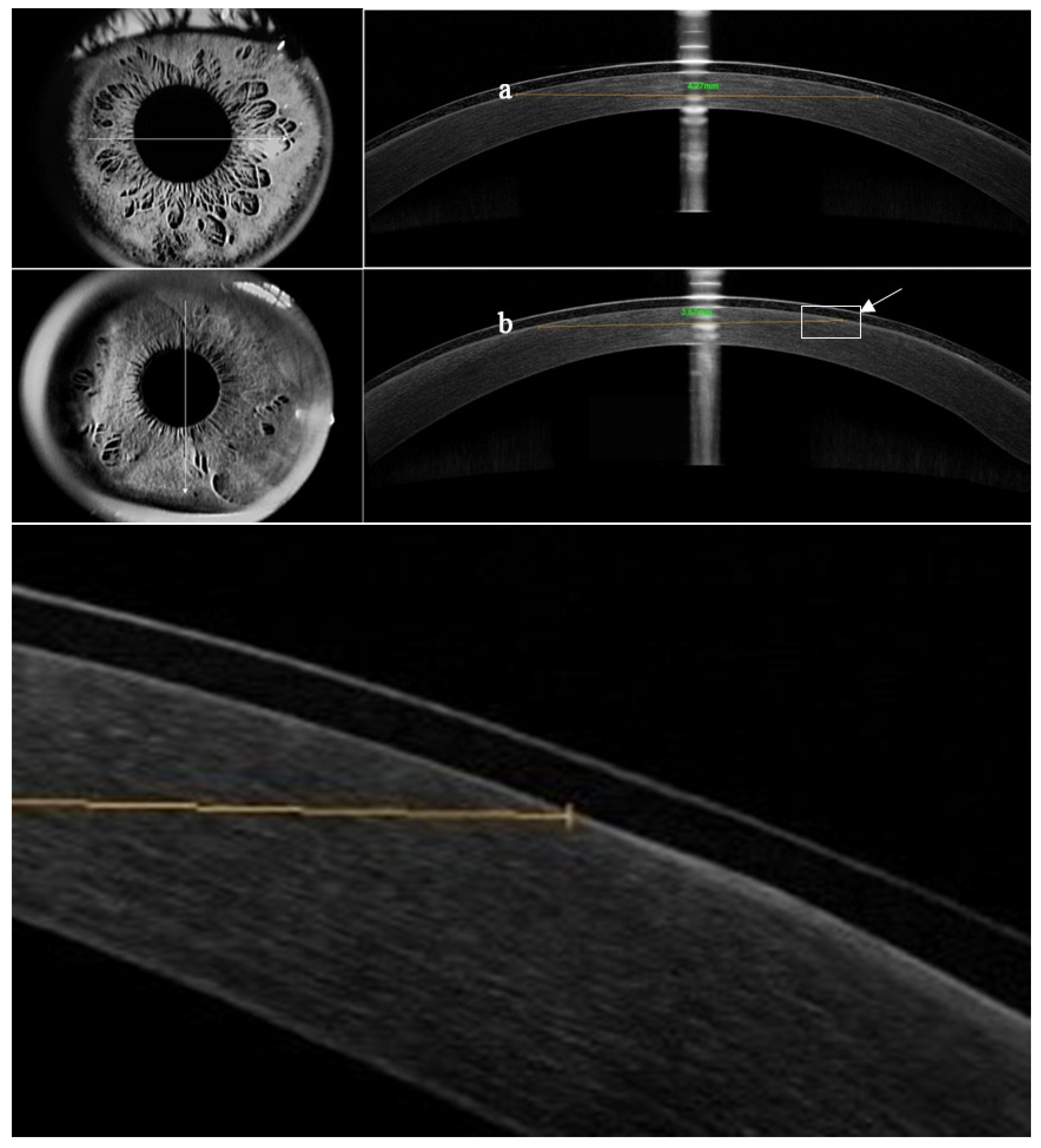
Figure 2.
The mean pain intensity of groups A and B on day one and three postoperatively.
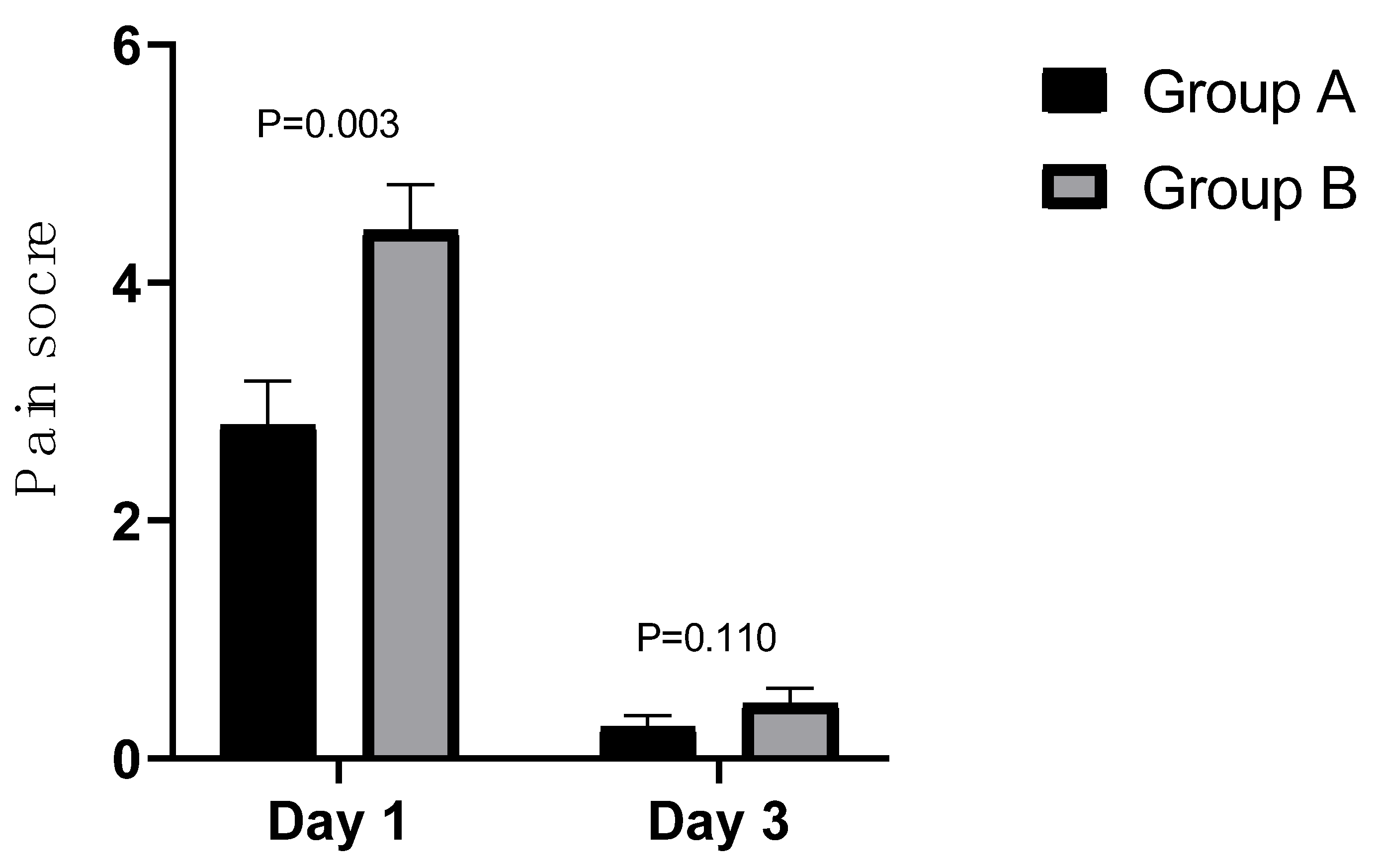
Figure 3.
Total protein precipitation in groups A and B.
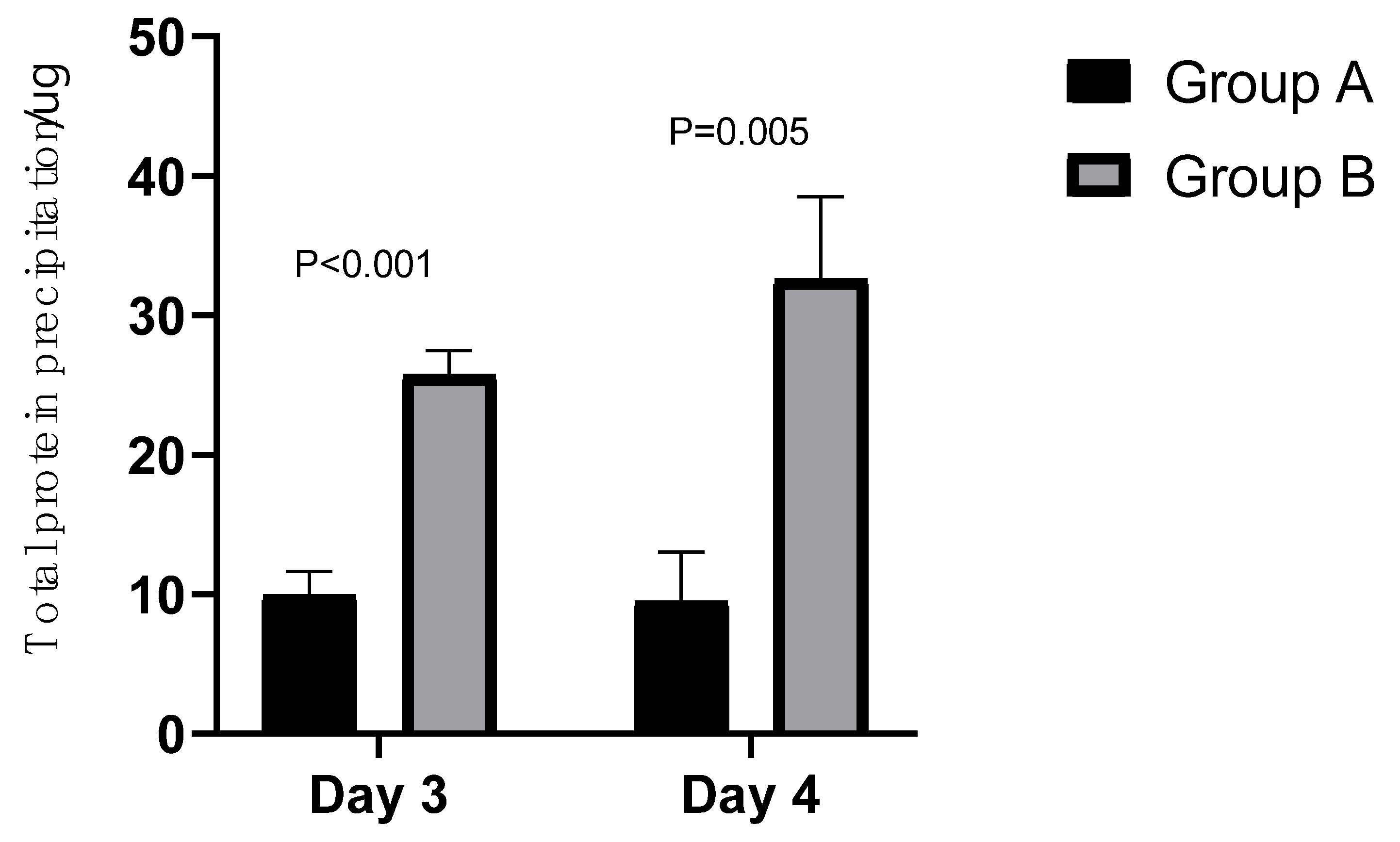

Table 1.
Characteristics of two silicone hydrogel lenses used in this study.
| Parameter | BCL A (Air Optix Night & Day) | BCL B (PureVision) |
|---|---|---|
| Material | Lotrafilcon A | Balafilcon A |
| Water content | 24% | 36% |
| Dk | 140 | 91 |
| Dk/t | 175 | 101 |
| Surface treatment | Plasma Coating | Plasma Oxidation |
| Elastic modulus | 1.4 MPa | 1.1 MPa |
| Central thickness | 0.08 mm | 0.09 mm |
| BOZR | 8.6 mm | 8.6 mm |
| TD | 13.8 mm | 14 mm |
| Continuous overnight wear | 28 days | 21 days |
| Dk: Oxygen permeability (×); Dk/t: Oxygen transmissibility (×;, BOZR: Back optic zone radius; TD: Total diameter; BCL: Bandage contact lens | ||
Table 2.
Lens movement grading scale.
| Grade | Lens movement |
|---|---|
| 0 | Extremely inadequate; lens does not move on blinking |
| 1 | Slightly inadequate; lens moves < 0.2 mm on blinking |
| 2 | Optimum; lens moves between 0.2 and 0.4 mm on blinking |
| 3 | Slightly excessive; lens moves between 0.4 and 1.0 mm on blinking |
| 4 | Extremely excessive; lens moves > 1.0 mm on blinking |
Table 3.
Lens surface deposits grading scale.
| Grade | Lens deposits |
|---|---|
| 0 | None |
| 1 | Five or fewer small (<0.1 mm) deposits or very slight film covering up to 25% of the lens surface |
| 2 | More than five small individual deposits, one individual deposit 0.1 to 0.5 mm in diameter, or film covering between 25%-50% of the lens surface area |
| 3 | Multiple deposits between 0.1 and 0.5 mm in diameter, one deposit larger than 0.5 mm in diameter, or moderate film covering between 50%-75% of the lens surface area |
| 4 | Multiple deposits of 0.5 mm in diameter or larger or film covering more than 75% of the lens surface area |
Table 4.
Patients' preoperative and surgical parameters.
| Group A | Group B | p | |
|---|---|---|---|
| Gender (n) | Male/female=18/23 | ||
| Age (y) | 25.46 ± 4.36 (range: 17–35) | ||
| SER (D) | −5.53 ± 1.94 | −5.51 ± 2.00 | 0.747 |
| Mean UDVA (logMAR) | 1.08 ± 0.30 | 1.10 ± 0.34 | 0.315 |
| Mean CDVA (logMAR) | -0.00 ± 0.01 | -0.01 ± 0.02 | 0.157 |
| Diameter of the operative Optical zone (mm) | 6.39 ± 0.52 | 6.38 ± 0.53 | 0.486 |
| Diameter of the surgical Treatment zone (mm) | 7.95 ± 0.37 | 7.94 ± 0.35 | 0.729 |
| Surgical ablation depth (μm) | 138.66 ± 18.10 | 138.27 ± 19.60 | 0.698 |
| SER: Spherical equivalent refraction; UDVA: Uncorrected Distance Visual Acuity; CDVA: Corrected Distance Visual Acuity. | |||
Table 5.
Comparison of the frequency of subjective eye irritation symptoms between groups A and B n (%).
Table 5.
Comparison of the frequency of subjective eye irritation symptoms between groups A and B n (%).
| Day 1 | Day 3 | |||||
| Grade | Group A | Group B | p | Group A | Group B | p |
| Foreign body sensation | 0.001 | 0.000 | ||||
| 0 | 12(29.3%) | 4(9.8%) | 16(39.0%) | 6(14.6%) | ||
| 1 | 9(22.0%) | 3(7.3%) | 19(46.3%) | 18(43.9%) | ||
| 2 | 8(19.5%) | 6(14.6%) | 2(4.9%) | 6(14.6%) | ||
| 3 | 6(14.6%) | 8(19.5%) | 4(9.8%) | 9(22.0%) | ||
| 4 | 6(14.6%) | 20(48.8%) | 0(0%) | 2(4.9%) | ||
| Pain | 0.001 | 0.705 | ||||
| 0 | 4(9.8%) | 0(0%) | 24(58.8%) | 23(56.1%) | ||
| 1 | 12(29.3%) | 9(22.0%) | 14(34.1%) | 14(34.1%) | ||
| 2 | 6(14.6%) | 4(9.8%) | 1(2.4%) | 3(7.3%) | ||
| 3 | 9(22.0%) | 9(22.0%) | 2(4.9%) | 1(2.4%) | ||
| 4 | 10(24.4%) | 19(46.3%) | 0(0%) | 0(0%) | ||
| Photophobia | 0.190 | 1.000 | ||||
| 0 | 4(9.8%) | 4(9.8%) | 4(9.8%) | 4(9.8%) | ||
| 1 | 3(7.3%) | 1(2.4%) | 22(53.7%) | 23(56.1%) | ||
| 2 | 7(17.1%) | 7(17.1%) | 9(22.0%) | 7(17.1%) | ||
| 3 | 12(29.3%) | 13(21.7%) | 5(12.2%) | 6(14.6%) | ||
| 4 | 15(36.6%) | 16(39.0%) | 1(2.4%) | 1(2.4%) | ||
| Dry eye | 0.066 | 0.157 | ||||
| 0 | 21(51.2%) | 20(48.8%) | 16(39.0%) | 15(36.6%) | ||
| 1 | 12(29.3%) | 10(24.4%) | 18(43.9%) | 18(43.9%) | ||
| 2 | 4(9.8%) | 5(12.2%) | 1(2.4%) | 2(4.9%) | ||
| 3 | 2(4.9%) | 3(7.3%) | 5(12.2%) | 5(12.2%) | ||
| 4 | 2(4.9%) | 3(7.3%) | 1(2.4%) | 1(2.4%) | ||
Table 6.
Comparison of subjective evaluation indexes under slit lamp between groups A and B n (%).
| Day 1 | Day 3 | |||||
| Grade | Group A | Group B | P | Group A | Group B | P |
| Limbal hyperemia | 0.157 | 0.257 | ||||
| 0 | 0(0%) | 0(0%) | 2(4.9%) | 1(2.4%) | ||
| 1 | 11(26.8%) | 8(19.5%) | 31(75.6%) | 30(73.2%) | ||
| 2 | 24(58.5%) | 26(63.4%) | 8(19.5%) | 10(24.4%) | ||
| 3 | 6(14.6%) | 7(17.1%) | 0(0%) | 0(0%) | ||
| 4 | 0(0%) | 0(0%) | 0(0%) | 0(0%) | ||
| Conjunctival hyperemia | 0.414 | 0.705 | ||||
| 0 | 0(0%) | 0(0%) | 4(9.8%) | 4(9.8%) | ||
| 1 | 9(22.0%) | 7(17.1%) | 29(70.7%) | 28(68.3%) | ||
| 2 | 25(61.0%) | 27(65.9%) | 8(19.5%) | 9(22.0%) | ||
| 3 | 7(17.1%) | 7(17.1%) | 0(0%) | 0(0%) | ||
| 4 | 0(0%) | 0(0%) | 0(0%) | 0(0%) | ||
| Lens movement | 0.527 | 0.132 | ||||
| 0 | 30(73.2%) | 31(75.6%) | 11(26.8%) | 16(39.0%) | ||
| 1 | 7(17.1%) | 6(14.6%) | 21(51.2%) | 16(39.0%) | ||
| 2 | 3(7.3%) | 4(9.8%) | 9(22.0%) | 9(22.0%) | ||
| 3 | 1(2.4%) | 0(0%) | 0(0%) | 0(0%) | ||
| 4 | 0(0%) | 0(0%) | 0(0%) | 0(0%) | ||
| Lens deposits | 0.796 | 0.071 | ||||
| 0 | 8(19.5%) | 4(9.8%) | 8(19.5%) | 3(7.3%) | ||
| 1 | 15(36.6%) | 21(51.2%) | 25(61.0%) | 28(68.3%) | ||
| 2 | 17(41.5%) | 16(39%) | 8(19.5%) | 10(24.4%) | ||
| 3 | 1(2.4%) | 0(0%) | 0(0%) | 0(0%) | ||
| 4 | 0(0%) | 0(0%) | 0(0%) | 0(0%) | ||
Disclaimer/Publisher’s Note: The statements, opinions and data contained in all publications are solely those of the individual author(s) and contributor(s) and not of MDPI and/or the editor(s). MDPI and/or the editor(s) disclaim responsibility for any injury to people or property resulting from any ideas, methods, instructions or products referred to in the content. |
© 2025 by the authors. Licensee MDPI, Basel, Switzerland. This article is an open access article distributed under the terms and conditions of the Creative Commons Attribution (CC BY) license (http://creativecommons.org/licenses/by/4.0/).
Copyright: This open access article is published under a Creative Commons CC BY 4.0 license, which permit the free download, distribution, and reuse, provided that the author and preprint are cited in any reuse.


