Submitted:
21 December 2023
Posted:
21 December 2023
You are already at the latest version
Abstract
Keywords:
1. Introduction
2. Materials and Methods
3. Results
4. Discussion
5. Conclusions
Author Contributions
Funding
Institutional Review Board Statement
Informed Consent Statement
Data Availability Statement
Acknowledgments
Conflicts of Interest
References
- Terio, K.A.; McAloose, D. ; St. Léger, J. Pathology of Wildlife and Zoo Animals; Academic press an imprint of Elsevier: London, UK, 2018; pp. 229–262. [Google Scholar]
- Lempp, C.; Jungwirth, N.; Grilo, M.L.; Reckendorf, A.; Ulrich, A.; van Neer, A.; Bodewes, R.; Pfankuche, V.M.; Bauer, C.; Osterhaus, A.D.M.E.; et al. Pathological Findings in the Red Fox (Vulpes vulpes), Stone Marten (Martes foina) and Raccoon Dog (Nyctereutes procyonoides), with Special Emphasis on Infectious and Zoonotic Agents in Northern Germany. PLoS One 2017, 12, e0175469. [Google Scholar] [CrossRef] [PubMed]
- Galov, A.; Sindičić, M.; Andreanszky, T.; Čurković, S.; Dežđek, D.; Slavica, A.; Hartl, G.B.; Krueger, B. High Genetic Diversity and Low Population Structure in Red Foxes (Vulpes vulpes) from Croatia. Mamm. Biol. 2014, 79, 77–80. [Google Scholar] [CrossRef]
- Slavica, A.; Severin, K.; Čač, Ž.; Cvetnić, Ž.; Lojkić, M.; Dežđek, D.; Konjević, D.; Pavlaka, M.; Bundišćak, Z. Model širenja silvatične bjesnoće na teritoriju Republike Hrvatske tijekom perioda od trideset godina. Vet.Stanica 2010, 41, 199–217. [Google Scholar]
- Ebani, V.V.; Trebino, C.; Guardone, L.; Bertelloni, F.; Cagnoli, G.; Nardoni, S.; Sel, E.; Wilde, E.; Poli, A.; Mancianti, F. Occurrence of Bacterial and Protozoan Pathogens in Red Foxes (Vulpes vulpes) in Central Italy. Animals 2022, 12, 2891. [Google Scholar] [CrossRef] [PubMed]
- Kelly, T.R.; Sleeman, J.M. Morbidity and Mortality of Red Foxes ( Vulpes vulpes ) and Gray Foxes ( Urocyon cinereoargenteus) Admitted to the Wildlife Center of Virginia, 1993–2001. J. Wildl. Dis. 2003, 39, 467–469. [Google Scholar] [CrossRef] [PubMed]
- Willebrand, T.; Samelius, G.; Walton, Z.; Odden, M.; Englund, J. Declining Survival Rates of Red Foxes Vulpes vulpes during the First Outbreak of Sarcoptic Mange in Sweden. Wildl. Biol. 2021, 2022. [Google Scholar] [CrossRef]
- Dias-Pereira, P. Morbidity and Mortality in Elderly Dogs – a Model for Human Aging. BMC Vet. Res. 2022, 18, 457. [Google Scholar] [CrossRef] [PubMed]
- McAloose, D.; Newton, A.L. Wildlife Cancer: A Conservation Perspective. Nat. Rev. Cancer 2009, 9, 517–526. [Google Scholar] [CrossRef] [PubMed]
- Tidière, M.; Gaillard, J.M.; Berger, V.; Müller, D.W.H.; Bingaman Lackey, L.; Gimenez, O.; Clauss, M.; Lemaître, J.F. Comparative Analyses of Longevity and Senescence Reveal Variable Survival Benefits of Living in Zoos across Mammals. Sci. Rep. 2016, 6, 36361. [Google Scholar] [CrossRef]
- Elvestad, K.; Henriques, U.V.; Kroustrup, J.P. Insulin-Producing Islet Cell Tumor in an Ectopic Pancreas of a Red Fox (Vulpes vulpes). J. Wildl. Dis. 1984, 20, 70–72. [Google Scholar] [CrossRef]
- Janovsky, M.; Steineck, T. Adenocarcinoma of the Mammary Gland in a Red Fox from Austria. J. Wildl. Dis. 1999, 35, 392–394. [Google Scholar] [CrossRef] [PubMed]
- Degli Uberti, B.; Mariani, U.; Restaino, G.; De Luca, G.; Anzalone, A.; Guida, M.; D’Alessio, N.; D’Amore, M. Caso report di un carcinoma renale in una volpe (Vulpes vulpes) in regione campania.; Società Italiana di Diagnostica di Laboratorio Veterinaria (SIDiLV): Perugia, 2018; p. 125. [Google Scholar]
- Hirayama, K.; Kagawa, Y.; Nihtani, K.; Taniyama, H. Thyroid C-Cell Carcinoma with Amyloid in a Red Fox ( Vulpes vulpes schrenchki ). Vet. Pathol. 1999, 36, 342–344. [Google Scholar] [CrossRef] [PubMed]
- Fukui, D.; Bando, G.; Ishikawa, Y.; Kadota, K. Adenosquamous Carcinoma with Cilium Formation, Mucin Production and Keratinization in the Nasal Cavity of a Red Fox (Vulpes vulpes schrencki). J. Comp. Pathol. 2007, 137, 142–145. [Google Scholar] [CrossRef] [PubMed]
- Hayes, H.M.; Sass, B. Testis Neoplasia in Captive Wildlife Mammals: Comparative Aspects and Review. J. Zoo Anim. Med. 1987, 18, 162. [Google Scholar] [CrossRef]
- Jeong, D.; Yang, J.; Kong, J.; Lee, B.; Lee, J.; Park, S.; Lee, S.; Seok, S.; Hong, I.; Lee, H.; et al. A Subcutaneous Lipoma in a Male Red Fox. J. Vet. Clin. 2015, 32, 278–281. [Google Scholar] [CrossRef]
- Monahan, C.F.; Garner, M.M.; Kiupel, M. Hepatocellular neoplasms in captive fennec foxes ( Vulpes zerda). J. Zoo Wildl. Med. 2018, 49, 996–1001. [Google Scholar] [CrossRef] [PubMed]
- Gray, K.N.; Harwell, G.; Tsai, C.C. Multiple primary tumors in a fennec fox (Fennecus zerda). J. Wildl. Dis. 1982, 18, 369–371. [Google Scholar] [CrossRef] [PubMed]
- Dillberger, J.E.; Citino, S.B. A Malignant Nephroblastoma in an Aged Fox (Fennecus zerda). J. Comp. Pathol. 1987, 97, 101–106. [Google Scholar] [CrossRef] [PubMed]
- Choudhary, S.; Andrews, G.A.; Carpenter, J.W. Small intestinal adenocarcinoma with carcinomatosis in a swift fox (Vulpes velox). J. Zoo Wildl. Med. 2015, 46, 596–600. [Google Scholar] [CrossRef] [PubMed]
- Šoštarić, I.-C.; Hohšteter, M.; Artukovi, B.; Sabo, R. Incidence and Types of Canine Tumours in Croatia. Vet. arhiv 2013, 83, 31–45. [Google Scholar]
- Roulichová, J.; Anděra, M. Simple Method of Age Determination in Red Fox, Vulpes vulpes. Folia Zool. 2007, 56, 440–444. [Google Scholar]
- Celva, R.; Crestanello, B.; Obber, F.; Dellamaria, D.; Trevisiol, K.; Bregoli, M.; Cenni, L.; Agreiter, A.; Danesi, P.; Hauffe, H.C.; et al. Assessing Red Fox (Vulpes vulpes) Demographics to Monitor Wildlife Diseases: A Spotlight on Echinococcus multilocularis. Pathogens 2022, 12, 60. [Google Scholar] [CrossRef] [PubMed]
- Bandelj, P.; Blagus, R.; Vengušt, G.; Žele Vengušt, D. Wild Carnivore Survey of Echinococcus Species in Slovenia. Animals 2022, 12, 2223. [Google Scholar] [CrossRef] [PubMed]
- Gambi, L.; Ravaioli, V.; Rossini, R.; Tranquillo, V.; Boscarino, A.; Mattei, S.; D’incau, M.; Tosi, G.; Fiorentini, L.; Donato, A.D. Prevalence of Different Salmonella enterica Subspecies and Serotypes in Wild Carnivores in Emilia-Romagna Region, Italy. Animals 2022, 12, 3368. [Google Scholar] [CrossRef]
- Pesavento, P.A.; Agnew, D.; Keel, M.K.; Woolard, K.D. Cancer in Wildlife: Patterns of Emergence. Nat. Rev. Cancer 2018, 18, 646–661. [Google Scholar] [CrossRef] [PubMed]
- Zagradišnik, M.; Židak, H.; Huber, D.; Buhin, I.M.; Vlahović, D.; Severin, K.; Hohšteter, M. Pojavnost neoplazija u hrvatskih autohtonih pasmina pasa: retrospektivna analiza tijekom desetogodišnjeg razdoblja. Hrvat. vet. vjesn. 2023, 31, 32–40. [Google Scholar]
- Wang, S.-L.; Dawson, C.; Wei, L.-N.; Lin, C.-T. The Investigation of Histopathology and Locations of Excised Eyelid Masses in Dogs. Vet. Rec. Open 2019, 6, e000344. [Google Scholar] [CrossRef]
- Goldschmidt, M. H. , Goldschmidt, K. H. Epithelial and Melanocytic Tumors of the Skin. In Tumors in Domestic Animals, 5th ed., Meuten, D. J., John Wiley & Sons Inc.: Ames, Iowa, 2017, pp. 111.
- Hendrick, M.J. Mesenchymal Tumors of the Skin and Soft Tissues. In Tumors in Domestic Animals, 5th ed., Meuten, D. J., John Wiley & Sons Inc.: Ames, Iowa, 2017, pp. 152.
- Gross, T. L.; Ihrke, P. J.; Walder, E. J.; Affolter, V. K. Skin Diseases of the Dog and Cat: Clinical and Histopathologic Diagnosis, 2nd ed.; Blackwell Science Ltd.: Oxford, UK, 2005; pp. 710–711. [Google Scholar]
- Foster, R. A. Male Genital System. In Jubb, Kennedy, and Palmer’s Pathology of Domestic Animals, 6th ed.; Maxie, M.G., Ed.; Elsevier: St. Louis, Missouri, 2016; Volume 3, pp. 465–510. [Google Scholar]
- Dalen, W. Agnew, D.W.; MacLachlan, N.J. Tumors of the Genital Systems. In Tumors in Domestic Animals, 5th ed., Meuten, D. J., John Wiley & Sons Inc.: Ames, Iowa, 2017; p. 711. [Google Scholar]
- North, S.; Banks, T.; Straw, R. Tumors of the urogenital tract. In Small Animal Oncology, an introduction; North, S., Banks, T., Straw, R., Eds.; Elsevier Saunders: Edinburgh, London, New York, Oxford, Philadelphia, St. Louis, Sydney, Toronto, 2009; pp. 151–172. [Google Scholar]
- Morris, J.; Dobson, J. Small Animal Oncology. 1st ed.; Blackwell Science Ltd.: Oxford, UK, 2001. [Google Scholar]
- Pêgas, G.R.A.; Monteiro, L.N.; Cassali, G.D. Extragonadal Malignant Teratoma in a Dog - Case Report. Arq. Bras. Med. Vet. Zootec. 2020, 72, 115–118. [Google Scholar] [CrossRef]
- Makovicky, P.; Makarevich, A.; Makovicky, P.; Seidavi, A.; Vannucci, L.; Rimarova, K. Benign Ovarian Teratoma in the Dog with Predominantly Nervous Tissue: A Case Report. Vet. Med. 2022, 67, 99–104. [Google Scholar] [CrossRef]
- Nagashima, Y.; Hoshi, K.; Tanaka, R.; Shibazaki, A.; Fujiwara, K.; Konno, K.; Machida, N.; Yamane, Y. Ovarian and Retroperitoneal Teratomas in a Dog. J. Vet. Med. Sci. 2000, 62, 793–795. [Google Scholar] [CrossRef]
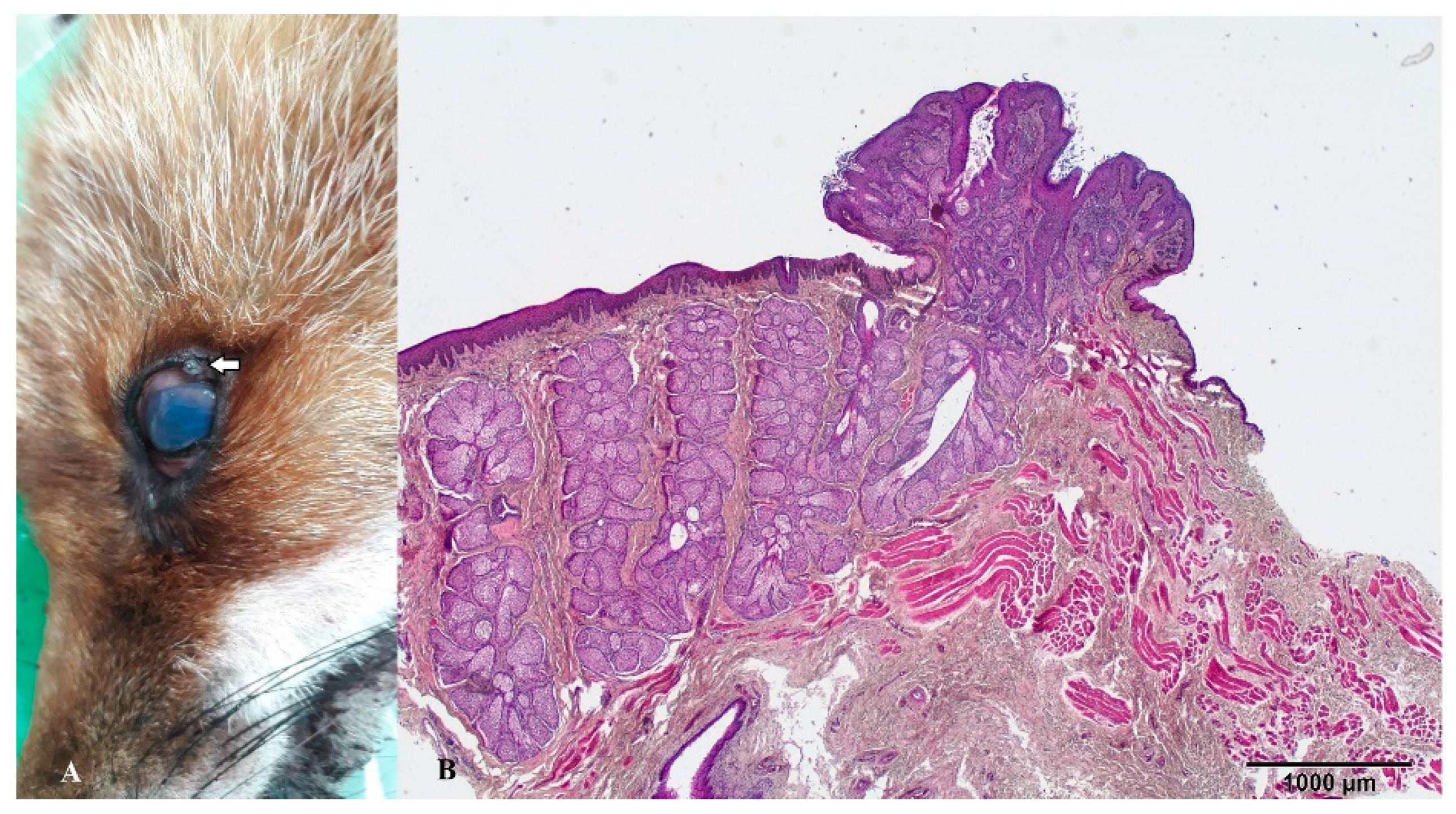
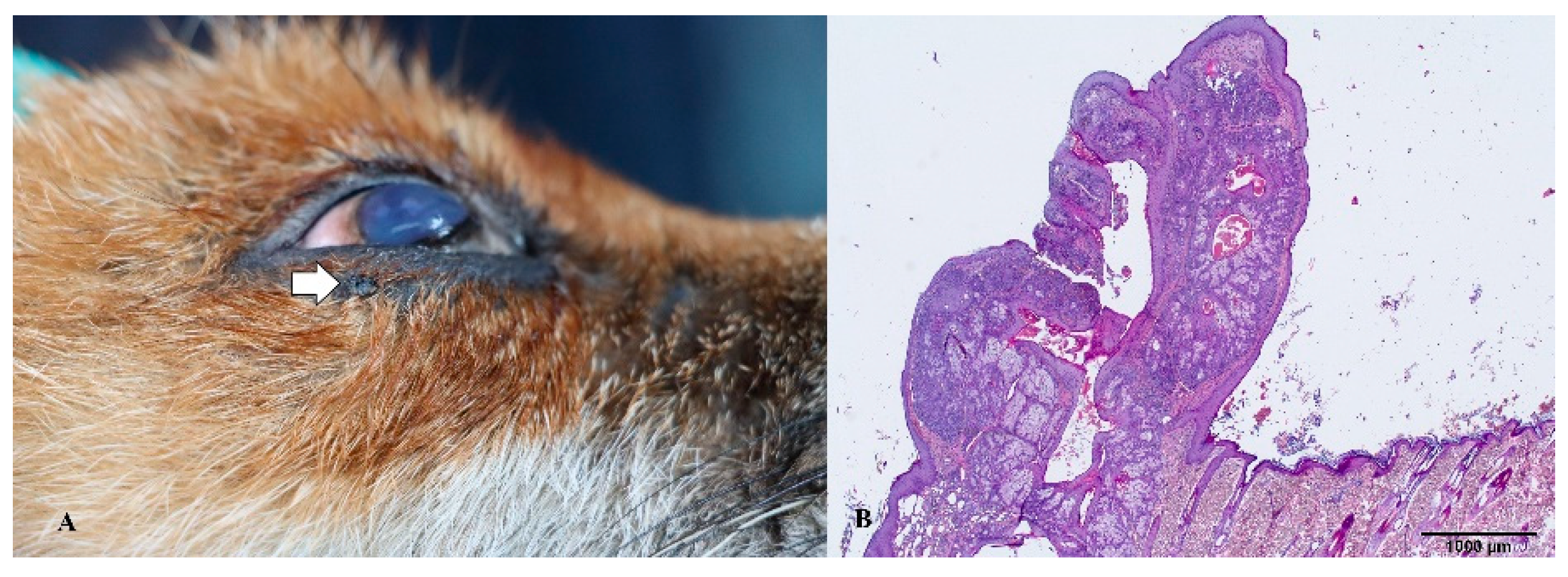
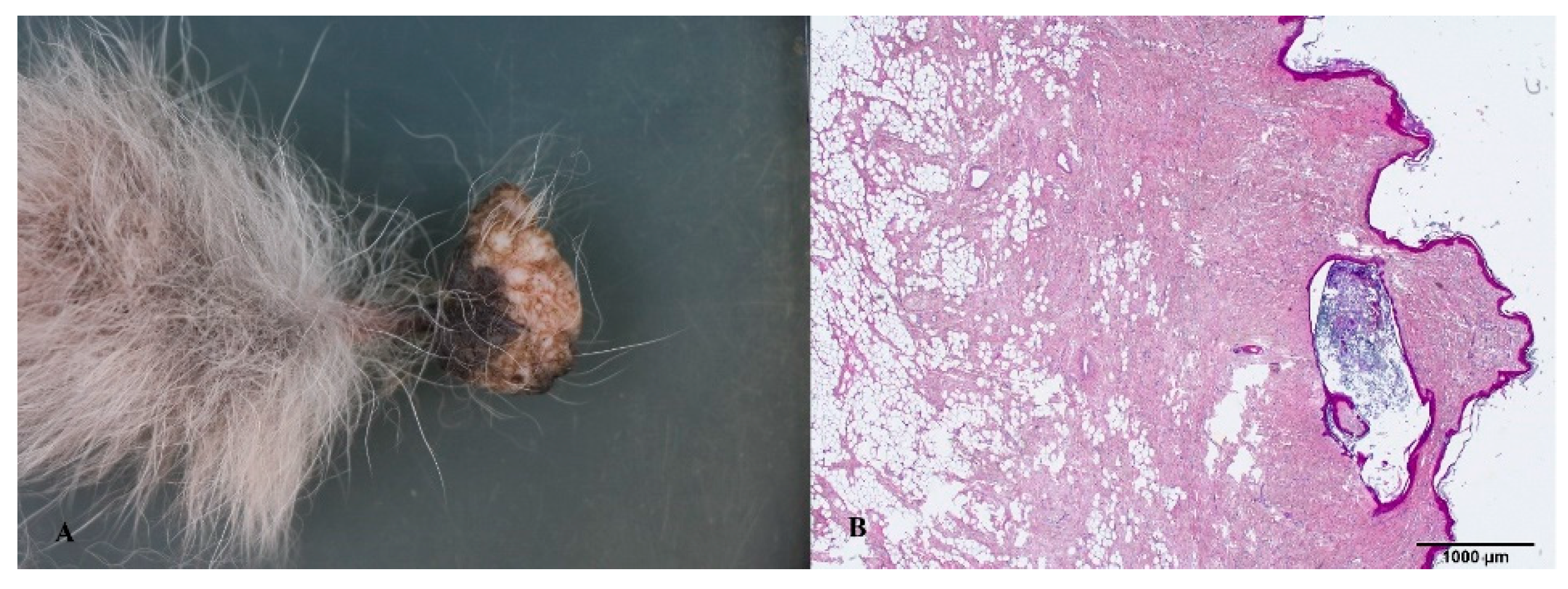
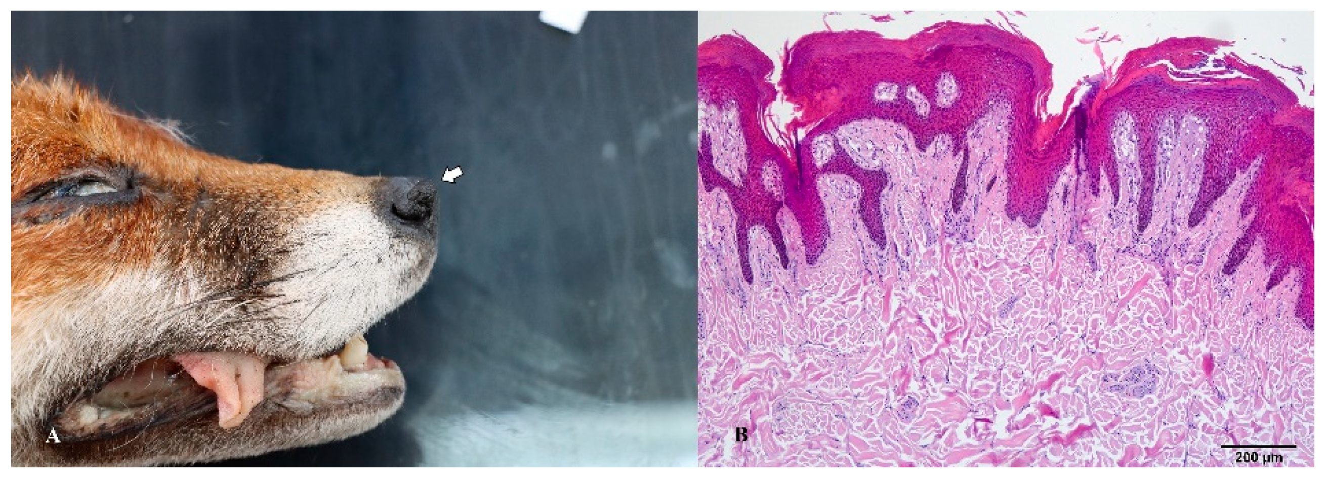
| Animals | Sex | Age | Parts of body | Necropsied | Diagnosis |
|---|---|---|---|---|---|
| Red fox (Vulpes vulpes) | M | 2 | Intraabdominal | 2020 | Teratoma |
| Red fox (Vulpes vulpes) | M | 4 | Upper eyelid | 2021 | Meibomian gland adenoma |
| Red fox (Vulpes vulpes) | F | 4 | Ventral abdomen | 2022 | Collagenous hamartoma |
| Red fox (Vulpes vulpes) | F | 5 | Lower eyelid | 2023 | Meibomian gland adenoma |
| Red fox (Vulpes vulpes) | M | 7 | Nose | 2023 | Collagenous hamartoma |
Disclaimer/Publisher’s Note: The statements, opinions and data contained in all publications are solely those of the individual author(s) and contributor(s) and not of MDPI and/or the editor(s). MDPI and/or the editor(s) disclaim responsibility for any injury to people or property resulting from any ideas, methods, instructions or products referred to in the content. |
© 2023 by the authors. Licensee MDPI, Basel, Switzerland. This article is an open access article distributed under the terms and conditions of the Creative Commons Attribution (CC BY) license (http://creativecommons.org/licenses/by/4.0/).


