Submitted:
23 December 2023
Posted:
26 December 2023
You are already at the latest version
Abstract
Chromosome segregation in female germ cells and early embryonic blastomeres is known to be highly prone to errors. The resulting aneuploidy is therefore the most frequent reason for termination of early development and embryo loss in mammals. And when compatible with embryonic and foetal development, aneuploidy leads into severe developmental disorders. The main surveillance mechanism, essential for protection of cells against aneuploidy, is the Spindle Assembly Checkpoint (SAC). And although all eukaryotic cells are perhaps able to mount SAC response, it is not clear, whether this pathway is active in all cell types, including blastomeres of early embryos. In this review, we will summarize and discuss the published literature with regards to mechanisms controlling chromosome segregation in early embryos. Our conclusion is that early embryos show limited capabilities to react to chromosome segregation defects, which might, at least partially, explain the widespread problem of aneuploidy during early development.
Keywords:
Spindle
; chromosome division
; segregation errors
; Spindle Assembly Checkpoint
; embryo
; CDK1
; cell size
; aneuploidy
1. The assembly of the spindle is under surveillance
Cell cycle represents an extremely complex cellular activity, which is engaged in order to create a new generation of cells. It requires precise spatial and temporal coordination of multiple processes, since the execution of all its parts or phases takes several hours, and some of the activities are separated within the cell by membranes at that time, for example by nuclear membrane. During this process, the crucial task is to preserve the integrity of the genome. The two steps, which are particularly risky, are replication of chromosomes during S phase and their segregation during M phase. Because of the complexity of cell cycle events, cells evolved surveillance control mechanisms, which in most of the cases ensure a flowless cell cycle progression. The important and dicey cell cycle events, such as entry into cell cycle, DNA replication, mitotic entry or assembly of the mitotic spindle, are therefore monitored by a specific checkpoint mechanism. In this review, we will focus on mechanisms controlling the assembly of the spindle. This is an essential organelle for correct segregation of chromosomes during anaphase and the surveillance mechanism is called the Spindle Assembly Checkpoint (SAC). And our goal is to discuss the importance of this mechanism for development of the early embryos, and mammalian embryos in particular.
Since the resolution of sister chromatids is irreversible, the errors during spindle assembly and connection of the spindle to kinetochores, have potentially severe consequences, not only for affected cell, but in multicellular organism, they can threaten its existence. If uncorrected, the daughter cells might suffer from the numerical chromosomal aberrations, or aneuploidy. Such situation leads to cell death, or in case of embryos, into termination of development. Aneuploidy is also very frequent in cancer cells [1,2,3], although sometimes it is not clear, whether it is a cause or consequence. To prevent errors during chromosome segregation, cells evolved SAC, which is also called the mitotic checkpoint [3,4,5]. The SAC is active, when cells enter mitosis. Prior to the entry into mitosis, some of the SAC players, namely Mad1, are sequestered from the chromosomes by nuclear membrane, however the SAC becomes active immediately after NEBD [6]. Then the SAC activity continues throughout mitosis, until the assembly of spindle is completed, when all kinetochores are accurately connected to the spindle apparatus. Then the SAC activity ceases and the cell is ready for entry into anaphase. The transition to anaphase is marked by activation of E3 ubiquitin ligase called Anaphase Promoting Complex/Cyclosome APC/C [7,8]. This large protein complex, together with proteasome, then removes CDC20-controlled multiple substrates, which were important for sustaining the preceding metaphase, including also Securin and Cyclin B [6]. The decrease of CDK1 activity and destruction of Securin activates Separase, which will cleave the cohesin holding together sister chromatids, triggering poleward movement of the sister chromatids (Figure 1).
The relationship between spindle morphology in prophase, prometaphase, metaphase and anaphase (upper panel), occupancy of kinetochores (middle panel) and SAC activity (lower panel) Mad2 change the conformation from open to closed form on unoccupied kinetochore, this stimulates the assembly of MCC consisting of Mad2, Bub3, BubR1, and Cdc20 and leads into inhibition of APC/C and also Separase (by CDK 1 and Securin). After attachment of all chromosomes to spindle microtubules and formation of the metaphase plate, the SAC signalling ceases and MCC is disassembled. Subsequently, CDC20 binds and activates APC/C, initiating destruction of cyclin B and securin. This activates separase which cleaves cohesin and promotes sister chromatid separation and anaphase. (based on [4])
Identification of proteins required in SAC signalling was instrumental for our understanding of its mechanism [9,10]. These proteins, and the complexes they form, are essential for monitoring of the attachment of the kinetochores to spindle microtubules, and for postponing the APC/C activity. The unoccupied kinetochores, specifically their outer layers, are essential for microtubule binding, and they interact via KMN network (KNL1, MIS12 and NDC80) [11] also with SAC components [12]. The recruitment of SAC subunits also requires the activity of Mps1 kinase [13,14,15]. The kinetochore then serves as a catalytic platform for assembling the Mitotic Checkpoint Complex (MCC) [16,17]. Building of MCC requires first loading of Mad1/Mad2 complex to the unattached kinetochore, more specifically to the KMN protein network, located in the outer kinetochore [18]. The Mad2 protein on the kinetochore undergoes conformational change from open to closed form, which allows its binding to CDC20 in cytoplasm that is an activator of APC/C [19,20,21]. Each MCC subunit contains two Cdc20 molecules [22] and then Mad2, BubR1 and Bub3 constitutes. The MCC inhibits APC/C by several means, including the sequestration of Cdc20, which makes it inaccessible for APC/C and also interact directly with APC/C [4]. The MCC is produced until all kinetochores are bound to microtubules. And throughout this time, the APC/C activation is delayed. SAC needs sufficient sensitivity, in order to respond to small changes in kinetochore occupancy. It seems that SAC is sufficiently sensitive, the cells are able to respond to quantitative changes in the number of attached kinetochores [23], as well as to the numbers of microtubules, attaching each specific kinetochore [24]. And the whole pathway therefore works in a gradual and quantitative manner [25]. The inactivation of SAC signalling is initiated by microtubule end binding to kinetochore, which interrupts the assembly of SAC subunits on this particular kinetochore that ends MCC production. For complete SAC inactivation is however also important to disassemble the existing cytoplasmic MCC blocking Cdc20, as well as MCC bound to APC/C and to prevent its rebinding [5]. The crucial step is to change back the Mad2 conformation to open, which requires set of interactors including TRIP13 and p31comet [26].
Another aspect, which is equally important for faithful chromosome segregation, as the attachment of kinetochores by microtubules, is actually the orientation of sister chromatids on the spindle [27]. The correct chromosome orientation in mitosis requires that the sister kinetochores are facing the opposite spindle poles. Such orientation is called bipolar, or amphitelic attachment, and in this case, there is a higher chance the sister chromatids are correctly segregated to both daughter cells during anaphase. When sister chromatids are engaged incorrectly, for example when they are both connected to the same pole of the spindle, which is called synthelic attachment, when one is not connected at all, which is called monotelic attachment, or when one kinetochore is connected to both poles, in the case of merotelic attachment, the attachment correction mechanisms are activated [27]. The merotelic attachment represents the most serious problem, since it escapes the detection by SAC [28,29]. The correction mechanisms are based on activity of chromosomal passenger complex (CPC) with Aurora B kinase, which is able to disengage the incorrect connections by phosphorylation of the components of the kinetochore [30,31]. It was however shown that the correction mechanisms are active even after SAC is being satisfied [32,33,34], which provides the opportunity to correct improper attachments even during anaphase.
2. Important aspects of early embryonic development with potential impact on chromosome segregation
Although the germ cells in mammals both undergo meiotic cell cycle, their development is strikingly different. In case of the oocytes, the cells enter meiosis during embryonic development of an individual and remain arrested in the prophase of the first meiotic division for prolonged time, in human even for decades. The sperm, on the other hand, develops after puberty in germinative epithelium in testes. Both are highly differentiated and specialized cells. Oocyte, after reaching its fully grown state, is one of the biggest cells in the body by volume, with cross diameter of 70-80 in mouse and up to 120-130 in human and cattle. Oocytes are also embedded by glycoprotein coat called zona pellucida, which plays essential role during fertilization and preserving the integrity of the embryo, during transition towards the uterus, when it is lost during the implantation [35]. The oocyte development critically depends on surrounding cells, which together form a follicle within the cortex of the ovary. The increasing pressure of the follicular fluid provides also a mechanical force and means of transportation for oocyte, after follicular rupture delivering it into infundibulum of the ovary, where the fertilization takes place. Sperm on the other hand, starts to appear after puberty, and its development requires specialized epithelium in the convoluted seminiferous tubules of the testes. Surrounding cells within the epithelium, namely the Sertoli cells, assist during transition of the spermatids throughout both meiotic divisions and also during spermiogenesis, which is the reduction of cytoplasm and development of flagella. Early embryonic development is then initiated by the fusion of oocyte and sperm, which leads to the formation of single cell embryo, called zygote. Fertilization in mammals is a very complex process, and it is important to ensure that only a single sperm penetrates inside the egg [36]. The principal part in preventing polyspermy is played by zona pellucida, as well as by cells protecting the egg from the outside, called cumulus cells [35]. The sperm and the egg are not fully synchronized in cell cycle during fusion. Whereas the sperm already completed both meiotic divisions and has haploid number of chromosomes wrapped around protamine-like proteins [37], largely replacing histones, the egg is arrested in metaphase II with intact spindle. Besides haploid genome, sperm also contributes by important molecule that is called phospholipase C zeta (PLCζ), and which is capable of abrogating the metaphase II block of the egg [38]. This molecule triggers signalling cascade, releasing calcium from the internal reserves in smooth endoplasmic reticulum. Then the APC/C is activated, which leads into degradation of cyclin B, and consequently into anaphase and the second polar body extrusion.
The zygote, now in interphase, quickly forms two pronuclei, which in some species, such as in mouse, differ in size. The pronuclei migrate towards each other, and when they are in a close proximity, they undergo NEBD. During their migration, the male pronucleus exchange protamines for histones and both pronuclei execute the first DNA replication after fertilization. The spindle in zygote arises from a complex process, which involves assembly of two separate spindles around each parental pronuclei first, followed by their fusion into a single spindle [39]. Therefore, the first mitosis is longer than the following mitoses in developing embryo [40]. Division of zygote is followed by rapid subsequent divisions of embryos called the cleavage cycles, during which the cell rapidly divides, without regrowing during subsequent G1. During the early embryogenesis, which lasts in mammals around 4-8 days, depending on the species, embryos move through the oviduct towards the uterus. The embryonic cells undergo profound changes, which might also have an impact on the fidelity of chromosome segregation as well [41]. We do not have a time to discuss all of them, we will just briefly mention those that might have impact on the fidelity of chromosome segregation. First, embryos need to activate their newly formed genome [42]. Fully grown oocytes arrest their transcription, and therefore the two meiotic divisions, fertilization and one to three divisions of the embryo, are executed without genomic transcription. The transcription is a principal part of cell cycle control in somatic cells, the oocytes and embryos therefore during transcriptional silencing use regulated translation instead [41]. Then there is a question of the spindle assembly in embryo and the fate of the sperm centriole during fertilization. In somatic cells, a specific organelle, called centrosome, are utilized to assembly a spindle [43]. In mammals however, the spindle assembly in oocytes is achieved by different mechanism [44]. The centrosomes are not present, and the spindle assembly depends on simplified Microtubule Organising Centres (MTOCs). Such spindle assembly seems to be error prone [45]. The morphology of the spindle seems to be also sensitive to in vitro culture [46]. After fertilization, the new centrosome (proximal centriole) is brought by the sperm [47]. However, when the embryo starts utilizing centrosomes from the sperm, remains still poorly understood. It is also important to mention other profound changes within the early embryo that we are unable to discuss here, although they might affect chromosome segregation as well. It is a complete remodelling of the epigenetic landmark of embryonic blastomeres, followed by the first and second round of differentiation, giving rise to several populations of differentiated cells, namely the trophoblast and embryoblast in the first, and epiblast and hypoblast epithelial layers in the second.
3. Control of chromosome segregation during early embryonic development
Chromosome segregation errors, undetected by surveillance mechanisms, might cause aneuploidy. Frequency of aneuploidy in oocytes and embryos is extremely high, in comparison to somatic cells, and it further increases with maternal age [48,49,50,51,52,53,54]. In fact, the aneuploidy represents the most frequent single cause of termination of embryonic development [55]. Arising of the aneuploidy during meiosis, or in the zygote, affects uniformly all cells within the embryo. And most frequently, it will result in embryo loss, only the specific chromosomal gains are tolerated during embryonic, and then foetal development. In human, in case of autosomes, these are the trisomies of chromosomes 21, 18 and 13, causing Down, Edwards, or Patau syndromes respectively. And although the embryos survive, the cell behaviour is changed profoundly, exhibiting a phenotypes related to the presence of an extra chromosome [56,57,58].
In most cases, the embryos are mosaic, containing a mixture of euploid and aneuploid cells. The aneuploidy is not just an artefact of in vitro culture, although in vitro conditions increase its frequency significantly [59]. Mouse embryos, developing in vivo, showed rate of aneuploidy per blastomere from 4% in zygotes to 7% in 8-cell embryos, with steep increase to 11% between 8 to 16-cell embryos [60]. The initial level of aneuploidy, up to the 8-cell stage, is not much different from the levels reported previously in meiosis II oocytes [61,62]. However, due to the increasing number of cells constituting the embryo, this translates into much higher frequency of embryos affected by aneuploidy. Similar frequencies were reported in embryos of other mammalian species, including human [63], cattle [59,64], pig [65] and rhesus monkey [66]. Experiments, in which aneuploid blastomeres were traced in live developing embryos showed that the aneuploid cells within the embryo survive and proliferate, and they are not eliminated until hatching [60,67]. We can only speculate what is behind their ability to survive (and multiply) for prolonged time. Is it the stockpile of maternal mRNAs and proteins accumulated during oocyte growth? Their survival might also indicate that the control mechanisms of chromosome segregation might be less active in early embryos. Nevertheless, in the situations, when the aneuploid blastomeres represent a significant fraction of cell constituting the embryos, the implantation and future development might be compromised. It all depends on a proportion between euploid and aneuploid cells. On the other hand, when the number of aneuploid cells within the embryo is not too high, the potential of such mosaic embryos to implant and develop further is similar to the euploid embryos [68,69]. It was shown that the cell cycle profile of aneuploid blastomeres within the embryo exhibit characteristic abnormalities, which can be used in non-invasive embryo screening [70]. In summary, it is obvious that the aneuploidy, caused by undetected or uncorrected chromosome segregations errors, represents a threat for embryonic development in mammals.
The mechanisms, monitoring chromosome segregation, were studied more extensively in mammalian oocytes than in embryos. From the initial studies it was clear, that although the SAC is operating in mouse oocytes [71], the chromosome segregation surveillance mechanisms are unable to postpone anaphase in cases, when the spindle is not properly assembled [72,73,74,75,76,77,78]. And this was similar between mouse and human, although their approach to build the spindle during meiosis I slightly differs. Similar insensitivity to obvious chromosomal and spindle defects was also recently reported in meiosis II oocytes [50]. Thus it seems possible, that in fact the failure of the sensing/correction mechanisms, such as the SAC, might contribute to frequent chromosome segregation errors in oocytes [79].
In contrast to the oocytes, in embryos the chromosomes segregation surveillance mechanisms were not extensively studied. Therefore, we have rather limited information whether, for example SAC, is fully functional during early embryonic development. The phylogenetic studies, concerning SAC in eukaryotes show that it is an evolutionary conserved mechanism. It is possible to trace its origin back to eukaryotic common ancestor [80,81,82], whose genome already harboured multiple components of the SAC pathway [81], functional APC/C [80] and evidence of a complex structure typical for the kinetochore [83,84]. There is only limited information from non-mammalian eukaryotes, whether they utilize SAC in response to errors in spindle assembly during the embryonic development (Figure 2). In C. elegans for example, the SAC in early embryos is functional, and capable to prevent chromosome segregation errors induced by disruption of the spindle [85]. And although the SAC pathway shows many similarities to the SAC known from mammalian cells, there are also important differences, namely in centralization of the response [86]. According to this study, the SAC in embryonic cells is weaker than in adult germline stem cell [87]. In C. elegans in general, the SAC strength seems to be linked to the cell fate and other cellular properties, such as the cell volume [88]. In zebrafish, the ability to use SAC is acquired with midblastula transition and its onset seems to be unlinked from the initiation of transcription from embryonic genome, or from changes in nuclear to cytoplasmic ratio [89]. Similar situation was observed in Xenopus embryos, which acquire sensitivity to microtubule poisons later during embryonic development, also in transcription independent manner [90]. And it was subsequently shown, that the SAC is absent in Xenopus early embryo [91]. However, in Xenopus oocyte extracts, a reaction similar to SAC can be induced by increasing the density of sperm nuclei [92]. In tunicates Phalusia mammilata, SAC is inactive or unresponsive until the 8th cleavage cycle, and its stringency afterwards depends on the cell size and fate [93]. Comparison of response to spindle disruption between various marine animals showed adequate response in sea urchins, mussels, and jellyfish and the absence of SAC in ascidian and amphioxus embryos [94]. The ascidian embryos also lack the ability to accumulate the core SAC proteins, such as Mad1 and Mad2 on unattached kinetochores, further supporting the evidence that the SAC in these animals is not active in early embryos.
Overview of Metazoan group with SAC activity present (ON) or absent (OFF) uring early development. In some species (based on [94,95])
Mammalian embryos are, in certain aspects concerning the cell cycle control, unique in comparison to somatic cells. For example, although they are able to quickly establish connections between spindle microtubules and kinetochores, and organize metaphase plate rather rapidly, without pronounced congression defects [96], they show tendency for prolonged mitoses, during which the cohesin connecting the sister chromatids is weakened, which increases probability of missegregation of chromosomes [97]. They are also characterized by presence of micronuclei, embedding the lagging chromosomes or chromosomal fragments in membrane [66,98,99,100]. In mouse, it was shown that unlike in somatic cells, the micronuclei in embryos do not contribute to chromothripsis [98].
In terms of functionality of SAC in mammalian early embryos, we have only limited information. Challenging SAC by spindle depolymerizing drugs in human embryos showed that they are sensitive and in later stages they undergo apoptosis [101]. The gene deletion studies of SAC components showed that SAC becomes limiting factor later during early development, for example in blastocyst stage in case of Mad2 [102] and even later, at stage 8.5 in case of BubR1 [103]. The phenotype in both cases pointed to problems with chromosome segregation and aneuploidy. Regarding the expression of SAC core components in the embryo, it was shown that Mad2, Bub3 and BubR1 are present in zygote and their depletion by RNAi caused acceleration of the first mitosis, insensitivity to nocodazole and chromosome segregation defects [104]. Other study however showed that the interaction of overexpressed Mad1 is only brief and the protein quicky disengage from the chromosomes after NEBD in zygotes and 2-cell embryos [105]. Recently published results showed that mouse embryos at morula stage, are insensitive to chromosome segregation defects [106] and the misaligned chromosomes do not delay the onset of anaphase. Interestingly, in this work authors show that a mild inhibition of APC/C by ProTame prolonged mitosis and simultaneously reduced the occurrence of micronuclei and misaligned chromosomes.
4. Is cell size important for the fidelity of chromosome segregation?
If there is one thing, which is remarkable in case of female germ cells, and then also in case of embryonic blastomeres, is the sheer size of these cells. They are among the biggest cells in the organism. The size of the germ cell is an advantage in animals with external development, such as fish, amphibians, reptiles, birds etc., because their early development depends on energy sources from the egg. It is perhaps less important in mammals. Although even in mammalian species, the meiosis and early embryogenesis is accomplished without transcription from the genome, and therefore dependent on maternal stockpile accumulated during oocyte growth. Nevertheless, the size might not be always advantageous, and the higher frequency of chromosome segregation errors might be the price. First it was noticed that in C. elegans embryos response to nocodazole and induced cell cycle delay, is proportional to the size of cell [107]. This would mean that the stringency of SAC would decrease with the cellular volume. This was subsequently tested and confirmed in mouse oocytes, in which cell volume manipulations led to the decrease of SAC stringency in larger cells [108]. Another work, aiming to address this link between cell size and SAC stringency showed that although smaller oocytes, obtained by manipulation, are capable to decrease their levels of APC/C substrate securin faster, incorrectly oriented bivalent chromosomes failed to prevent anaphase in meiosis I [109]. The link between the cell size and SAC stringency was recently tested also in mouse 2-cell embryos [106]. The results showed no difference in response to the low levels of nocodazole in cell with normal volume and in cells with reduced volume, indicating that the cell size in mouse embryo is not linked to the stringency of SAC. In conclusion, more experimental work will be needed to finally confirm the effect of cell size in SAC stringency.
5. Conclusions
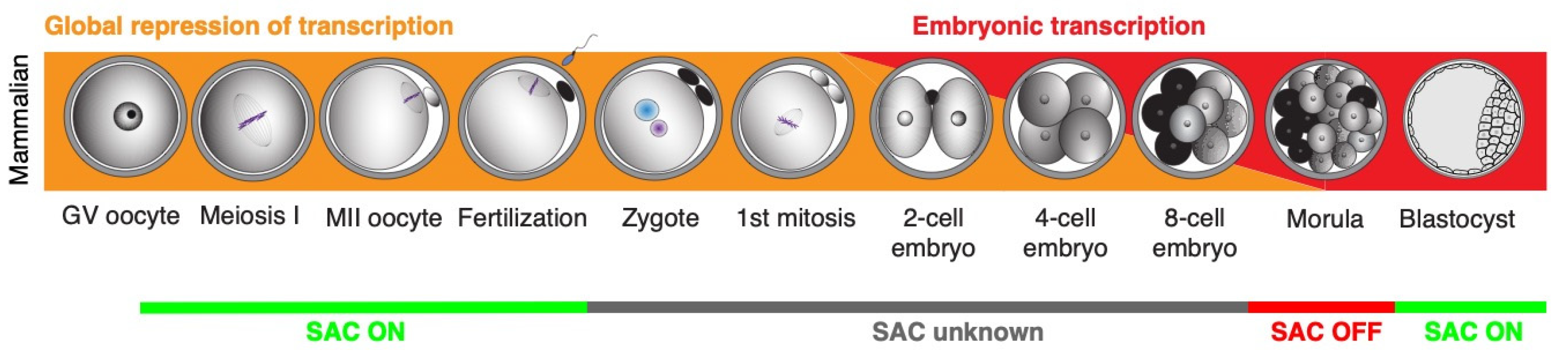
The published work summarized here shows clearly that our knowledge about the control mechanisms of chromosome segregation in early embryos is fairly limited (Figure 3). And considering how important problem the aneuploidy in early embryo represents, this gap in our knowledge should be addressed urgently. Studies addressing the expression and localization pattern of SAC proteins in early embryonic mitoses, gene depletion studies specifically targeting the SAC components and also functional studies challenging the SAC, would help to obtain better picture about SAC function during early development. It is conceivable that the results might in fact reveal, that similarly to xenopus and zebrafish, the SAC in early mammalian embryos is inactive. If this will be the case, it will be important to address when its functionality is regained and, more importantly, how the chromosome segregation in early embryos is controlled without SAC.
Funding
This work was supported by the Czech Science Foundation project 19-24528S and by Grant RO 0518 of the Ministry of Agriculture of the Czech Republic.
Conflicts of Interest
The authors declare no conflict of interest.
References
- Kops, G.J.; Weaver, B.A.; Cleveland, D.W. On the road to cancer: aneuploidy and the mitotic checkpoint. Nat Rev Cancer 2005, 5, 773–785. [Google Scholar] [CrossRef] [PubMed]
- Lakhani, A.A.; Thompson, S.L.; Sheltzer, J.M. Aneuploidy in human cancer: new tools and perspectives. Trends Genet 2023, 39, 968–980. [Google Scholar] [CrossRef] [PubMed]
- Mazzagatti, A.; Engel, J.L.; Ly, P. Boveri and beyond: Chromothripsis and genomic instability from mitotic errors. Mol Cell 2023, S1097-2765(23)00918. [Google Scholar] [CrossRef]
- Musacchio, A. The Molecular Biology of Spindle Assembly Checkpoint Signaling Dynamics. Curr Biol 2015, 25, R1002–18. [Google Scholar] [CrossRef] [PubMed]
- McAinsh, A.D.; Kops GJ, P.L. Principles and dynamics of spindle assembly checkpoint signalling. Nat Rev Mol Cell Biol, 3696. [Google Scholar]
- Jackman, M.; Marcozzi, C.; Barbiero, M.; Pardo, M.; Yu, L.; Tyson, A.L.; Choudhary, J.S.; Pines, J. Cyclin B1-Cdk1 facilitates MAD1 release from the nuclear pore to ensure a robust spindle checkpoint. J Cell Biol 2020, 219, http. [Google Scholar] [CrossRef] [PubMed]
- Sivakumar, S.; Gorbsky, G.J. Spatiotemporal regulation of the anaphase-promoting complex in mitosis. Nat Rev Mol Cell Biol 2015, 16, 82–94. [Google Scholar] [CrossRef] [PubMed]
- Watson, E.R.; Brown, N.G.; Peters, J.M.; Stark, H.; Schulman, B.A. Posing the APC/C E3 Ubiquitin Ligase to Orchestrate Cell Division. Trends Cell Biol 2019, 29, 117–134. [Google Scholar] [CrossRef]
- Hoyt, M. A. , Totis, L. and Roberts, B. T. S. cerevisiae genes required for cell cycle arrest in response to loss of microtubule function. Cell 1991, 66, 507–517. [Google Scholar] [CrossRef]
- Li, R.; Murray, A.W. Feedback control of mitosis in budding yeast. Cell 1991, 66, 519–531. [Google Scholar] [CrossRef] [PubMed]
- Foley, E.A.; Kapoor, T.M. Microtubule attachment and spindle assembly checkpoint signalling at the kinetochore. Nat Rev Mol Cell Biol 2013, 14, 25–37. [Google Scholar]
- Lara-Gonzalez, P.; Pines, J.; Desai, A. Spindle assembly checkpoint activation and silencing at kinetochores. Semin Cell Dev Biol 2021, S1084-9521(21)00160. [Google Scholar] [CrossRef]
- Santaguida, S.; Tighe, A.; D’Alise, A.M.; Taylor, S.S.; Musacchio, A. Dissecting the role of MPS1 in chromosome biorientation and the spindle checkpoint through the small molecule inhibitor reversine. J Cell Biol 2010, 190, 73–87. [Google Scholar] [CrossRef]
- Yamagishi, Y. , Yang, C. H., Tanno, Y. and Watanabe, Y. MPS1/Mph1 phosphorylates the kinetochore protein KNL1/Spc7 to recruit SAC components. Nat Cell Biol 2012, 14, 746–752. [Google Scholar] [CrossRef] [PubMed]
- Raisch, T.; Ciossani, G.; d’Amico, E.; Cmentowski, V.; Carmignani, S.; Maffini, S.; Merino, F.; Wohlgemuth, S.; Vetter, I.R.; Raunser, S.; Musacchio, A. Structure of the RZZ complex and molecular basis of Spindly-driven corona assembly at human kinetochores. EMBO J 2022, 41, e110411. [Google Scholar] [CrossRef]
- Sudakin, V.; Chan, G.K.; Yen, T.J. Checkpoint inhibition of the APC/C in HeLa cells is mediated by a complex of BUBR1, BUB3, CDC20, and MAD2. J Cell Biol 2001, 154, 925–936. [Google Scholar] [CrossRef] [PubMed]
- Fischer, E.S.; Yu CW, H.; Hevler, J.F.; McLaughlin, S.H.; Maslen, S.L.; Heck AJ, R.; Freund SM, V.; Barford, D. Juxtaposition of Bub1 and Cdc20 on phosphorylated Mad1 during catalytic mitotic checkpoint complex assembly. Nat Commun 2022, 13, 6381. [Google Scholar] [CrossRef] [PubMed]
- Varma, D.; Salmon, E.D. The KMN protein network--chief conductors of the kinetochore orchestra. J Cell Sci 2012, 125, 5927–5936. [Google Scholar] [CrossRef]
- Mapelli, M.; Massimiliano, L.; Santaguida, S.; Musacchio, A. The Mad2 conformational dimer: structure and implications for the spindle assembly checkpoint. Cell 2007, 131, 730–743. [Google Scholar] [CrossRef] [PubMed]
- Yang, M. , Li, B., Tomchick, D. R., Machius, M., Rizo, J., Yu, H. and Luo, X. p31comet blocks Mad2 activation through structural mimicry. Cell 2007, 131, 744–755. [Google Scholar] [CrossRef]
- Luo, X.; Fang, G.; Coldiron, M.; Lin, Y.; Yu, H.; Kirschner, M.W.; Wagner, G. Structure of the Mad2 spindle assembly checkpoint protein and its interaction with Cdc20. Nat Struct Biol 2000, 7, 224–229. [Google Scholar]
- Izawa, D.; Pines, J. The mitotic checkpoint complex binds a second CDC20 to inhibit active APC/C. Nature 2015, 517, 631–634. [Google Scholar] [CrossRef] [PubMed]
- Dick, A.E.; Gerlich, D.W. Kinetic framework of spindle assembly checkpoint signalling. Nat Cell Biol 2013, 15, 1370–1377. [Google Scholar] [CrossRef] [PubMed]
- Kuhn, J.; Dumont, S. Mammalian kinetochores count attached microtubules in a sensitive and switch-like manner. J Cell Biol 2019, 218, 3583–3596. [Google Scholar] [CrossRef] [PubMed]
- Collin, P.; Nashchekina, O.; Walker, R.; Pines, J. The spindle assembly checkpoint works like a rheostat rather than a toggle switch. Nat Cell Biol 2013, 15, 1378–1385. [Google Scholar] [CrossRef] [PubMed]
- Alfieri, C.; Chang, L.; Barford, D. Mechanism for remodelling of the cell cycle checkpoint protein MAD2 by the ATPase TRIP13. Nature 2018, 559, 274–278. [Google Scholar] [CrossRef]
- Kelly, A.E.; Funabiki, H. Correcting aberrant kinetochore microtubule attachments: an Aurora B-centric view. Curr Opin Cell Biol 2009, 21, 51–58. [Google Scholar] [CrossRef]
- Cimini, D.; Howell, B.; Maddox, P.; Khodjakov, A.; Degrassi, F.; Salmon, E.D. Merotelic kinetochore orientation is a major mechanism of aneuploidy in mitotic mammalian tissue cells. The Journal of cell biology 2001, 153, 517–528. [Google Scholar] [CrossRef] [PubMed]
- Gregan, J.; Polakova, S.; Zhang, L.; Tolić-Nørrelykke, I.M.; Cimini, D. Merotelic kinetochore attachment: causes and effects. Trends Cell Biol 2011, 21, 374–381. [Google Scholar] [CrossRef]
- Lampson, M.A.; Cheeseman, I.M. Sensing centromere tension: Aurora B and the regulation of kinetochore function. Trends Cell Biol 2011, 21, 133–140. [Google Scholar] [CrossRef]
- Carmena, M.; Wheelock, M.; Funabiki, H.; Earnshaw, W.C. The chromosomal passenger complex (CPC): from easy rider to the godfather of mitosis. Nat Rev Mol Cell Biol 2012, 13, 789–803. [Google Scholar] [CrossRef]
- Cimini, D.; Cameron, L.A.; Salmon, E.D. Anaphase spindle mechanics prevent mis-segregation of merotelically oriented chromosomes. Curr Biol 2004, 14, 2149–2155. [Google Scholar] [CrossRef]
- Kamenz, J.; Hauf, S. Slow checkpoint activation kinetics as a safety device in anaphase. Curr Biol 2014, 24, 646–651. [Google Scholar] [CrossRef]
- Maiato, H.; Silva, S. Double-checking chromosome segregation. J Cell Biol 2023, 222, e202301106. [Google Scholar] [CrossRef]
- Rankin, T.; Soyal, S.; Dean, J. The mouse zona pellucida: folliculogenesis, fertility and pre-implantation development. Mol Cell Endocrinol 2000, 163, 21–25. [Google Scholar] [CrossRef]
- Bhakta, H.H.; Refai, F.H.; Avella, M.A. The molecular mechanisms mediating mammalian fertilization. Development 2019, 146, dev176966. [Google Scholar] [CrossRef]
- Gaspa-Toneu, L.; Peters, A.H. Nucleosomes in mammalian sperm: conveying paternal epigenetic inheritance or subject to reprogramming between generations. Curr Opin Genet Dev 2023, 79, 102034. [Google Scholar] [CrossRef]
- Saunders, C. M. , Larman, M. G., Parrington, J., Cox, L. J., Royse, J., Blayney, L. M., Swann, K. and Lai, F. A. PLC zeta: a sperm-specific trigger of Ca(2+) oscillations in eggs and embryo development. Development 2002, 129, 3533–3544. [Google Scholar] [CrossRef]
- Reichmann, J.; Nijmeijer, B.; Hossain, M.J.; Eguren, M.; Schneider, I.; Politi, A.Z.; Roberti, M.J.; Hufnagel, L.; Hiiragi, T.; Ellenberg, J. Dual-spindle formation in zygotes keeps parental genomes apart in early mammalian embryos. Science 2018, 361, 189–193. [Google Scholar] [CrossRef]
- Ciemerych, M.A.; Maro, B.; Kubiak, J.Z. Control of duration of the first two mitoses in a mouse embryo. Zygote 1999, 7, 293–300. [Google Scholar] [CrossRef]
- Anger, M.; Radonova, L.; Horakova, A.; Sekach, D.; Charousova, M. Impact of Global Transcriptional Silencing on Cell Cycle Regulation and Chromosome Segregation in Early Mammalian Embryos. Int J Mol Sci 2021, 22, 9073. [Google Scholar] [CrossRef]
- Schulz, K.N.; Harrison, M.M. Mechanisms regulating zygotic genome activation. Nat Rev Genet 2018. [Google Scholar] [CrossRef]
- Aljiboury, A.; Hehnly, H. The centrosome - diverse functions in fertilization and development across species. J Cell Sci 2023, 136, jcs261387. [Google Scholar] [CrossRef]
- Mogessie, B.; Scheffler, K.; Schuh, M. Assembly and Positioning of the Oocyte Meiotic Spindle. Annu Rev Cell Dev Biol, 3002. [Google Scholar]
- Bennabi, I.; Terret, M.E.; Verlhac, M.H. Meiotic spindle assembly and chromosome segregation in oocytes. J Cell Biol 2016, [http://www.ncbi.nlm.nih.gov/entrez/query.fcgi?cmd=Retrieve&db=PubMed&dopt=Citation&list_uids=27879467].
- Kovacovicova, K.; Awadova, T.; Mikel, P.; Anger, M. In Vitro Maturation of Mouse Oocytes Increases the Level of Kif11/Eg5 on Meiosis II Spindles. Biol Reprod 2016, 95, 18. [Google Scholar] [CrossRef]
- Amargant, F.; Pujol, A.; Ferrer-Vaquer, A.; Durban, M.; Martínez, M.; Vassena, R.; Vernos, I. The human sperm basal body is a complex centrosome important for embryo preimplantation development. Mol Hum Reprod 2021, 27, gaab062. [http://www.ncbi.nlm.nih.gov/entrez/query.fcgi–cmd=Retrieve&db=PubMed&dopt=Citation&list_uids=34581808]. [Google Scholar] [CrossRef]
- Vázquez-Diez, C.; FitzHarris, G. Causes and consequences of chromosome segregation error in preimplantation embryos. Reproduction 2018, 155, R63–R76. [Google Scholar] [CrossRef]
- Nagaoka, S.I.; Hassold, T.J.; Hunt, P.A. Human aneuploidy: mechanisms and new insights into an age-old problem. Nat Rev Genet 2012, 13, 493–504. [Google Scholar] [CrossRef]
- Mihajlovic, A.I.; Byers, C.; Reinholdt, L.; FitzHarris, G. Spindle assembly checkpoint insensitivity allows meiosis-II despite chromosomal defects in aged eggs. EMBO Rep 2023, 24, e57227. [Google Scholar] [CrossRef]
- Yin, L. , Mihajlović, A. I., Yang, G. and FitzHarris, G. Kinetochore deterioration concommitant with centromere weakening during aging in mouse oocyte meiosis-I. FASEB J 2023, 37, e22922. [Google Scholar] [CrossRef]
- Charalambous, C.; Webster, A.; Schuh, M. Aneuploidy in mammalian oocytes and the impact of maternal ageing. Nat Rev Mol Cell Biol 2022. [Google Scholar] [CrossRef]
- Wartosch, L.; Schindler, K.; Schuh, M.; Gruhn, J.R.; Hoffmann, E.R.; McCoy, R.C.; Xing, J. Origins and mechanisms leading to aneuploidy in human eggs. Prenat Diagn 2021, 41, 620–630. [Google Scholar] [CrossRef]
- Mihajlovic, A.I.; Haverfield, J.; FitzHarris, G. Distinct classes of lagging chromosome underpin age-related oocyte aneuploidy in mouse. Dev Cell 2021, 56, 2273–2283. [Google Scholar] [CrossRef]
- Hassold, T.; Hunt, P. To err (meiotically) is human: the genesis of human aneuploidy. Nat Rev Genet 2001, 2, 280–291. [Google Scholar] [CrossRef]
- Shahbazi, M.N.; Wang, T.; Tao, X.; Weatherbee BA, T.; Sun, L.; Zhan, Y.; Keller, L.; Smith, G.D.; Pellicer, A.; Scott, R.T.; Seli, E.; Zernicka-Goetz, M. Developmental potential of aneuploid human embryos cultured beyond implantation. Nat Commun 2020, 11, 3987. [Google Scholar] [CrossRef]
- Torres, E.M. Consequences of gaining an extra chromosome. Chromosome Res 2023, 31, 24. [Google Scholar] [CrossRef]
- Krivega, M.; Stiefel, C.M.; Storchova, Z. Consequences of chromosome gain: A new view on trisomy syndromes. The American Journal of Human Genetics 2022, 109, 2126–2140. [Google Scholar] [CrossRef]
- Tšuiko, O.; Catteeuw, M.; Zamani Esteki, M.; Destouni, A.; Bogado Pascottini, O.; Besenfelder, U.; Havlicek, V.; Smits, K.; Kurg, A.; Salumets, A.; D’Hooghe, T.; Voet, T.; Van Soom, A.; Robert Vermeesch, J. Genome stability of bovine in vivo-conceived cleavage-stage embryos is higher compared to in vitro-produced embryos. Hum Reprod 2017, 32, 2348–2357. [Google Scholar] [CrossRef]
- Pauerova, T.; Radonova, L.; Kovacovicova, K.; Novakova, L.; Skultety, M.; Anger, M. Aneuploidy during the onset of mouse embryo development. Reproduction 2020, 160, 773–782. [Google Scholar] [CrossRef]
- Duncan, F.E.; Chiang, T.; Schultz, R.M.; Lampson, M.A. Evidence that a defective spindle assembly checkpoint is not the primary cause of maternal age-associated aneuploidy in mouse eggs. Biol Reprod 2009, 81, 768–776. [Google Scholar] [CrossRef]
- Danylevska, A.; Kovacovicova, K.; Awadova, T.; Anger, M. The frequency of precocious segregation of sister chromatids in mouse female meiosis I is affected by genetic background. Chromosome Res 2014, 22, 365–373. [Google Scholar] [CrossRef]
- Carbone, L.; Chavez, S.L. Mammalian pre-implantation chromosomal instability: species comparison, evolutionary considerations, and pathological correlations. Syst Biol Reprod Med 2015, 61, 321–335. [Google Scholar] [CrossRef]
- Destouni, A.; Zamani Esteki, M.; Catteeuw, M.; Tšuiko, O.; Dimitriadou, E.; Smits, K.; Kurg, A.; Salumets, A.; Van Soom, A.; Voet, T.; Vermeesch, J.R. Zygotes segregate entire parental genomes in distinct blastomere lineages causing cleavage-stage chimerism and mixoploidy. Genome Res 2016, 26, 567–578. [Google Scholar] [CrossRef] [PubMed]
- Hornak, M.; Oracova, E.; Hulinska, P.; Urbankova, L.; Rubes, J. Aneuploidy detection in pigs using comparative genomic hybridization: from the oocytes to blastocysts. PLoS One 2012, 7, e30335. [Google Scholar] [CrossRef] [PubMed]
- Daughtry, B.L.; Rosenkrantz, J.L.; Lazar, N.H.; Fei, S.S.; Redmayne, N.; Torkenczy, K.A.; Adey, A.; Yan, M.; Gao, L.; Park, B.; Nevonen, K.A.; Carbone, L.; Chavez, S.L. Single-cell sequencing of primate preimplantation embryos reveals chromosome elimination via cellular fragmentation and blastomere exclusion. Genome Res 2019, 29, 367–382. [Google Scholar] [CrossRef] [PubMed]
- Bolton, H.; Graham, S.J.; Van der Aa, N.; Kumar, P.; Theunis, K.; Fernandez Gallardo, E.; Voet, T.; Zernicka-Goetz, M. Mouse model of chromosome mosaicism reveals lineage-specific depletion of aneuploid cells and normal developmental potential. Nat Commun 2016, 7, 11165. [Google Scholar] [CrossRef] [PubMed]
- Yang, M.; Rito, T.; Metzger, J.; Naftaly, J.; Soman, R.; Hu, J.; Albertini, D.F.; Barad, D.H.; Brivanlou, A.H.; Gleicher, N. Depletion of aneuploid cells in human embryos and gastruloids. Nat Cell Biol 2021, 23, 314–321. [Google Scholar] [CrossRef] [PubMed]
- Capalbo, A.; Poli, M.; Jalas, C.; Forman, E.J.; Treff, N.R. On the reproductive capabilities of aneuploid human preimplantation embryos. Am J Hum Genet 2022, 109, 1572–1581. [Google Scholar] [CrossRef]
- Vera-Rodriguez, M.; Chavez, S.L.; Rubio, C.; Reijo Pera, R.A.; Simon, C. Prediction model for aneuploidy in early human embryo development revealed by single-cell analysis. Nat Commun 2015, 6, 7601. [Google Scholar] [CrossRef]
- McGuinness, B. E. , Anger, M., Kouznetsova, A., Gil-Bernabé, A. M., Helmhart, W., Kudo, N. R., Wuensche, A., Taylor, S., Hoog, C., Novak, B. and Nasmyth, K. Regulation of APC/C activity in oocytes by a Bub1-dependent spindle assembly checkpoint. Curr Biol 2009, 19, 369–380. [Google Scholar] [CrossRef]
- Nagaoka, S.I.; Hodges, C.A.; Albertini, D.F.; Hunt, P.A. Oocyte-specific differences in cell-cycle control create an innate susceptibility to meiotic errors. Curr Biol 2011, 21, 651–657. [Google Scholar] [CrossRef]
- Sebestova, J.; Danylevska, A.; Novakova, L.; Kubelka, M.; Anger, M. Lack of response to unaligned chromosomes in mammalian female gametes. Cell Cycle 2012, 11, 3011–3018. [Google Scholar] [CrossRef]
- Kolano, A.; Brunet, S.; Silk, A.D.; Cleveland, D.W.; Verlhac, M.H. Error-prone mammalian female meiosis from silencing the spindle assembly checkpoint without normal interkinetochore tension. Proc Natl Acad Sci U S A 2012, 109, E1858–67. [Google Scholar] [CrossRef]
- Lane, S.I.; Yun, Y.; Jones, K.T. Timing of anaphase-promoting complex activation in mouse oocytes is predicted by microtubule-kinetochore attachment but not by bivalent alignment or tension. Development 2012, 139, 1947–1955. [Google Scholar] [CrossRef]
- Gui, L.; Homer, H. Spindle assembly checkpoint signalling is uncoupled from chromosomal position in mouse oocytes. Development 2012, 139, 1941–1946. [Google Scholar] [CrossRef]
- Holubcová, Z. , Blayney, M., Elder, K. and Schuh, M. Human oocytes. Error-prone chromosome-mediated spindle assembly favors chromosome segregation defects in human oocytes. Science 2015, 348, 1143–1147. [Google Scholar] [CrossRef]
- Haverfield, J.; Dean, N.L.; Nöel, D.; Rémillard-Labrosse, G.; Paradis, V.; Kadoch, I.J.; FitzHarris, G. Tri-directional anaphases as a novel chromosome segregation defect in human oocytes. Hum Reprod, /: [http, 2844. [Google Scholar]
- Mihajlovic, A.I.; FitzHarris, G. Segregating Chromosomes in the Mammalian Oocyte. Curr Biol 2018, 28, R895–R907. [Google Scholar] [CrossRef]
- Eme, L.; Trilles, A.; Moreira, D.; Brochier-Armanet, C. The phylogenomic analysis of the anaphase promoting complex and its targets points to complex and modern-like control of the cell cycle in the last common ancestor of eukaryotes. BMC Evol Biol 2011, 11, 265. [Google Scholar] [CrossRef]
- Vleugel, M.; Hoogendoorn, E.; Snel, B.; Kops, G.J. Evolution and function of the mitotic checkpoint. Dev Cell 2012, 23, 239–250. [Google Scholar] [CrossRef]
- Kops GJ, P.L.; Snel, B.; Tromer, E.C. Evolutionary Dynamics of the Spindle Assembly Checkpoint in Eukaryotes. Curr Biol 2020, 30, R589–R602. [Google Scholar] [CrossRef]
- van Hooff, J. J. , Tromer, E., van Wijk, L. M., Snel, B. and Kops, G. J. Evolutionary dynamics of the kinetochore network in eukaryotes as revealed by comparative genomics. EMBO Rep, 2864. [Google Scholar]
- Tromer, E.C.; van Hooff JJ, E.; Kops GJ, P.L.; Snel, B. Mosaic origin of the eukaryotic kinetochore. Proc Natl Acad Sci U S A 2019, 116, 12873–12882. [Google Scholar] [CrossRef]
- Encalada, S.E.; Willis, J.; Lyczak, R.; Bowerman, B. A spindle checkpoint functions during mitosis in the early Caenorhabditis elegans embryo. Mol Biol Cell 2005, 16, 1056–1070. [Google Scholar] [CrossRef]
- Essex, A.; Dammermann, A.; Lewellyn, L.; Oegema, K.; Desai, A. Systematic analysis in Caenorhabditis elegans reveals that the spindle checkpoint is composed of two largely independent branches. Mol Biol Cell 2009, 20, 1252–1267. [Google Scholar] [CrossRef] [PubMed]
- Gerhold, A. R. , Ryan, J., Vallée-Trudeau, J. N., Dorn, J. F., Labbé, J. C. and Maddox, P. S. Investigating the regulation of stem and progenitor cell mitotic progression by in situ imaging. Curr Biol 2015, 25, 1123–1134. [Google Scholar] [CrossRef] [PubMed]
- Gerhold, A. R. , Poupart, V., Labbé, J. C. and Maddox, P. S. Spindle assembly checkpoint strength is linked to cell fate in the Caenorhabditis elegans embryo. Mol Biol Cell 2018, 29, 1435–1448. [Google Scholar] [CrossRef] [PubMed]
- Zhang, M.; Kothari, P.; Lampson, M.A. Spindle assembly checkpoint acquisition at the mid-blastula transition. PLoS One 2015, 10, e0119285. [Google Scholar] [CrossRef] [PubMed]
- Clute, P.; Masui, Y. Regulation of the appearance of division asynchrony and microtubule-dependent chromosome cycles in Xenopus laevis embryos. Dev Biol 1995, 171, 273–285. [Google Scholar] [CrossRef] [PubMed]
- Shao, H.; Li, R.; Ma, C.; Chen, E.; Liu, X.J. Xenopus oocyte meiosis lacks spindle assembly checkpoint control. J Cell Biol 2013, 201, 191–200. [Google Scholar] [CrossRef]
- Minshull, J.; Sun, H.; Tonks, N.K.; Murray, A.W. A MAP kinase-dependent spindle assembly checkpoint in Xenopus egg extracts. Cell 1994, 79, 475–486. [Google Scholar] [CrossRef] [PubMed]
- Roca, M.; Besnardeau, L.; Christians, E.; McDougall, A.; Chenevert, J.; Castagnetti, S. Acquisition of the spindle assembly checkpoint and its modulation by cell fate and cell size in a chordate embryo. Development 2023, 150, dev201145. [http://www.ncbi.nlm.nih.gov/entrez/query.fcgi–cmd=Retrieve&db=PubMed&dopt=Citation&list_uids=36515557]. [Google Scholar] [CrossRef]
- Chenevert, J.; Roca, M.; Besnardeau, L.; Ruggiero, A.; Nabi, D.; McDougall, A.; Copley, R.R.; Christians, E.; Castagnetti, S. The Spindle Assembly Checkpoint Functions during Early Development in Non-Chordate Embryos. Cells 2020, 9, 1087. [Google Scholar] [CrossRef]
- Paps, J.; Rossi, M.E.; Bowles, A.; Álvarez-Presas, M. Assembling animals: trees, genomes, cells, and contrast to plants. Frontiers in Ecology and Evolution 2023, 11, 1185566. [Google Scholar] [CrossRef]
- Macaulay, A.D.; Allais, A.; FitzHarris, G. Chromosome dynamics and spindle microtubule establishment in mouse embryos. FASEB J 2020, 34, 8057–8067. [Google Scholar] [CrossRef] [PubMed]
- Allais, A.; FitzHarris, G. Absence of a robust mitotic timer mechanism in early preimplantation mouse embryos leads to chromosome instability. Development 2022, 149, dev200391. [http://www.ncbi.nlm.nih.gov/entrez/query.fcgi–cmd=Retrieve&db=PubMed&dopt=Citation&list_uids=35771634]. [Google Scholar] [CrossRef]
- Vázquez-Diez, C.; Yamagata, K.; Trivedi, S.; Haverfield, J.; FitzHarris, G. Micronucleus formation causes perpetual unilateral chromosome inheritance in mouse embryos. Proc Natl Acad Sci U S A 2016, 113, 626–631. [Google Scholar] [CrossRef] [PubMed]
- Kort, D.H.; Chia, G.; Treff, N.R.; Tanaka, A.J.; Xing, T.; Vensand, L.B.; Micucci, S.; Prosser, R.; Lobo, R.A.; Sauer, M.V.; Egli, D. Human embryos commonly form abnormal nuclei during development: a mechanism of DNA damage, embryonic aneuploidy, and developmental arrest. Hum Reprod 2016, 31, 312–323. [Google Scholar] [CrossRef] [PubMed]
- Yao, T.; Ueda, A.; Khurchabilig, A.; Mashiko, D.; Tokoro, M.; Nagai, H.; Sho, T.; Matoba, S.; Yamagata, K.; Sugimura, S. Micronucleus formation during early cleavage division is a potential hallmark of preimplantation embryonic loss in cattle. Biochem Biophys Res Commun 2022, 617, 25–32. [Google Scholar] [CrossRef] [PubMed]
- Jacobs, K.; Van de Velde, H.; De Paepe, C.; Sermon, K.; Spits, C. Mitotic spindle disruption in human preimplantation embryos activates the spindle assembly checkpoint but not apoptosis until Day 5 of development. Mol Hum Reprod 2017, 23, 321–329. [Google Scholar] [CrossRef] [PubMed]
- Dobles, M.; Liberal, V.; Scott, M.L.; Benezra, R.; Sorger, P.K. Chromosome missegregation and apoptosis in mice lacking the mitotic checkpoint protein Mad2. Cell 2000, 101, 635–645. [Google Scholar] [CrossRef] [PubMed]
- Wang, Q. , Liu, T., Fang, Y., Xie, S., Huang, X., Mahmood, R., Ramaswamy, G., Sakamoto, K. M., Darzynkiewicz, Z., Xu, M. and Dai, W. BUBR1 deficiency results in abnormal megakaryopoiesis. Blood 2004, 103, 1278–1285. [Google Scholar] [CrossRef] [PubMed]
- Wei, Y.; Multi, S.; Yang, C.R.; Ma, J.; Zhang, Q.H.; Wang, Z.B.; Li, M.; Wei, L.; Ge, Z.J.; Zhang, C.H.; Ouyang, Y.C.; Hou, Y.; Schatten, H.; Sun, Q.Y. Spindle assembly checkpoint regulates mitotic cell cycle progression during preimplantation embryo development. PLoS One 2011, 6, e21557. [Google Scholar] [CrossRef]
- Radonova, L.; Svobodova, T.; Skultety, M.; Mrkva, O.; Libichova, L.; Stein, P.; Anger, M. ProTAME Arrest in Mammalian Oocytes and Embryos Does Not Require Spindle Assembly Checkpoint Activity. Int J Mol Sci 2019, 20, http. [Google Scholar] [CrossRef]
- Vázquez-Diez, C.; Paim LM, G.; FitzHarris, G. Cell-Size-Independent Spindle Checkpoint Failure Underlies Chromosome Segregation Error in Mouse Embryos. Curr Biol 2019, 29, 865–873. [Google Scholar] [CrossRef] [PubMed]
- Galli, M.; Morgan, D.O. Cell Size Determines the Strength of the Spindle Assembly Checkpoint during Embryonic Development. Dev Cell 2016, 36, 344–352. [Google Scholar] [CrossRef] [PubMed]
- Kyogoku, H.; Kitajima, T.S. Large Cytoplasm Is Linked to the Error-Prone Nature of Oocytes. Dev Cell 2017, 41, 287–298. [Google Scholar] [CrossRef]
- Lane SI, R.; Jones, K.T. Chromosome biorientation and APC activity remain uncoupled in oocytes with reduced volume. J Cell Biol 2017, 216, 3949–3957. [Google Scholar] [CrossRef] [PubMed]
Figure 1.
Control of spindle assembly by SAC .
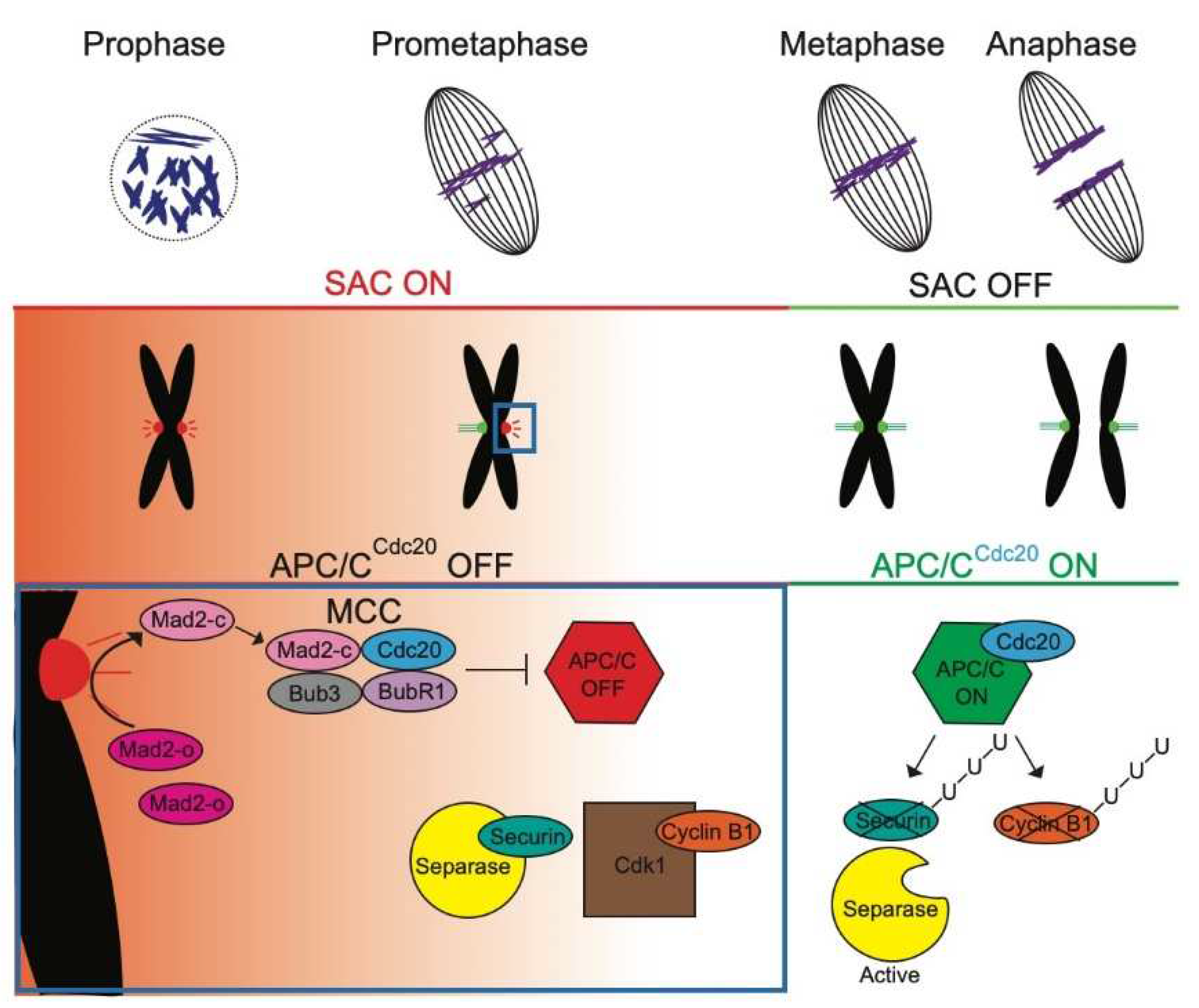
Figure 2.
The SAC activity in Metazoans.
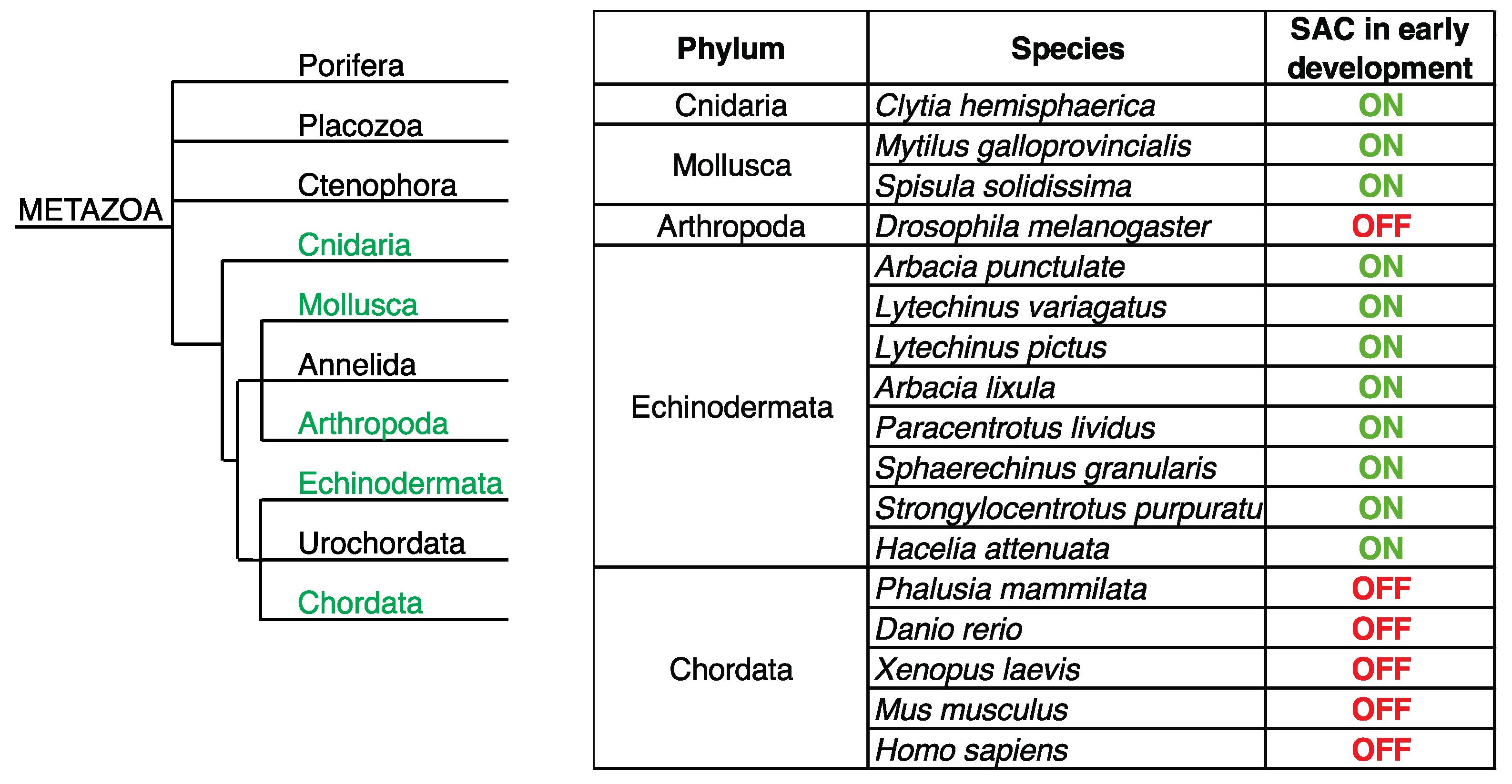
Figure 3.
The overview of early development in mammals and SAC activity.
Disclaimer/Publisher’s Note: The statements, opinions and data contained in all publications are solely those of the individual author(s) and contributor(s) and not of MDPI and/or the editor(s). MDPI and/or the editor(s) disclaim responsibility for any injury to people or property resulting from any ideas, methods, instructions or products referred to in the content. |
© 2023 by the authors. Licensee MDPI, Basel, Switzerland. This article is an open access article distributed under the terms and conditions of the Creative Commons Attribution (CC BY) license (http://creativecommons.org/licenses/by/4.0/).
Copyright: This open access article is published under a Creative Commons CC BY 4.0 license, which permit the free download, distribution, and reuse, provided that the author and preprint are cited in any reuse.


