Submitted:
01 December 2024
Posted:
02 December 2024
Read the latest preprint version here
Abstract
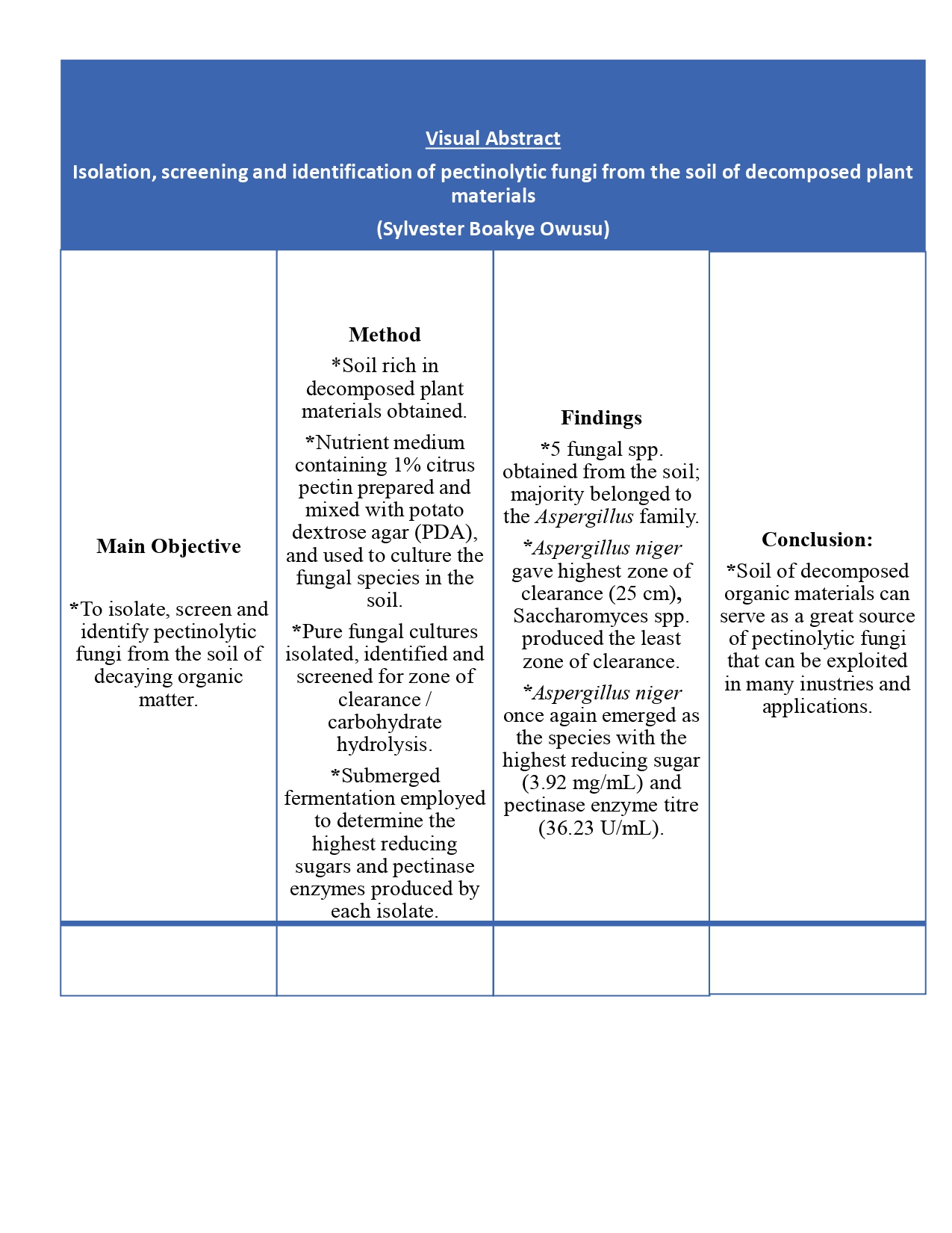
Pectinases are essential enzymes and command a quarter of all food enzymes sold globally, and are recognised as excellent workhorses in plethora of industries. These enzymes are mainly sourced from animals, microorganisms and plants, with microbes serving as the main sources owing to the quality, quantity, ease of extraction and economic considerations. This research study isolated, screened, and identified five pectinase-producing fungi from the soil of decaying fruits and vegetables. The isolated fungal species were cultured and screened for highest pectinase production using pectinase screening agar medium containing 1% citrus pectin. Four isolates identified as Aspergillus niger, Fusarium species, Trichoderma species and Aspergillus flavus, exhibited very high values of pectinase hydrolysis based on clear zone method, which were 25, 23, 20, and 20 mm for the respective isolates. A secondary pectin hydrolysis screening exercise was conducted afterwards to obtain the highest level of reducing sugars and pectinase released by the isolates. Aspergillus niger, once again, proved to be the best as it recorded maximum reducing sugar of 3.92 mg/mL and pectinase enzyme activity of 36.23 U/mL. Soil have the potential to serve as the primary source of fungal species capable of degrading pectin to release pectinase enzymes, which have numerous applications in the production of pharmaceuticals, food, beverages, animal feed, textiles, detergents, protoplast fusion, pulp and paper and biofuels.
Keywords:
Pectinase
; fungal species
; Aspergillus niger
; pectin
; screening
1. Introduction
Pectin is a hecteropolysaccharide that serves as an important component of the cell wall and other internal components of higher plants. It confers mechanical support and flexibility to plants by acting as a cementing material that holds the cell walls of adjacent cells together [1,2]. Pectin is made up of numerous units that consists of D-galacturonic acids with carboxyl groups mostly esterified with methanol, with the main chain being made up of α-(1, 4) glycosidic bonds. Pectin with its methyl groups removed is called pectate or pectic acid, which is also called polygalacturonic acid [3]. The structure of pectin contains homogalacturonan which appears as linear biopolymer and is made up of methyl esterified or acetylated D-galacturonic acid residues or monomers containing α-(1, 4) glycosidic linkages. Pectin is formed when about 75% of carboxylic acids found in homogalacturonans become methylated. Pectic acid is also formed when less than 75% of the carboxylic acids are methylated. Polygalacturonic acid results when there is no methyl esterification [4]. Also, protopectin represents the pectic substances that are not soluble in water [5]. Soluble pectic substances can be formed after limited hydrolysis of protopectin [6]. Pectinic substances, which act as the methylated form of galacturonic acid are formed when there is enzymatic hydrolysis of protopectin in ripe fruits [7].
Pectin is non-toxic, biocompatible and biodegradable and can dissolve in water. It has crucial stabilizing, hydration, gelling, thickening and swelling properties, which makes it very useful in many commercial applications. A few of the commercial applications include food designs, production of fruits juices, drinks, and fermented, confectionary pharmaceutical and health products. Pectin is also utilized as nano-encapsulator, drug delivery agent, constituent of pro-health formulations, dietary fibre, anti-cancer agent, and hydrogel formulation material. It is also used to lower blood cholesterol and low density lipoprotein levels as well as being used in the treatment of diarrhoea and constipation [8,9,10]. The primary sources of pectin have been citrus fruits, apple pomace, mango peels and sugar beet pulp [11,12,13].
Pectin can be hydrolyzed by a group of enzymes called pectinases by splitting the glycosidic bonds existing between pectin molecules [14]. Pectinases are able to break down pectin oligo-D-galacturonate present in plant cell walls into monogalacturonic acids. The hydrolysis is achieved when pectinolytic enzymes cleave the α-1, 4 glycosidic linkages of polygalacturonic acid [15]. Pectinases can be classified as extracellular and intracellular. Extracellular pectinase enzymes represent those secreted outside of the cell into the surrounding media in which the microbes live, whereas intracellular pectinase enzymes are those secreted and act within the cell membrane [16]. Nevertheless, extracellular and intracellular pectinases are also classified into three broader groups based on their mode of action on pectic substances [16,17], which include Protopectinases, Esterases, and Depolymerases [18,19,20,21]. Pectin esterases are able to catalyze the de-esterification of methyl groups in pectin to form pectic acids. Pectin depolymerases, depending on their mechanism of action, are able to cleave the glycosidic bonds found in pectic acids. Also, protopectinases are able to break down protopectin [22]. In terms of their nature, pectinases can be categorized as acidic and alkaline. Acidic pectinases usually enjoy their applications in wine, beverage and fruit juice industries for clarification and purification of fruit juices and wines [13,23]. Alkaline pectinases on the other hand, find their applications in textile industries and wastewater treatments, [4,24,25].
Among the various pectinases, extracellular pectinases derived from fungal species have been known to possess excellent abilities compared to pectinases from other sources such as plants, animals and bacterial [26] and their method of extraction is very simple as the integrity of the cells remains intact. Other added advantages include high yields, economic feasibility, stability and easy genetic manipulation. Also, fungal-derived pectinolytic enzymes have generally regarded as safe (GRAS) status [22]. For these and many more reasons, extracellular pectinases isolated from fungal species especially that of Aspergillus spp. are in high demand in various applications [27,28]. The applications extend across food/feed, beverage, pulp and paper, biofuel, textile, oil extraction and waste and environmental remediation [13,21,29,30]. Pectinases employed in fruit and wine industries can enhance taste and clarity and overall chromatic appearance compared to untreated ones [24].
The soil, as part of the natural environment, serves as a huge reservoir for fungal species that can be isolated and employed in the production of pectinase enzymes. Microorganisms isolated from the soil are mesophilic, can tolerate stress and have the ability to synthesize pectinase enzymes in large quantities in wider spectrum of growth parameters [31]. Soils particularly found in plant food processing locations, markets and organic waste dumpsites have been proven to be very much dominant with pectin-degrading fungus [5]. Recent advances indicate that the trend to identify novel enzymes from microorganisms from such environments is accelerating due to their inherent stability and efficiency [32,33]. Hence, pectinolytic microbes were obtained from the soils exposed to high and low temperatures, and subsequent identification was carried out in this current investigation.
2. Materials and Methods
2.1. Collection of Soil Sample
Soil composed of plant-decayed materials were collected from the dumpsites of fruits, vegetables and plant materials in some markets in Kumasi as these places contained a lot of spoiled fruits and vegetable wastes and decaying plant materials. All the samples were pooled together and kept in sterile plastic containers and carried to the microbiology laboratories of Kwame Nkrumah University of Science and Technology where fungal species were isolated from them.
2.2. Preparation of Medium for Isolation of Fungal Species
A sterile nutrient medium containing 1% citrus pectin; FeSO4.7H2O (5 g/L), MgSO4.7H2O (0.02%), (NH4)2SO4 (0.14%), K2HPO4 (0.2%) as well as nutrient solution (0.1%) containing: ZnSO4.7H2O (1.4 mg/L), MnSO4.H2O (1.6 mg/L), CoCl2 (2.0 mg/L) was prepared, and mixed with 3% agar-agar (the gelling agent) and autoclaved at 121oC for 20 minutes [20]. It was then allowed to cool to around 45°C before being placed into Petri dishes to gel. To ensure sterility, the plates were incubated overnight in an incubator at 37oC.
2.3. Preparation of Potato Dextrose Agar for Isolation of Fungal Species
Potato Dextrose Agar (PDA) was made from a commercial dehydrated OXOID product. Thirty-nine PDA powder (39.0 g) was dissolved in 1 L of distilled water, and sterilized at 121oC for 20 minutes and at pressure of 1.2 g/cm2 according to the product recommendations.
2.4. Isolation and Sub-Culturing of Fungal Species
The simple dilution plate method was adopted here. One (1) g of soil sample was diluted ten-fold in 9 mL of sterile distilled water. One (1) mL of the 6th dilution (10-6) was flooded onto three replicates of the previously prepared sterile Potato Dextrose Agar (PDA) nutrient medium, which had been supplemented with chloramphenicol antibiotic to inhibit bacterial growth. The plates were then incubated at room temperature (25oC - 28oC) for 7 days, until visible fungal colonies appeared. All morphologically distinct colonies were purified by sub-culturing on different plates and streaking with a sterile inoculation loop. This process proceeded until pure fungal isolates were obtained. Pure cultures were maintained on PDA slants, refrigerated and sub-cultured at regular intervals throughout the study and were used as stock cultures. All pure fungal growths were counted with a colony counter, and identified appropriately. Their frequency of occurrence were subsequently established as well.
2.5. Identification of Fungal Isolates
The identification procedure for all the fungal isolates was based on macromorphological and micromorphological characteristics. Macromorphological identification was done by observing colony characteristics such as colour, texture, and spore structure, the form of mycelia and/or pattern of growth. The micromorphological characteristics like separation, spore shapes, among others were determined using the usual lactophenol cotton blue procedure (LPCB). All the fungal isolates had been defined in accordance with the manual for determining the identity of fungi [34] and Illustrated Genera of Imperfect Fungi by Barnett and Hunter, 1972 [35]. In summary, a three-day-old pure culture was utilized to prepare slides for microscopic examination. A small amount of mycelia was placed on the slide, along with a drop of lactophenol cotton blue reagent. A cover slip was laid over it, and the specimen was examined under a light microscope at X400 magnification. Identification of the species was accomplished by comparing features to the micrographs, and the photographs taken included at the end of this section.
2.6. Qualitative (Primary) Screening for Pectinolytic Fungal Species
The process of screening for pectinase synthesis was carried out with the use of the prepared cultivation medium (in the previous section) by applying the spot inoculation method. In brief, pure cultures of the various fungal species were introduced into the already prepared sterile and solidified pectin agar medium. The plates that were inoculated then incubated at a temperature of 30°C for four days (96 hours), and following that, clear observable colony of fungi could be seen on every petri dish. Thereafter, the petri dishes were soaked with Iodine-Potassium iodide solutions (a mixture of 1.0g of iodine and 5.0g of potassium iodide dissolved in 330 mL distilled water) for about 10 minutes in order to boost visibility for measuring the zone of clearance / carbohydrate hydrolysis [20]. A distinct zone formed surrounding the pectinase-producing colonies was observed and subsequently measured. The fungal isolates with the most promising clear zone of pectinase hydrolysis were chosen and preserved under 4oC in the refrigerator on pectin agar and later used in the next step.
2.7. Quantitative (Secondary) Screening for Pectinolytic Fungal Species
This experiment utilized the protocol established by Kaur and Kaur [36] with some modifications. The four isolates with higher degrees of pectinolytic activity showing larger zones of pectin hydrolysis obtained in the previous experiment were used in a submerged fermentation process. 50 mL of the fermentation medium (without the 3% agar agar) prepared earlier underwent inoculation with 1mL of spore suspension of each of the culture isolates. It was made to incubate at 30°C for 7 days on a rotary shaker at 120 revolutions per minute. Throughout the experiment, triplicate Erlenmeyer flasks (250mL) were used for each species of fungus. Once the incubation period elapsed, the crude pectinolytic enzyme underwent extraction. The extraction was done by mixing 50mLof sodium acetate buffer with the fermented broth cultures. The mixture was centrifuged for 15 minutes at 4000 revolutions per minute to remove the debris and cells. The supernatants were labeled as crude enzymes. The pectinolytic activities and reducing sugars of the enzyme were measured by estimating the reducing sugars generated from pectin using the 3, 5-dinitrosalicylic acid (DNS) technique. Glucose was employed as the benchmark. Each sample’s pectinase activity was defined as the quantity of enzyme required to produce 1µmol reducing groups/minute.
3. Results and Discussion
3.1. Isolation and Sub-Culturing of Fungal Species
Table 1 presents the results of identified species of fungus from the soil samples sourced from the various locations. In all, 5 fungal isolates were obtained from the pooled soil samples. The microbes were designated as FS1, FS2, FS3, FS4 and FS5 based on the petri dishes they were cultured on. This result is clearly in line with recent developments which have involved screening a significant number of microbial species from diverse soil resources, agrowastes and environments. Such developments are aimed at finding microbial species capable of degrading polysaccharides present in plant biomass that would illicit synthesis of pectinases [5,37,38,39]. These microbial creatures play crucial roles in the maintenance of nutrient cycles, biodiversity and degradation of organic matter. Microbes have also been shown to be very useful for industrial processes. The fact that, all the 5 isolates being fungal species perfectly agrees with Meyer et al., 2018. According to them, fungal species are the most dominant microbial consortia present in the environment. The dominance is as a result of their robustness in their lifecycle and activities [31].
The wide range of fungal species in the natural environment, particularly in plant food processing locations, markets and organic waste dumpsites, is also in strong alignment with Thakur et al., 2021 [5]. It is of no doubt that, soil serves as a reservoir for plant-degrading microorganisms that can be employed to carry out many processes. Previous investigations established by Ezenwelu et al., 2022 [41] demonstrated the existence of Aspergillus niger as well as Rhizopus spp. within the environments composed of decaying plant materials. Ametefe et al., 2021 [42], recoded Aspergillus niger and Penicillium spp as part of the fungi they isolated from the soil. Aside the soil, decaying fruits have also been implicated to harbour many microbial consortia especially fungal species. For instance, Okonji et al., 2019 [31] isolated Aspergillus fumigatus from decaying citrus. Mat Jalil et al., 2021[43] also isolated Aspergillus niger (LFP-1 strain) from decomposed citrus fruits. Budu et al., 2023 [39], showed the abundance of fungal spp. in decaying cassava peels. Plant-decaying soils (especially that of fruits and vegetables) are attractive to the growth and survival of fungi. This is because, they serve as a rich source of nutrients and the soil creates conducive environment that is highly favourable to fungal growth and survival [40].
As a result, such soils have the potential to draw fungi to colonize and flourish on them. Secondly, the physical structure of fruits and vegetables, which is usually characterized by their fibrous and porous nature provide a generous surface area and soil environment for fungal spores to land and establish their growth and thrive successfully. The physical structure in addition to texture, pH, oxygen content, cation exchange capacity and redox reactions of the soil provide synergistic conditions for microbe wellbeing. As such, their presence on organic matter in the soil, as a natural consequence, underscores the roles fungi play as the agents involved in breaking down and recycling of organic components and nutrients.
3.2. Identification of Fungal Isolates
Fungal isolates FS1, FS2, FS3, FS4 and FS5 were thoroughly identified in accordance with their macromorphological and micromorphological features [34,35]. At the end of this section, Aspergillus flavus, Fusarium spp., Aspergillus niger, Trichoderma spp. and saccharomyces spp. were realized. Table 1 below demonstrates a summary of the various fungal species successfully obtained and identified and their characteristics.
3.3. Qualitative (primary) screening for pectinolytic fungal species
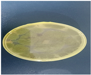
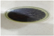
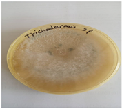
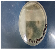
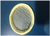
One of the numerous ways to show pectinase production ability of microorganisms especially fungal species is the method popularly known as pectin/carbohydrate hydrolysis or zone of clearance. Such procedures have been known to be very effective in determining and selecting the best fungal species needed to carry out pectinase production [19,31,37]. By adopting similar paths, this current study conducted a protocol showing carbohydrate hydrolysis. The aim was to select the isolates with the highest pectin degradation potential, after 5 days of incubation. After subjecting the isolates to pectin hydrolysis, the result showed that, all the fungal species possessed various degrees of pectinolytic activity (Table 2 and Figure 1). Aspergillus niger (FS2) emerged slightly on top as the most efficient microbial species (25.0 mm) among the tested species. Fusarium species (FS4) followed second with (23.0 mm), then Trichoderma species (FS3) and Aspergillus flavus (FS1) produced (20.0 mm each). Saccharomyces species (FS5) gave 7. 0 mm, hence it was left out in subsequent experiment.
It is well noted that, the strains of Aspergillus spp. have been known to dominate pectinase enzyme manufacturing in various studies and industrial applications [31]. The fact that, fungal species dominated the synthesis of pectinase in terms of zone of clearance in current study is favourably supported by El-Ghomary et al., 2021 [38]. In their work, they opined that, majority of commercial pectinase enzymes employed in industries are generated from species of fungal sources including Rhizopus stolonifer, Aspergillus and Penicillium spp. Oliyad and Dawit, 2018 [27] and Adedayo et al., 2021 [37], also asserted that, commercial pectinase enzyme production has recognized Aspergillus and Penicillium spp. as the main sources of pectinase enzymes. El-Ghomary et al., 2021 [38] isolated four strains of Aspergillus spp. that showed clear zone of pectin hydrolysis of 3.62 mm, 3.70 mm, 3.91 mm, and 4.75 mm for the respective strains, with Aspergillus flavus emerging as the species with highest zone of clearance. Current result is also, in agreement with Okonji et al., 2019 [31] when fungal isolate identified as Aspergillus fumigatus showed the highest pectin hydrolysis. Other numerous earlier investigations have also indicated the vital role played by Aspergillus spp. [13,19,29,43].
Furthermore, the relative dominance of Aspergillus niger, in current investigation, after exhibiting highest activity of pectinase in relation to zone of clearance (25 mm) is no fluke at all. This assertion is true as its ability to hydrolyze pectin-rich media has also been agreeably demonstrated in series of previous studies cited in Satapathy et al., 2019 [20]. In another closely related study conducted by Adedayo et al. (2021) [37], Aspergillus niger showed a better zone of clearance of 7.0 mm whereas Aspergillus flavus produced 5.5 mm. In addition, Kamalambigeswari et al., 2018 [45], examined three distinct strains of Aspergillus niger (F-3, F-4, and F-P), which revealed isolate F-4 as one that exhibited the largest region of pectin breakdown (9 mm).
3.4. Quantitative screening of potent pectinolytic fungal species (secondary screening)
Due to their promising nature, all the four isolates with comparatively higher zones of pectin hydrolysis were selected for further work on pectinase production (secondary screening). The four isolates that were deemed the best at pectinolytic ability exhibiting a clear zone of clearance were scrutinized for quantitative evaluation of pectinase synthesis utilizing a selective enrichment technique to generate comparative assessments. The secondary pectin hydrolysis screening exercise was then performed to identify the greatest amount of reducing sugars generated by the isolates with their respective enzyme activity via enzymatic breakdown of pectin. The galacturonic acid released was measured spectrophotometrically. The result (Table 3) showed that among the four (4) fungal isolates, FS2, which was identified as Aspergillus niger, once again, emerged as the most potent fungal species for the production of pectinase after recording the highest value reducing sugar formation (3.92 mg/mL) and pectinase activity (36.23 U/mL) under submerged fermentation after seven (7) days of incubation.
This finding fits in with El-Ghomary et al., 2021 [38], who found that, fungal isolates tagged as F9 showed relatively the highest titre of pectinase (35.83 U/mL) than that of all the other isolates. Okonji et al. (2019) [31] also discovered that Aspergillus fumigatus yielded 42 U/mL of pectinase enzyme activity in submerged fermentation conditions. The results achieved in this study and subsequent selection of fungal strains is also in line with Sudeep et al., 2020, who in their research, isolated 4 strains of fungal species with high pectinolytic activities from the soil. After thorough primary and secondary screening procedure, Aspergillus spp. was observed as the most potent fungus for pectinase enzyme production.
4. Conclusions
Soils obtained from the selected markets in Kumasi were found to be great source of plethora of diverse pectin-degrading microorganisms especially fungal species. A. niger has proven to be very dominant probably as a result of its ubiquitous nature and its ability to colonize diverse environments and substrate as it thrives well in many growth media or substrates as proven by this and other studies. A successful collection of these industrially-relevant strains of microorganisms from the soil could very well contribute to the increasing demand and adequate supply of pectinase enzymes in commercial quantities by related industries. Pectinase enzymes from microbes would also be very useful in remediation of areas polluted with plant waste. Further researches should be directed towards the mechanisms involved in the molecular regulation of pectinase enzymes production among fungal species especially A. niger. With such knowledge, scientists would be able to optimize the production of pectinase for commercial purposes. Also, subsequent studies could be invested in screening of extreme environments with the aim of finding fungal species with enhanced capabilities that can synthesize pectinase with novel properties.
Author Contributions
Conceptualization and design of the study were done by all authors. Material preparation, data collection and analysis as well as the first draft of the manuscript were performed by Owusu Sylvester Boakye, Zakpaa Hilary Domakyaara reviewed, edited and provided valuable comments and guidance whilst Malik Borigu supervised the laboratory experiments. Abudu Ballu Duwiejuah reviewed and provided valuable comments. All authors read and approved the final manuscript.
Funding
This research received no external funding.
References
- Shilpa, M.K.; Jason, Y. Isolation and screening of pectinase-producing bacteria from soil sample. CGC Int. j. contemp. res. eng. technol. 2021, 3, 166–170. [Google Scholar]
- Belkheiri, A.; Forouhar, A.; Ursu, A.V.; Dubessay, P.; Pierre, G.; Delattre, C.; Djelveh, G.; Abdelkafi, S.; Hamdami, N.; Michaud, P. Extraction, Characterization, and Applications of Pectins from Plant By-Products. Appl. Sci. 2021, 11, 6596. [Google Scholar] [CrossRef]
- Rokade, D.D.; Vaidya, S.L.; Naziya, M.A.; Dixit, P.P. Screening of Pectinase Producing Bacteria, Isolated from Osmanabad fruit market soil. Int. J. Interdiscip. Multidiscip. 2015, 2, 141–141. [Google Scholar]
- Tabssum, F.; Ali, S.S. Screening of Pectinase Producing Gram Positive Bacteria: Isolation and Characterization. Punjab- Univ. J. Zoöl. 2018, 33. [Google Scholar] [CrossRef]
- Thakur, P.; Singh, A.K.; Mukherjee, G. Isolation and Characterization of Alkaline Pectinase Productive Bacillus tropicus from Fruit and Vegetable Waste Dump Soil. Braz. Arch. Biol. Technol. 2021, 64. [Google Scholar] [CrossRef]
- Rakitikul, W.; Tammasat, T.; Udjai, J.; Nimmanpipug, P. Experimental and DFT study of gelling factor of pectin. Suranaree J. Sci. Technol. 2016, 23, 421–428. [Google Scholar]
- Food Science Avenue (2016): Pectinic acid. Pectinic-acid.html (Date assessed: 20/02/2023).
- Freitas, C.M.P.; Coimbra, J.S.R.; Souza, V.G.L.; Sousa, R.C.S. Structure and Applications of Pectin in Food, Biomedical, and Pharmaceutical Industry: A Review. Coatings 2021, 11, 922. [Google Scholar] [CrossRef]
- Lucarini, M.; Durazzo, A.; Bernini, R.; Campo, M.; Vita, C.; Souto, E.B.; Lombardi-Boccia, G, Ramadan; Santini, A.; Romani, A. Fruit Wastes as a Valuable Source of Value-Added Compounds: A Collaborative Perspective. Molecules 2021, 26, 6338. [Google Scholar] [CrossRef]
- Chandel, Vinay, Deblina Biswas, Swarup Roy, Devina Vaidya, Anil Verma, and Anil Gupta. (2022). “Current Advancements in Pectin: Extraction, Properties and Multifunctional Applications” Foods 11, no. 17: 2683.
- Tan, J.; Hua, X.; Liu, J.; Wang, M.; Liu, Y.; Yang, R.; Cao, Y. Extraction of sunflower head pectin with superfine grinding pretreatment. Food Chem. 2020, 320, 126631. [Google Scholar] [CrossRef]
- Picot-Allain, M.C.N.; Ramasawmy, B.; Emmambux, M.N. Extraction, Characterisation, and Application of Pectin from Tropical and Sub-Tropical Fruits: A Review. Food Rev. Int. 2020, 38, 282–312. [Google Scholar] [CrossRef]
- Roman-Benn, A.; Contador, C.A.; Li, M.-W.; Lam, H.-M.; Ah-Hen, K.; Ulloa, P.E.; Ravanal, M.C. Pectin: An overview of sources, extraction and applications in food products, biomedical, pharmaceutical and environmental issues. Food Chem. Adv. 2023, 2. [Google Scholar] [CrossRef]
- Hassan, S. (2020). Development of novel Pectinase and Xylanase juice clarifcation enzymes via a combined biorefnery and immobilization approach, Doctoral Thesis, Technological University Dublin. [CrossRef]
- Baljinder Singh Kauldhar, Harpreet Kaur, Venkatesh Meda, Balwinder Singh Sooch (2022). Chapter 12 - Insights into upstreaming and downstreaming processes of microbial extremozymes. Extremozymes and Their Industrial Applications, Academic Press, Pages 321-352, ISBN 9780323902748. [CrossRef]
- Shet AR, Desai SV, Achappa S. (2018). Pectinolytic enzymes: classification, production, purification and applications. Res. J. Life Sci Bioinform Pharm Chem Sci.; 4:337 –48.
- Soorej M. Basheer, Sreeja Chellappan, A. Sabu (2022). Chapter 8 - Enzymes in fruit and vegetable processing, Editor(s): Mohammed Kuddus, Cristobal Noe Aguilar, Value-Addition in Food Products and Processing through Enzyme Technology, Academic Press, Pages 101-110, ISBN 9780323899291.
- Garg, G.; Singh, A.; Kaur, A.; Singh, R.; Kaur, J.; Mahajan, R. Microbial pectinases: an ecofriendly tool of nature for industries. 3 Biotech 2016, 6, 1–13. [Google Scholar] [CrossRef] [PubMed]
- Oumer, O.J.; Abate, D. Screening and Molecular Identification of Pectinase Producing Microbes from Coffee Pulp. BioMed Res. Int. 2018, 2018, 1–7. [Google Scholar] [CrossRef] [PubMed]
- Satapathy, S.; Behera, P.M.; Tanty, D.K.; Srivastava, S.; Thatoi, H.; Dixit, A.; Sahoo, S.L. Isolation and molecular identification of pectinase producing Aspergillus species from different soil samples of Bhubaneswar regions. BioRxiv 2019, 1, 1–22. [Google Scholar]
- Sarita Shrestha; Chonlong Chio; Janak Raj Khatiwada; Aristide Laurel Mokale Kognou; Xuantong Chen; Wensheng Qin (2023). Optimization of Cultural Conditions for Pectinase Production by Streptomyces sp. and Characterization of Partially Purified Enzymes. Microb Physiol 33 (1): 12–26. PubMed: 36417846.
- Xiangyang Liu, Chandrakant Kokare, (2023). Chapter 17 - Microbial enzymes of use in industry, Editor(s): Goutam Brahmachari, Biotechnology of Microbial Enzymes (Second Edition), Academic Press, Pages 405-444, ISBN 9780443190599.
- Osete-Alcaraz, A.; Gómez-Plaza, E.; Pérez-Porras, P.; Bautista-Ortín, A.B. Revisiting the use of pectinases in enology: A role beyond facilitating phenolic grape extraction. Food Chem. 2022, 372, 131282. [Google Scholar] [CrossRef]
- Haile, S.; Ayele, A. Pectinase from Microorganisms and Its Industrial Applications. Sci. World J. 2022, 2022, 1–15. [Google Scholar] [CrossRef] [PubMed]
- Nazneen Inamdar, Asmita Dike, Amol Jadhav (2024). Isolation, Screening, and Identification of Pectin Degrading Bacteria from Soil. Journal of Chemical Health Risks, JCHR 14(2), 930-938 | ISSN: 2251-6727.
- Ivarsson, M.; Drake, H.; Bengtson, S.; Rasmussen, B. A Cryptic Alternative for the Evolution of Hyphae. BioEssays 2020, 42, e1900183. [Google Scholar] [CrossRef]
- Oliyad, J.O., Dawit, A. (2018). Screening and molecular identification of pectinase producing microbes from coffee pulp. Biomed Research International: 2961767.
- Carlos, D.L.-M.J.; Leonardo, S.; Jesús, M.-C.; Paola, M.-R.; Alejandro, Z.-C.; Juan, A.-V.; Noé, A.C. Solid-State Fermentation with Aspergillus niger GH1 to Enhance Polyphenolic Content and Antioxidative Activity of Castilla Rose (Purshia plicata). Plants 2020, 9, 1518. [Google Scholar] [CrossRef]
- Sudeep, K.C, Jitendra U. Dev R. J., Binod L, Dhiraj K. C., Bhoj R. P, Tirtha R. B., Rajiv D. Santosh K., Niranjan K., 10,Vijaya R. (2020). Production, Characterization, and Industrial Application of Pectinase Enzyme Isolated from Fungal Strains. Fermentation, 6, 59; 6.
- Singh, A.; Varghese, L.M.; Yadav, R.D.; Mahajan, R. A pollution reducing enzymatic deinking approach for recycling of mixed office waste paper. Environ. Sci. Pollut. Res. 2020, 27, 45814–45823. [Google Scholar] [CrossRef] [PubMed]
- Okonji, R.E.; Itakorode, B.O.; Ovumedia, J.O.; Adedeji, O.S. Purification and biochemical characterization of pectinase produced by Aspergillus fumigatus isolated from soil of decomposing plant materials. Journal of Applied Biology & Biotechnology 2019, 7, 1–8. [Google Scholar]
- Mrinmoy Ghosh and Krishna Kanth Pulicherla (2021). Psychrophiles as the source for potential industrial psychrozymes. Recent Developments in Microbial Technologies, Environmental and Microbial Biotechnology. [CrossRef]
- Ram Prasad, Vivek Kumar, Joginder Singh, Chandrama Prakash Upadhyaya (2021). Recent Developments in Microbial Technologies. Environmental and Microbial Biotechnology. ISSN 2662-169X.
- Alexopoulos, C.J., Mims, C.W., Blackwell, M. (1996). Intro. Mycology. Vol. 4. New York: Wiley.
- Barnett, H.L.; Hunter, B.B. Illustrated Genera of Imperfect Fungi, 4th ed.; Practice Hall: New York, NY, USA, 1987. [Google Scholar]
- Kaur, H.P.; Kaur, G. Optimization of cultural conditions for pectinase produced by fruit spoilage fungi. International journal of advances in research 2014, 3, 851–859. [Google Scholar]
- Adedayo, M.R.; Mohammed, M.T.; Ajiboye, A.E.; A Abdulmumini, S. Pectinolytic activity of aspergillus niger and Aspergillus flavus grown on grapefruit (citrus Parasidis) peel in solid state fermentation. Glob. J. Pure Appl. Sci. 2021, 27, 93–105. [Google Scholar] [CrossRef]
- El-Ghomary, A.E.; Shoukry, A.A.; El-Kotkat, M.B. Productivity of pectinase enzymes by Aspergillus sp. isolated from Egyptian soil. Al-Azhar J. Agric. Res. 2021, 46, 79–87. [Google Scholar] [CrossRef]
- Martison Budu, Patrick Boakye, Joseph Asankomah Bentil (2023). Microbial diversity, enzyme profile and substrate concentration for bioconversion of cassava peels to bioethanol. Preprints. [CrossRef]
- Timothy C. Cairns, Corrado Nai and Vera Meyer (2018). Aspergillus niger: A Hundred Years of Contribution to the Natural Products Chemistry. Fungal Biol Biotechnol, 5:13.
- Ezenwelu, C.O.; Afeez, O.A.; Anthony, O.U.; Promise, O.A.; Mmesoma, U.-E.C.; Henry, O.E. Studies on Properties of Lipase Produced from Aspergillus sp. Isolated from Compost Soil. Adv. Enzym. Res. 2022, 10, 49–60. [Google Scholar] [CrossRef]
- Ametefe, G.D., Oluwadamilare, L.A., Ifeoma, C. J., Olubunmi, I. I., Ofoegbu, V. O., Folake, F., Orji, F. A., Iweala, E.E.J., and Chinedu, S. N. (2021). Optimization of pectinase activity from locally isolated fungi and agrowastes. Research square.
- Jalil, M.T.M.; Ibrahim, D. Partial Purification and Characterisation of Pectinase Produced by Aspergillus niger LFP-1 Grown on Pomelo Peels as a Substrate. Trop. Life Sci. Res. 2021, 32, 1–22. [Google Scholar] [CrossRef]
- Oumer, O.J. Pectinase: Substrate, Production and their Biotechnological Applications. Int. J. Environ. Agric. Biotechnol. 2017, 2, 1007–1014. [Google Scholar] [CrossRef]
- Kamalambigeswari, R.; Sangilimuthu, A.Y.; Narender, S.; Ushani, U. Isolation, identification, screening and optimization of pectinase producing soil fungi (Aspergillus niger). International Journal of Research in Pharmaceutical Sciences 2018, 9, 762–762. [Google Scholar]
Figure 1.
Qualitative analysis of fungal species based on zone of pectin hydrolysis. (A) Fusarium spp.; (B) Aspergillus niger; (C) Aspergillus flavus; (D) Trichoderma spp.; (E) Saccharomyces spp.
Figure 1.
Qualitative analysis of fungal species based on zone of pectin hydrolysis. (A) Fusarium spp.; (B) Aspergillus niger; (C) Aspergillus flavus; (D) Trichoderma spp.; (E) Saccharomyces spp.
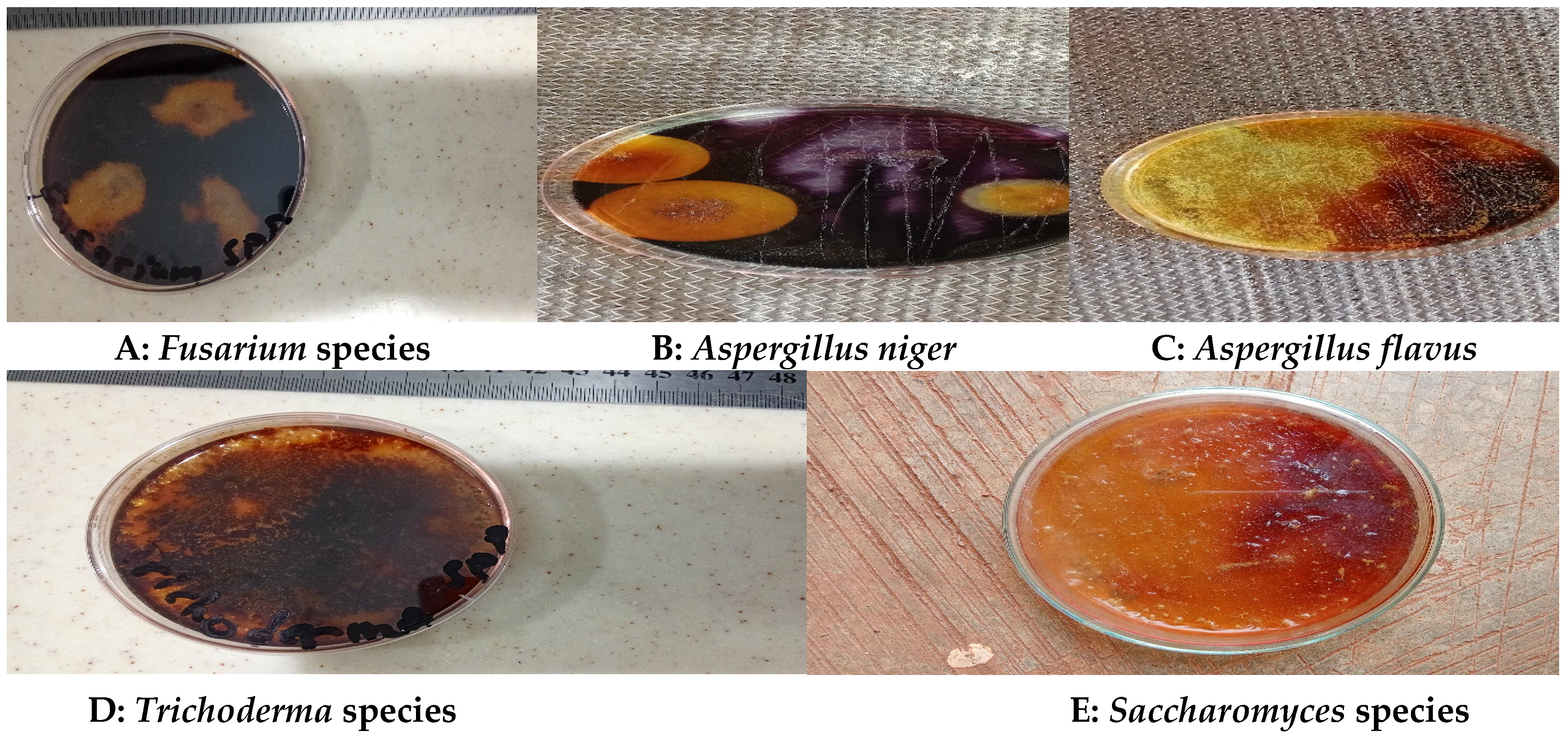

Table 1.
Characteristics of the fungal isolates.
| Isolate | Characteristics (Macro-and Micromorphological) | Possible Identity |
|---|---|---|
|
FS1 |
Appeared white initially and later turned grey or yellowish brown whilst pale yellow appeared as the reverse; colonies were fast growing with the texture appearing velvety or powdery, with a cottony appearance; conidial heads appeared as compact clusters, typically yellowish-green to yellow in color; large spore-bearing heads, spherical, and densely packed; clear, thin, and short hyphae; greenish mycelia; branched greenish conidia and septate hyphae; dense unbranched conidiophores with rounded or flask-shaped vesicle; elongated Phialides with constricted neck. |
 Aspergillus flavus Aspergillus flavus
|
|
FS2 |
Colony had fast and aggressive growth and looked fluffy white initially, turned black later; generated enormous black spores; reverse appeared pale yellow; wrinkled mycelia with visible dense and lengthy aerial hyphae; conidia were unbranched; texture looked velvety or powdery and the conidial produced heads with compact clusters, typically dark green to black in colour; hyphae looked septate, dense and branched; long, unbranched, and terminate conidiophores and gave rise to the conidial heads; rounded or flask-shaped vesicle at the end of conidiophore; Phialides were elongated with constricted neck. |
 Aspergillus niger. |
|
FS3 |
Colony was fast-growing and dominant with white appearance; sticky green phialoconidia clusters formed within a few days; flask-like Phialides with an enlarged base; woolly colonies formed initially and got compacted over time; as the conidia developed, dispersed blue green or yellow-green spots appeared visible, the reverse was pale, tan, or yellowish; conidia usually appeared within a week in dense or loose tufts of green, yellow, or white.; a yellow pigment was observed to have been released into the agar medium; an irregular yellow zone without conidia was present around the colony; Some concentric circles/rings also appeared. |
 Trichoderma spp. Trichoderma spp. |
|
FS4 |
Colonies grew very fast, and looked pale or brightly-coloured with the mycelia appearing like cotton; the thallus looked whitish in colour and showed presence of conidia from slender phialides; conidiophores looked solitary, short, lateral monophialides in the airborne mycelium, but were later observed as thickly branching groups; macroconidia looked fusiform, slightly curved and pointed at the tip and also appeared plentiful, largely non-septate, and straight or curved and looked cylindrically-shaped; chlamydospores appeared terminal, smooth and rough-walled; the phialides were short and mainly non-septate. |
 Fusarium spp. Fusarium spp. |
|
FS5 |
Colonies of Saccharomyces grew rapidly and matured in three days; they looked small, flat, smooth, moist and creamy or white in colour, raised and clustered, exhibiting oval or spherical to ellipsoidal shapes. |
 Saccharomyces spp. Saccharomyces spp. |
Table 2.
Qualitative analysis of fungal species based on zone of pectin hydrolysis achieved.
| Microorganism | Zone of Pectinolytic activity/mm |
|---|---|
| Aspergillus niger | 25.00 |
| Fusarium spp. | 23.00 |
| Trichoderma spp. | 20.00 |
| Aspergillus flavus | 20.00 |
| Saccharomyces spp. | 7.00 |
| P-value = 0.0511. | |
Table 3.
shows the figures obtained during the secondary screening.
| Microorganism | Reducing sugar (mg/mL) | Enzyme activity (U/mL) |
|---|---|---|
| Aspergillus niger (FS2) | 3.92 | 36.23 |
| Aspergillus flavus (FS1) | 3.68 | 32.52 |
| Trichoderma spp. (FS3) | 3.23 | 29.74 |
| Fusarium spp. (FS4) | 3.16 | 28.66 |
| P-value = 0.0260. | ||
Disclaimer/Publisher’s Note: The statements, opinions and data contained in all publications are solely those of the individual author(s) and contributor(s) and not of MDPI and/or the editor(s). MDPI and/or the editor(s) disclaim responsibility for any injury to people or property resulting from any ideas, methods, instructions or products referred to in the content. |
© 2024 by the authors. Licensee MDPI, Basel, Switzerland. This article is an open access article distributed under the terms and conditions of the Creative Commons Attribution (CC BY) license (http://creativecommons.org/licenses/by/4.0/).
Copyright: This open access article is published under a Creative Commons CC BY 4.0 license, which permit the free download, distribution, and reuse, provided that the author and preprint are cited in any reuse.


