
Submitted:
11 April 2025
Posted:
14 April 2025
Read the latest preprint version here
Abstract
While life expectancy has risen, healthspan—the period of life spent in good functional health—has not kept pace. Age-related and chronic diseases are often seen as inevitable decline, but more accurately reflect the cumulative cost of chronic stress adaptation under conditions of bioenergetic and substrate depletion. This review presents a systems-level framework that links aging and chronic disease to prolonged energy and nutrient trade-offs across physiological systems. We introduce the concept of Exposure-Related Malnutrition (ERM)—a subclinical state in which metabolic resources are silently diverted from maintenance and repair toward stress response. Drawing on classical stress models and evidence from immunometabolism, endocrinology, and systems biology, we describe how persistent stress drives resource reallocation across neuroendocrine, immune, muscular, and mitochondrial systems. We outline a shared adaptive trajectory—Respond → Adapt → Resolve—with outcomes shaped by both exposure intensity and metabolic availability. Progressive ERM manifests as anabolic resistance, low-grade inflammation, impaired healing, and lean mass loss—despite adequate intake. Clinical recognition of ERM using tools like pattern-based biomarkers, bioelectrical impedance, and adrenal reserve testing may allow early intervention. By identifying ERM as a reversible precursor to maladaptation, this framework reframes resilience as a metabolically costly process and highlights opportunities to preserve healthspan through upstream strategies.
Keywords:
Energy Metabolism
; Adaptation
; Chronic Disease
; Malnutrition
; Aging
; Resilience
1. Introduction
The paradox of modern resilience: longer life, less health
Human longevity has increased dramatically over the past century. Yet this achievement has revealed a troubling paradox: lifespan has outpaced healthspan—the portion of life spent in good functional health (Crane et al., 2022). Non-communicable diseases (NCDs) such as cardiovascular disease, diabetes, obesity, and neurodegeneration now account for over 70% of global deaths, with prevalence continuing to rise (WHO, 2025).
This growing gap raises fundamental questions about resilience: the body’s ability to withstand and recover from stress. In this review, we examine how responses to chronic stress follow a trajectory first outlined in Hans Selye’s General Adaptation Syndrome (GAS)—Alarm, Resistance, Exhaustion—now reframed through the lens of energy availability and recovery potential. We explore how the distribution of metabolic substrates across systems shapes whether stress resolves in recovery or decline.
The Exposome and Adaptive Responses: Drivers of Metabolic Burden
The body exists in continuous interaction with a dynamic exposome—encompassing diet, inactivity, psychosocial stress, circadian disruption, toxins, and infections (Vermeulen et al., 2020). While short-term exposures can trigger beneficial adaptations, chronic exposures impose a cumulative metabolic toll. These persistent demands drain energy reserves and force biological trade-offs that may not cause immediate dysfunction but gradually erode resilience.
Crucially, health outcomes are not determined by exposure alone, but by the individual’s capacity to adapt. For example, Baldwin et al. (2021) found that higher adverse childhood experience (ACE) scores correlate with disease risk at a population level but fail to predict individual trajectories. This underscores a critical insight: resilience is shaped by how energy and substrates are triaged across competing priorities—respond, adapt, and recover (Baldwin et al., 2021). When adaptive capacity is exceeded—due to limited metabolic reserves or inefficient allocation, maladaptation leads to arise of chronic dysfunction and disease: not from failure to respond, but from failure to resolve.
Healthspan as a Function of Adaptive Capacity
According to the World Health Organization, health is “a state of complete physical, mental, and social well-being and not merely the absence of disease or infirmity” (WHO, 1948).” In this context, healthspan reflects the body’s sustained ability to restore balance under stress, across multiple systems (Calabrese & Agathokleous, 2019). This ability hinges not only on energy availability but also on how efficiently it is allocated to meet fluctuating physiological demands.
The GAS provides a useful model. During the Alarm phase, resources are mobilized for defense, and performance often declines. In the Resistance phase, the body attempts to restore stability. Recovery depends on whether energy and substrates are sufficient—and properly directed—for repair. Successful adaptation may restore baseline or even enhance capacity (hormesis); if not, persistent stress leads to maladaptation and decline (Selye, 1950).
This trajectory parallels athletic training, where initial performance dips under load but may rebound with proper recovery or worsen if demands outpace regeneration (West, 2008). These outcomes reveal a critical insight: resilience is not a passive capacity but a metabolically expensive investment. It requires continuous investment of energy, often at the expense of long-term maintenance (Ryan & Ryznar, 2022).
Aging and Chronic Disease as the Cost of Adaptation
Aging and chronic disease are not simply the result of isolated system failures but reflect the accumulated toll of energy-conserving trade-offs. Under sustained stress, the body prioritizes short-term survival over long-term maintenance—diverting resources from repair, immunity, and regeneration.
This persistent triage drives functional decline over time. For example, skeletal muscle catabolism, inflammation, and mitochondrial dysfunction all reflect energetic reallocations made during chronic adaptation (Shaulson et al., 2024). At the cellular level, stress impairs mitochondrial efficiency, increases reliance on glycolysis, and accelerates epigenetic aging (Bobba-Alves et al., 2023). These changes represent not just damage but a biological reprogramming that favors short-term adaptation at the expense of long-term resilience.
Toward a systems view of metabolic trade-offs
This review adopts a systems-level framework to examine chronic stress triggers, energy availability, and substrate allocation shape adaptive outcomes across physiological domains—including neuroendocrine, immune, muscular, and mitochondrial networks. We propose reframing aging and chronic disease as the cumulative metabolic cost of unresolved adaptation to stress.
In the final sections, we introduce the concept of Exposure-Related Malnutrition (ERM)—a proposed early-stage, subclinical energetic deficit that arises from chronic stress adaptation—highlighting its role as a precursor to overt dysfunction and a potential target for preventive intervention. By identifying early signs of subclinical maladaptation, we aim to inform strategies that preserve adaptive capacity, improve recovery potential, and extend healthspan through timely intervention and support of metabolic resilience.
2. Methodology: A Thematic Narrative Review
This review uses a thematic narrative synthesis to investigate how chronic stress, energy availability, and metabolic resource allocation shape resilience, aging, and disease across multiple levels of biological organization—from systemic and organ-level processes to cellular mechanisms. The aim is to identify recurring adaptive patterns and the determinants that shift outcomes toward recovery, compensation, or decline.
Literature Selection
We used purposive sampling to identify relevant peer-reviewed studies and conceptual frameworks across physiology, aging research, systems biology, molecular signaling, immunometabolism, and exposomics. Priority was given to work that addressing:
- energy availability and substrate partitioning,
- adaptive stress responses across biological levels,
- chronic stress or allostatic load,
- aging mechanisms and chronic disease progression.
Sources were identified through targeted searches in PubMed, Web of Science, and Scopus, complemented by backward citation tracking. While not systematic, this approach emphasized conceptual depth and interdisciplinary integration.
Thematic Synthesis
We employed a three-phase thematic coding strategy:
- Open coding identified recurrent concepts such as metabolic trade-offs, substrate reallocation, and hormesis.
- Axial coding mapped relationships among energy dynamics, stress adaptation, and physiological outcomes.
- Narrative integration constructed a coherent, system-level framework that reflects a consistent adaptive trajectory: Respond → Adapt → Recover, culminating in either homeostasis, hormesis, or maladaptation.
Cross-Domain Conceptual Integration
This model emerged inductively through repeated analysis cycles across diverse domains and was cross-compared with established frameworks such as the GAS and hormesis theory, and emerging models including immunometabolic reprogramming, integrated stress response (ISR and ISRmt), brain–body energy conservation, and insulin-regulated substrate dynamics to ensure conceptual coherence and validity. These perspectives, drawn from molecular to whole-organism levels, reveal a common stress trajectory shaped by energetic and substrate availability and prioritization. The model is visually represented in Figure 1, which synthesizes the energetic trajectory of adaptation and its divergent outcomes:
Adaptive outcomes following stress are contingent on energy and substrate sufficiency. The resolution phase can lead to hormesis, homeostasis, or maladaptation. See Table 2 for comparative system outcomes.
3. The Energetic Trajectory of Stress Adaptation: From Response to Resolution
Stress adaptation is not merely a biochemical or hormonal cascade—it is an energetically expensive process requiring the coordinated mobilization, prioritization, and allocation of metabolic resources (Monzel et al., 2023). Across organismal, cellular, and subcellular levels, adaptive outcomes hinge on substrate availability and distribution efficiency. When demands exceed energy supply, systems are forced into trade-offs that preserve immediate function at the expense of long-term health.
General Adaptation Syndrome and Allostatic Load
GAS, proposed by Hans Selye, remains foundational for understanding systemic stress responses. It outlines three stages: Alarm (emergency mobilization), Resistance (sustained adaptation), and Exhaustion (breakdown from prolonged strain) (Selye, 1950). Contemporary models such as allostasis refine this view, emphasizing dynamic resource reallocation to maintain stability through change (McEwen & Wingfield, 2003).
The Resistance phase may lead not only to delay of collapse but to one of three outcomes:
- Restored homeostasis
- Adaptive overcompensation (hormesis)
- Maladaptation and decline (exhaustion)
This non-linear, reversible trajectory underscores that exhaustion is not inevitable—given sufficient resources and recovery capacity, systems may recover or even strengthen.
Hormesis: Adaptive Activation and Thresholds of Strain
Hormesis describes how low-dose or transient stress can activate adaptive responses that enhance long-term resilience (Calabrese & Agathokleous, 2019). Initially observed in toxicology, it is now recognized across contexts like exercise, caloric restriction, and thermal exposure. These stressors impose short-term metabolic costs in exchange for durable gains in tolerance, repair, or capacity.
However, the hormetic response depends on dose, timing, and recovery. The same exposure that promotes adaptation under one condition can trigger dysfunction in another. Hormesis is thus best understood as an energetic gamble—one that pays off only if recovery is supported.
Energy and Substrates as Limiting Factors in Resilience
All stress responses require substrates—ATP, amino acids, glucose, lipids, and micronutrients. These are reallocated to meet immediate demands, often diverting resources from longer-term processes like growth, cognition, and immune surveillance (Zera & Harshman, 2001). Under chronic or cumulative stress, this redistribution becomes a zero-sum game.
The Concept of Metabolic Triage and Functional Trade-Offs
When resources are constrained, the body activates conserved survival hierarchies—prioritizing critical functions while deferring regeneration and maintenance. This is captured by the nutrient triage hypothesis, which posits that even marginal deficits prompt protection of short-term survival at the expense of longevity-promoting pathways (Ames, 2006). Over time, this results in degradation of systems like skeletal muscle, reproductive health, and neuroplasticity.
We introduce the idea of subclinical adaptation failure—early physiological inefficiency where resilience erodes before overt disease manifests. Fatigue, low-grade inflammation, anabolic resistance, and poor recovery may reflect this hidden cost. Detecting such trade-offs offers a window for preclinical intervention.
Unifying System-Specific Adaptation: Respond, Adapt, Resolve
A consistent stress trajectory emerges across physiological systems:
- Phase 1: Respond – Emergency activation and energy mobilization.
- Phase 2: Adapt – Metabolic prioritization and reprogramming.
- Phase 3: Resolve – Transition toward recovery or dysfunction.
At each phase, outcomes depend on energetic capacity and regulatory flexibility. Systems that restore balance may regain function (homeostasis) or strengthen (hormesis); those that fail to resolve may enter a maladaptive state marked by dysfunction, inflammation, or degeneration.
4. The Energetic Architecture of Adaptation: Substrate Reallocation in Systemic Stress Responses
This section explores how the energetic economy of stress adaptation plays out across five key systems: neuroendocrine, immune, muscular, mitochondrial, and cellular integrated stress response (ISR) networks. Despite differing mechanisms, all follow the shared trajectory outlined earlier: Respond → Adapt → Resolve, culminating in one of three outcomes—Homeostasis, Hormesis, or Maladaptation. The quality and direction of this resolution depend on substrate availability, system-specific regulatory capacity, and the degree of sustained stress.
Phase 1: Respond — Emergency Signaling and Energy Mobilization
In the initial response phase, systems activate high-priority defense mechanisms, diverting energy from non-essential functions to immediate survival needs:
- Neuroendocrine System activates the hypothalamic-pituitary-adrenal (HPA) axis and sympathetic-adrenal-medullary (SAM) system, rapidly mobilizing glucose and suppressing growth and reproduction (Tsigos & Chrousos, 2002).
- Immune System initiates acute inflammation via pattern recognition receptors (PRRs), releasing pro-inflammatory cytokines (e.g., TNF-α, IL-6) and shifting immune metabolism toward glycolysis (Alack et al., 2019; Straub, 2017).
- Muscle tissue supplies gluconeogenic substrates by breaking down amino acids via proteolytic pathways (Cahill, 2006; Wolfe, 2006).
- Cellular ISR halts general protein synthesis via eIF2α phosphorylation while promoting selective translation of stress-resilient genes (Pakos-Zebrucka et al., 2016).
- Mitochondria shift metabolic output and activate antioxidant signaling and the mitochondrial unfolded protein response (UPRmt), increase ATP production, and activate antioxidant signaling (Picard & Shirihai, 2022).
This phase reflects acute catabolic triage: stress overrides baseline priorities, rerouting energy toward preservation of core functions.
Phase 2: Adapt — Metabolic Prioritization and Stress Programming
If stress persists, systems enter an adaptive phase, reallocating resources to optimize survival under constraint:
- Neuroendocrine System– Cortisol orchestrates systemic prioritization, supporting cerebral glucose supply while suppressing insulin, growth, and reproduction (McEwen & Wingfield, 2003).
- Immune cells undergo metabolic polarization: pro-inflammatory cells rely on glycolysis, while regulatory or reparative cells depend on oxidative phosphorylation (Olenchock et al., 2017; Willmann & Moita, 2024) (Geric et al., 2019; Olenchock et al., 2017). Chronic stress can trap cells in inflammatory states.
- Skeletal muscle, a major metabolic sink, attempts to transition from catabolism to repair. This shift requires amino acid availability and immune–muscle coordination, both of which are impaired under conditions of anabolic resistance. Anabolic resistance is not only a consequence but also a signal of unresolved adaptation, a state in which substrates and signaling are insufficient to restore muscle regeneration (Paulussen et al., 2021).
- Cellular ISR, when energetically supported, transitions from acute translation suppression to remodeling via autophagy, stress granule formation, and selective translation of repair-promoting factors (Gambardella et al., 2020). This metabolic reprioritization also drives epigenetic remodeling that accelerates cellular aging, especially under persistent stress (Gambardella et al., 2020).
- Mitochondria undergo remodeling, including mitophagy, fission/fusion dynamics, and shifts in substrate utilization to meet tissue-specific energy demands (Lockhart et al., 2020).
At this stage, systems are not yet failing, but function is constrained. Sustained imbalance may entrench dysfunction or promote resilience—depending on recovery support.

Table 1.
provides a comparative overview of how different systems prioritize, adapt, and resolve under stress conditions, based on their energetic thresholds and functional roles.
Table 1.
provides a comparative overview of how different systems prioritize, adapt, and resolve under stress conditions, based on their energetic thresholds and functional roles.
| System | Stress Priority Function | Energy Source Preference | Resolution Potential | Vulnerabilities Under Deficit |
|---|---|---|---|---|
| Neuroendocrine | Glucose mobilization, survival triage | Gluconeogenesis, lipolysis | Moderate (via cortisol tapering) | HPA overactivation, insulin resistance |
| Immune | Inflammation, defense | Glycolysis (pro-inflammatory), OXPHOS (repair) | High if balance restored | Chronic inflammation, immune senescence |
| Muscle | Amino acid reservoir, repair coordination | Glycogen, fatty acids, structural protein | High if nutrients available | Anabolic resistance, sarcopenia |
| Cellular ISR | Proteostasis, autophagy | Internal recycling, selective translation | Moderate tohigh | Persistent translation block, apoptosis |
| Mitochondria | Energy production, redox signaling | OXPHOS, glycolysis, fatty acids | High if fission/fusion restored | ROS overload, mitokine dysfunction |
Table 1. Comparative features of adaptive stress responses across physiological systems. Resolution capacity depends on energy substrate availability and the ability to deactivate stress programs. Vulnerabilities reflect common outcomes of unresolved or prolonged adaptation.
Phase 3: Resolve — Transitioning from Adaptation to Outcome
The Resolve phase marks a pivotal point in the stress response—where systems either recover, overcompensate, or deteriorate. Unlike the rapid mobilization of Phase 1 or the constrained remodeling of Phase 2, this phase involves deactivating stress programs and restoring metabolic balance. Crucially, resolution is not binary. Outcomes are contingent on energy availability, substrate reallocation, and system-specific capacity for repair.
When recovery is successful, systems withdraw catabolic signals—cortisol declines, parasympathetic tone increases, and inflammatory responses resolve. Tissue remodeling, mitochondrial recalibration, and restoration of anabolic processes follow—but only if metabolic reserves are sufficient (McEwen, 2007; Sapolsky, 2004).
In this phase:
- Neuroendocrine systems reduce HPA axis activity and re-establish circadian rhythm and metabolic homeostasis. Flattened or delayed cortisol recovery signals impaired resolution.
- Immune systems transition from inflammation to repair, with M1 macrophages converting to M2 phenotypes and resolution pathways (e.g., resolvins, lipoxins) facilitating tissue remodeling (Olenchock et al., 2017). Micronutrient sufficiency—particularly zinc, selenium, and iron—is critical to this process.
- Skeletal muscle resumes protein synthesis and regeneration, but only if inflammation resolves and energy/nutrient levels support mTORC1 and satellite cell activation. Without adequate support, fibrosis or sarcopenia may ensue (Paulussen et al., 2021).
- Cellular ISR mechanisms, such as GADD34-mediated dephosphorylation of eIF2α, permit selective restoration of protein synthesis. This reactivation depends on sufficient ATP, proteostasis, and redox control (Gambardella et al., 2020).
- Mitochondria stabilize through restored fission/fusion dynamics and mitophagy, allowing redox homeostasis and efficient energy production. Transient mitokine signaling subsides as systemic demands normalize (Picard & Shirihai, 2022).
These transitions are dynamic and reversible. Systems can shift toward recovery—or regress toward dysfunction—based on fluctuations in substrate availability and stressor duration. Even partially stalled recovery can be redirected if energetic and regulatory conditions improve.
Outcomes fall into one of three categories:
- Homeostasis – Return to pre-stress function and balance.
- Hormesis – Functional overcompensation and increased resilience.
- Maladaptation – Incomplete resolution, leading to persistent dysfunction.
These are not uniform across systems. As shown in Table 1, resolution pathways vary in efficiency and vulnerability depending on tissue type, stress history, and energetic flexibility. This variability explains why stress recovery may be complete in one domain (e.g., immune) but incomplete or pathological in another (e.g., muscle or mitochondrial networks) (McEwen & Wingfield, 2003; Sapolsky, 2004).
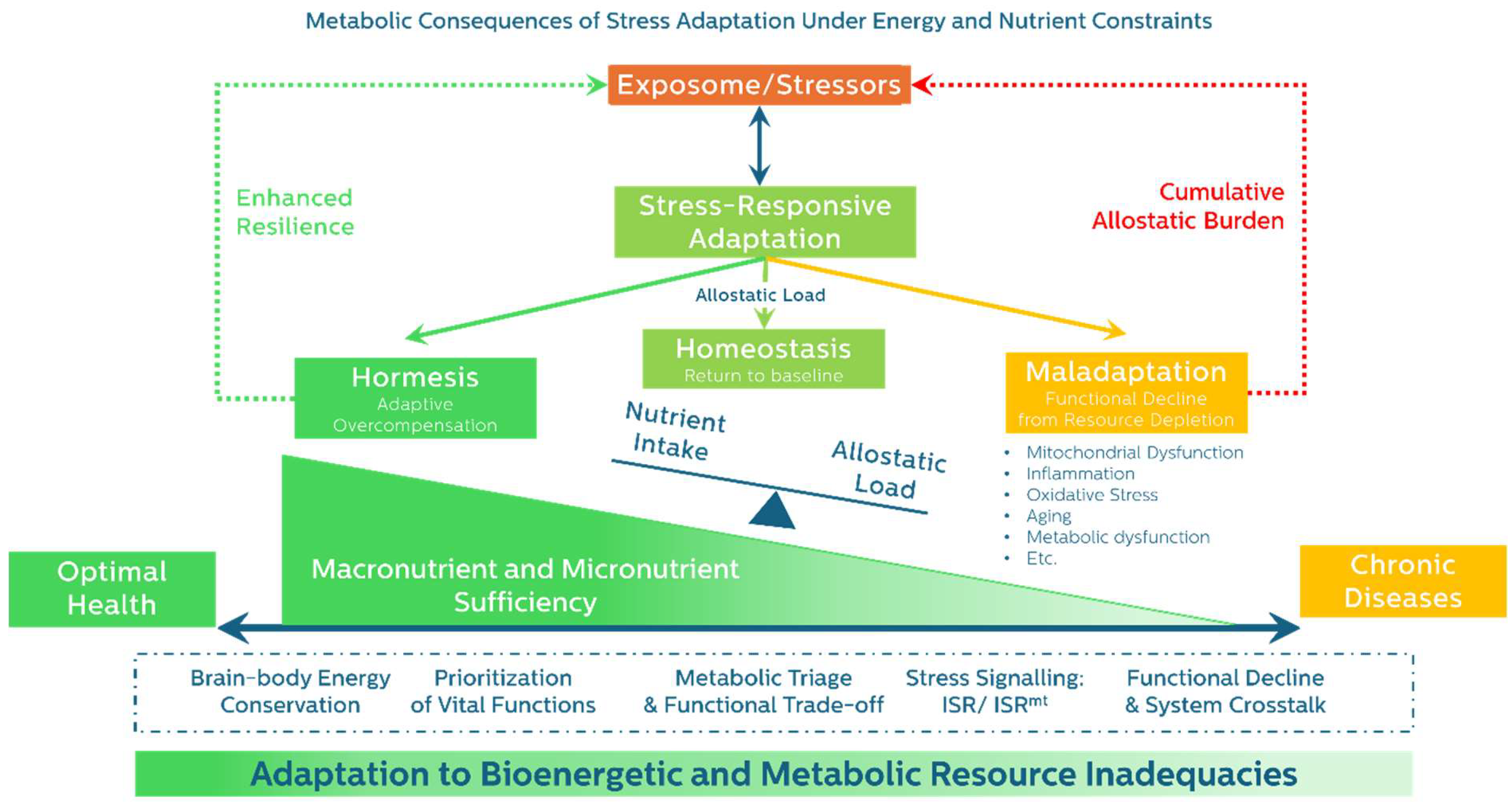
To visualize how stress responses unfold through these stages, we reintroduce the broader energetic trajectory of adaptation in Figure 2 below. This model illustrates how Respond → Adapt → Resolve leads to divergent outcomes, shaped by metabolic availability and trade-offs at each stage.
Ultimately, the Resolve phase offers a critical window for intervention—the point at which resilience can be reinforced or decline accelerated. Early detection of stalled resolution, paired with targeted substrate support and stressor mitigation, may shift outcomes from maladaptive to restorative.
5. Resolution and Its Consequences: Divergent Outcomes Shaped by Energy and Resource Allocation.
Following the adaptive phase, stress resolution proceeds along one of three general paths: Homeostasis, Hormesis, or Maladaptation. These outcomes depend not on exposure alone, but on energy status, substrate distribution, and system-specific flexibility. While all systems share basic resolution pathways, their capacity to complete this process varies depending on metabolic reserve and prior stress burden.
5.1. Homeostasis — Energetic Recovery and Structural Recalibration
Homeostasis reflects a return to pre-stress equilibrium. This requires sufficient energy and nutrients to downregulate catabolic pathways, restore baseline signaling, and re-engage long-term maintenance.
Shared features across systems:
- Cortisol and inflammatory cytokines decline, parasympathetic tone is restored, and insulin sensitivity improves (Bobba-Alves et al., 2022).
- Immune resolution involves clearance of apoptotic cells, matrix remodeling, and macrophage transition from M1 to M2 phenotypes—processes that rely on mitochondrial OxPhos, redox regulation, and micronutrients like zinc, iron, and selenium (Alack et al., 2019; Laurent et al., 2017; Olenchock et al., 2017).
- In skeletal muscle, recovery depends on satellite cell activation and nutrient-sensitive pathways such as mTORC1, supported by leucine, vitamin D, and redox cofactors (Beaudart et al., 2017; Careccia et al., 2023; Paulussen et al., 2021).
- Cellular ISR resolves through GADD34-mediated dephosphorylation of eIF2α, enabling proteostasis and selective translation restoration (Gambardella et al., 2020; Novoa et al., 2001).
- Mitochondrial recovery via mitophagy and biogenesis restores ATP production and oxidative balance; transient ROS bursts activate adaptive pathways via NRF2 and FOXO, while sustained oxidative stress impairs recovery (Picard & Shirihai, 2022).
These changes re-establish systemic integrity but depend heavily on substrate sufficiency and stress program withdrawal.
5.2. Hormesis — Energetic Overcompensation and Adaptive Remodeling
Hormesis occurs when moderate, transient stress paired with adequate recovery produces adaptive overcompensation—resulting in enhanced function and resilience (Calabrese & Agathokleous, 2019). Hormesis is not a reward but a cost-paid outcome—it emerges only when the energetic investment required for overcompensation is met and recovery pathways are intact.
Examples of hormetic remodeling:
- Trained immunity: Monocytes, macrophages, and NK cells undergo glycolytic and epigenetic reprogramming via mTOR–HIF-1α signaling, increasing responsiveness and tolerance (Netea et al., 2016; Ochando et al., 2023; Vuscan et al., 2024).
- Immune resolution and tolerance: Regulatory T cells and M2 macrophages mediate inflammation resolution and tissue repair via mitochondrial metabolism (Vuscan et al., 2024).
- Exercise-induced muscle remodeling: IL-13–producing ILC2s, IL-33–expressing stromal cells, and macrophage–Treg signaling coordinate mitochondrial biogenesis and type 2 immunity in recovery (Langston & Mathis, 2024; Metallo & Vander Heiden, 2013).
- Mild ISR activation: Transient eIF2α phosphorylation enhances redox balance, proteostasis, and metabolic flexibility via ATF4/CHOP signaling (Costa-Mattioli & Walter, 2020; Sparkenbaugh et al., 2011).
- Mitohormesis: Low-level ROS from mitochondrial stress induces biogenesis, antioxidant upregulation, and mitokine release (e.g., FGF21, MOTS-c) for systemic coordination (Lockhart et al., 2020; Ristow & Schmeisser, 2014).
These adaptations are metabolically expensive and depend on sufficient ATP, intact signaling, and micronutrient availability.
5.3. Maladaptation — Energetic Collapse and Structural Degeneration
Maladaptation reflects failed recovery—where energy reserves are insufficient or misdirected, and stress programs remain chronically active. This outcome is not a failure to respond, but a failure to resolve.
Key system manifestations:
- Neuroendocrine: Sustained cortisol, insulin resistance, hippocampal atrophy, and central fatigue due to prolonged stress signaling (Chrousos, 2009; Meeusen et al., 2006; Shaulson et al., 2024).
- Immune: Inflammaging and immunosenescence from persistent IL-6, TNF-α, SASP signaling, and impaired clearance of senescent cells(Franceschi et al., 2018; Fulop et al., 2018; Wang et al., 2024).
- Skeletal muscle: Anabolic resistance, mitochondrial dysfunction, and catabolism lead to sarcopenia and frailty, compounded by aging, nutrient deficits, and inflammation(Cruz-Jentoft et al., 2023; Walrand et al., 2021).
- Cellular ISR: Chronic eIF2α phosphorylation impairs translation, promotes apoptosis, and drives redox imbalance and mitochondrial damage (Hetz & Papa, 2018; Wek, 2018).
- Mitochondria: PGAM5-driven mitochondrial fragmentation, ROS generation, and mtDNA-triggered inflammasome activation fuel a cycle of mitophagy failure, pyroptosis, and degeneration (Qi et al., 2025; Youle & van der Bliek, 2012; Yuk et al., 2020)
This phase marks the energetic and structural tipping point beyond which spontaneous recovery is unlikely without targeted intervention. Maladaptation arises not from insufficient signaling, but from a systemic failure to allocate energy and substrates toward recovery—a breakdown in bioenergetic governance.
To synthesize and compare these divergent outcomes,
Table 2.
summarizes the key physiological and metabolic characteristics of Homeostasis, Hormesis, and Maladaptation, highlighting how energy status and adaptive resolution differ across systems. This table encapsulates the core thesis of the review: that resilience, while biologically attainable, carries a metabolic cost—and that outcome hinges not just on the presence of stress but on the energetic resources available to recover from it.
| Feature |
 Homeostasis Homeostasis |
 Hormesis Hormesis |
 Maladaptation Maladaptation |
|---|---|---|---|
| Energy Availability | Restored baseline levels | Sufficient with transient surplus | Depleted or misallocated |
| Functional Outcome | Re-equilibration | Enhanced resilience or capacity | Persistent dysfunction |
| Immune Response | Inflammation resolves | Trained immunity and regulatory tolerance | Chronic inflammation, immune exhaustion |
| Muscle Remodeling | Repair of damaged fibers | Functional hypertrophy, mitochondrial gains | Catabolism, fibrosis, sarcopenia |
| ISR Recovery | Reinstated proteostasis | Increased stress resilience, adaptive memory | Persistent translation block, apoptosis |
| Mitochondrial Dynamics | Normalized bioenergetics | Improved redox balance, adaptive signaling | ROS overload, mitophagy failure |
| Recovery Dependency | Energy repletion, stress withdrawal | Surplus energy, time, micronutrient support | Insufficient recovery, chronic demand |
Table 2. Comparative features of adaptive resolution outcomes across systems.
Each outcome—homeostasis, hormesis, or maladaptation—reflects differences in energy availability, recovery quality, and system-specific plasticity.
6. Energy and Substrate Insufficiency in High-Demand States
Across a wide range of physiological and clinical conditions, nutrient insufficiency often arises not from inadequate intake, but from elevated metabolic demand. In such states, energy and substrates may be diverted toward acute survival functions at the expense of long-term maintenance, gradually undermining resilience.
Three classical syndromes illustrate how malnutrition can be demand-driven, even in the presence of apparently adequate intake:
- Disease-Related Malnutrition (DRM) occurs in patients with acute or chronic disease where inflammation increases resting energy expenditure and protein breakdown, often despite ongoing feeding (Cederholm & Bosaeus, 2024; Muscaritoli et al., 2023).
- Chronic Energy Deficiency (CED) reflects persistent nutritional insufficiency during states of heightened need—such as pregnancy—where energy and nutrient reallocation can compromise maternal and fetal health even when BMI appears normal (Prisabela et al., 2023; Taylor-Baer & Herman, 2018).
- Relative Energy Deficiency in Sport (REDs) results from chronic low energy availability in athletes, leading to impaired endocrine, immune, bone, and cognitive function, often with normal or low-normal body weight (Cabre et al., 2022; Mountjoy et al., 2018).
Each reflects a state where high demand, rather than insufficient supply, drives a critical mismatch between energy needs and availability. These conditions demonstrate that energy deficiency can be demand-driven, even in nutrient-replete environments.
Table 3.
compares features of these classical high-demand malnutrition.
| Feature | DRM | CED | REDs |
|---|---|---|---|
| Primary Affected Populations | Hospitalized or chronically ill patients | Pregnant women, children, low-resource settings | Endurance athletes, dancers, military recruits |
| Onset | Acute or chronic illness | Gradual under chronic stress (e.g., pregnancy) | Subacute with high training load |
| Triggers | Inflammation, disease burden | Increased physiological need, low intake | Prolonged mismatch between training intensity and caloric intake |
| Nutritional Markers | Often abnormal (e.g., prealbumin ↓) | Subclinical changes; may appear normal in standard labs | May have normal BMI, altered hormones |
| Clinical Presentation | Weight loss, immune dysfunction, poor healing | Maternal fatigue, micronutrient depletion, fetal risk | Performance decline, bone loss, endocrine suppression |
| Response to Nutrition | Requires nutritional support alongside anti-inflammatory therapy | Improves with energy/nutrient repletion | Requires coordinated refeeding and training balance |
Table 3. Comparative features of classical high-demand energy deficiency syndromes.
While differing in onset and context, all reflect a mismatch between energy demand and metabolic availability—often in the absence of overt undernutrition.
These syndromes illustrate how substrate insufficiency can develop independently of food scarcity—an insight that underpins the emerging concept of Exposure-Related Malnutrition.
Exposure-Related Malnutrition: The Preclinical Cost of Chronic Stress Adaptation
We propose the term Exposure-Related Malnutrition (ERM) to describe a subclinical, early-stage form of nutrient insufficiency that emerges in response to prolonged metabolic and environmental stress.
ERM differs from traditional malnutrition in three key ways:
- No overt intake deficit – energy intake may appear normal, and BMI may be stable or elevated.
- Triggered by cumulative exposome burden – including inflammation, toxin exposure, psychosocial stress, circadian disruption, or chronic low-grade infections (Pizzorno, 2020; Vermeulen et al., 2020).
- Manifests through the metabolic trade-offs pattern – such as impaired muscle recovery, fatigue, immunosuppression, or anabolic resistance, often before clinical thresholds of dysfunction are met.
ERM represents the front end of the malnutrition trajectory. It is the physiological cost of prolonged adaptation under constraint, when substrates are increasingly diverted from maintenance and repair toward stress-response and immune activation. Over time, this hidden deficit can fuel the transition to maladaptation and eventual disease.
Clinically, ERM helps explain why individuals with normal labs and anthropometrics may still present with chronic fatigue, sarcopenia, frequent infections, or poor healing—hallmarks of compromised metabolic resilience (Ames, 2006; Sganga et al., 1985; Wang & Zhang, 2025).
By identifying and intervening during this early stage—before DRM, CED, or REDs develop—clinicians have a window to preserve adaptive capacity and restore metabolic flexibility.
7. Metabolic Substrate Misallocation, Aging, and the Modern Disease Burden
Modern chronic diseases are often attributed to caloric excess. But beneath this lies a deeper dysfunction: the misallocation of metabolic substrates. This process—driven by chronic stress, insulin resistance, and hormonal signaling errors—leads to energy being sequestered in storage rather than delivered to tissues that need it most(Ludwig, 2023).
Insulin Signaling and Substrate Trapping
Insulin plays a central role in coordinating nutrient flow—promoting glucose uptake, lipid storage, and protein synthesis. In insulin-resistant states, however, this coordination breaks down. Skeletal muscle and liver become desensitized, while adipose tissue often retains partial insulin sensitivity, favoring fat storage over energy mobilization (Friedman et al., 2024). As a result, glucose and amino acids fail to reach active tissues, even amid nutrient abundance (Chen & Kahn, 2024).
This mismatch—known as substrate trapping—impairs mitochondrial function, promotes chronic inflammation, and fuels energy-sensing stress responses. Defend and repair systems become substrate-starved, contributing to anabolic resistance, immune dysfunction, and cellular rigidity. Over time, this persistent misallocation undercuts systemic resilience, laying the groundwork for chronic disease (Kalinkovich & Livshits, 2017; Speakman & Hall, 2021).
Fructose Survival Hypothesis: A Maladaptive Energy Lock-In
The Fructose Survival Hypothesis proposes that fructose—whether ingested or endogenously produced—activates a conserved metabolic program designed to maximize energy storage and suppress non-survival functions (Johnson et al., 2024). This same metabolic logic underlies insulin-induced substrate partitioning with depleted circulating fuel despite caloric sufficiency. Fructose metabolism triggers ATP depletion, impairs mitochondrial function, elevates oxidative stress, and shifts energy away from processes like reproduction, cognition, and muscle maintenance. While adaptive during food scarcity, this response becomes maladaptive in modern environments of caloric surplus and chronic stress. Repeated activation—via dietary fructose or stress-induced glucocorticoid pathways—drives a “storage-locked” state, amplifying insulin resistance and reinforcing substrate misallocation across systems.
This energy-locking phenomenon contributes to systemic metabolic rigidity—a hallmark of aging and chronic disease—driven in part by impaired nutrient-sensing pathways such as mTOR–AMPK imbalance and SIRT1 downregulation. These regulatory disruptions further suppress adaptive flexibility, hindering the body's ability to shift from survival metabolism to repair, growth, or recovery.
Chronic Maladaptation and the Progression of Aging and NCDs
This metabolic rigidity mirrors the broader theme of maladaptation: the collapse of flexible energy allocation in favor of chronic survival signaling. In this framework, NCDs reflect not only nutrient overload, but the chronic failure to deliver substrates to where they are needed most—a breakdown in energetic governance across the body’s most adaptive systems.
8. Recognizing ERM as the Cost of Adaptation in Clinical Practice
ERM represents the early, subclinical consequence of prolonged stress adaptation, in which metabolic substrates are continuously diverted toward immediate survival functions at the expense of repair, regeneration, and long-term resilience. Unlike classical malnutrition, ERM often arises in individuals with normal caloric intake and body weight, making it difficult to detect using conventional anthropometric or laboratory markers. Rather than reflecting nutrient scarcity, ERM is defined by persistent bioenergetic trade-offs. Over time, this silent reallocation erodes physiological reserve and impairs recovery capacity, especially when exposure burdens are sustained and unresolved.
Recognizing ERM Through Functional Patterns
Early detection of ERM requires a shift from static diagnostic thresholds to recognition of dynamic shifts in metabolism, recovery, and stress-response efficiency. Key features reflect subtle metabolic compromises across systems:
Clinical clues of ERM:
- Chronic fatigue despite adequate sleep and nutrition
- Poor exercise recovery or delayed wound healing
- Frequent mild infections or persistent low-grade inflammation
- Difficulty maintaining or gaining lean mass despite adequate intake and physical activity
- Biomarker patterns suggesting inflammation-driven nutrient diversion
When these signs co-occur—particularly in the context of known exposome stressors—ERM should be suspected, even in the absence of weight loss or overt nutritional deficiency.
Drivers of ERM: The Cumulative Exposome
ERM emerges from chronic exposure to a wide range of environmental, lifestyle, and internal stressors that challenge metabolic regulation and repair capacity. These include:
- External triggers: air pollution, persistent organic pollutants (POPs), heavy metals (e.g., lead, arsenic, mercury), microplastics, endocrine-disrupting chemicals, and circadian rhythm disruption
- Internal triggers: chronic inflammation, dysbiosis, latent infections, psychosocial stress, and trauma
These stressors interact with metabolic and immune systems, shifting nutrient allocation toward short-term defense and away from tissue maintenance (Pizzorno, 2020; Vermeulen et al., 2020). Without addressing underlying exposures, nutrition alone is often insufficient to restore resilience.
Staging ERM: A Continuum of Energetic Decline
ERM unfolds along a progressive trajectory of adaptive compromise, characterized by increasing metabolic inefficiency and loss of recovery capacity:
- Mild ERM: Subtle reductions in physical performance, cognitive clarity, or recovery from exertion; typically reversible with adequate substrate support and removal of stressors
- Moderate ERM: Emergence of functional trade-offs, including low-grade inflammation, anabolic resistance, downregulated housekeeping and transport proteins, or early hormonal imbalances
- Severe ERM: Entrenched catabolism, sarcopenia, immune dysfunction, and metabolic rigidity, often accompanied by structural decline and reduced adaptability
Each stage is marked by measurable physiological changes, including elevated inflammatory markers, reprioritization of protein synthesis, slowed cellular turnover, and blunted adrenal output. Recognizing these patterns across systems enables timely, targeted intervention—potentially preventing irreversible dysfunction and the transition to overt disease.
Biomarkers of Nutrient Triage and Trade-Offs
ERM reflects an altered hepatic protein synthesis profile in response to inflammation. Positive acute-phase proteins (APPs)—such as C-reactive protein (CRP), ferritin, and fibrinogen—rise to support immune defense, while negative APPs—albumin, prealbumin, transferrin, and retinol-binding protein— decline as the liver reallocates amino acids away from homeostatic functions (Cederholm & Bosaeus, 2024; Gulhar et al., 2024; Sganga et al., 1985).
This trade-off is adaptive in the acute phase but becomes maladaptive when sustained, contributing to impaired recovery and long-term functional decline (Bresnahan & Tanumihardjo, 2014). Importantly, these biomarkers fluctuate based on the phase of adaptation. For example, prealbumin may transiently increase during early resolution if substrate availability is sufficient but will decline if metabolic reserves are not restored. As highlighted in the ASPEN guidelines, these proteins are best interpreted as indicators of inflammatory protein redistribution rather than standalone measures of nutritional status (Evans et al., 2021).
In addition, increased activity of glycolytic enzymes—such as lactate dehydrogenase (LDH) and neuron-specific enolase (NSE) —may signal a shift toward glycolytic predominance or impaired OXPHOS. These changes reflect a broader metabolic reprogramming under sustained stress, favoring rapid ATP generation at the cost of mitochondrial efficiency and long-term resilience (Donnelly & Finlay, 2015; Fang et al., 2024; Olcay Güngör et al., 2018).
Despite widespread clinical use, serum and plasma nutrient levels are limited in their ability to detect subtle or early nutrient insufficiencies. These levels primarily reflect short-term intake and systemic circulation, which may appear normal despite intracellular depletion or increased demand under chronic stress. While valuable for population-level trends—such as in NHANES—these markers lack the sensitivity to identify functional deficits in individuals (Adams et al., 2020; Peeri et al., 2021).
Moreover, comprehensive serum testing for all nutrients is costly and impractical in clinical practice. Emerging tools like urine organic acid profiling provide insights into intermediary metabolism but are expensive, not standardized, and lack reference ranges for early-stage dysfunction. Variability in sample preparation and metabolite recovery further reduces reliability in mild cases (Carling et al., 2025).
In contrast, recognizing trade-off patterns in standard clinical biomarkers offers a more practical and cost-effective strategy for detecting ERM (Foy et al., 2024). These changes provide an actionable window into physiological compromise, enabling earlier intervention to restore resilience before irreversible dysfunction develops.
Cellular Turnover and Intracellular Proteins
A less commonly recognized sign of maladaptive stress is the elevation of intracellular proteins and enzymes—such as alanine transaminase (ALT), aspartate transaminase (AST), and creatine phosphokinase (CPK)—without clear organ-specific pathology or causes. These elevations may also indicate slow cell turnover, impaired proteostasis, or early signs of catabolic stress, accompanying by gradual stage of ERM. When persistent, these elevations may reflect insufficient support of bioenergetic and metabolic substrates to sustain normal cellular turnover under metabolic strain, rather than overt tissue injury (Aujla et al., 2025; Nakajima et al., 2022).
Bioimpedance: A Window into Subclinical Body Composition Changes
Bioelectrical impedance analysis (BIA) provides a non-invasive, early detection tool for identifying subclinical changes associated with ERM. A declining phase angle indicates compromised cellular membrane integrity and reduced cellular health (Lee et al., 2014). A stable or increasing BMI accompanied by a loss of lean mass suggests covert nutrient redistribution and a shift toward catabolic dominance. Additionally, elevated extracellular water may reflect low-grade inflammation, edema, or protein loss (Branco et al., 2023). When interpreted alongside clinical symptoms and biochemical markers, BIA enhances early recognition, staging, and monitoring of ERM before overt dysfunction appears.
Endocrine Clues: Adrenal Reserve and Allostatic Load
Another essential dimension of physiological adaptation is adrenal reserve—the capacity of the adrenal glands to sustain glucocorticoid output under chronic or repeated stress. The HPA axis is central to coordinating systemic stress responses, regulating glucose metabolism, inflammation, protein turnover, and circadian synchrony (Herman, 2013). In the context of ERM, the functionality of this axis plays a decisive role in determining whether the body can successfully adapt and recover or begins to decompensate.
In early ERM, adrenal output generally supports stability, enabling sufficient cortisol-mediated mobilization of substrates. However, as adaptive demands accumulate, the HPA axis may become dysregulated. Clinical signs of this shift include flattened diurnal cortisol rhythms, exaggerated fatigue, stress intolerance, and delayed recovery from illness or exertion (Herman, 2013). Aging compounds this burden: while basal cortisol may remain stable or rise slightly, dynamic responsiveness often declines. Decreased DHEA production and a higher cortisol to DHEA ratio reflect a catabolic endocrine profile associated with frailty and immune senescence (Yiallouris et al., 2019).
In this context, elevated cholesterol may serve as an indirect marker of adrenal demand, as it is the precursor for cortisol and DHEA. Similarly, sustain elevation of glycated hemoglobin level despite adequate diet may reflect cortisol-driven gluconeogenesis and emerging insulin resistance—both signs of stress-induced substrate mobilization (Bar-Ziv et al., 2020; Seiler et al., 2020; Yiallouris et al., 2019).
Functional adrenal assessments—such as morning cortisol and DHEA-S, or ACTH stimulation testing—can help evaluate adrenal reserve, particularly in patients with unexplained fatigue, poor recovery, or paradoxically elevated lipids despite lifestyle intervention (Warde et al., 2023). When interpreted alongside inflammatory markers, BIA trends, and clinical history, these assessments contribute to a broader picture of stress-related endocrine and metabolic burden.
Clinical Implication: Interpreting Patterns, Not Points
To recognize ERM before it progresses to overt dysfunction, clinicians must move beyond interpreting individual lab values in isolation and instead focus on identifying meaningful patterns over time. For example:
- Persistent elevation of CRP alongside declining prealbumin or transferrin
- Declining phase angle and lean body mass, even with preserved or rising body weight
- Elevated intracellular enzymes (e.g., ALT, AST, CPK) without clear organ-specific pathology
- Persistent hypercholesterolemia or hyperglycemia despite appropriate dietary and lifestyle interventions
These trends suggest systemic metabolic trade-offs—hallmarks of adaptation under constraint—and can help differentiate functional compensation from emerging maladaptation.
The most important clinical questions shift from asking “Is this patient malnourished?” to:
- “What phase of adaptation is this patient in?”
- “What exposures, stressors, or nutritional deficits are sustaining this trade-off—and what interventions could restore metabolic balance?”
This perspective emphasizes dynamic evaluation, enabling earlier and more effective intervention to preserve resilience and prevent the progression toward irreversible dysfunction
Reversibility of ERM and the Importance of Timing
The window to reverse ERM is greatest in its early stages. When substrate flow is restored and stressors are reduced, recovery is possible. Physiological resilience can be rebuilt through targeted intervention—restoring immune tolerance, mitochondrial flexibility, and protein turnover.
This underscores the need for vigilance: the earlier ERM is recognized and addressed, the greater the potential for recovery. Timely intervention not only halts progression to disease but reestablishes resilience before deeper dysfunction takes hold.
Toward Resilience-Informed Healthspan Interventions
Preserving healthspan requires a fundamental shift in clinical strategy—from treating disease endpoints to proactively supporting the body’s adaptive capacity. The goal is not merely to correct deficiencies, but to maintain or restore the metabolic flexibility essential for responding to stress, repairing damage, and sustaining long-term resilience.
Intervention strategies include:
- Targeted Dietary support: Emphasize high-quality protein and healthy fats, with controlled and context-specific carbohydrate intake
- Micronutrient repletion: Address subclinical deficiencies in protein and their critical cofactors for metabolic and immune functions such as zinc, selenium, magnesium, and iron
- Exposome reduction: Minimize environmental and dietary stressors through clean air and water, toxin avoidance, and anti-inflammatory, nutrient-dense foods
- Circadian and metabolic tempo optimization: align light exposure, sleep-wake cycles, and feeding-fasting windows to support hormonal and metabolic coherence
- Lifestyle-based resilience building: Encourage regular physical activity, stress reduction techniques, restorative sleep, and social connectedness
- Functional monitoring tools: utilize technologies such as BIA and AI-powered wearables to track recovery and adaptation capacity in real time
Together, these interventions help shift the clinical paradigm from reactive care to proactive resilience—supporting the body’s ability to adapt, repair, and thrive across the lifespan.
9. Conclusion: The Metabolic Cost of Resilience
This review reframes aging and chronic disease as the cumulative metabolic cost of unresolved adaptation. Under conditions of chronic or repeated stress—whether inflammatory, psychological, environmental, or metabolic—the body reallocates limited energy and substrates toward immediate survival functions, including immune activation, neural vigilance, and glucose mobilization. While initially protective, these shifts come at the expense of long-term maintenance, regeneration, and repair.
Over time, this persistent triage leads to anabolic resistance, immune dysfunction, impaired mitochondrial function, and structural decline across systems. These are not isolated failures, but systemic outcomes of energetic reallocation under constraint. Resilience, in this framework, is not a default state—it is an energetically costly, actively maintained capacity.
When substrate availability is inadequate or allocation becomes dysregulated—through mechanisms such as insulin resistance, inflammation, or substrate trapping—adaptive processes stall. What begins as a healthy stress response transitions into a trajectory of progressive maladaptation and subclinical dysfunction.
By introducing the concept of ERM, this review offers a unifying framework to recognize and intervene in the early, reversible stages of this trajectory. ERM reflects a state in which the body’s adaptive systems remain intact but are operating under silent energy constraints. Identifying ERM opens a new window for restoring resilience—before sarcopenia, immune failure, or chronic metabolic disease emerge.
Ultimately, this perspective calls for a transformation in clinical thinking: from static diagnostics to dynamic pattern recognition; from treating symptoms to tracking substrate trade-offs; from reactive care to resilience-informed, bioenergetically aware prevention.
Aging and chronic illness are not inevitable endpoints. They represent the long-term cost of unrecognized and unaddressed energy and substrate imbalance. Preserving healthspan will require that we learn to recognize—and repay—that metabolic debt before critical systems begin to fail.
List of Abbreviations
| Abbreviation | Full Term | |
| ACE | Adverse Childhood Experiences | |
| ACTH | Adrenocorticotropic Hormone | |
| ALT | Alanine Transaminase | |
| AMPK | AMP-Activated Protein Kinase | |
| APP | Acute Phase Proteins | |
| AST | Aspartate Transaminase | |
| ATP | Adenosine Triphosphate | |
| BIA | Bioelectrical Impedance Analysis | |
| BMI | Body Mass Index | |
| CED | Chronic Energy Deficiency | |
| CPK | Creatine Phosphokinase | |
| CRP | C-Reactive Protein | |
| DHEA | Dehydroepiandrosterone | |
| DRM | Disease-Related Malnutrition | |
| eIF2α | Eukaryotic Initiation Factor 2 Alpha | |
| ERM | Exposure-Related Malnutrition | |
| FGF21 | Fibroblast Growth Factor 21 | |
| GAS | General Adaptation Syndrome | |
| GDF15 | Growth Differentiation Factor 15 | |
| HIF-1α | Hypoxia-Inducible Factor 1-Alpha | |
| HPA axis | Hypothalamic–Pituitary–Adrenal Axis | |
| IL | Interleukin (e.g., IL-6, IL-13, IL-33) | |
| ISR | Integrated Stress Response | |
| ISRmt | Mitochondrial Integrated Stress Response | |
| LDH | Lactate Dehydrogenase | |
| M1/M2 | Macrophage Polarization States (Pro-inflammatory / Anti-inflammatory) | |
| mTORC1 | Mechanistic Target of Rapamycin Complex 1 | |
| mtDNA | Mitochondrial DNA | |
| NK cells | Natural Killer Cells | |
| NCDs | Non-Communicable Diseases | |
| NRF2 | Nuclear Factor Erythroid 2–Related Factor 2 | |
| OxPhos | Oxidative Phosphorylation | |
| POPs | Persistent Organic Pollutants | |
| PRR | Pattern Recognition Receptor | |
| REDs | Relative Energy Deficiency in Sport | |
| ROS | Reactive Oxygen Species | |
| SAM system | Sympathetic–Adrenal–Medullary System | |
| SASP | Senescence-Associated Secretory Phenotype | |
| Treg | Regulatory T Cells | |
| TNF-α | Tumor Necrosis Factor Alpha | |
| UPRmt | Mitochondrial Unfolded Protein Response | |
Funding
The authors received no financial support from any organization for the submitted work.
Conflicts of interest/Competing interests
The authors have no relevant financial or non-financial interests to disclose.
Written Consent for publication
Written consent for publication from all authors involved in this study is available upon request.
Availability of data and material
The datasets used and/or analysed during the current study are available from the corresponding author on reasonable request.
Code availability
Not applicable.
Authors' contributions
TT designed and supervised the study. PH, AS assisted in data collection. PT contributed to data analysis and drafted the manuscript. All authors reviewed and approved the final manuscript.
Acknowledgments
We appreciated the continuing support of the team of nurses and supporting staff in the Nutritional and Environment Medicine department, HP Medical Center, on this paper.
Declaration of generative AI and AI-assisted technologies in the writing process
During the preparation of this work the authors used Elicit’s Notebooks to locate relevant papers, refine research questions, and Chat GPT for paraphrasing sentences. After using this tool/service, the authors reviewed and edited the content as needed and take full responsibility for the content of the publication.
References
- Adams, S. H.; Anthony, J. C.; Carvajal, R.; Chae, L.; Khoo, C. S. H.; Latulippe, M. E.; Yan, W. Perspective: Guiding Principles for the Implementation of Personalized Nutrition Approaches That Benefit Health and Function. Adv Nutr 2020, 11(1), 25–34. [Google Scholar] [CrossRef] [PubMed]
- Alack, K.; Pilat, C.; Krüger K*Shared, a. Current Knowledge and New Challenges in Exercise Immunology. Deutsche Zeitschrift für Sportmedizin 2019, 70(No. 10), 250–260. [Google Scholar] [CrossRef]
- Ames, B. N. Low micronutrient intake may accelerate the degenerative diseases of aging through allocation of scarce micronutrients by triage. Proc Natl Acad Sci U S A 2006, 103(47), 17589–17594. [Google Scholar] [CrossRef] [PubMed]
- Aujla, R. S.; Zubair, M.; Patel, R. Creatine Phosphokinase. In StatPearls; StatPearls Publishing, 2025. [Google Scholar]
- Baldwin, J. R.; Caspi, A.; Meehan, A. J.; Ambler, A.; Arseneault, L.; Fisher, H. L.; Danese, A. Population vs Individual Prediction of Poor Health From Results of Adverse Childhood Experiences Screening. JAMA Pediatrics 2021, 175(4), 385–393. [Google Scholar] [CrossRef]
- Bar-Ziv, R.; Bolas, T.; Dillin, A. Systemic effects of mitochondrial stress. EMBO reports 2020, 21(6), e50094. [Google Scholar] [CrossRef] [PubMed]
- Beaudart, C.; Dawson, A.; Shaw, S. C.; Harvey, N. C.; Kanis, J. A.; Binkley, N.; Dennison, E. M. Nutrition and physical activity in the prevention and treatment of sarcopenia: systematic review. Osteoporos Int 2017, 28(6), 1817–1833. [Google Scholar] [CrossRef]
- Bobba-Alves, N.; Juster, R.-P.; Picard, M. The energetic cost of allostasis and allostatic load. Psychoneuroendocrinology 2022, 146, 105951. [Google Scholar] [CrossRef]
- Bobba-Alves, N.; Sturm, G.; Lin, J.; Ware, S. A.; Karan, K. R.; Monzel, A. S.; Picard, M. Cellular allostatic load is linked to increased energy expenditure and accelerated biological aging. Psychoneuroendocrinology 2023, 155, 106322. [Google Scholar] [CrossRef]
- Branco, M. G.; Mateus, C.; Capelas, M. L.; Pimenta, N.; Santos, T.; Mäkitie, A.; Ravasco, P. Bioelectrical Impedance Analysis (BIA) for the Assessment of Body Composition in Oncology: A Scoping Review. Nutrients 2023, 15(22), 4792. Available online: https://www.mdpi.com/2072-6643/15/22/4792. [CrossRef]
- Bresnahan, K. A.; Tanumihardjo, S. A. Undernutrition, the acute phase response to infection, and its effects on micronutrient status indicators. Adv Nutr 2014, 5(6), 702–711. [Google Scholar] [CrossRef]
- Cabre, H. E.; Moore, S. R.; Smith-Ryan, A. E.; Hackney, A. C. Relative Energy Deficiency in Sport (RED-S): Scientific, Clinical, and Practical Implications for the Female Athlete. Dtsch Z Sportmed 2022, 73(7), 225–234. [Google Scholar] [CrossRef] [PubMed]
- Cahill, G. F., Jr. Fuel metabolism in starvation. Annu Rev Nutr 2006, 26, 1–22. [Google Scholar] [CrossRef]
- Calabrese, E. J.; Agathokleous, E. Building Biological Shields via Hormesis. Trends Pharmacol Sci 2019, 40(1), 8–10. [Google Scholar] [CrossRef]
- Careccia, G.; Mangiavini, L.; Cirillo, F. Regulation of Satellite Cells Functions during Skeletal Muscle Regeneration: A Critical Step in Physiological and Pathological Conditions. Int J Mol Sci 2023, 25(1). [Google Scholar] [CrossRef] [PubMed]
- Carling, R. S.; Witek, K.; Emmett, E. C.; Gallagher, C.; Moat, S. J. Urine organic acid metabolomic profiling by gas chromatography mass spectrometry: Assessment of solvent extract evaporation parameters on the recovery of key diagnostic metabolites. Clinica Chimica Acta 2025, 565, 120015. [Google Scholar] [CrossRef] [PubMed]
- Cederholm, T.; Bosaeus, I. Malnutrition in Adults. New England Journal of Medicine 2024, 391(2), 155–165. [Google Scholar] [CrossRef]
- Chen, W.; Kahn, C. R. Insulin. Trends in Endocrinology & Metabolism 2024. [Google Scholar] [CrossRef]
- Chrousos, G. P. Stress and disorders of the stress system. Nat Rev Endocrinol 2009, 5(7), 374–381. [Google Scholar] [CrossRef]
- Costa-Mattioli, M.; Walter, P. The integrated stress response: From mechanism to disease. Science 2020, 368(6489), eaat5314. [Google Scholar] [CrossRef]
- Crane, P. A.; Wilkinson, G.; Teare, H. Healthspan versus lifespan: new medicines to close the gap. Nature Aging 2022, 2(11), 984–988. [Google Scholar] [CrossRef]
- Cruz-Jentoft, A. J.; Gonzalez, M. C.; Prado, C. M. Sarcopenia ≠ low muscle mass. European Geriatric Medicine 2023, 14(2), 225–228. [Google Scholar] [CrossRef] [PubMed]
- Donnelly, R. P.; Finlay, D. K. Glucose, glycolysis and lymphocyte responses. Mol Immunol 2015, 68 2 Pt C, 513–519. [Google Scholar] [CrossRef] [PubMed]
- Evans, D. C.; Corkins, M. R.; Malone, A.; Miller, S.; Mogensen, K. M.; Guenter, P.; Committee, A. M. The Use of Visceral Proteins as Nutrition Markers: An ASPEN Position Paper. Nutr Clin Pract 2021, 36(1), 22–28. [Google Scholar] [CrossRef]
- Fang, Y.; Li, Z.; Yang, L.; Li, W.; Wang, Y.; Kong, Z.; Zeng, L. Emerging roles of lactate in acute and chronic inflammation. Cell Communication and Signaling 2024, 22(1), 276. [Google Scholar] [CrossRef]
- Foy, B. H.; Petherbridge, R.; Roth, M. T.; Zhang, C.; De Souza, D. C.; Mow, C.; Higgins, J. M. Haematological setpoints are a stable and patient-specific deep phenotype. Nature 2024. [Google Scholar] [CrossRef]
- Franceschi, C.; Garagnani, P.; Parini, P.; Giuliani, C.; Santoro, A. Inflammaging: a new immune–metabolic viewpoint for age-related diseases. Nature Reviews Endocrinology 2018, 14(10), 576–590. [Google Scholar] [CrossRef] [PubMed]
- Friedman, M. I.; Sørensen, T. I. A.; Taubes, G.; Lund, J.; Ludwig, D. S. Trapped fat: Obesity pathogenesis as an intrinsic disorder in metabolic fuel partitioning. Obesity Reviews 2024. n/a(n/a). [Google Scholar] [CrossRef]
- Fulop, T.; Larbi, A.; Dupuis, G.; Le Page, A.; Frost, E. H.; Cohen, A. A.; Franceschi, C. Immunosenescence and Inflamm-Aging As Two Sides of the Same Coin: Friends or Foes? [Review]. Front Immunol 2018, 8. [Google Scholar] [CrossRef]
- Gambardella, G.; Staiano, L.; Moretti, M. N.; De Cegli, R.; Fagnocchi, L.; Di Tullio, G.; di Bernardo, D. GADD34 is a modulator of autophagy during starvation. Science Advances 2020, 6(39), eabb0205. [Google Scholar] [CrossRef]
- Geric, I.; Schoors, S.; Claes, C.; Gressens, P.; Verderio, C.; Verfaillie, C. M.; Baes, M. Metabolic Reprogramming during Microglia Activation. Immunometabolism 2019, 1(1), e190002, Article e190002. [Google Scholar] [CrossRef]
- Gulhar, R.; Ashraf, M. A.; Jialal, I. Physiology, Acute Phase Reactants. In StatPearls; StatPearls Publishing, 2024. [Google Scholar]
- Herman, J. P. Neural control of chronic stress adaptation. Front Behav Neurosci 2013, 7, 61. [Google Scholar] [CrossRef] [PubMed]
- Hetz, C.; Papa, F. R. The Unfolded Protein Response and Cell Fate Control. Molecular Cell 2018, 69(2), 169–181. [Google Scholar] [CrossRef] [PubMed]
- Johnson, R. J.; Sánchez-Lozada, L. G.; Lanaspa, M. A. The fructose survival hypothesis as a mechanism for unifying the various obesity hypotheses. Obesity 2024, 32(1), 12–22. [Google Scholar] [CrossRef]
- Kalinkovich, A.; Livshits, G. Sarcopenic obesity or obese sarcopenia: A cross talk between age-associated adipose tissue and skeletal muscle inflammation as a main mechanism of the pathogenesis. Ageing Res Rev 2017, 35, 200–221. [Google Scholar] [CrossRef] [PubMed]
- Langston, P. K.; Mathis, D. Immunological regulation of skeletal muscle adaptation to exercise. Cell Metab 2024. [Google Scholar] [CrossRef]
- Laurent, P.; Jolivel, V.; Manicki, P.; Chiu, L.; Contin-Bordes, C.; Truchetet, M.-E.; Pradeu, T. Immune-Mediated Repair: A Matter of Plasticity [Mini Review]. Front Immunol 2017, 8. [Google Scholar] [CrossRef]
- Lee, S. Y.; Lee, Y. J.; Yang, J. H.; Kim, C. M.; Choi, W. S. The Association between Phase Angle of Bioelectrical Impedance Analysis and Survival Time in Advanced Cancer Patients: Preliminary Study. Korean J Fam Med 2014, 35(5), 251–256. [Google Scholar] [CrossRef]
- Lockhart, S. M.; Saudek, V.; O'Rahilly, S. GDF15: A Hormone Conveying Somatic Distress to the Brain. Endocr Rev 2020, 41(4). [Google Scholar] [CrossRef]
- Ludwig, D. S. Carbohydrate-insulin model: does the conventional view of obesity reverse cause and effect? Philosophical Transactions of the Royal Society B: Biological Sciences 2023, 378(1888), 20220211. [Google Scholar] [CrossRef]
- McEwen, B. S. Physiology and neurobiology of stress and adaptation: central role of the brain. Physiol Rev 2007, 87(3), 873–904. [Google Scholar] [CrossRef]
- McEwen, B. S.; Wingfield, J. C. The concept of allostasis in biology and biomedicine. Hormones and Behavior 2003, 43(1), 2–15. [Google Scholar] [CrossRef] [PubMed]
- Meeusen, R.; Watson, P.; Hasegawa, H.; Roelands, B.; Piacentini, M. F. Central fatigue: the serotonin hypothesis and beyond. Sports Med 2006, 36(10), 881–909. [Google Scholar] [CrossRef]
- Metallo, Christian M.; Vander Heiden, Matthew G. Understanding Metabolic Regulation and Its Influence on Cell Physiology. Molecular Cell 2013, 49(3), 388–398. [Google Scholar] [CrossRef] [PubMed]
- Monzel, A. S.; Levin, M.; Picard, M. The energetics of cellular life transitions. Life Metabolism 2023, 3(3). [Google Scholar] [CrossRef]
- Mountjoy, M.; Sundgot-Borgen, J. K.; Burke, L. M.; Ackerman, K. E.; Blauwet, C.; Constantini, N.; Budgett, R. IOC consensus statement on relative energy deficiency in sport (RED-S): 2018 update. Br J Sports Med 2018, 52(11), 687–697. [Google Scholar] [CrossRef] [PubMed]
- Muscaritoli, M.; Imbimbo, G.; Jager-Wittenaar, H.; Cederholm, T.; Rothenberg, E.; di Girolamo, F. G.; Molfino, A. Disease-related malnutrition with inflammation and cachexia. Clin Nutr 2023, 42(8), 1475–1479. [Google Scholar] [CrossRef]
- Nakajima, K.; Yuno, M.; Tanaka, K.; Nakamura, T. High Aspartate Aminotransferase/Alanine Aminotransferase Ratio May Be Associated with All-Cause Mortality in the Elderly: A Retrospective Cohort Study Using Artificial Intelligence and Conventional Analysis. Healthcare (Basel) 2022, 10(4). [Google Scholar] [CrossRef]
- Netea, M. G.; Joosten, L. A.; Latz, E.; Mills, K. H.; Natoli, G.; Stunnenberg, H. G.; Xavier, R. J. Trained immunity: A program of innate immune memory in health and disease. Science 2016, 352(6284), aaf1098. [Google Scholar] [CrossRef]
- Novoa, I.; Zeng, H.; Harding, H. P.; Ron, D. Feedback inhibition of the unfolded protein response by GADD34-mediated dephosphorylation of eIF2alpha. J Cell Biol 2001, 153(5), 1011–1022. [Google Scholar] [CrossRef]
- Ochando, J.; Mulder, W. J. M.; Madsen, J. C.; Netea, M. G.; Duivenvoorden, R. Trained immunity—basic concepts and contributions to immunopathology. Nature Reviews Nephrology 2023, 19(1), 23–37. [Google Scholar] [CrossRef]
- Güngör, Olcay; Kıreker Köylü, Oya; Dalkıran, Tahir; Kırık, Serkan; Tepe, Elif; Cevizli, Derya; Dilber, C. Evaluation of blood neuron specific enolase and S-100 beta protein levels in acute mercury toxicity. Trace Elements and Electrolytes 2018, 35, 131–136. [Google Scholar] [CrossRef]
- Olenchock, B. A.; Rathmell, J. C.; Vander Heiden, M. G. Biochemical Underpinnings of Immune Cell Metabolic Phenotypes. Immunity 2017, 46(5), 703–713. [Google Scholar] [CrossRef] [PubMed]
- Pakos-Zebrucka, K.; Koryga, I.; Mnich, K.; Ljujic, M.; Samali, A.; Gorman, A. M. The integrated stress response. EMBO reports 2016, 17(10), 1374–1395. [Google Scholar] [CrossRef]
- Paulussen, K. J. M.; McKenna, C. F.; Beals, J. W.; Wilund, K. R.; Salvador, A. F.; Burd, N. A. Anabolic Resistance of Muscle Protein Turnover Comes in Various Shapes and Sizes. Front Nutr 2021, 8, 615849. [Google Scholar] [CrossRef]
- Peeri, N. C.; Chai, W.; Cooney, R. V.; Tao, M. H. Association of serum levels of antioxidant micronutrients with mortality in US adults: National Health and Nutrition Examination Survey 1999-2002. Public Health Nutr 2021, 24(15), 4859–4868. [Google Scholar] [CrossRef] [PubMed]
- Picard, M.; Shirihai, O. S. Mitochondrial signal transduction. Cell Metab 2022, 34(11), 1620–1653. [Google Scholar] [CrossRef] [PubMed]
- Pizzorno, J. Thoughts on a Unified Theory of Disease. Integrative medicine (Encinitas, Calif.) 2020, 19(6), 8–17. [Google Scholar]
- Prisabela, M.; Nadhiroh, S. R.; Isaura, E. R. Characteristics of Pregnant Woman with Chronic Energy Deficiency in Puskesmas Gesang, Lumajang on 2020: Descriptive Analysis. Media Gizi Kesmas 2023, 12(2), 643–648. [Google Scholar] [CrossRef]
- Qi, Y.; Rajbanshi, B.; Hao, R.; Dang, Y.; Xu, C.; Lu, W.; Zhang, X. The dual role of PGAM5 in inflammation. Exp Mol Med 2025, 57(2), 298–311. [Google Scholar] [CrossRef]
- Ristow, M.; Schmeisser, K. Mitohormesis: Promoting Health and Lifespan by Increased Levels of Reactive Oxygen Species (ROS). Dose-Response 2014, 12(2), dose-response.13-035.Ristow. [Google Scholar] [CrossRef]
- Ryan, M.; Ryznar, R. The Molecular Basis of Resilience: A Narrative Review [Review]. Frontiers in Psychiatry 2022, 13. [Google Scholar] [CrossRef] [PubMed]
- Sapolsky, R. M. Why zebras don’t get ulcers: The acclaimed guide to stress, stress-related diseases, and coping, 3rd ed.; Henry Holt, 2004. [Google Scholar]
- Seiler, A.; Fagundes, C. P.; Christian, L. M. Choukèr, A., Ed.; The Impact of Everyday Stressors on the Immune System and Health. In Stress Challenges and Immunity in Space: From Mechanisms to Monitoring and Preventive Strategies; Springer International Publishing, 2020; pp. 71–92. [Google Scholar] [CrossRef]
- Selye, H. Stress and the General Adaptation Syndrome. British Medical Journal 1950, 1(4667), 1383–1392. Available online: http://www.ncbi.nlm.nih.gov/pmc/articles/PMC2038162/. [CrossRef]
- Sganga, G.; Siegel, J. H.; Brown, G.; Coleman, B.; Wiles, C. E., 3rd; Belzberg, H.; Placko, R. Reprioritization of hepatic plasma protein release in trauma and sepsis. Arch Surg 1985, 120(2), 187–199. [Google Scholar] [CrossRef]
- Shaulson, E. D.; Cohen, A. A.; Picard, M. The brain–body energy conservation model of aging. Nature Aging 2024, 4(10), 1354–1371. [Google Scholar] [CrossRef] [PubMed]
- Sparkenbaugh, E. M.; Saini, Y.; Greenwood, K. K.; LaPres, J. J.; Luyendyk, J. P.; Copple, B. L.; Roth, R. A. The Role of Hypoxia-Inducible Factor-1α in Acetaminophen Hepatotoxicity. J Pharmacol Exp Ther 2011, 338(2), 492–502. [Google Scholar] [CrossRef] [PubMed]
- Speakman, J. R.; Hall, K. D. Carbohydrates, insulin, and obesity. Science 2021, 372(6542), 577–578. [Google Scholar] [CrossRef]
- Straub, R. H. The brain and immune system prompt energy shortage in chronic inflammation and ageing [Perspective]. Nat Rev Rheumatol 2017, 13, 743–751. [Google Scholar] [CrossRef]
- Taylor-Baer, M.; Herman, D. Halfon, N., Forrest, C. B., Lerner, R. M., Faustman, E. M., Eds.; From Epidemiology to Epigenetics: Evidence for the Importance of Nutrition to Optimal Health Development Across the Life Course. In Handbook of Life Course Health Development; Springer International Publishing, 2018; pp. 431–462. [Google Scholar] [CrossRef]
- Tsigos, C.; Chrousos, G. P. Hypothalamic–pituitary–adrenal axis, neuroendocrine factors and stress. Journal of Psychosomatic Research 2002, 53(4), 865–871. [Google Scholar] [CrossRef]
- Vermeulen, R.; Schymanski, E. L.; Barabási, A. L.; Miller, G. W. The exposome and health: Where chemistry meets biology. Science 2020, 367(6476), 392–396. [Google Scholar] [CrossRef]
- Vuscan, P.; Kischkel, B.; Joosten, L. A. B.; Netea, M. G. Trained immunity: General and emerging concepts. Immunological Reviews 2024, 323(1), 164–185. [Google Scholar] [CrossRef]
- Walrand, S.; Guillet, C.; Boirie, Y. Nutrition, Protein Turnover and Muscle Mass. In Sarcopenia; 2021; pp. 45–62. [Google Scholar] [CrossRef]
- Wang, L.; Hong, W.; Zhu, H.; He, Q.; Yang, B.; Wang, J.; Weng, Q. Macrophage senescence in health and diseases. Acta Pharm Sin B 2024, 14(4), 1508–1524. [Google Scholar] [CrossRef] [PubMed]
- Wang, X.; Zhang, G. The mitochondrial integrated stress response: A novel approach to anti-aging and pro-longevity. Ageing Res Rev 2025, 103, 102603. [Google Scholar] [CrossRef] [PubMed]
- Warde, K. M.; Smith, L. J.; Basham, K. J. Age-related Changes in the Adrenal Cortex: Insights and Implications. Journal of the Endocrine Society 2023, 7(9), bvad097. [Google Scholar] [CrossRef]
- Wek, R. C. Role of eIF2α Kinases in Translational Control and Adaptation to Cellular Stress. Cold Spring Harb Perspect Biol 2018, 10(7). [Google Scholar] [CrossRef]
- West, J. Effects of a cognitive behavioural intervention on athletes' stress, recovery and performance; The University of Queensland, 2008. Available online: https://espace.library.uq.edu.au/view/UQ:151292.
- WHO. Constitution of the World Health Organization; World Health Organization, 1948. Available online: https://www.who.int/about/governance/constitution.
- WHO. Noncommunicable diseases; WHO, 2025. Available online: https://www.who.int/health-topics/noncommunicable-diseases#tab=tab_1.
- Willmann, K.; Moita, L. F. Physiologic disruption and metabolic reprogramming in infection and sepsis. Cell Metab 2024. [Google Scholar] [CrossRef] [PubMed]
- Wolfe, R. R. The underappreciated role of muscle in health and disease. Am J Clin Nutr 2006, 84(3), 475–482. [Google Scholar] [CrossRef]
- Yiallouris, A.; Tsioutis, C.; Agapidaki, E.; Zafeiri, M.; Agouridis, A. P.; Ntourakis, D.; Johnson, E. O. Adrenal Aging and Its Implications on Stress Responsiveness in Humans [Review]. Frontiers in Endocrinology 2019, 10(54). [Google Scholar] [CrossRef]
- Youle, R. J.; van der Bliek, A. M. Mitochondrial Fission, Fusion, and Stress. Science 2012, 337(6098), 1062–1065. [Google Scholar] [CrossRef]
- Yuk, J.-M.; Silwal, P.; Jo, E.-K. Inflammasome and Mitophagy Connection in Health and Disease. Int J Mol Sci 2020, 21(13), 4714. Available online: https://www.mdpi.com/1422-0067/21/13/4714. [CrossRef]
- Zera, A. J.; Harshman, L. G. The Physiology of Life History Trade-Offs in Animals. Annual Review of Ecology, Evolution, and Systematics 2001, 32(2001), 95–126. [Google Scholar] [CrossRef]
Figure 1.
Energetic Adaptation Arc: Respond → Adapt → Resolve outcomes shaped by energy availability.
Figure 1.
Energetic Adaptation Arc: Respond → Adapt → Resolve outcomes shaped by energy availability.
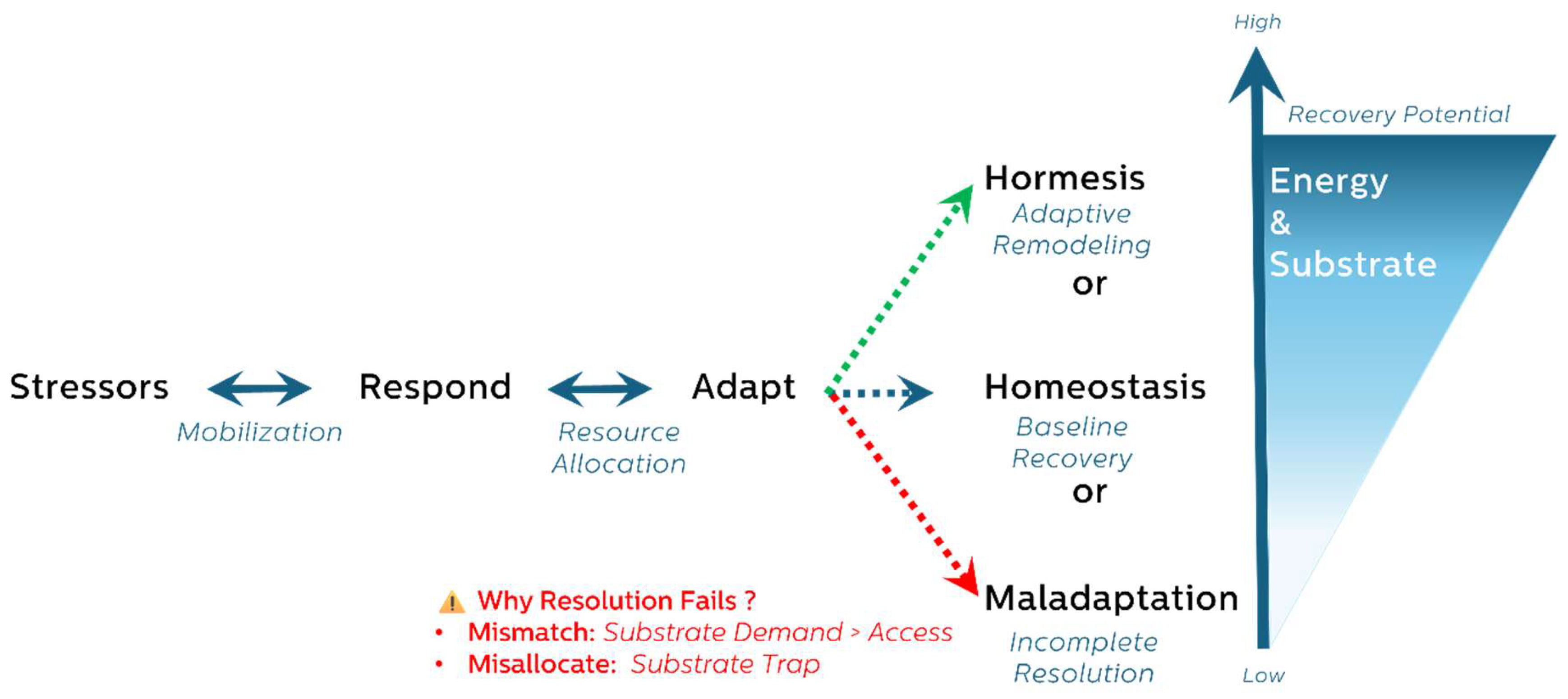
Figure 2.
Metabolic Outcomes of Stress-Responsive Adaptation Under Bioenergetic Constraints.
Disclaimer/Publisher’s Note: The statements, opinions and data contained in all publications are solely those of the individual author(s) and contributor(s) and not of MDPI and/or the editor(s). MDPI and/or the editor(s) disclaim responsibility for any injury to people or property resulting from any ideas, methods, instructions or products referred to in the content. |
© 2025 by the authors. Licensee MDPI, Basel, Switzerland. This article is an open access article distributed under the terms and conditions of the Creative Commons Attribution (CC BY) license (http://creativecommons.org/licenses/by/4.0/).
Copyright: This open access article is published under a Creative Commons CC BY 4.0 license, which permit the free download, distribution, and reuse, provided that the author and preprint are cited in any reuse.


